Embed Size (px)
Citation preview
Case reports 817
total calcium with overt bone disease is hard toexplain. The history of constipation, nocturia andpolyuria, albeit mild, do suggest hypercalcaemia andit may be that the ionized calcium was elevatedwithout the total calcium being raised.There are many points of similarity in the patient
reported here with the case reported by Mather in1953. His patient was a 39-year-old woman who haddeveloped diffuse aching pains in the lower limbsand back 6 months before admission to hospital.His patient was shown to have evidence of osteitisfibrosa both by skeletal X-ray survey and sternalbone biopsy. Four values for the serum calcium con-centration during the 3 months prior to the removalof a Wasserhelle cell parathyroid adenoma were allwithin the normal range. Studies of intestinalfunction were not, however, reported. Mather'spatient is of considerable interest, not only becauseof the short duration of symptoms but also because,as far as we are aware, it is the only other reportedcase that was persistently normocalcaemic until thetime of operation.The importance of the detection of cases of nor-
mocalcaemic primary hyperparathyroidism is stressedby the report of Nichols & Flanagan (1967). Theyreported six patients, with this syndrome, whichrepresented 37% of all the patients in whom theyhad made a diagnosis of parathyroid hyperfunctionin a 3-year period. Similarly the occurrence of eleven
cases with this syndrome seen in one centre (Georgeet al., 1965; Wills et al., 1969) over a 9-year periodsuggests a high incidence among patients with recur-rent renal calculi. The patient reported here, togetherwith that of Mather (1953), suggest that normo-calcaemia in primary hyperparathyroidism may alsobe present in some patients with osteitis fibrosa.
AcknowledgmentsWe wish to thank Professor C. B. Perry and Dr M. Hartog
for permission to report this case.
ReferencesDAVIEs, D.R., DENT, C.E. & WATSON, L. (1968) Tertiary
hyperparathyroidism. British Medical Journal, 3, 395.EISENBERG, E. & GOTCH, F.A. (1968) Normocalcaemic hyper-
parathyroidism culminating in hypercalcaemic crisis.Archives of Internal Medicine, 122, 258.
GEORGE, J.M., RABSON, A.S., KETCHAM, A. & BARTTER,F.C. (1965) Calcareous renal disease and hyperparathyroid-ism. Quarterly Journal of Medicine., 34, 291.
MATHER, H.G. (1953) Hyperparathyroidism with normalserum calcium. British Medical Journal, 2, 424.
NICHOLS, G., Jr & FLANAGAN, B. (1967) Normocalcaemichyperparathyroidism. Transactions of the Association ofAmerican Physicians, 80, 314.
WILLS, M.R. (1969) The urinary calcium/creatinine ratio asa measure of urinary calcium excretion. Journal of ClinicalPathology, 22, 287.
WILLS, M.R., PAK, C.Y.C., HAMMOND, W.C. & BARTTER,F.C. (1969) Normocalcaemic primary hyperparathyroid-ism. American Journal of Medicine, 47, 384.
Axial osteomalacia
JOHN R. CONDONB.Sc., M.B., M.R.C.P.
St George's Hospital,London
J. R. NASSIMF.R.C.P.
Royal National Orthopaedic Hospital,London and St George's Hospital, London
IN 1961 Frame and his co-workers reported threepatients who presented with mild back discomfortand in whom radiological studies showed a coarseningand distortion of the trabecular pattern of the cervicalvertebrae, lumbar vertebrae, ribs and pelvis. Iliaccrest biopsy in each of these patients showed widen-ing of the osteoid seams and because all the knowncauses of osteomalacia were excluded, the disorderwas called 'atypical osteomalacia involving the axialskeleton'. With the exception of a slightly raisedplasma alkaline phosphatase in one subject, plasmacalcium, phosphate and alkaline phosphatase valueswere normal in all patients; 24-hr urinary calcium
estimations were also normal. Only two otherexamples of this syndrome have been reported(Arnstein, Frame & Frost, 1967).We are reporting a sixth patient who has had this
disorder for 18 years and, therefore, gives someindication of the natural history of the disease. Theresults of calcium, phosphorus and nitrogen balancestudies are documented; the effect of prolongedtherapy with vitamin D on the symptoms, radio-logical picture and bone biopsy histology arereported. The possibility that the disorder is not dueto any abnormal metabolism ofcalcium, phosphorus,alkaline phosphatase and vitamin D is discussed.
818 Case reports
Case reportThe patient, a 71-year-old man, was first admitted
to hospital when aged 53 years. At this time hecomplained of low back pain which had beenpresent for 18 months.On examination there were no abnormal physical
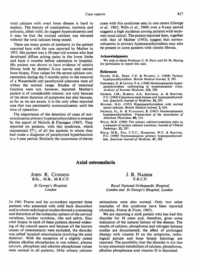
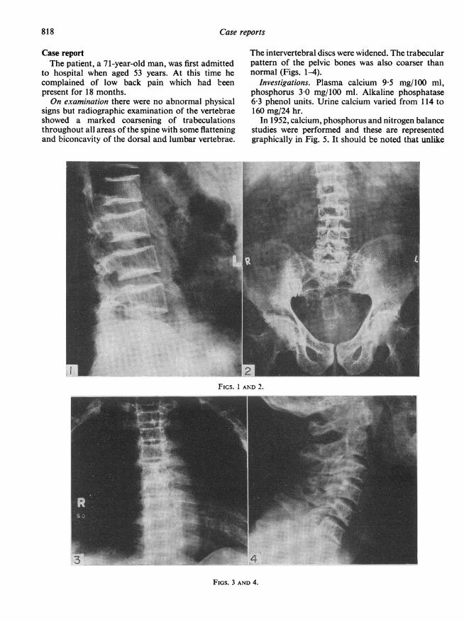
signs but radiographic examination of the vertebraeshowed a marked coarsening of trabeculationsthroughout all areas of the spine with some flatteningand biconcavity of the dorsal and lumbar vertebrae.
The intervertebral discs were widened. The trabecularpattern of the pelvic bones was also coarser thannormal (Figs. 1-4).
Investigations. Plasma calcium 9'5 mg/100 ml,phosphorus 30 mg/100 ml. Alkaline phosphatase6&3 phenol units. Urine calcium varied from 114 to160 mg/24 hr.
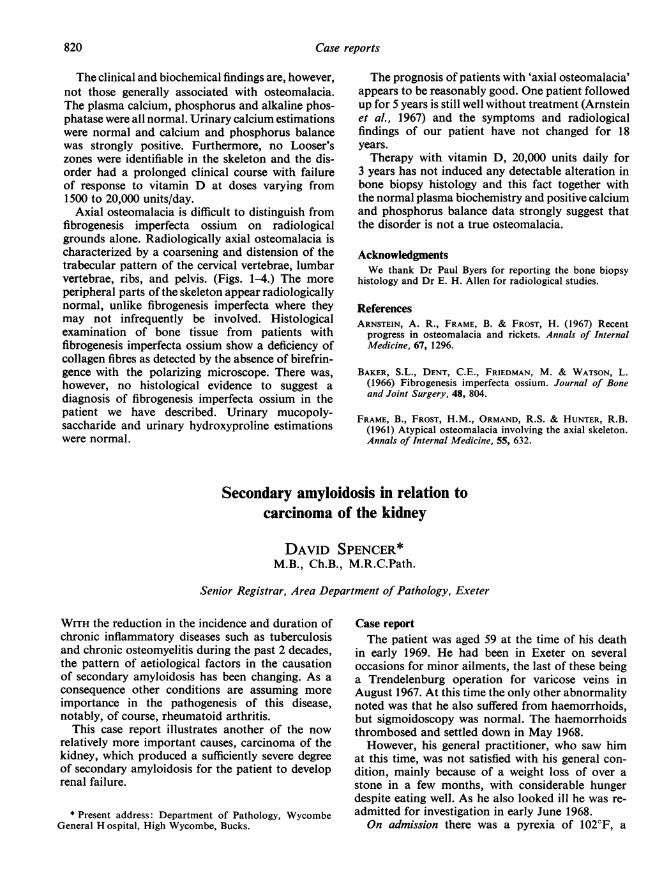
In 1952, calcium, phosphorus and nitrogen balancestudies were performed and these are representedgraphically in Fig. 5. It should be noted that unlike
.... ^*II....
Fics. I AND 2.
3
FIGS. 3 AND 4.

Case reports 819
patients with 'typical osteomalacia' this individualwas in strongly positive calcium and phosphorusbalance with a normal urinary calcium.
In 1959, the patient had acute pancreatitis whichwas confirmed at laparotomy. In 1960, he had aright middle cerebral artery thrombosis and in 1963an impacted fracture of the left femur. Three yearslater he began developing bilateral Dupuytren'scontractures.
In 1966, a bone biopsy examination was per-formed. Normal bone trabeculae were replaced by anetwork of thickened structures which gave thespecimen a grossly abnormal appearance on nakedeye examination. Examination of decalcified sections
S t ilboestrol(0-5mg/day)
Methyltestosterone
Control (10 0 mg/day)
-200
-200EE
z' 600
-200
0 600 ....
0
1000
0-
LI~~~~~~~~~~~~~~~~~~~~~~~~~~~
a 6
0 8 16 24Days
FIG. S. Open columns, faeces; hatched columns, urine.
showed thick irregular bone trabeculae. Much ofthe trabecular surface was covered by osteoid tissuewhich in places formed layers of up to 100 ,. inthickness. Some areas ofbone resorption were presentbut there was no evidence of fibrous replacement ofbone. Microradiographs showed the smudgy outlineof osteocytic lacunae found in rickets and osteo-malacia. All the osteoid tissue had a normal lamellarstructure; there was nothing to suggest a diagnosisof fibrogenesis imperfecta ossium.
Increased bone density is known to occur in someexamples of long standing osteomalacia and thepresent case appears to be an example of thisassociation.From 1952 to 1970 plasma calcium, phosphorus
and alkaline phosphatase levels were measured at3-6-monthly intervals and were invariably normal.Twenty-four-hour urine hydroxyproline values wereall within the normal range.
Treatment. Treatment with vitamin D at a dosageof 1500 units and later 10,000 units/day, was begunin 1952 and continued for 6 months. This wasfollowed by stilboestrol 1 mg/day combined withmethyltestosterone 10 mg/day and later methyl-testosterone alone. There was no improvement ofsymptoms and no change in radiological appear-ances of the bone following this therapy.
In 1966, the dosage of vitamin D was increased to20,000 units/day and continued for 3 years. Therewas still no improvement in symptoms and theradiological bone picture remained unchanged.Repeat iliac crest bone biopsy was performed in1970 and reported as follows:'The material, a small piece of thin cortical bone
and a few narrow widely separated trabeculae wasinadequate for proper histological assessment. Therewere few osteoid seams, but the widtlh of thosepresent was similar to the width of seams seen inareas of the bone biopsy taken in 1966.
Microradiographs again show the smudgy outlineof the osteocyte lacunae to be present.'
It is difficult to draw any definite conclusionsconcerning the effect of therapy.
DiscussionThe patient we have reported came under medical
supervision when aged 53 years, because of backpain. At that time the abnormal trabecular patternof the axial skeleton was noted but regarded as aform of osteoporosis. In 1966, the radiologicalevidence was reviewed and a diagnosis of fibro-genesis imperfecta ossium (Baker et al., 1966)suggested. Bone biopsy, which was performed in1966 to confirm this diagnosis, revealed histologicalfeatures which are generally accepted to be diagnosticof osteomalacia.
820 Case reports
The clinical and biochemical findings are, however,not those generally associated with osteomalacia.The plasma calcium, phosphorus and alkaline phos-phatase were all normal. Urinary calcium estimationswere normal and calcium and phosphorus balancewas strongly positive. Furthermore, no Looser'szones were identifiable in the skeleton and the dis-order had a prolonged clinical course with failureof response to vitamin D at doses varying from1500 to 20,000 units/day.Axial osteomalacia is difficult to distinguish from
fibrogenesis imperfecta ossium on radiologicalgrounds alone. Radiologically axial osteomalacia ischaracterized by a coarsening and distension of thetrabecular pattern of the cervical vertebrae, lumbarvertebrae, ribs, and pelvis. (Figs. 1-4.) The moreperipheral parts of the skeleton appear radiologicallynormal, unlike fibrogenesis imperfecta where theymay not infrequently be involved. Histologicalexamination of bone tissue from patients withfibrogenesis imperfecta ossium show a deficiency ofcollagen fibres as detected by the absence of birefrin-gence with the polarizing microscope. There was,however, no histological evidence to suggest adiagnosis of fibrogenesis imperfecta ossium in thepatient we have described. Urinary mucopoly-saccharide and urinary hydroxyproline estimationswere normal.
The prognosis of patients with 'axial osteomalacia'appears to be reasonably good. One patient followedup for 5 years is still well without treatment (Arnsteinet al., 1967) and the symptoms and radiologicalfindings of our patient have not changed for 18years.Therapy with vitamin D, 20,000 units daily for
3 years has not induced any detectable alteration inbone biopsy histology and this fact together withthe normal plasma biochemistry and positive calciumand phosphorus balance data strongly suggest thatthe disorder is not a true osteomalacia.
AcknowledgmentsWe thank Dr Paul Byers for reporting the bone biopsy
histology and Dr E. H. Allen for radiological studies.
ReferencesARNSTEIN, A. R., FRAME, B. & FROST, H. (1967) Recent
progress in osteomalacia and rickets. Annals of InternalMedicine, 67, 1296.
BAKER, S.L., DENT, C.E., FRIEDMAN, M. & WATSON, L.(1966) Fibrogenesis imperfecta ossium. Journal of Boneand Joint Surgery, 48, 804.
FRAME, B., FROST, H.M., ORMAND, R.S. & HUNTER, R.B.(1961) Atypical osteomalacia involving the axial skeleton.Annals of Internal Medicine, 55, 632.
Secondary amyloidosis in relation tocarcinoma of the kidney
DAVID SPENCER*M.B., Ch.B., M.R.C.Path.
Senior Registrar, Area Department of Pathology, Exeter
WITH the reduction in the incidence and duration ofchronic inflammatory diseases such as tuberculosisand chronic osteomyelitis during the past 2 decades,the pattern of aetiological factors in the causationof secondary amyloidosis has been changing. As aconsequence other conditions are assuming moreimportance in the pathogenesis of this disease,notably, of course, rheumatoid arthritis.
This case report illustrates another of the nowrelatively more important causes, carcinoma of thekidney, which produced a sufficiently severe degreeof secondary amyloidosis for the patient to developrenal failure.
* Present address: Department of Pathology, WycombeGeneral H ospital, High Wycombe, Bucks.
Case reportThe patient was aged 59 at the time of his death
in early 1969. He had been in Exeter on severaloccasions for minor ailments, the last of these beinga Trendelenburg operation for varicose veins inAugust 1967. At this time the only other abnormalitynoted was that he also suffered from haemorrhoids,but sigmoidoscopy was normal. The haemorrhoidsthrombosed and settled down in May 1968.However, his general practitioner, who saw him
at this time, was not satisfied with his general con-dition, mainly because of a weight loss of over astone in a few months, with considerable hungerdespite eating well. As he also looked ill he was re-admitted for investigation in early June 1968.On admission there was a pyrexia of 102°F, a