Embed Size (px)
Citation preview
Bacterial Recognition of Silicon Nanowire ArraysHoon Eui Jeong,†,‡ Ilsoo Kim,§ Pierre Karam,† Heon-Jin Choi,§ and Peidong Yang*,†
†Department of Chemistry, University of California, Berkeley, California 94720, United States‡School of Mechanical and Advanced Materials Engineering, Ulsan National Institute of Science and Technology, Ulsan 689-798,South Korea§Department of Materials Science and Engineering, Yonsei University, Seoul 120-749, South Korea
*S Supporting Information
ABSTRACT: Understanding how living cells interact with nanostructures is integral to abetter understanding of the fundamental principles of biology and the development ofnext-generation biomedical/bioenergy devices. Recent studies have demonstrated thatmammalian cells can recognize nanoscale topographies and respond to these structures.From this perspective, there is a growing recognition that nanostructures, along with theirspecific physicochemical properties, can also be used to regulate the responses and motionsof bacterial cells. Here, by utilizing a well-defined silicon nanowire array platform andsingle-cell imaging, we present direct evidence that Shewanella oneidensis MR-1 canrecognize nanoscale structures and that their swimming patterns and initial attachmentlocations are strongly influenced by the presence of nanowires on a surface. Analyses ofbacterial trajectories revealed that MR-1 cells exhibited a confined diffusion mode in thepresence of nanowires and showed preferential attachment to the nanowires, whereas asuperdiffusion mode was observed in the absence of nanowires. These results demonstratethat nanoscale topography can affect bacterial movement and attachment and play an important role during the early stages ofbiofilm formation.
KEYWORDS: Nanowires, bacteria, diffusion, single-cell imaging, trajectory analysis
Nanoengineered surfaces that can regulate bacterialattachment are potentially useful for developing micro-
bial fuel cells with high power densities and antibacterialbiomedical devices.1−5 Nanostructured electrodes have beendeveloped to enhance the power density of microbial fuel cellsby promoting bacterial attachment to the electrode surface.1,2
Nanostructured materials have also been explored asantibacterial surfaces that are resistant to biofilm formation.3−5
While much effort has been expended on the development ofnanoengineered surfaces that can control bacterial motion andattachment on surfaces, the fundamental principles of bacteria−nanostructure interactions remain poorly understood. Differentbacterial behaviors on nanostructured substrates have beenreported. Some studies reported that nanostructures had nosignificant effects on bacterial attachment, whereas othersreported that nanoscale topographies did influence theattachment and growth of bacteria.4−9 Moreover, most previousworks were based on observations of large bacterial populationscultured on a surface with random nanostructures.5−9 Whilethese approaches are useful for studying bacterial filmformation at the macroscopic level, randomly oriented, high-density nanostructures and massive bacterial populations makeit difficult to investigate bacteria−nanostructure interactions ina systematic manner.4−9
In this respect, nanowires can be a powerful platform forstudying bacteria−nanostructure interactions at the single-celllevel. Recent studies have shown that nanowires can interface
with mammalian cells in a minimally invasive manner becauseof their nanoscale dimensions.10−13 In addition to mammaliancells, nanowires are also promising for interfacing with bacterialcells, since their nanoscale dimensions (100−300 nm) arecomparable to those of individual bacterial cells. Interfacingnanowires with single bacterial cells will enable us to investigatein detail how individual bacteria interact with surfaces. Inaddition, nanowires can be produced in precisely ordered arrayswith accurate size and position control, allowing systematicstudy of bacteria−nanostructure interactions for nanowires withdifferent geometries.14 Furthermore, the unique electricalproperties of nanowires and surface functionalizations ofnanowires would potentially allow electrical and chemicalstimuli to be applied through the nanowires.12
Here, by combining a precisely defined silicon nanowirearray platform and real-time optical imaging, we explorebacteria−nanostructure interactions at the single-cell level. Inthis study, Shewanella oneidensis MR-1, a gram-negativefacultative bacterium, was used as a model microorganism tostudy bacteria−nanostructure interactions, since S. oneidensisMR-1 is well-known for its biofilm formation on mineralsurfaces and electrodes of microbial fuel cells.15−17 Thetrajectories of MR-1 cells on substrates with Si nanowire arrays
Received: April 4, 2013Revised: May 8, 2013Published: May 17, 2013
Letter
pubs.acs.org/NanoLett
© 2013 American Chemical Society 2864 dx.doi.org/10.1021/nl401205b | Nano Lett. 2013, 13, 2864−2869

were analyzed and quantified by mathematical models. Weshow that MR-1 cells can recognize nanowires and that theirswimming and attachment behaviors are drastically altered bythe presence of nanowires on a substrate. Our novel approachbased on interfacing between a nanowire and a single bacterialcell offers a new set of tools for systematically studyingbacteria−surface interactions at the single-cell level.Figure 1a shows the patterned Si nanowire arrays with a 10
μm pitch. The diameter and length of the individual nanowiresare ∼300 nm and ∼3 μm, respectively. Shewanella oneidensisMR-1 was cultured on the Si nanowire arrays under aerobic oranaerobic conditions, and its motion was observed usingupright microscopy. Interestingly, the majority of MR-1 cellsattached directly to the Si nanowires (Figure 1b) rather thanthe bottom substrate. The scanning electron microscopy(SEM) images and their corresponding merged optical images(fluorescence and dark field) (Figure 1c−e) clearly indicate that
the MR-1 bacterium attached preferentially to the Si nanowires.More interestingly, single MR-1 cells attached to individual Sinanowires in perfect alignment with the Si nanowires (Figure1c). Occasionally, MR-1 cells were also observed sitting on thebottom substrate (Figure 1b, e).When compared with prior approaches using substrates with
random nanostructures,5−9 our approach using patterned Sinanowire arrays allows accurate analyses of bacteria−nano-structure interactions to be performed at the single-cell level.Moreover, the surfaces of the Si nanowires can be easilyfunctionalized with different chemicals. The size of thenanowires, including the diameter as well as the length, canbe readily controlled by modulating the growth conditions.14
Interestingly, when the size of a nanowire was comparable tothat of an individual MR-1 bacterium and the bacterial densitywas low, we could readily induce a single MR-1 cell to attach toan individual Si nanowire. To examine the bacteria−nanowire
Figure 1. Shewanella oneidensisMR-1 grown on patterned Si nanowire arrays. (a) Titled SEM image of the patterned Si nanowire arrays on a Si(111)substrate. The pitch of the arrays is 10 μm and the diameter and length of the individual nanowires are ∼300 nm and ∼3 μm, respectively. (b)Bright-field micrograph of Shewanella oneidensis MR-1 grown on the Si nanowire arrays. Scale bars in panels a and b, 5 μm. (c−e) SEM imagesshowing MR-1 cells attached on the Si nanowires and their corresponding merged images of fluorescence micrographs of stained MR-1 and dark-field micrographs of Si nanowires. Single (c) or multiple (d) MR-1 cells were preferentially attached on the individual nanowires with alignmentsalong the length direction of the nanowires. Some cells were observed on the bottom substrate, but many of them still maintained close contact withthe nanowires (e). Scale bars in panels c−e, 500 nm.
Nano Letters Letter
dx.doi.org/10.1021/nl401205b | Nano Lett. 2013, 13, 2864−28692865
interactions at the single-cell level, we made the dimension ofthe Si nanowires (∼300 nm in diameter and ∼3 μm in length)similar to that of an MR-1 bacterium (400−700 nm in widthand 2−3 μm in length).To confirm the observation of preferential attachment of
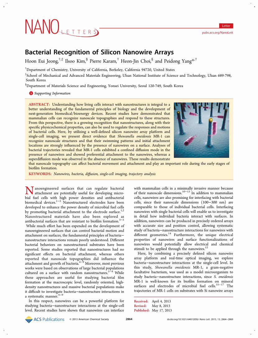
MR-1 cells to Si nanowires, we performed real-time opticalimaging of the MR-1 movements on the nanowire arrays.Nanowire arrays with relatively long pitches (10 and 15 μm)were used in these experiments. Figure 2a shows time-lapseimages of the movements of a single MR-1 cell on an array witha 15 μm pitch. At the initial stage of the observation (Figure2a), the MR-1 cell swam freely without binding to or sitting onthe substrate. However, after the MR-1 cell encountered one ofthe nanowires at ∼15 s, the cell stayed on the nanowire for along period of time (>5 min) while changing the orientation ofits body (see Supporting Information Figure S1 and Movie S1).To further investigate bacteria−nanowire interactions, weanalyzed the trace of the bacterial movements using particle-tracking software. The analyzed trajectory (Figure 2b) clearlyshowed that the MR-1 bacterium stayed around the nanowireand had a diminished velocity after it approached the nanowire.Additional X, Y displacement analysis with time (Figure 2c,d)further confirmed that the MR-1 bacterium strongly preferredto stay on the Si nanowires rather than travel to the planarbottom Si substrate, although they both have the same Si/SiO2
surface. As shown in Figure 2c,d, the position of the cell wasalmost fixed to the specific coordinate values where the Sinanowire was located after the cell encountered the nanowire at∼15 s, suggesting there is a distinct interaction between theMR-1 bacterium and the Si nanowire.To investigate the effects of the pitch of the nanowire arrays
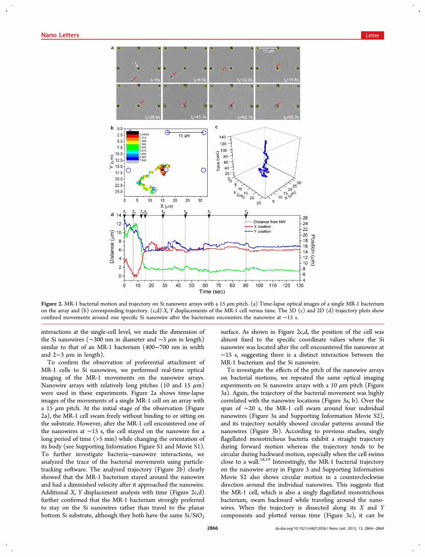
on bacterial motions, we repeated the same optical imagingexperiments on Si nanowire arrays with a 10 μm pitch (Figure3a). Again, the trajectory of the bacterial movement was highlycorrelated with the nanowire locations (Figure 3a, b). Over thespan of ∼20 s, the MR-1 cell swam around four individualnanowires (Figure 3a and Supporting Information Movie S2),and its trajectory notably showed circular patterns around thenanowires (Figure 3b). According to previous studies, singlyflagellated monotrichous bacteria exhibit a straight trajectoryduring forward motion whereas the trajectory tends to becircular during backward motion, especially when the cell swimsclose to a wall.18,19 Interestingly, the MR-1 bacterial trajectoryon the nanowire array in Figure 3 and Supporting InformationMovie S2 also shows circular motion in a counterclockwisedirection around the individual nanowires. This suggests thatthe MR-1 cell, which is also a singly flagellated monotrichousbacterium, swam backward while traveling around the nano-wires. When the trajectory is dissected along its X and Ycomponents and plotted versus time (Figure 3c), it can be
Figure 2. MR-1 bacterial motion and trajectory on Si nanowire arrays with a 15 μm pitch. (a) Time-lapse optical images of a single MR-1 bacteriumon the array and (b) corresponding trajectory. (c,d) X, Y displacements of the MR-1 cell versus time. The 3D (c) and 2D (d) trajectory plots showconfined movements around one specific Si nanowire after the bacterium encounters the nanowire at ∼15 s.
Nano Letters Letter
dx.doi.org/10.1021/nl401205b | Nano Lett. 2013, 13, 2864−28692866
further confirmed that the cell dwelled on each nanowire forabout 2−8 s before it went through a quick transition state (<1s) and then stayed around another nanowire. No bacterialconfinement was observed in the absence of nanowires. Asshown in Figure 3d, the swimming traces of the MR-1 cells on aplanar surface exhibited random-walk patterns. A directcomparison between Figure 3 panels b and d also shows thatthe swimming and attachment behaviors of the MR-1 bacteriaare influenced significantly by the presence of nanoscaletopography on a surface.Additional optical imaging experiments were also performed
to investigate the detailed process of how individual MR-1 cellsattached to the Si nanowires over a longer observation time(Supporting Information Figure S2). During the observation,the MR-1 cells preferentially attached to the nanowires, butsome of the attached MR-1 cells left the nanowires after just afew seconds or a few tens of seconds. This means that thebacterial attachment is reversible at this stage, which is acharacteristic of the early stages of biofilm formations.20,21
However, the number of cells attached to the nanowire arraysgradually increased with time (Supporting Information FigureS2). Interestingly, when we increased the length of the Sinanowires from 3 to 15−20 μm, multiple cells were observed toselectively sit on a single Si nanowire (Supporting InformationFigure S2b). This initial but reversible MR-1 attachment to theSi nanowires observed here corresponds to the first step ofbacterial biofilm formation. At this stage, bacteria use a varietyof extracellular appendages and proteins for sensing and
attaching to solid surfaces, including flagella, pili, fimbriae,curlifibers, and outer membrane proteins.5,21−23 To investigatethe relevant biological mechanisms, mutants of MR-1 bacterialacking outer membrane cytochromes OmcA and MtrC, whichare in charge of electron transfer across the cell membranes,24
were cultured on the nanowire arrays. However, the MR-1mutants still exhibited preferential movement toward andattachment to the Si nanowires. Experiments with flagellamutants have been excluded in our study because mutants ofMR-1 bacteria lacking flagella are not able to swim and most ofcells settle on the bottom substrate.25,26 However, the SEMimage shown in Figure 1c in which the MR-1 bacteriumattached to the Si nanowire wraps the nanowire with its flagellamay suggest the possibility that the bacteria sense the nanoscalestructures by utilizing their cellular appendages as suggested inother studies.22,23 Interestingly, the Si nanowire-confinedswimming behaviors were also observed for other strains ofbacteria such as Escherichia coli K-12 strains W3110 andMG1655 in separate experiments (data not shown). Thissuggests that the ability to sense nanoscale topographies isgeneral for many bacterial strains that have evolved mechanismsfor attaching to surfaces and forming biofilms for survival indiverse environmental conditions.5,16,20−23 More studies arerequired to reveal the detailed biological mechanisms of thebacteria−nanostructure recognitions.To quantify how the MR-1 bacteria are confined around
nanowires, a set of swimming trajectories on planar Sisubstrates and substrates with nanowire arrays of 10 and 15
Figure 3. MR-1 bacterial motion and trajectory on Si nanowire arrays with a 10 μm pitch. (a) Time-lapse optical images of a single MR-1 bacteriumon the array. The analyzed trajectory (b) shows confined bacterial motions around the Si nanowires. (c) X, Y displacements of the MR-1 cell versustime. Gray lines and navy arrows indicate positions where individual Si nanowires are located. (d) Trajectories of multiple MR-1 cells on a planar Sisubstrate showing random swimming patterns.
Nano Letters Letter
dx.doi.org/10.1021/nl401205b | Nano Lett. 2013, 13, 2864−28692867
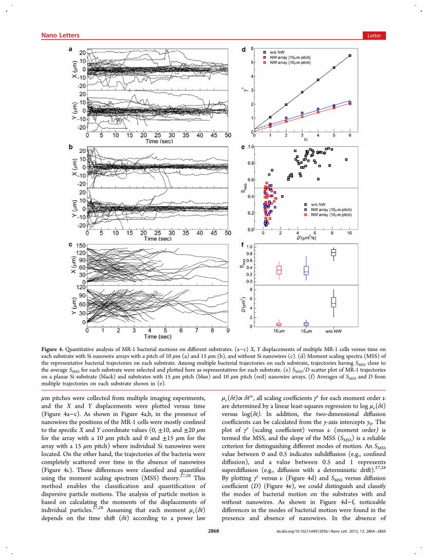
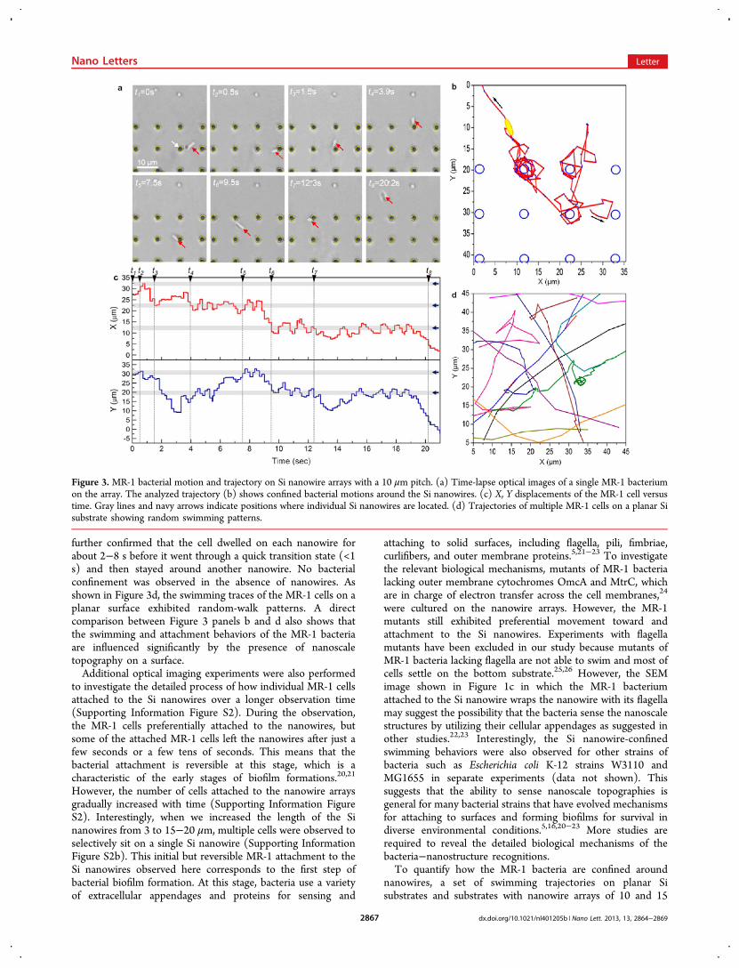
μm pitches were collected from multiple imaging experiments,and the X and Y displacements were plotted versus time(Figure 4a−c). As shown in Figure 4a,b, in the presence ofnanowires the positions of the MR-1 cells were mostly confinedto the specific X and Y coordinate values (0, ±10, and ±20 μmfor the array with a 10 μm pitch and 0 and ±15 μm for thearray with a 15 μm pitch) where individual Si nanowires werelocated. On the other hand, the trajectories of the bacteria werecompletely scattered over time in the absence of nanowires(Figure 4c). These differences were classified and quantifiedusing the moment scaling spectrum (MSS) theory.27,28 Thismethod enables the classification and quantification ofdispersive particle motions. The analysis of particle motion isbased on calculating the moments of the displacements ofindividual particles.27,28 Assuming that each moment μν(δt)depends on the time shift (δt) according to a power law
μν(δt)∝ δtγν, all scaling coefficients γν for each moment order νare determined by a linear least-squares regression to log μν(δt)versus log(δt). In addition, the two-dimensional diffusioncoefficients can be calculated from the y-axis intercepts y0. Theplot of γν (scaling coefficient) versus ν (moment order) istermed the MSS, and the slope of the MSS (SMSS) is a reliablecriterion for distinguishing different modes of motion. An SMSS
value between 0 and 0.5 indicates subdiffusion (e.g., confineddiffusion), and a value between 0.5 and 1 representssuperdiffusion (e.g., diffusion with a deterministic drift).27,28
By plotting γν versus ν (Figure 4d) and SMSS versus diffusioncoefficient (D) (Figure 4e), we could distinguish and classifythe modes of bacterial motion on the substrates with andwithout nanowires. As shown in Figure 4d−f, noticeabledifferences in the modes of bacterial motion were found in thepresence and absence of nanowires. In the absence of
Figure 4. Quantitative analysis of MR-1 bacterial motions on different substrates. (a−c) X, Y displacements of multiple MR-1 cells versus time oneach substrate with Si nanowire arrays with a pitch of 10 μm (a) and 15 μm (b), and without Si nanowires (c). (d) Moment scaling spectra (MSS) ofthe representative bacterial trajectories on each substrate. Among multiple bacterial trajectories on each substrate, trajectories having SMSS close tothe average SMSS for each substrate were selected and plotted here as representatives for each substrate. (e) SMSS/D scatter plot of MR-1 trajectorieson a planar Si substrate (black) and substrates with 15 μm pitch (blue) and 10 μm pitch (red) nanowire arrays. (f) Averages of SMSS and D frommultiple trajectories on each substrate shown in (e).
Nano Letters Letter
dx.doi.org/10.1021/nl401205b | Nano Lett. 2013, 13, 2864−28692868
nanowires, the MR-1 cells were observed to have a super-diffusion mode of motion with an SMSS of 0.821 ± 0.130 and adiffusion coefficient (D) of 5.279 ± 1.538 μm2/s (n = 92).However, in the presence of nanowires, significant drops in theslope of the MSS and the diffusion coefficient were observed(Figure 4d−f). Most of the analyzed trajectories exhibited thecharacteristics of a confined diffusion mode. For the 10 and 15μm pitch array, the SMSS was 0.334 ± 0.138 and 0.314 ± 0.188,respectively and the diffusion coefficient was 0.546 ± 0.323μm2/s (n = 36) and 0.616 ± 0.370 μm2/s (n = 28),respectively. The decreases in the diffusion constant and SMSSare mainly due to the dwelling times that each MR-1 cell spentin the vicinity of the nanowires.Our results demonstrate that nanoscale topographies on
surfaces play an important role during the early stage of biofilmformation. Understanding the origins of biofilm formation atthe single-cell level could provide a wealth of information foradvancing the fundamentals of microbiology and designingnext-generation bioenergy and biomedical devices. Our currentapproach based on precisely defined nanowire arrays and real-time single-cell imaging can serve as a powerful platform forstudying bacteria−surface interactions under varying chemical,physical, electrical, and optical conditions at the single-cell level.
■ ASSOCIATED CONTENT*S Supporting InformationAdditional information and figures. This material is availablefree of charge via the Internet at http://pubs.acs.org.
■ AUTHOR INFORMATIONCorresponding Author*E-mail: [email protected] authors declare no competing financial interest.
■ ACKNOWLEDGMENTSP.Y. Thanks the National Science Foundation for the A. T.Waterman award. The authors thank the CISMM at UNC−CHfor video spot tracking software, and Y. J. Hwang for discussion.H.J.C. and I.K. thank the National Research Foundation ofKorea for the grant (No. 2012R1A2A1A03010558) and thePioneer Research Program for Converging Technology (No.2009-008-1529).
■ REFERENCES(1) Xie, X.; Hu, L. B.; Pasta, M.; Wells, G. F.; Kong, D. S.; Criddle, C.S.; Cui, Y. Nano Lett. 2011, 11, 291−296.(2) Mink, J. E.; Rojas, J. P.; Logan, B. E.; Hussain, M. M. Nano Lett.2012, 12, 791−795.(3) Mitik-Dineva, N.; Wang, J.; Truong, V. K.; Stoddart, P.;Malherbe, F.; Crawford, R. J.; Ivanova, E. P. Curr. Microbiol. 2009, 58,268−273.(4) Hochbaum, A. I.; Aizenberg, J. Nano Lett. 2010, 10, 3717−3721.(5) Renner, L. D.; Weibel, D. B. MRS Bull. 2011, 36, 347−355.(6) Puckett, S. D.; Taylor, E.; Raimondo, T.; Webster, T. J.Biomaterials 2010, 31, 706−713.(7) Jeyachandran, Y. L.; Venkatachalam, S.; Karunagaran, B.;Narayandass, S. K.; Mangalaraj, D.; Bao, C. Y.; Zhang, C. L. Mater.Sci. Eng., C 2007, 27, 35−41.(8) Rizzello, L.; Sorce, B.; Sabella, S.; Vecchio, G.; Galeone, A.;Brunetti, V.; Cingolani, R.; Pompa, P. P. ACS Nano 2011, 5, 1865−1876.(9) Akesso, L.; Pettitt, M.; Callow, J.; Callow, M.; Stallard, J.; Teer,D.; Liu, C.; Wang, S.; Zhao, Q.; D’Souza, F.; Willemsen, P.; Donnelly,
G.; Donik, C.; Kocijan, A.; Jenko, M.; Jones, L.; Guinaldo, P. C.Biofouling 2009, 25, 55−67.(10) Yan, R.; Park, J.-H.; Choi, Y.; Heo, C.-J.; Yang, S.-M.; Lee, L. P.;Yang, P. Nat. Nanotechnol. 2012, 7, 191−196.(11) Kim, W.; Ng, J. K.; Kunitake, M. E.; Conklin, B. R.; Yang, P. J.Am. Chem. Soc. 2007, 129, 7228−7729.(12) Yang, P. D.; Yan, R. X.; Fardy, M. Nano Lett. 2010, 10, 1529−1536.(13) Shalek, A. K.; Robinson, J. T.; Karp, E. S.; Lee, J. S.; Ahn, D. R.;Yoon, M. H.; Sutton, A.; Jorgolli, M.; Gertner, R. S.; Gujral, T. S.;MacBeath, G.; Yang, E. G.; Park, H. Proc. Natl. Acad. Sci. U.S.A. 2010,107, 1870−1875.(14) Hochbaum, A. I.; Fan, R.; He, R.; Yang, P. Nano Lett. 2005, 5,457−460.(15) Gorby, Y. A.; Yanina, S.; McLean, J. S.; Rosso, K. M.; Moyles,D.; Dohnalkova, A.; Beveridge, T. J.; Chang, I. S.; Kim, B. H.; Kim, K.S.; Culley, D. E.; Reed, S. B.; Romine, M. F.; Saffarini, D. A.; Hill, E.A.; Shi, L.; Elias, D. A.; Kennedy, D. W.; Pinchuk, G.; Watanabe, K.;Ishii, S.; Logan, B.; Nealson, K. H.; Fredrickson, J. K. Proc. Natl. Acad.Sci. U.S.A. 2006, 103, 11358−11363.(16) Lower, S. K.; Hochella, M. F.; Beveridge, T. J. Science 2001, 292,1360−1363.(17) Lovley, D. R. Nat. Rev. Microbiol. 2006, 4, 497−508.(18) Goto, T.; Nakata, K.; Baba, K.; Nishimura, M.; Magariyama, Y.Biophys. J. 2005, 89, 3771−3779.(19) Taylor, B. L.; Koshland, D. E. J. Bacteriol. 1974, 119, 640−642.(20) Kolter, R.; Greenberg, E. P. Nature 2006, 441, 300−302.(21) McDougald, D.; Rice, S. A.; Barraud, N.; Steinberg, P. D.;Kjelleberg, S. Nat. Rev. Microbiol. 2012, 10, 39−50.(22) Thormann, K. M.; Saville, R. M.; Shukla, S.; Pelletier, D. A.;Spormann, A. M. J. Bacteriol. 2004, 186, 8096−8104.(23) Pratt, L. A.; Kolter, R. Mol. Microbiol. 1998, 30, 285−293.(24) Xiong, Y. J.; Shi, L.; Chen, B. W.; Mayer, M. U.; Lower, B. H.;Londer, Y.; Bose, S.; Hochella, M. F.; Fredrickson, J. K.; Squier, T. C.J. Am. Chem. Soc. 2006, 128, 13978−13979.(25) Bouhenni, R. A.; Vora, G. J.; Biffinger, J. C.; Shirodkar, S.;Brockman, K.; Ray, R.; Wu, P.; Johnson, B. J.; Biddle, E. M.; Marshall,M. J.; Fitzgerald, L. A.; Little, B. J.; Fredrickson, J. K.; Beliaev, A. S.;Ringeisen, B. R.; Saffarini, D. A. Electroanalysis 2010, 22, 856−864.(26) Friedlander, R. S.; Vlamakis, H.; Kim, P.; Khan, M.; Kolter, R.;Aizenberg, J. Proc. Natl. Acad. Sci. U.S.A. 2013, 110, 5624−5629.(27) Ferrari, R.; Manfroi, A. J.; Young, W. R. Phys. D 2001, 154,111−137.(28) Ewers, H.; Smith, A. E.; Sbalzarini, I. F.; Lilie, H.;Koumoutsakos, P.; Helenius, A. Proc. Natl. Acad. Sci. U.S.A. 2005,102, 15110−15115.
■ NOTE ADDED AFTER ASAP PUBLICATIONThe Acknowledgments have been updated. The revised versionwas re-posted on May 29, 2013.
Nano Letters Letter
dx.doi.org/10.1021/nl401205b | Nano Lett. 2013, 13, 2864−28692869