Embed Size (px)
DESCRIPTION
mnjhgff
Citation preview

Al-Azhar University of Gaza
Faculty of Dentistry
DR/ ASHRAF SHAMIA
DR/ NOOR M. ABU AMARA
Sep. 2015
ORAL RADIOLOGY
Introduction To Oral Radiology

1- Radiology
2- Roentgenology
3- Dental radiology
4- Dental radiography
5- Radiograph
6- Radiation
7- Radiolucent
8- Radiopaque
Terminology

Radiology
Science that deals with application of high- energy radiation in diagnosis, therapeutic and researches.
Roentgenology
Science that deals with application of X-ray on any field.

Dental radiology
It is the branch of science that deals with the use of radiation in diagnosis of dental diseases.
Dental radiography
It is the art of producing an image or picture for intra- or extra-oral structures on a dental film using X-ray.

Radiograph
It is the shadow features (image) received on a radiation-sensitive film emulsion by exposure to ionizing radiation directed through an area or region or substance of interest, followed by chemical processing of the film. It is basically dependent on the differential absorption of radiation directed through heterogeneous media.
Radiation
It is the process of emission, propagation and transmission of energy by atoms in the form of waves.

Radiolucent
Objects that permitting the passage of radiant energy with relatively little attenuation by absorption and appear black on the film, such as silicate restoration, pulp tissues, gingiva, and carious lesion.
Another definition; Objects partly or wholly penetrable by roentgen rays; the image of such a material on the film ranges from dark gray to black.
Radiopaque Objects that absorb X-rays and appear white on radiograph, such as amalgam restoration, enamel, and bone.
Another def. : Objects that not freely penetrable by radiation.
OR Objects highly resistant to penetration by roentgen rays; the image of such a material appears on the film within range of gray to white.


The purpose of dental radiography is to record images of a patient's oral structures on film by using X-rays. When the X-ray films are processed, the resulting radiographs provide the dental officer with a valuable diagnostic aid.
In the case of death, radiographs can be used to aid in identification "Forensic Dentistry."
Purposes of Dental Radiology

Radiographic examination provides important anatomical information to determine the different pathologies and conditions of the teeth's and tissues like bone supports:
Defects and Variations in Tooth Density Trauma and Exodontia Fractures Endodontic Treatment of Teeth Developmental Defects and Anomalies Swellings, Cysts and Neoplasm Metabolic Diseases Orthodontic treatment
Presence and severity of periodontal diseases, abscesses or infections
Shape and sizes of the roots
Maxillary sinus disease
The exact locations of impacted teeth, position of third molars and the status of develop
Cavities that can not be seen directly or in early stage, allowing patients to receive preventive treatment.


FRACTURE

PERIAPICAL RADIOLUCENCY

ENDODONTICS

DENTINOGENESIS IMPERFECTA

DENTIGEROUS CYST

MALOCCLUSION

??

??
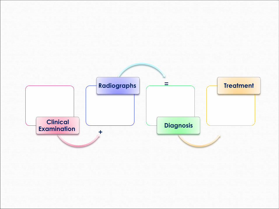
Clinical Examination
Radiographs
Diagnosis
Treatment
+
=

Are a form of pure energy units belonging to
electromagnetic spectrum characterized by having a
very short wave length and have the ability of producing
images of the body tissues.
They are invisible, penetrative and travel with the same
speed as visible light. They are usually produced by
bombarding a target of high atomic number with fast
electrons in a high vacuum
X-Rays

How are X-rays created?
When fast-moving electrons collide with matter, X-radiation is produced.
The most efficient means of generating X-rays is an X-ray tube.
In it, X-rays are produced by directing a high- speed stream of electrons against a metal target. As they strike the atoms of the target, the electrons are stopped. Most of their energy is transformed into heat, but a small proportion is transformed into X-rays.

X-ray machine consists of :
1. Tubehead : produces the x-rays
Tube: Cathode + Anode
Accessories: Filters + collimators + cones
2. Control panel: which allows you to alter the duration of the x-ray beam (exposure time) and, on some x-ray machines, the intensity (energy) of the x-ray beam.
3. Support arms: which allow you to move the tubehead
around the patient’s head .


PID = position indicating device
BID = beam indicating device
PID, BID (cone)
X-ray Tubehead
degrees


Control panel

Timer :
A/ Automatic timers
1- Direct or immediate timers: It attached to a long cord to enable the operator to go away from the field of radiation. Operator press on a button just to activate the exposure while the time is pre adjusted and the exposure will stop automatically even if the operator continuous to press the button.
2- Delayed timers: This type provide about 9 second before the start of exposure.
B/ Manual timers
Direct type in old x-ray machines. The exposure is controlled manually (like the clock alarm) and exposure will stop only if the operator stop pressing on the button.

Intraoral Radiographic Techniques:
films located inside the mouth
Extra-oral Radiographic Techniques:
films located outside the mouth
Classification of Dental Radiographic
Techniques

Intra-oral Radiographic Techniques
A- Periapical B- Bite-wing C- Occlusal
Paralleling
technique
Bisecting angle
technique

USES
Extent of alveolar bone loss
Periapical film (Parallel method)
The whole length of teeth, Periapical area
Periapical film (Bisection method)
Interproximal Caries
Alveolar bone involvement
Bitewing film
A section of maxilla or mandible Occlusal film

Highlights the entire tooth.
Shows tooth apices and surrounding structures in a particular
intra oral area.
Detects changes in the bone surrounding the roots of the tooth.
Used to study crown & root length and tooth morphology.
To evaluate root apex formation.
Periapical Radiography


PERIAPICAL RADIOGRAPHIC TECHNIQUES
1- BISECTING ANGLE TECNIQUE

PERIAPICAL RADIOGRAPHIC TECHNIQUES
2- The paralleling technique (long cone tech.) or
Right angle technique

Reveals the crown, neck and coronal third roots of both upper
and lower posterior teeth and dental arches.
Detects inter-proximal lesions.
Useful for determining the proper fit of a crown/cast
restoration and marginal integrity of fillings.
Bitewing Radiography

Shows relationship of the teeth to underlying structures in the
alveolar process (such as cysts, abscesses)
Nearly the full upper or lower arch is shown.
To determine bucco-lingual position of impacted teeth
To examine cleft palate.
Occlusal Radiography

They allow the Dentist to view large areas of the jaws and skull
on a single radiograph not covered by intraoral films.
Purpose and uses:
Examine large areas of the jaws and Skull.
Study growth and development of bone and teeth.
Detect fractures and evaluate trauma
Detect pathological lesions and Diseases of the jaws.
Detect and evaluate impacted teeth.
Evaluate TMJ Disorders.
Extra-oral Radiographic Techniques


Except for the panoramic radiographs, extra-oral radiographs are
not frequently used by General practitioners, Major users are
orthodontists, prosthodontists, oral surgeons.
Orthodontists: uses lateral cephalometric radiograph to:
Measure and compare changes in Growth and development of
bone and the teeth through pre & progress and post treatment
records.
Prosthodontists: Use Facial profile radiographs (lateral
cephalometric ) to record :
The contour of the lips and the face
The relationship of the teeth before removal, this will help
them construct prosthetic appliances that look natural.

Oral surgeons: use Extra-oral radiographs extensively to:
Evaluate trauma.
Determine the location and extent of fractures.
Locate impacted teeth & abnormalities .
Malignancies .
Injuries to TMJ

Extra-oral Radiographic Techniques:
• Panoramic radiograph
• Skull views
PA Skull PA Cephalogram
AP Skull Towne’s view
Submento-vertex view (base of the skull)
Lateral skull Lateral cephalogram
• Maxillary sinus
PA Water’s view
Modifications - Grenger’s view
Caldwell’s projection
• Mandible
PA Mandible
Lateral oblique views
Body
Ramus
• TMJ views
Transcranial
Transpharyngeal
Transorbital
Reverse Towne’s view


Panoramic Radiography (OPG)
Panorex
Shows a 2-dimensional view of a half circle from ear to ear.
Purpose:
Evaluation of trauma and 3rd molars.
Evaluation of teeth development & developmental anomalies.
Evaluation of cysts , tumors.
Temporomandibular Joint Dysfunction and ankylosis.
Examination of maxillary sinuses.


• It shows the entire skull from the side and the X-ray passes
from the lateral side
Purpose:
• Orthodontic purpose
1.Pre and post treatment records.
2.Evaluate the growth and development
3.Facial soft tissue profile of the face
• Surgeons also use it for pre and post treatment records
• Trauma
• Pathology
• Developmental Abnormalities
( Lateral skull )
Cephalometric Radiography

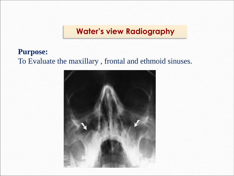
Purpose:
To Evaluate the maxillary , frontal and ethmoid sinuses.
Water’s view Radiography
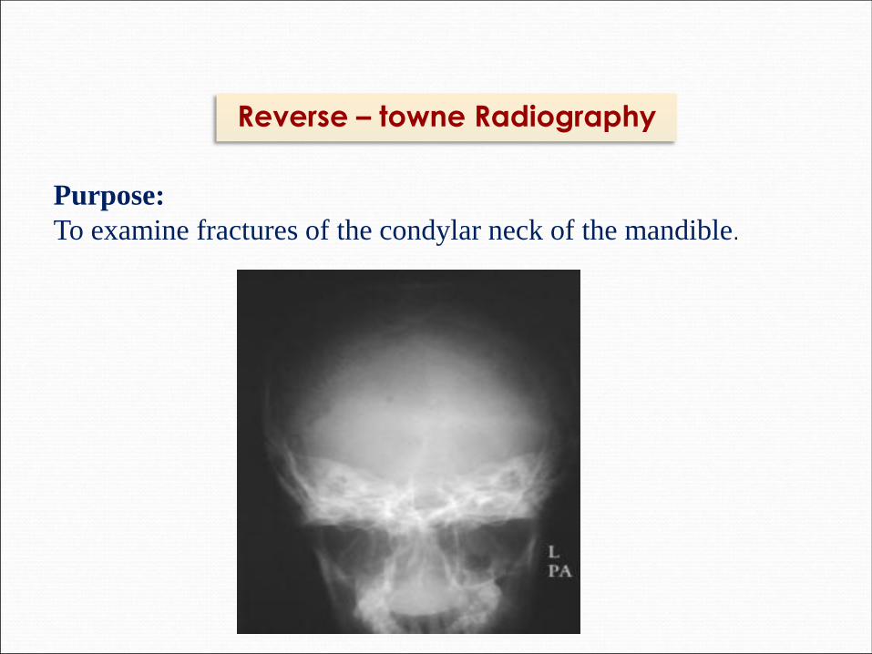
Purpose:
To examine fractures of the condylar neck of the mandible.
Reverse – towne Radiography
Purpose:
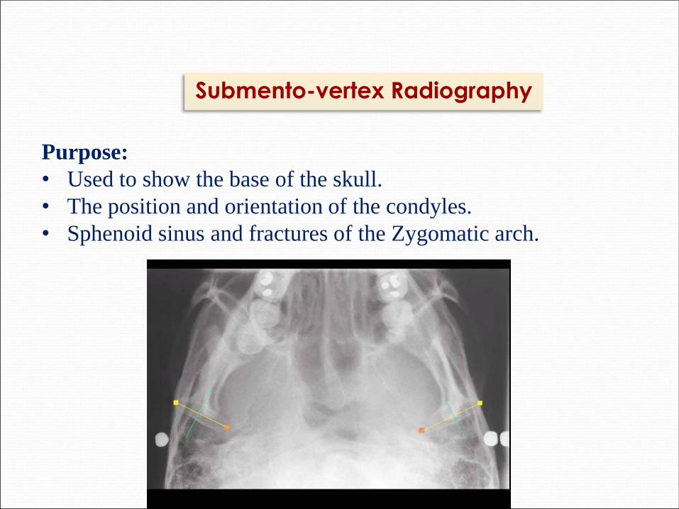
• Used to show the base of the skull.
• The position and orientation of the condyles.
• Sphenoid sinus and fractures of the Zygomatic arch.
Submento-vertex Radiography

• Tomograms
• Sialography
• Computed tomography
• MRI
• Ultrasonography (USG)
• Cone beam CT
Advanced Radiographic Techniques

Show a particular layer or "slice" of the mouth while blurring
out all other layers. This type of X-ray is useful for examining
structures that are difficult to clearly see … for instance,
because other structures are in very close proximity to the
structure to be viewed .
Tomograms

Involves visualization of the salivary glands following the
injection of a dye. The dye, called a radiopaque contrast
agent, is injected into the salivary glands so that the organ can
be seen on the X-ray film (the organ is a soft tissue that would
not otherwise be seen with an X-ray). Dentists might order this
type of test to look for salivary gland problems, such as
blockages or Sjogren's syndrome.
Sialography

Otherwise known as CT scanning, shows the body's interior
structures as a three-dimensional image. This type of X-ray is
performed in a hospital rather than a dentist's office.
Indications:
The diagnosis and extent of -
Variety of infections
Osteomyelitis
Cysts
Benign and malignant tumors
Trauma in the maxillofacial region
Lesions involving the bone
3D CT has been applied to trauma and craniofacial reconstructive surgery and used
for treatment of congenital and acquired deformities
Computed tomography

Indications:
To evaluate the position and integrity of the disk in the TMJ.
Neoplasia involving the soft tissues, such as tongue, cheek, salivary glands,
and neck.
Determining malignant involvement of lymphnodes.
Determining peri-neural invasion by malignant neoplasms.
With contrast, enhances the image resolution of neoplasia.
MRI

Indications:
For the evaluation of
Neoplasms in the thyroid, parathyroid or salivary glands or
lymph-nodes.
Stones in salivary glands or ducts
Vessels of neck
To guide fine-needle aspiration in the neck
Ultrasonography (USG)

A recent technology initially
developed for angiography in
1982 and subsequently applied to
maxillofacial imaging.
It constitutes of:
1. Two-dimensional digital array
providing an area detector fixed
on a rotating gantry
2. A three-dimensional (3D) cone
shaped x-ray beam
CONE-BEAM Computed
Tomography

PRINCIPLE :
Cone-beam scanners use a two-dimensional digital array
providing an area detector rather than a linear detector as CT
does.
This is combined with a three-dimensional (3D) x-ray beam
with circular collimation so that the resultant beam is in the
shape of a cone, hence the name "cone beam."
APPLICATIONS :
Orthodontic treatment planning
Dental implants
Temporomandibular joints for osseous degenerative changes
Evaluation of wisdom teeth vs. mandibular nerve
Endodontic assessment
Assessment of impacted teeth, fractured teeth and jaws,
periapical infections and periodontal diseases.
Benign calcifications ( tonsilloliths, lymph-nodes, salivary
gland stones) can also be identified.

C B C T - ORTHO ,,,, Cephalo Tracing

C B C T ,,, Nerve Mapping

CBCT- TMJ view

ADVANTAGES:
Lower dose than helical
Compact design
Superior images to Panoramic
Low cost
Low heat load
High speed scanning (less than 30 secs)
DISADVANTAGES:
Worse low contrast detectability
Poor soft tissue contrast
Long scan times = motion artifacts
Slightly Inferior quality to conventional CT
Image noise
Metal artifacts

Dental radiographs produced with a special computer create
digital images (computerized dental radiographs) that can be
displayed and enhanced on the computer monitor.
It involves the use of a radiography machine like that used for
conventional x-rays. But instead of using films, the clinician
makes digital images using a small electronic sensor or an
image receptor placed in mouth to capture the image.
Digital Radiography


•DOSE REDUCTION
• IMAGE MANIPULATION
• TIME
• STORAGE
• TELERADIOLOGY
• ENVIRONMENTALLY FRIENDLY
ADVANTAGES
•COST
•CROSS-INFECTION CONTROL DISADVANTAGES


