Embed Size (px)
Citation preview
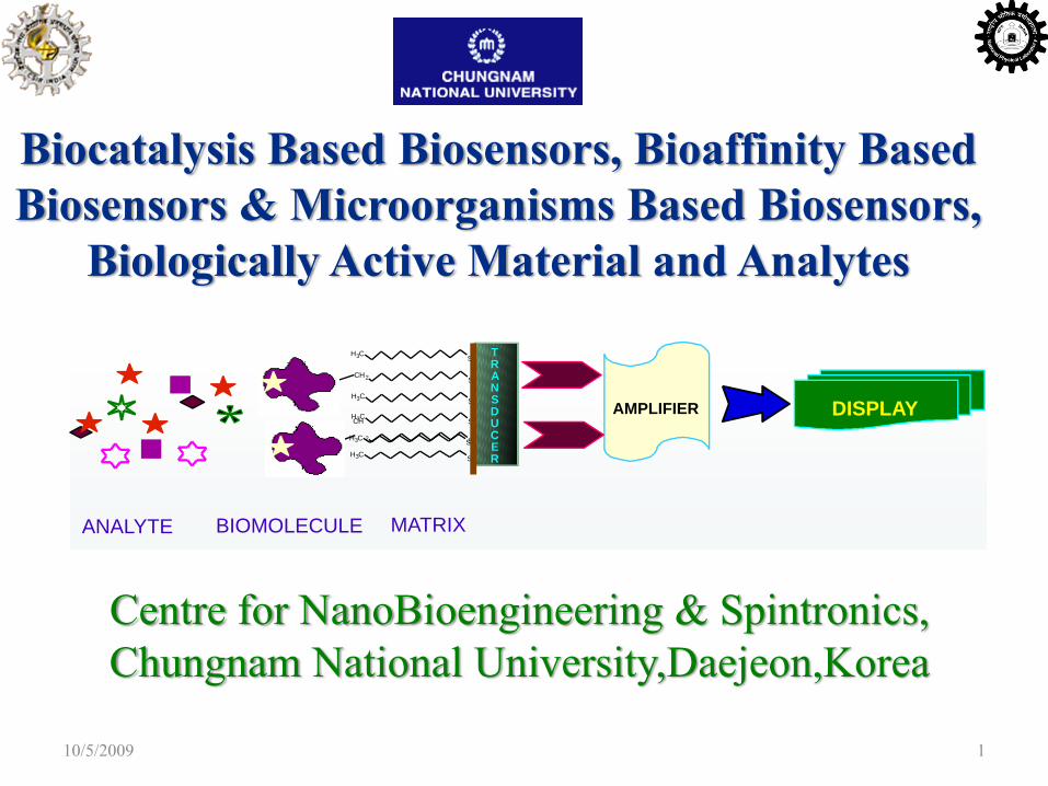
Biocatalysis Based Biosensors, Bioaffinity Based Biosensors & Microorganisms Based Biosensors,
Biologically Active Material and Analytes
TRANSDUCER
AMPLIFIER DISPLAY
CH3 S
CH3 S
CH
2 S
CH3 S
CH3 S
CH2 S
CH3 S
MATRIXBIOMOLECULEANALYTE
Centre for NanoBioengineering & Spintronics, Chungnam National University,Daejeon,Korea
10/5/2009 1
10/5/2009 WCU Project, CNU,[email protected] 2
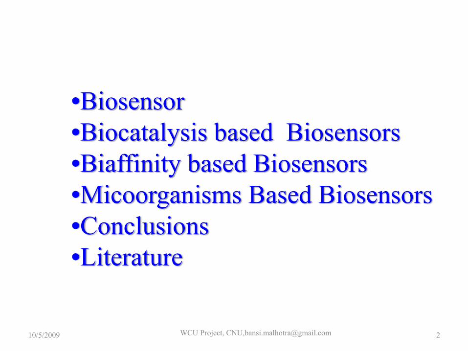
•Biosensor•Biocatalysis based Biosensors•Biaffinity based Biosensors•Micoorganisms Based Biosensors•Conclusions•Literature
BIOSENSOR
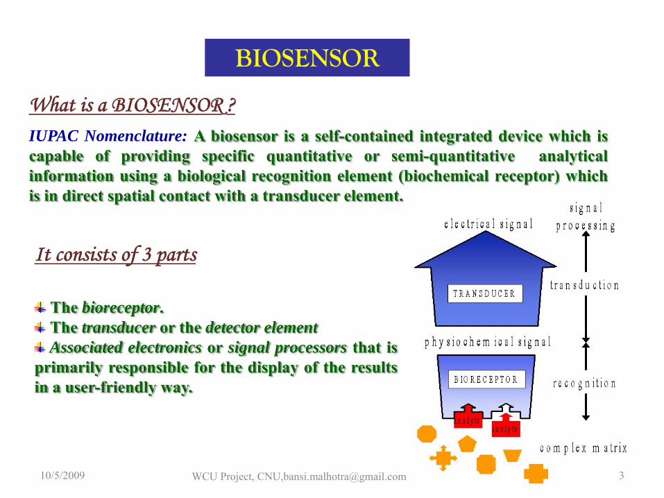
IUPAC Nomenclature: A biosensor is a self-contained integrated device which iscapable of providing specific quantitative or semi-quantitative analyticalinformation using a biological recognition element (biochemical receptor) whichis in direct spatial contact with a transducer element.
What is a BIOSENSOR ?
The bioreceptor.The transducer or the detector elementAssociated electronics or signal processors that is
primarily responsible for the display of the resultsin a user-friendly way.
It consists of 3 parts
10/5/2009 WCU Project, CNU,[email protected] 3
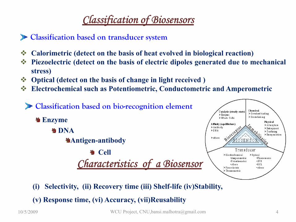
Calorimetric (detect on the basis of heat evolved in biological reaction)Piezoelectric (detect on the basis of electric dipoles generated due to mechanicalstress)Optical (detect on the basis of change in light received )Electrochemical such as Potentiometric, Conductometric and Amperometric
Classification of Biosensors
Characteristics of a Biosensor
Classification based on transducer system
Classification based on bio-recognition element
Antigen-antibody
(i) Selectivity, (ii) Recovery time (iii) Shelf-life (iv)Stability,
(v) Response time, (vi) Accuracy, (vii)Reusability
EnzymeDNA
Cell
10/5/2009 WCU Project, CNU,[email protected] 4
Biocatalysis based sensor
Biocatalysis-based biosensors depend universally on the use of enzymes.
The field of biocatalysis is open. This frontier of research is racingahead, propelled by advances in the database-supported analysis ofsequences and structures as well as the designability of genes &proteins.
Biocatalytic processes differ from conventional chemical processes, owingmainly to enzyme kinetics, protein stability under technical conditions andcatalyst features that derive from their role in the cell’s physiology, such asgrowth, induction of enzyme activity or the use of metabolic pathways formultistep reactions.
10/5/2009 WCU Project, CNU,[email protected] 5
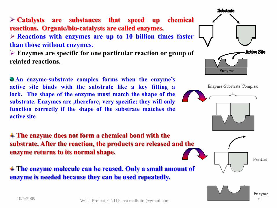
Catalysts are substances that speed up chemicalreactions. Organic/bio-catalysts are called enzymes.
Reactions with enzymes are up to 10 billion times fasterthan those without enzymes.
Enzymes are specific for one particular reaction or group ofrelated reactions.
An enzyme-substrate complex forms when the enzyme’sactive site binds with the substrate like a key fitting alock. The shape of the enzyme must match the shape of thesubstrate. Enzymes are ,therefore, very specific; they will onlyfunction correctly if the shape of the substrate matches theactive site
The enzyme does not form a chemical bond with the substrate. After the reaction, the products are released and the enzyme returns to its normal shape.
The enzyme molecule can be reused. Only a small amount of enzyme is needed because they can be used repeatedly.
10/5/2009 WCU Project, CNU,[email protected] 6
Effect of Temperature
Increase in the temperature of a system results from increases in the kinetic energy of the system. This has several effects on the rates of reactions.
1. More energetic collisionsThe greater the kinetic energy of the molecules in a system, the greater is the resulting chemical potential energy when two molecules collide .
2 The number of collisions per unit time will increase.
In order to convert substrate into product, enzymes must collide with and bind to the substrate atthe active site. Increasing the temperature of a system will increase the number of collisions ofenzyme and substrate per unit time. Thus, within limits, the rate of the reaction will increase.
3 The heat of the molecules in the system will increase As the temperature of the system is increased, internal energy of the molecules in the system will increase.The internal energy of the molecules may include the translational energy, vibrational energy and rotationalenergy of the molecules. Some of this may be converted into chemical potential energy. If this chemicalpotential energy increase is great enough , some of the weak bonds that determine the three dimensional shapeof the active proteins may be broken. This could lead to a thermal denaturation of the protein and thusinactivate the protein. Thus too much heat can cause the rate of an enzyme catalyzed reaction to decreasebecause the enzyme or substrate becomes denatured and inactive.
10/5/2009 WCU Project, CNU,[email protected] 7
pH can affect the ionization of the amino acid side chain, which in turn change the secondary, tertiary and quaternary structures of the protein molecule. This will change the enzyme's active site and consequently its activity.
Effect of pH
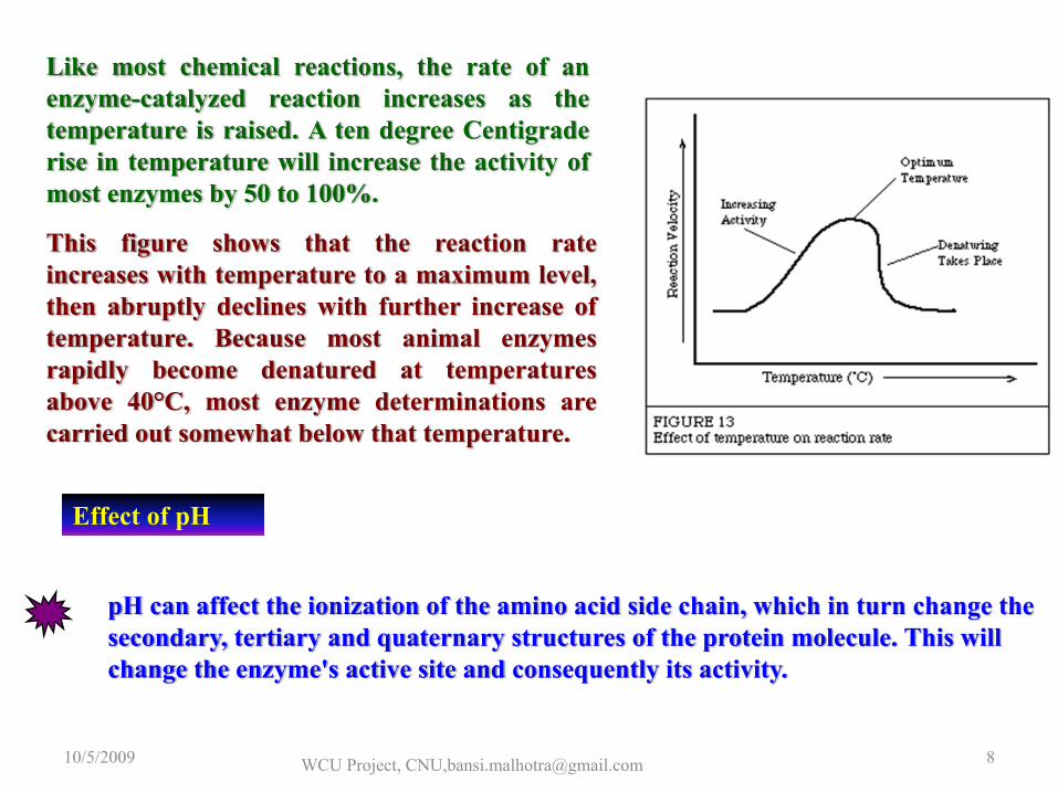
Like most chemical reactions, the rate of anenzyme-catalyzed reaction increases as thetemperature is raised. A ten degree Centigraderise in temperature will increase the activity ofmost enzymes by 50 to 100%.
This figure shows that the reaction rateincreases with temperature to a maximum level,then abruptly declines with further increase oftemperature. Because most animal enzymesrapidly become denatured at temperaturesabove 40°C, most enzyme determinations arecarried out somewhat below that temperature.
10/5/2009 WCU Project, CNU,[email protected] 8
Enzyme ClassificationThere are approximately 3000 known enzymes. These enzymes are classified into six categories based on the type of reaction they catalyze.
1. Oxido- reductase: Oxidizes or reduces by transfer of hydrogen or electrons.(a) Dehydrogenases:SH2 + A ↔ S + AH2 (S: Substrate, A: acceptor)Example:Lactate dehydrogenase: L-lactate + NAD ↔ Pyruvate + NADH + H+
(b) Oxidases:SH2 + 1/2 O2 → S + H2O orSH2 + O2 → S + H2O2Example:Glucose oxidase: β-D-glucose + O2 → Gluconolactone + H2O2
(c) Peroxidases:2SH + H2O2 → 2S + 2H2O or2S + 2H+ + H2O2 → 2S+ + 2H2OExample: Horse radish peroxidase:2[Fe(CN)6]4- + 2H+ → 2[Fe(CN)6]3- + 2H2O
(d) Oxygenases:SH + DH + O2 → S-OH + D + H2OExample: Lactate 2-monooxygenase:L-lactate + O2 → acetate + CO2 + H2O
10/5/2009 WCU Project, CNU,[email protected] 9
2. Transferase: Transfers C-, N-, P-, or S-containing functional groups such as aldehydesand ketones, glycosils, acyls, phosphates, and sulfur containing groups.
AX + B ↔ A + BXExample: Hexokinase: D-hexose + ATP ↔ D-hexose-6-phosphate + ADP
3. Hydrolase: Hydrolyses esters, anhydrides, peptide bonds, other C-Nbonds, glycosidesExample: Cholesterol esterase:Cholesterol ester + H2O → cholesterol + fatty acidGlucoamylase:Amylose + n H2O → n β-D-glucose
4. Lyase: Adds to double bonds:
> C = C <> C = O> C = N
5. Isomerase: Isomerizes optical iomersExampleGlucose isomerase: D-glucose ↔ D-fructose
10/5/2009 WCU Project, CNU,[email protected] 10
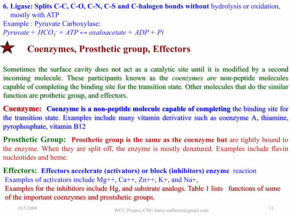
6. Ligase: Splits C-C, C-O, C-N, C-S and C-halogen bonds without hydrolysis or oxidation, mostly with ATP
Example : Pyruvate Carboxylase:Pyruvate + HCO3
- + ATP ↔ oxaloacetate + ADP + Pi
Coenzymes, Prosthetic group, Effectors
Sometimes the surface cavity does not act as a catalytic site until it is modified by a secondincoming molecule. These participants known as the coenzymes are non-peptide moleculescapable of completing the binding site for the transition state. Other molecules that do the similarfunction are prothetic group, and effectors.
Coenzyme: Coenzyme is a non-peptide molecule capable of completing the binding site forthe transition state. Examples include many vitamin derivative such as coenzyme A, thiamine,pyrophosphate, vitamin B12
Prosthetic Group: Prosthetic group is the same as the coenzyme but are tightly bound tothe enzyme. When they are split off, the enzyme is mostly denatured. Examples include flavinnucleotides and heme.
Effectors: Effectors accelerate (activators) or block (inhibitors) enzyme reactionExamples of activators include Mg++, Ca++, Zn++, K+, and Na+,Examples for the inhibitors include Hg, and substrate analogs. Table 1 lists functions of someof the important coenzymes and prostshetic groups.
10/5/2009 WCU Project, CNU,[email protected] 11
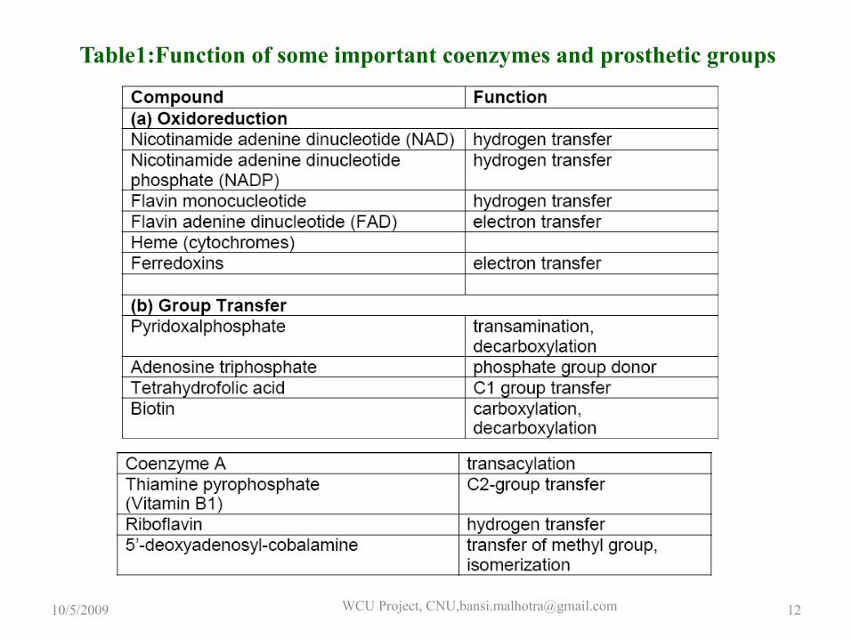
Table1:Function of some important coenzymes and prosthetic groups
10/5/2009 WCU Project, CNU,[email protected] 12
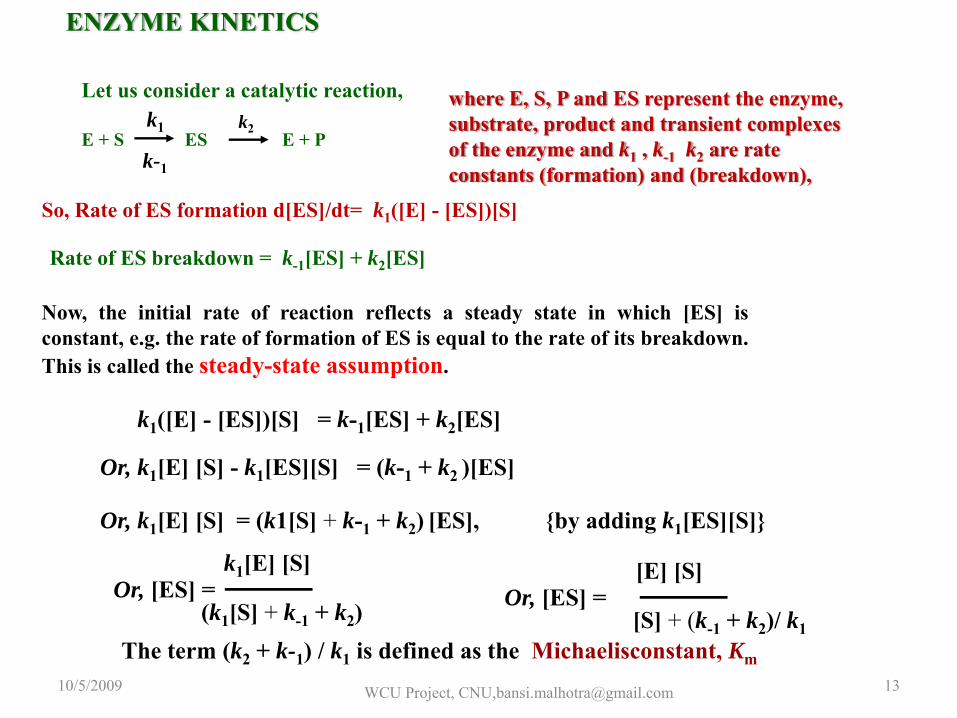
Let us consider a catalytic reaction,
E + S ES E + Pk1 k2
k-1
where E, S, P and ES represent the enzyme, substrate, product and transient complexes of the enzyme and k1 , k-1 k2 are rate constants (formation) and (breakdown),
So, Rate of ES formation d[ES]/dt= k1([E] - [ES])[S]
Rate of ES breakdown = k-1[ES] + k2[ES]
Now, the initial rate of reaction reflects a steady state in which [ES] isconstant, e.g. the rate of formation of ES is equal to the rate of its breakdown.This is called the steady-state assumption.
k1([E] - [ES])[S] = k-1[ES] + k2[ES]
Or, k1[E] [S] - k1[ES][S] = (k-1 + k2 )[ES]
Or, k1[E] [S] = (k1[S] + k-1 + k2) [ES], {by adding k1[ES][S]}
Or, [ES] =(k1[S] + k-1 + k2)
k1[E] [S]Or, [ES] =
[S] + (k-1 + k2)/ k1
[E] [S]
The term (k2 + k-1) / k1 is defined as the Michaelisconstant, Km
ENZYME KINETICS
10/5/2009 WCU Project, CNU,[email protected] 13
Assumptions
10/5/2009 WCU Project, CNU,[email protected] 14
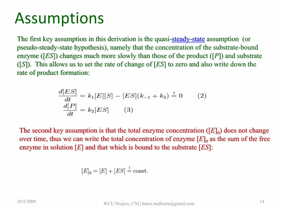
The first key assumption in this derivation is the quasi-steady-state assumption (or pseudo-steady-state hypothesis), namely that the concentration of the substrate-bound enzyme ([ES]) changes much more slowly than those of the product ([P]) and substrate ([S]). This allows us to set the rate of change of [ES] to zero and also write down the rate of product formation:
The second key assumption is that the total enzyme concentration ([E]0) does not change over time, thus we can write the total concentration of enzyme [E]0 as the sum of the free enzyme in solution [E] and that which is bound to the substrate [ES]:
The validity of the following derivation rests on the reaction Scheme givenbelow and two key assumptions: that the total enzyme concentration andthe concentration of the intermediate complex do not change over time.
The most convenient derivation of the Michaelis–Menten equation,described by Briggs andHaldane, is obtained as follows (Note that often the experimentalparameter kcat is used but in this simple case it is equal tothe kinetic parameter k2):
The enzymatic reaction is assumed to be irreversible, and the product doesnot bind to the enzyme.
WCU Project, CNU,[email protected]
GLUCOSE
CHOLESTEROL
UREA
Biocatalysis based Biosensor at NPL
10/5/2009 WCU Project, CNU,[email protected] 16
GLUCOSE SENSOR
GLUCOSE + GLUCOSE OXIDASEOXIDIZED ↓
PRODUCT +GLUCOSE OXIDASE REDUCED
GLUCOSE OXIDASEREDUCED +MEDIATOROXIDIZED
↓MEDIATORREDUCED + GLUCOSE
OXIDASEOXIDIZED + MEDIATOR REDUCED
↓MEDIATOROXIDIZED+2e-
10/5/2009WCU Project, CNU,[email protected]
17
Glucose Oxidase and the biochemical reactions involved during the Glucose sensing
The enzymatic reaction catalysed by glucose oxidase (GOx)
Structure of glucose oxidase
Active site structure of GOx enzyme

Polythiophene Gold Nanoparticles
Composite
Iron OxideNanoparticles-Chitosan Composite
Au nanoparticle/ PolyanilineComposite
Au-nanoparticles/ PolypyrroleComposite
Matrices for Glucose
biosensor
10/5/2009 WCU Project, CNU,[email protected] 19
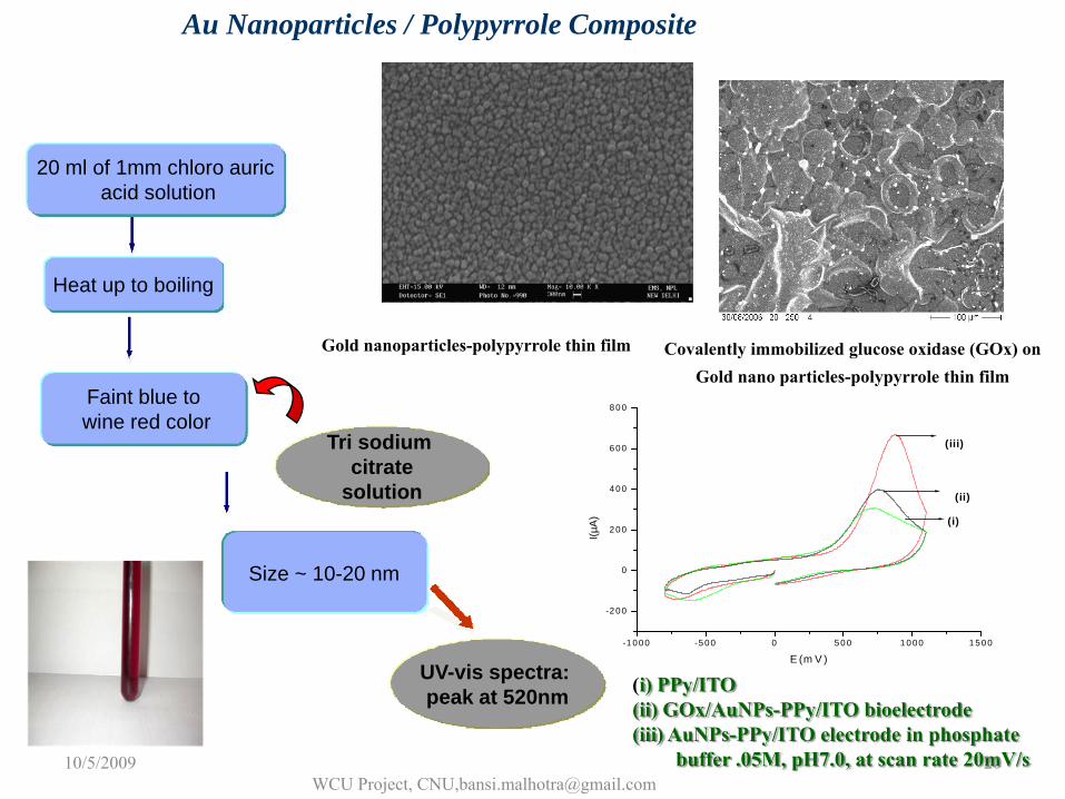
Gold nanoparticles-polypyrrole thin film Covalently immobilized glucose oxidase (GOx) on Gold nano particles-polypyrrole thin film
20 ml of 1mm chloro auricacid solution
Heat up to boiling
Tri sodium citrate
solution
Faint blue towine red color
Size ~ 10-20 nm
UV-vis spectra: peak at 520nm
-1 0 00 -5 00 0 5 0 0 1 0 00 1 5 00
-2 00
0
2 00
4 00
6 00
8 00
(iii)
(ii)
(i)
I(µA
)
E (m V )
(i) PPy/ITO(ii) GOx/AuNPs-PPy/ITO bioelectrode(iii) AuNPs-PPy/ITO electrode in phosphate
buffer .05M, pH7.0, at scan rate 20mV/s
Au Nanoparticles / Polypyrrole Composite
10/5/2009WCU Project, CNU,[email protected]
20
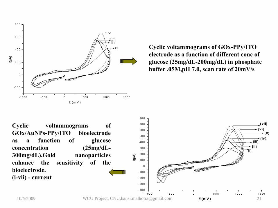
Cyclic voltammograms ofGOx/AuNPs-PPy/ITO bioelectrodeas a function of glucoseconcentration (25mg/dL-300mg/dL).Gold nanoparticlesenhance the sensitivity of thebioelectrode.(i-vii) - current
Cyclic voltammograms of GOx-PPy/ITO electrode as a function of different conc of glucose (25mg/dL-200mg/dL) in phosphate buffer .05M,pH 7.0, scan rate of 20mV/s
10/5/2009 WCU Project, CNU,[email protected] 21
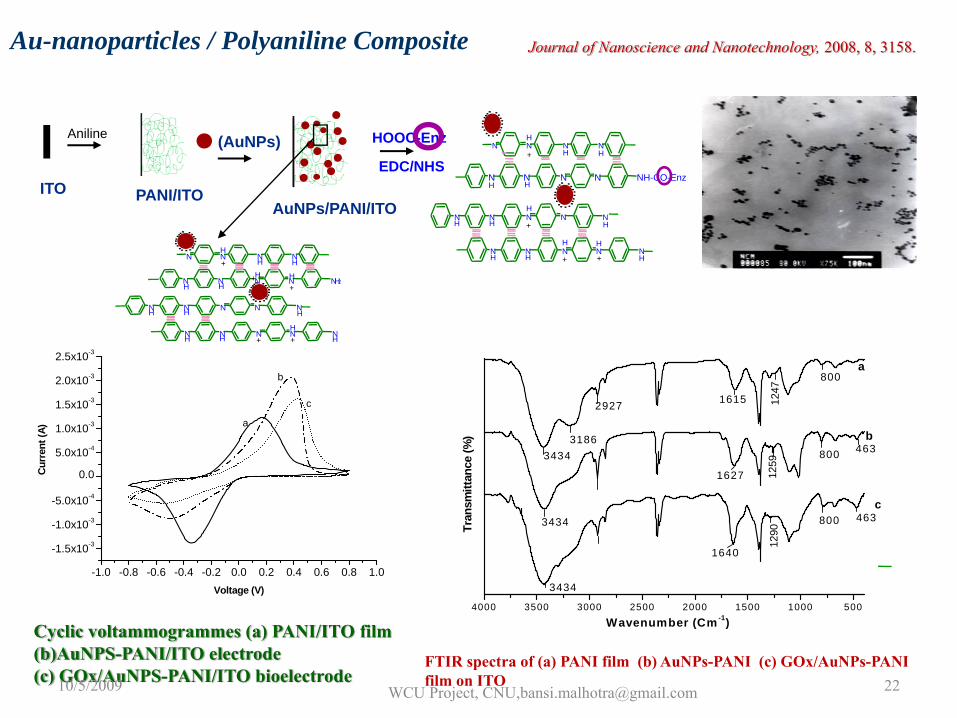
Aniline
ITO
(AuNPs)
PANI/ITOAuNPs/PANI/ITO
N N N N NH-CO-EnzH H
N N N N NH H
H
+ H
N N N N NH H
H H
+ + H
HH
H
+NNNN
N N N N NH2H H
H H++
N N N N NH H H
N N N N NH HH
+ + H
HHH
+NNNN
EDC/NHS
HOOC-Enz
Au-nanoparticles / Polyaniline Composite
4000 3500 3000 2500 2000 1500 1000 500
b
c
a
2927
1290
1259
1247
1615
1627
1640
800
800
800
3186
3434
3434
3434
463
463
Tran
smitt
ance
(%)
Wavenumber (Cm -1)
FTIR spectra of (a) PANI film (b) AuNPs-PANI (c) GOx/AuNPs-PANI film on ITO
-1.0 -0.8 -0.6 -0.4 -0.2 0.0 0.2 0.4 0.6 0.8 1.0
-1.5x10⁻³
-1.0x10⁻³
-5.0x10⁻⁴
0.0
5.0x10⁻⁴
1.0x10⁻³
1.5x10⁻³
2.0x10⁻³
2.5x10⁻³
c
b
a
Curr
ent (
A)
Voltage (V)
Cyclic voltammogrammes (a) PANI/ITO film(b)AuNPS-PANI/ITO electrode(c) GOx/AuNPS-PANI/ITO bioelectrode
Journal of Nanoscience and Nanotechnology, 2008, 8, 3158.
10/5/2009 WCU Project, CNU,[email protected] 22
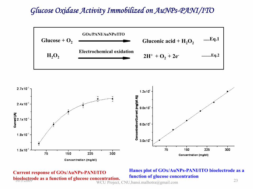
Glucose Oxidase Activity Immobilized on AuNPs-PANI/ITO
Current response of GOx/AuNPs-PANI/ITO bioelectrode as a function of glucose concentration.
Hanes plot of GOx/AuNPs-PANI/ITO bioelectrode as a function of glucose concentration
GOx/PANI/AuNPs/ITOGlucose + O2 Gluconic acid + H2O2
.....Eq.1
Electrochemical oxidation2H+ + O2 + 2e-- .........Eq.2H2O2
10/5/2009 WCU Project, CNU,[email protected] 23
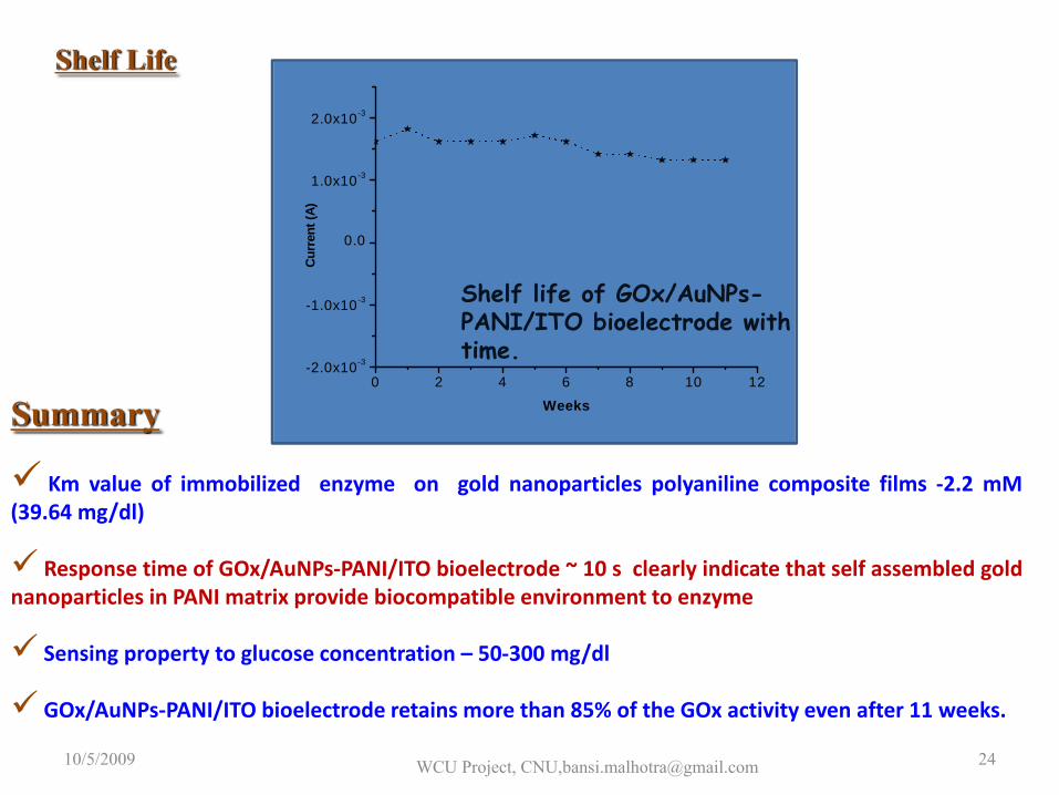
Summary
Km value of immobilized enzyme on gold nanoparticles polyaniline composite films ‐2.2 mM(39.64 mg/dl)
Response time of GOx/AuNPs‐PANI/ITO bioelectrode ~ 10 s clearly indicate that self assembled goldnanoparticles in PANI matrix provide biocompatible environment to enzyme
Sensing property to glucose concentration – 50‐300 mg/dl
GOx/AuNPs‐PANI/ITO bioelectrode retains more than 85% of the GOx activity even after 11 weeks.
0 2 4 6 8 10 12-2.0x10⁻³
-1.0x10⁻³
0.0
1.0x10⁻³
2.0x10⁻³
Curr
ent (
A)
Weeks
Shelf Life
Shelf life of GOx/AuNPs-PANI/ITO bioelectrode with time.
10/5/2009 WCU Project, CNU,[email protected] 24
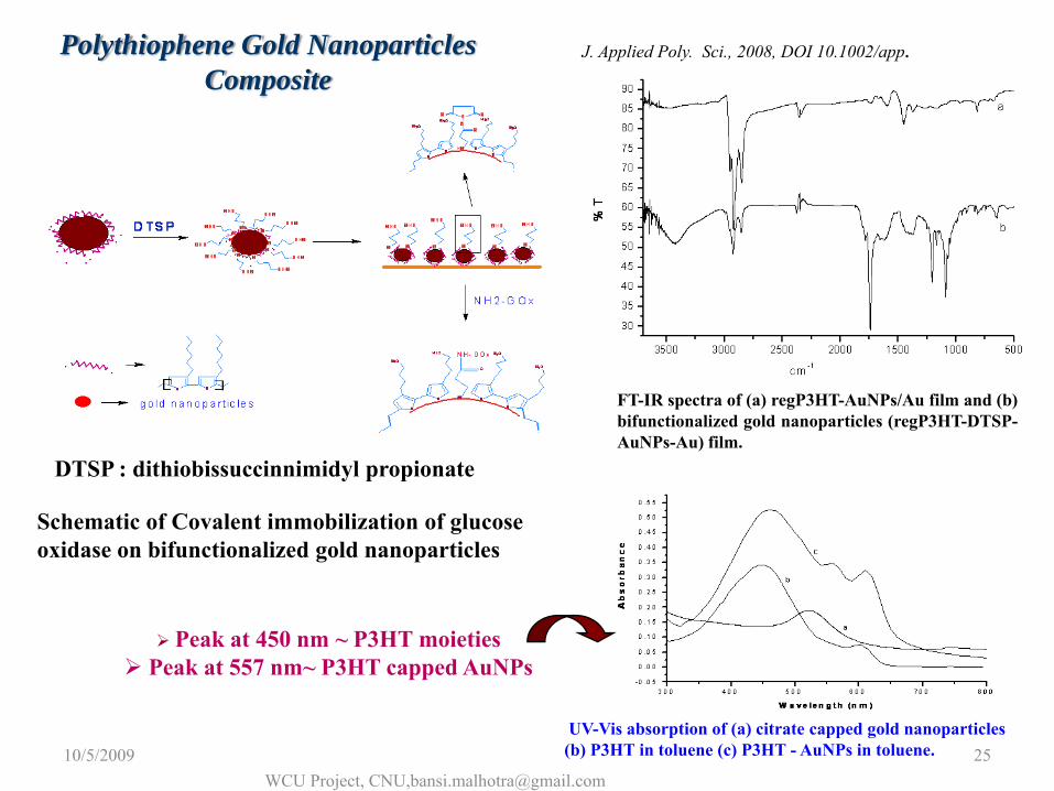
Polythiophene Gold NanoparticlesComposite
DTSP : dithiobissuccinnimidyl propionate
Schematic of Covalent immobilization of glucose oxidase on bifunctionalized gold nanoparticles
FT-IR spectra of (a) regP3HT-AuNPs/Au film and (b)bifunctionalized gold nanoparticles (regP3HT-DTSP-AuNPs-Au) film.
UV-Vis absorption of (a) citrate capped gold nanoparticles(b) P3HT in toluene (c) P3HT - AuNPs in toluene.
Peak at 450 nm ~ P3HT moietiesPeak at 557 nm~ P3HT capped AuNPs
J. Applied Poly. Sci., 2008, DOI 10.1002/app.
10/5/2009WCU Project, CNU,[email protected]
25
0 50 100 150 200 250 300 350 400 4500.002
0.004
0.006
0.008
0.010
0.012
0.014
Abso
rban
ce (5
00 n
m)
Conc (mg/dL)
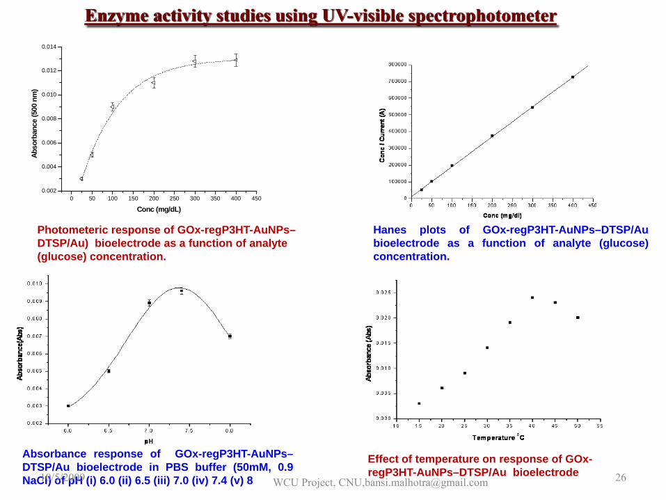
Photometeric response of GOx-regP3HT-AuNPs–DTSP/Au) bioelectrode as a function of analyte(glucose) concentration.
Hanes plots of GOx-regP3HT-AuNPs–DTSP/Aubioelectrode as a function of analyte (glucose)concentration.
Enzyme activity studies using UV-visible spectrophotometer
Absorbance response of GOx-regP3HT-AuNPs–DTSP/Au bioelectrode in PBS buffer (50mM, 0.9NaCl) of pH (i) 6.0 (ii) 6.5 (iii) 7.0 (iv) 7.4 (v) 8
Effect of temperature on response of GOx-regP3HT-AuNPs–DTSP/Au bioelectrode10/5/2009 WCU Project, CNU,[email protected] 26
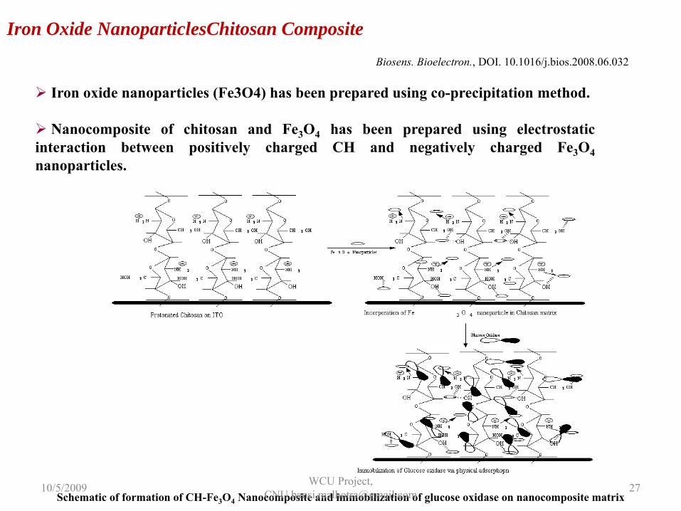
Iron Oxide NanoparticlesChitosan Composite
Iron oxide nanoparticles (Fe3O4) has been prepared using co-precipitation method.
Nanocomposite of chitosan and Fe3O4 has been prepared using electrostaticinteraction between positively charged CH and negatively charged Fe3O4nanoparticles.
Schematic of formation of CH-Fe3O4 Nanocomposite and immobilization of glucose oxidase on nanocomposite matrix
Biosens. Bioelectron., DOI. 10.1016/j.bios.2008.06.032
10/5/2009 WCU Project, CNU,[email protected] 27
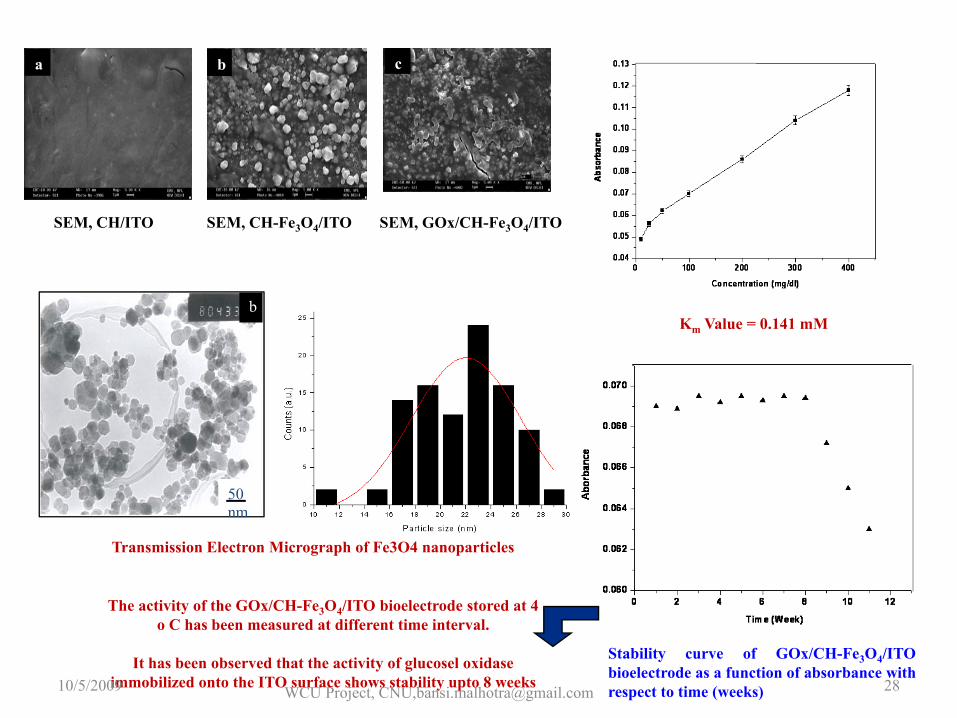
a b c
SEM, CH/ITO SEM, CH-Fe3O4/ITO SEM, GOx/CH-Fe3O4/ITO
50 nm
b
Transmission Electron Micrograph of Fe3O4 nanoparticles
Km Value = 0.141 mM
Stability curve of GOx/CH-Fe3O4/ITObioelectrode as a function of absorbance withrespect to time (weeks)
The activity of the GOx/CH-Fe3O4/ITO bioelectrode stored at 4 o C has been measured at different time interval.
It has been observed that the activity of glucosel oxidaseimmobilized onto the ITO surface shows stability upto 8 weeks10/5/2009 WCU Project, CNU,[email protected] 28
10/5/2009 WCU Project, CNU,[email protected] 29
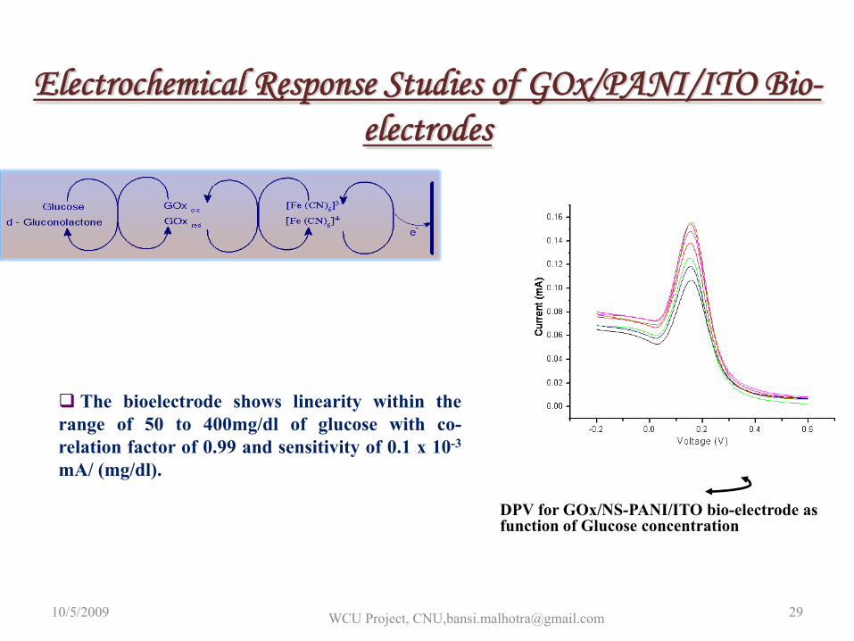
The bioelectrode shows linearity within therange of 50 to 400mg/dl of glucose with co-relation factor of 0.99 and sensitivity of 0.1 x 10⁻³
mA/ (mg/dl).
Electrochemical Response Studies of GOx/PANI/ITO Bio-electrodes
DPV for GOx/NS-PANI/ITO bio-electrode as function of Glucose concentration
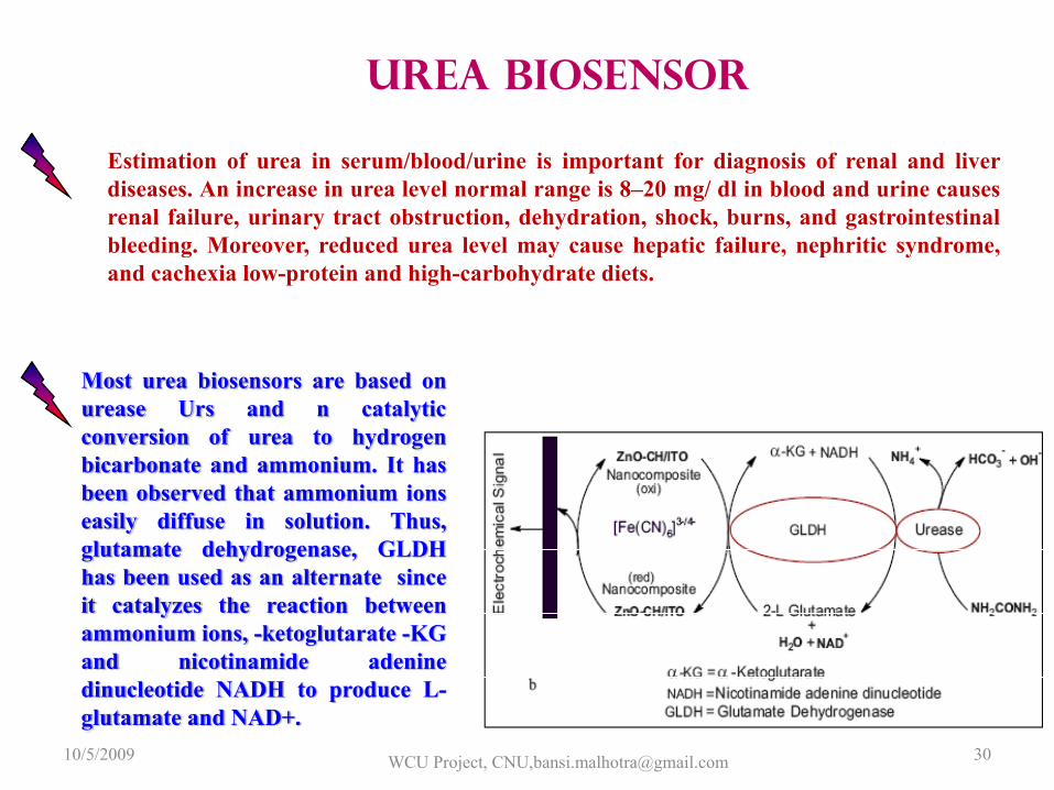
Most urea biosensors are based onurease Urs and n catalyticconversion of urea to hydrogenbicarbonate and ammonium. It hasbeen observed that ammonium ionseasily diffuse in solution. Thus,glutamate dehydrogenase, GLDHhas been used as an alternate sinceit catalyzes the reaction betweenammonium ions, -ketoglutarate -KGand nicotinamide adeninedinucleotide NADH to produce L-glutamate and NAD+.
Estimation of urea in serum/blood/urine is important for diagnosis of renal and liverdiseases. An increase in urea level normal range is 8–20 mg/ dl in blood and urine causesrenal failure, urinary tract obstruction, dehydration, shock, burns, and gastrointestinalbleeding. Moreover, reduced urea level may cause hepatic failure, nephritic syndrome,and cachexia low-protein and high-carbohydrate diets.
Urea Biosensor
10/5/2009 WCU Project, CNU,[email protected] 30
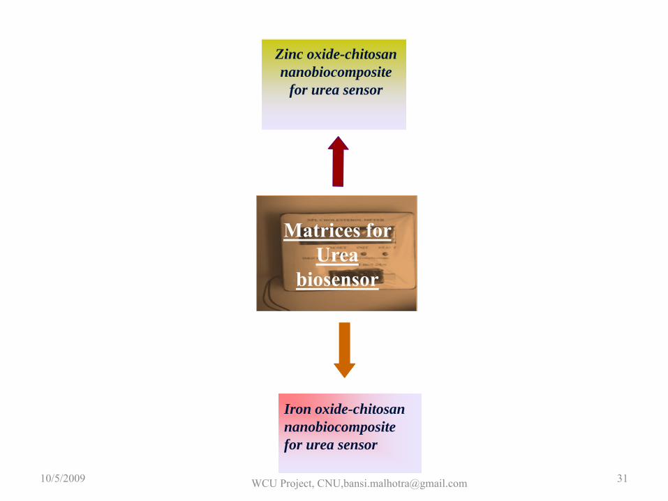
Iron oxide-chitosan nanobiocomposite for urea sensor
P3HT - SAM
Zinc oxide-chitosan nanobiocomposite
for urea sensor
Matrices for Urea
biosensor
10/5/2009 WCU Project, CNU,[email protected] 31
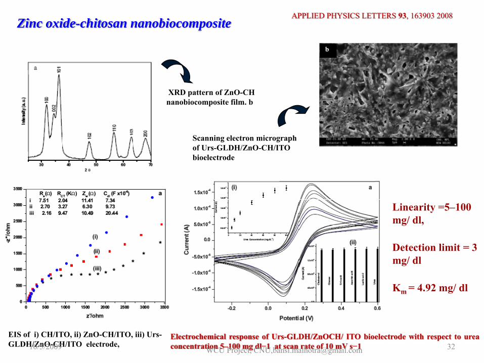
Zinc oxide-chitosan nanobiocomposite
XRD pattern of ZnO-CH nanobiocomposite film. b
Scanning electron micrographof Urs-GLDH/ZnO-CH/ITO bioelectrode
EIS of i) CH/ITO, ii) ZnO-CH/ITO, iii) Urs-GLDH/ZnO-CH/ITO electrode,
Electrochemical response of Urs-GLDH/ZnOCH/ ITO bioelectrode with respect to ureaconcentration 5–100 mg dl−1 at scan rate of 10 mV s−1
Km = 4.92 mg/ dl
Linearity =5–100 mg/ dl,
Detection limit = 3 mg/ dl
APPLIED PHYSICS LETTERS 93, 163903 2008
10/5/2009 WCU Project, CNU,[email protected] 32
Iron oxide-chitosan nanobiocomposite
X-ray diffraction pattern of Fe3O4nanoparticles.
Ur-GLDH/CH-Fe3O4nanobiocomposite/ITO electrode.
SEM images of CH-Fe3O4nanobiocomposite/ITO electrode
Sensors and Actuators B 138 (2009) 572–580
10/5/2009 WCU Project, CNU,[email protected] 33
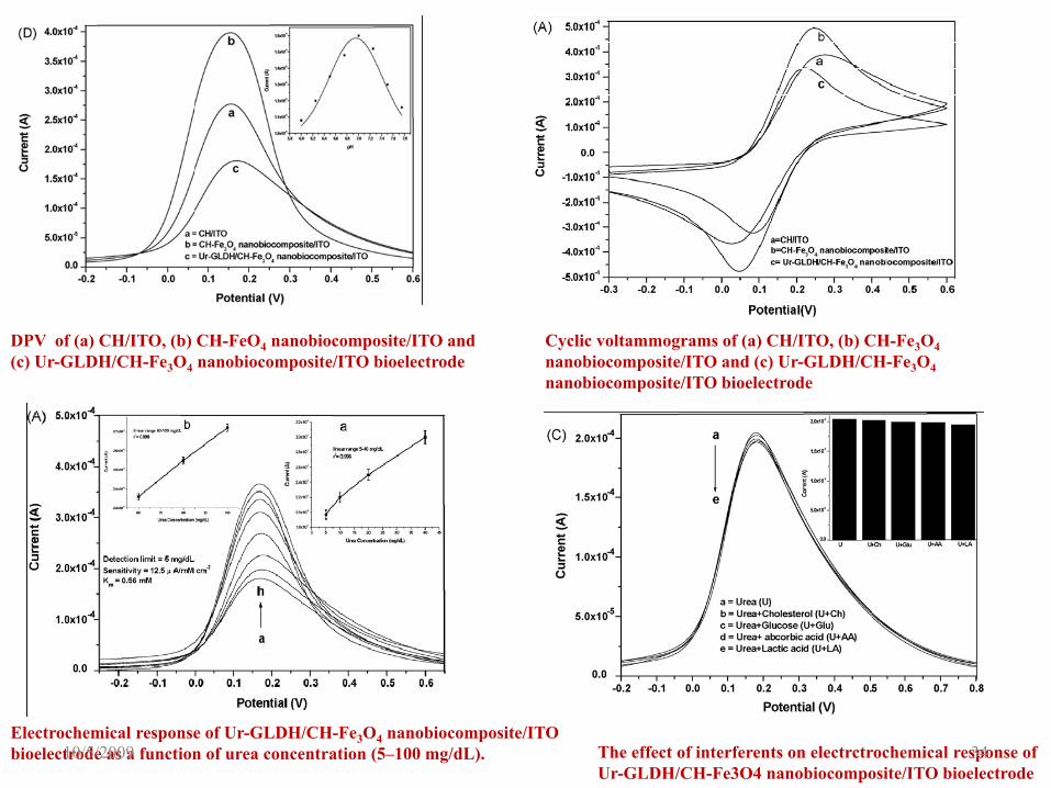
DPV of (a) CH/ITO, (b) CH-FeO4 nanobiocomposite/ITO and (c) Ur-GLDH/CH-Fe3O4 nanobiocomposite/ITO bioelectrode
Cyclic voltammograms of (a) CH/ITO, (b) CH-Fe3O4nanobiocomposite/ITO and (c) Ur-GLDH/CH-Fe3O4nanobiocomposite/ITO bioelectrode
Electrochemical response of Ur-GLDH/CH-Fe3O4 nanobiocomposite/ITO bioelectrode as a function of urea concentration (5–100 mg/dL). The effect of interferents on electrctrochemical response of
Ur-GLDH/CH-Fe3O4 nanobiocomposite/ITO bioelectrode10/5/2009 34
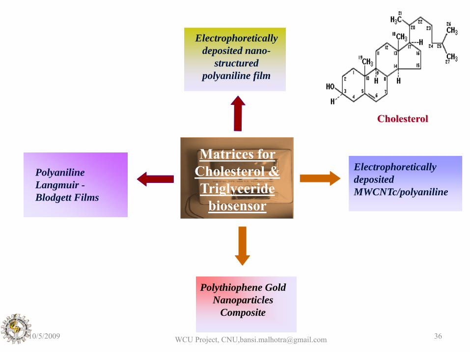
Polythiophene Gold Nanoparticles
Composite
P3HT - SAM
Polyaniline Langmuir -Blodgett Films
Electrophoretically deposited MWCNTc/polyaniline
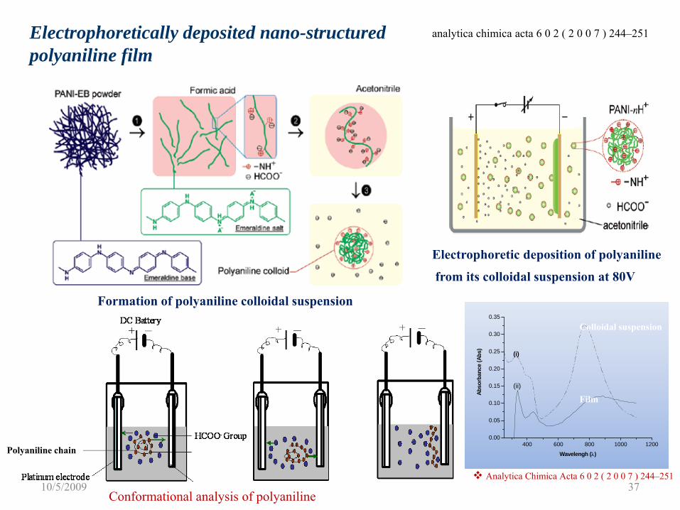
Electrophoretically deposited nano-
structured polyaniline film
Matrices for Cholesterol & Triglyceride
biosensor
Cholesterol
10/5/2009 WCU Project, CNU,[email protected] 36
400 600 800 1000 12000.00
0.05
0.10
0.15
0.20
0.25
0.30
0.35
(ii)
(i)
Abso
rban
ce (A
bs)
Wavelengh (λ)
Formation of polyaniline colloidal suspension
Electrophoretically deposited nano-structured polyaniline film
Analytica Chimica Acta 6 0 2 ( 2 0 0 7 ) 244–251
Electrophoretic deposition of polyaniline
from its colloidal suspension at 80V
Polyaniline chain
ITO
-NH+
Colloidal suspension
Film
Conformational analysis of polyaniline
analytica chimica acta 6 0 2 ( 2 0 0 7 ) 244–251
10/5/2009 37
Adduct-II
Adduct-I
NHS
Polyaniline
EDC Enzyme O
OHEnz CH3CH2-N=C=N-(CH2)3N(CH3)2
O
O
O
O
P NH2P NH
O
Amide bond formation
NEnzEnz
O
O NNH
N-(CH2)3N(CH3)2
-CH2CH3
O O
OHN
Enz
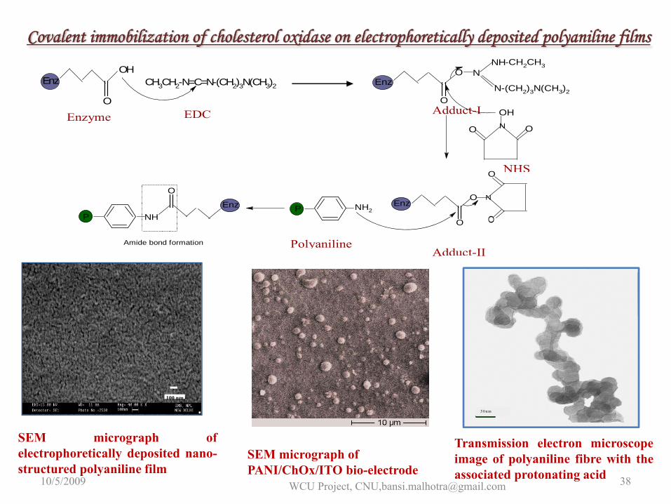
Covalent immobilization of cholesterol oxidase on electrophoretically deposited polyaniline films
5 0 nm
SEM micrograph of PANI/ChOx/ITO bio-electrode
Transmission electron microscopeimage of polyaniline fibre with theassociated protonating acid
100 nm
SEM micrograph ofelectrophoretically deposited nano-structured polyaniline film
10/5/2009 WCU Project, CNU,[email protected] 38
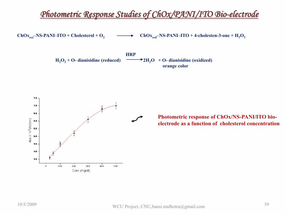
Photometric Response Studies of ChOx/PANI/ITO Bio-electrode
HRPH2O2 + O- dianisidine (reduced) 2H2O + O- dianisidine (oxidized)
orange color
Photometric response of ChOx/NS-PANI/ITO bio-electrode as a function of cholesterol concentration
ChOxoxi–NS-PANI–ITO + Cholesterol + O2 ChOxred–NS-PANI–ITO + 4-cholesten-3-one + H2O2
Optimum pH 6.5
10/5/2009 WCU Project, CNU,[email protected] 39
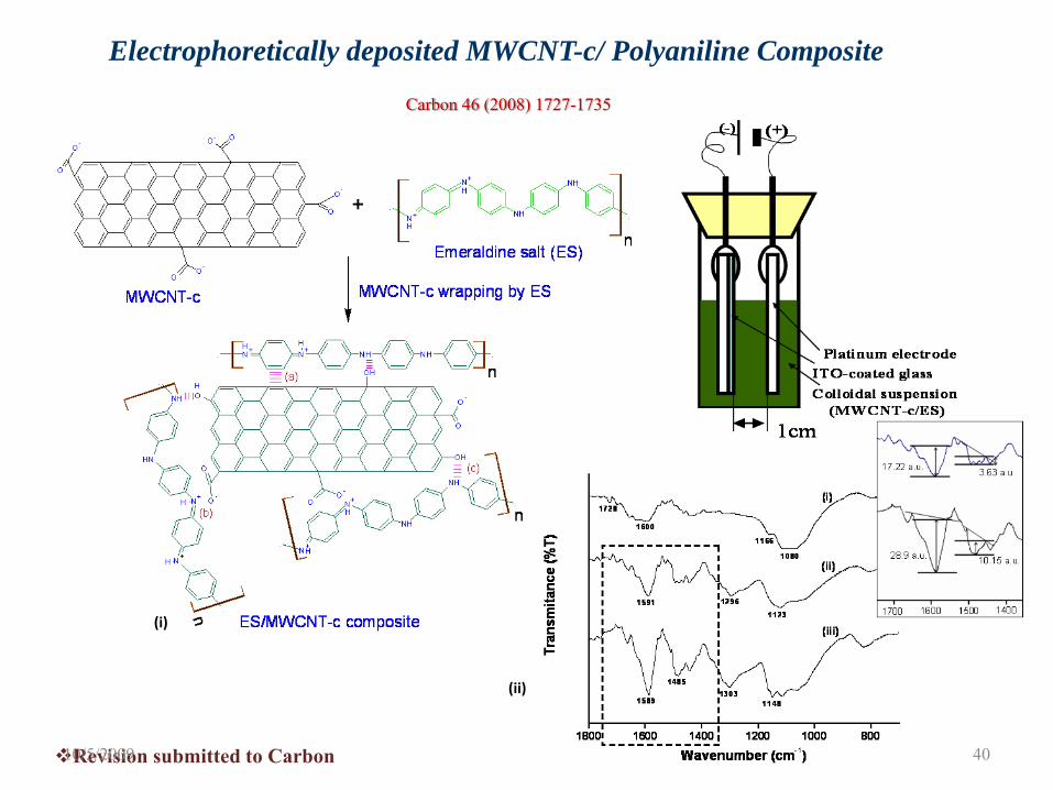
Electrophoretically deposited MWCNT-c/ Polyaniline Composite
(i)
(ii)
Revision submitted to Carbon
Carbon 46 (2008) 1727-1735
10/5/2009 40
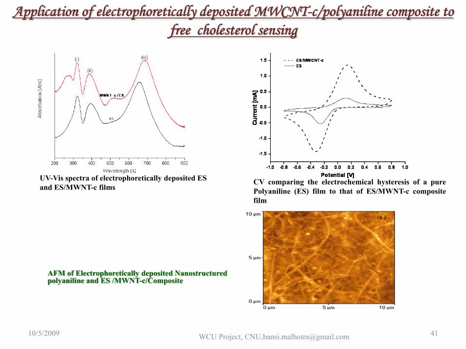
Application of electrophoretically deposited MWCNT-c/polyaniline composite to free cholesterol sensing
UV-Vis spectra of electrophoretically deposited ES and ES/MWNT-c films
AFM of Electrophoretically deposited Nanostructuredpolyaniline and ES /MWNT-c/Composite
CV comparing the electrochemical hysteresis of a purePolyaniline (ES) film to that of ES/MWNT-c compositefilm
10/5/2009 WCU Project, CNU,[email protected] 41
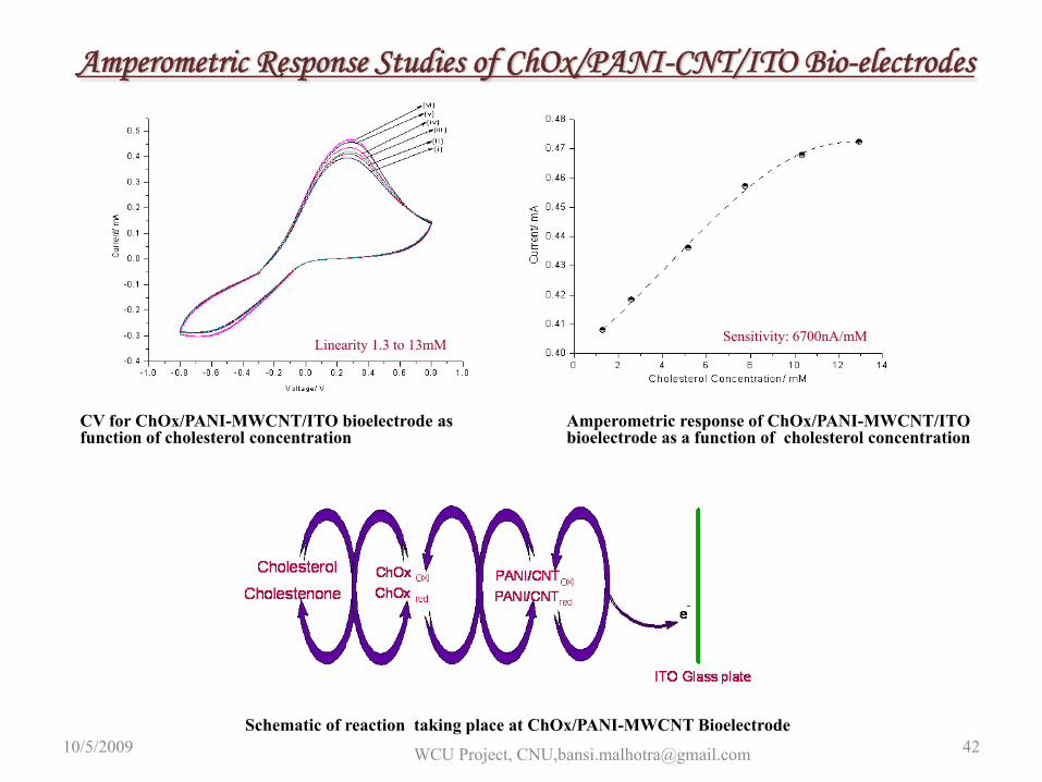
Amperometric Response Studies of ChOx/PANI-CNT/ITO Bio-electrodes
CV for ChOx/PANI-MWCNT/ITO bioelectrode as function of cholesterol concentration
Amperometric response of ChOx/PANI-MWCNT/ITO bioelectrode as a function of cholesterol concentration
Linearity 1.3 to 13mM Sensitivity: 6700nA/mM
Schematic of reaction taking place at ChOx/PANI-MWCNT Bioelectrode10/5/2009 WCU Project, CNU,[email protected] 42
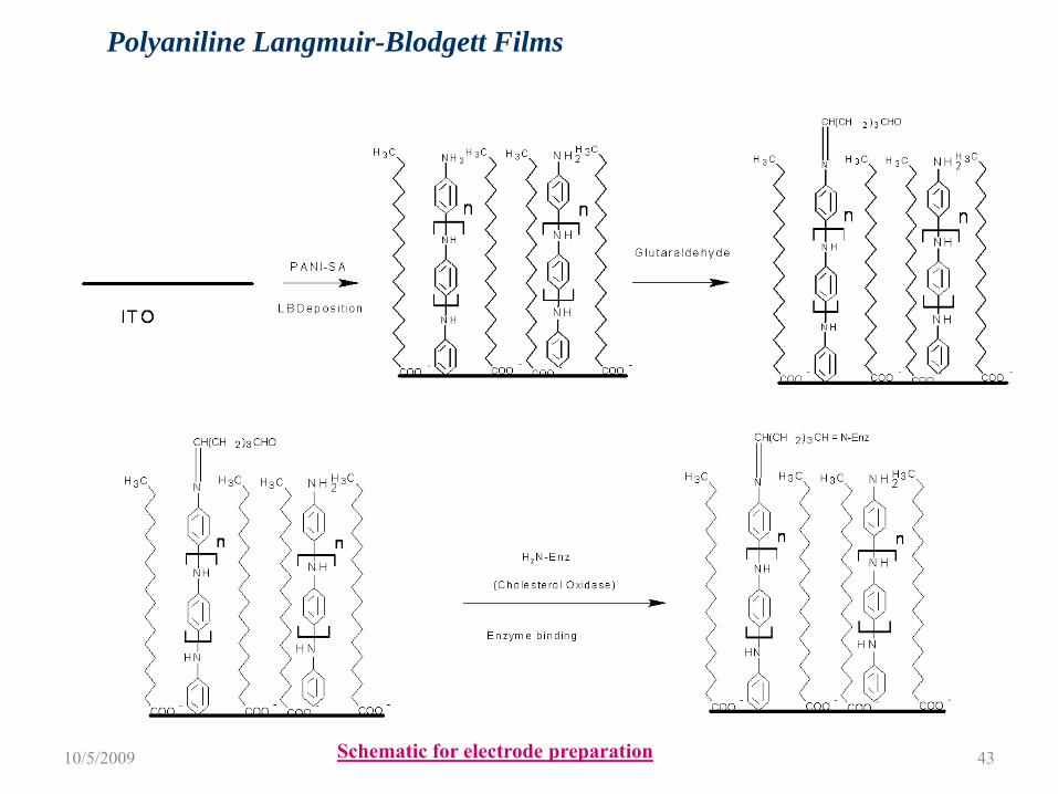
Polyaniline Langmuir-Blodgett Films
Schematic for electrode preparation10/5/2009 43
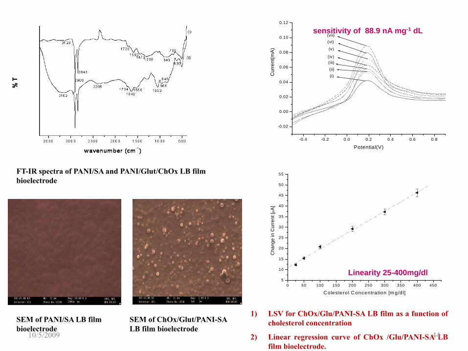
FT-IR spectra of PANI/SA and PANI/Glut/ChOx LB film bioelectrode
SEM of PANI/SA LB film bioelectrode
SEM of ChOx/Glut/PANI-SA LB film bioelectrode
-0.4 -0.2 0.0 0.2 0.4 0.6 0.8
-0.02
0.00
0.02
0.04
0.06
0.08
0.10
0.12
(vii)(vi)
(iv)
(v)
(iii)(ii)(i)C
urre
nt(m
A)
Potential(V)
sensitivity of 88.9 nA mg-1 dL
0 50 100 150 200 250 300 350 400 4505
10
15
20
25
30
35
40
45
50
55
Cha
nge
in C
urre
nt [μ
A]
Colestero l Concentration [m g/d l]
Linearity 25-400mg/dl
1) LSV for ChOx/Glu/PANI-SA LB film as a function ofcholesterol concentration
2) Linear regression curve of ChOx /Glu/PANI-SA LBfilm bioelectrode.
10/5/2009 44
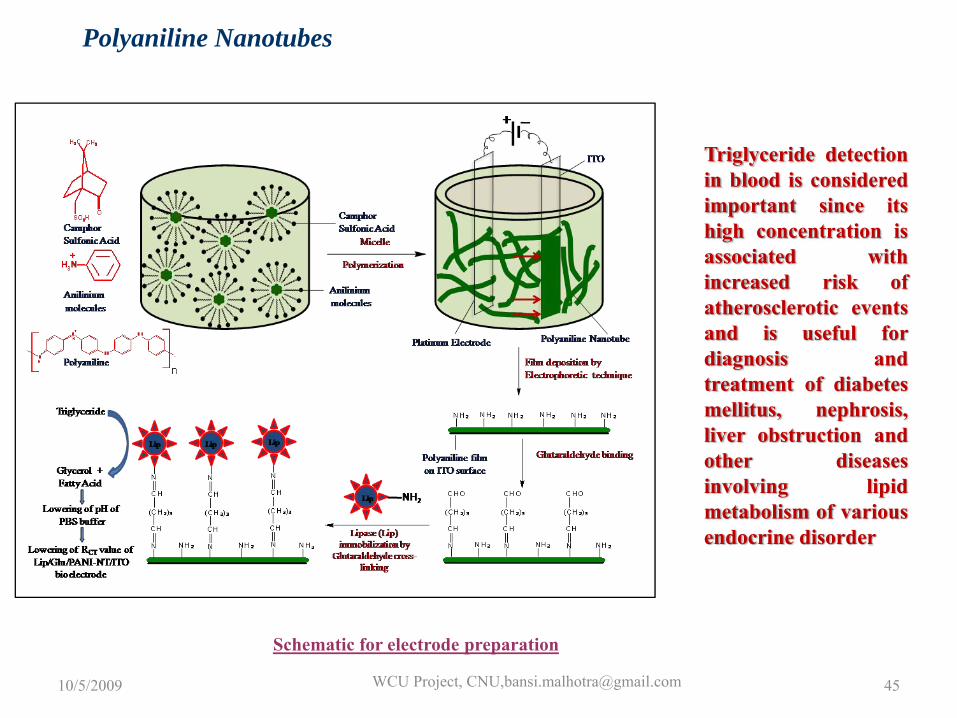
Polyaniline Nanotubes
Triglyceride detectionin blood is consideredimportant since itshigh concentration isassociated withincreased risk ofatherosclerotic eventsand is useful fordiagnosis andtreatment of diabetesmellitus, nephrosis,liver obstruction andother diseasesinvolving lipidmetabolism of variousendocrine disorder
Schematic for electrode preparation
10/5/2009 WCU Project, CNU,[email protected] 45
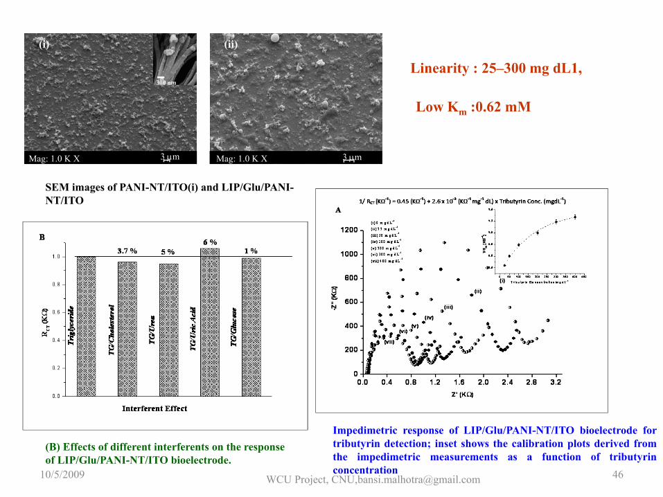
3 μmMag: 1.0 K X Mag: 1.0 K X 3 μm
300 nm
(i) (ii)
SEM images of PANI-NT/ITO(i) and LIP/Glu/PANI-NT/ITO
(B) Effects of different interferents on the response of LIP/Glu/PANI-NT/ITO bioelectrode.
Impedimetric response of LIP/Glu/PANI-NT/ITO bioelectrode fortributyrin detection; inset shows the calibration plots derived fromthe impedimetric measurements as a function of tributyrinconcentration
Linearity : 25–300 mg dL1,
Low Km :0.62 mM
10/5/2009 WCU Project, CNU,[email protected] 46
Bioaffinity Based Sensor
N
NH
NH2
O
N
N
NH
N
NH2
NH
NH
O
O
CH3
N NH
NHN
NH2
O
N
NH
NH
N
NH2
O
OOOH
OH
O+
P
OOOH
OH
O+
P
OOOH
OH
O
P
OO
OH
OH
OH
O+
P
OOOH
OH
O+
P
OOOH
OH
O+
P
CH3
CH3
CH3
CH3
NN
NH2
O
CH3
10/5/2009 WCU Project, CNU,[email protected] 47
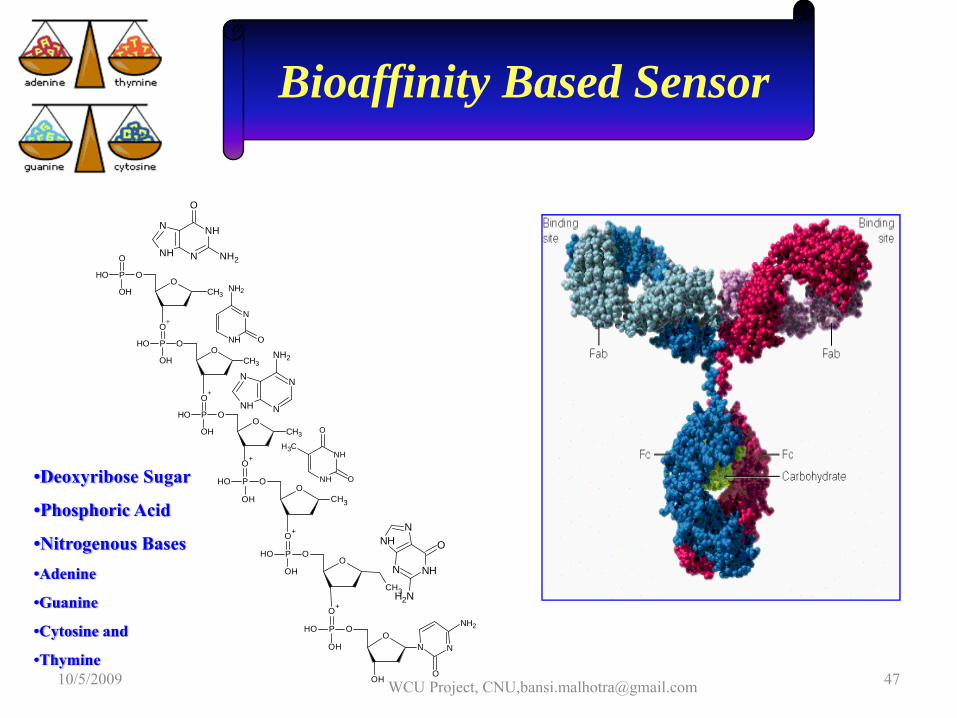
•Deoxyribose Sugar
•Phosphoric Acid
•Nitrogenous Bases•Adenine
•Guanine
•Cytosine and
•Thymine
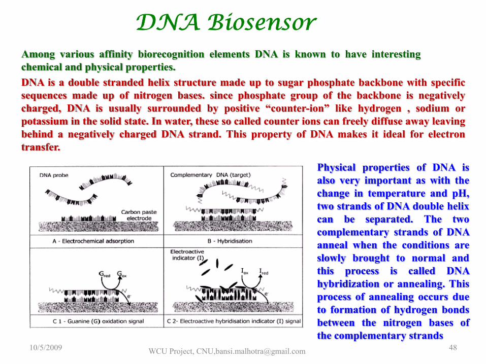
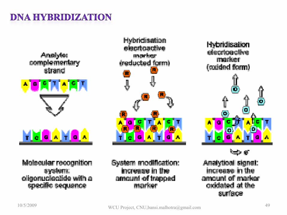
Among various affinity biorecognition elements DNA is known to have interestingchemical and physical properties.DNA is a double stranded helix structure made up to sugar phosphate backbone with specificsequences made up of nitrogen bases. since phosphate group of the backbone is negativelycharged, DNA is usually surrounded by positive “counter-ion” like hydrogen , sodium orpotassium in the solid state. In water, these so called counter ions can freely diffuse away leavingbehind a negatively charged DNA strand. This property of DNA makes it ideal for electrontransfer.
Physical properties of DNA isalso very important as with thechange in temperature and pH,two strands of DNA double helixcan be separated. The twocomplementary strands of DNAanneal when the conditions areslowly brought to normal andthis process is called DNAhybridization or annealing. Thisprocess of annealing occurs dueto formation of hydrogen bondsbetween the nitrogen bases ofthe complementary strands
DNA Biosensor
10/5/2009 WCU Project, CNU,[email protected] 48
10/5/2009 WCU Project, CNU,[email protected] 49
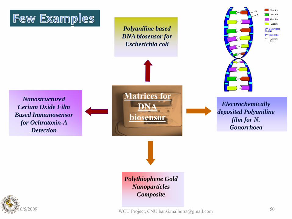
Polythiophene Gold Nanoparticles
Composite
P3HT - SAM
NanostructuredCerium Oxide Film
Based Immunosensorfor Ochratoxin-A
Detection
Electrochemically deposited Polyaniline
film for N.Gonorrhoea
Polyaniline based DNA biosensor for
Escherichia coli
Matrices for DNA
biosensor
10/5/2009 WCU Project, CNU,[email protected] 50
C=O
C=O
C=O
C=O
N N N N
5’biotin end labeled BdE probe
Avidin
Covalent bond between –COOH of avidin and -NH of PANI PANI film onto
Pt disc electrode
C=O
Hybridization with complementary DNA
C=O C=OC=O
N N N N
Complementary target DNA
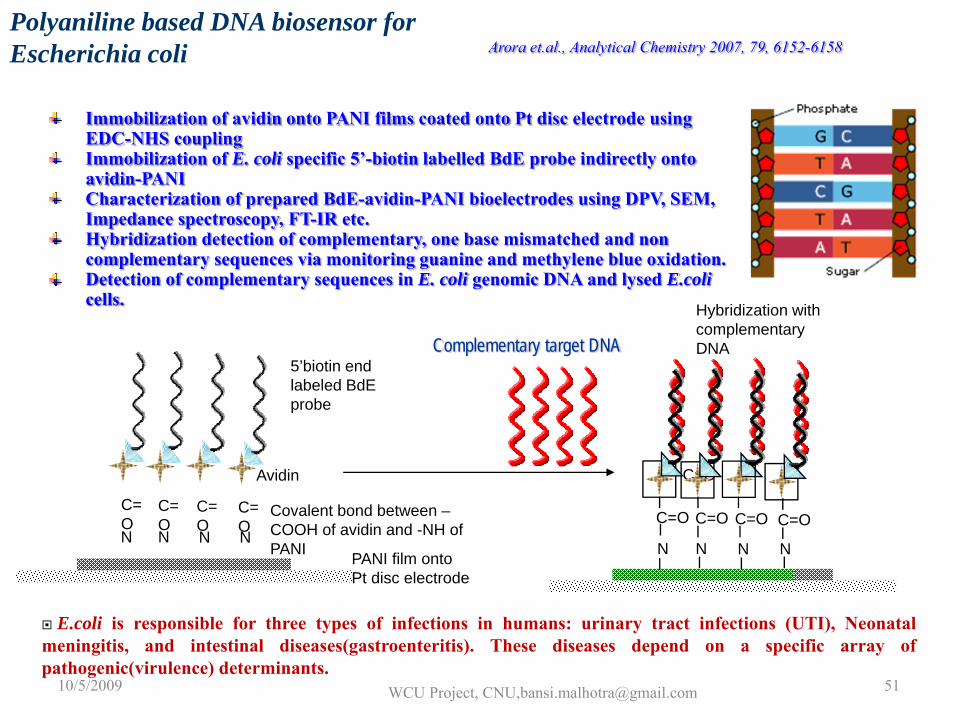
C=O
Immobilization of avidin onto PANI films coated onto Pt disc electrode using EDC-NHS couplingImmobilization of E. coli specific 5’-biotin labelled BdE probe indirectly onto avidin-PANICharacterization of prepared BdE-avidin-PANI bioelectrodes using DPV, SEM, Impedance spectroscopy, FT-IR etc.Hybridization detection of complementary, one base mismatched and non complementary sequences via monitoring guanine and methylene blue oxidation.Detection of complementary sequences in E. coli genomic DNA and lysed E.colicells.
Arora et.al., Analytical Chemistry 2007, 79, 6152-6158Polyaniline based DNA biosensor for Escherichia coli
E.coli is responsible for three types of infections in humans: urinary tract infections (UTI), Neonatalmeningitis, and intestinal diseases(gastroenteritis). These diseases depend on a specific array ofpathogenic(virulence) determinants.
10/5/2009 WCU Project, CNU,[email protected] 51
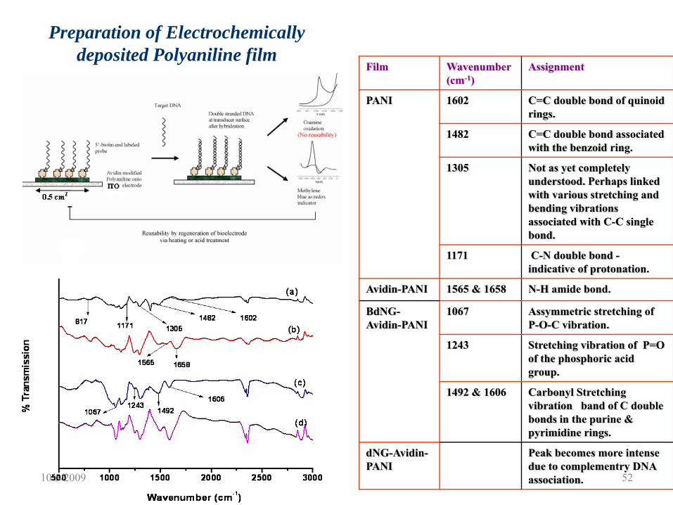
Preparation of Electrochemically deposited Polyaniline film
Film Wavenumber(cm-1)
Assignment
PANI 1602 C=C double bond of quinoid rings.
1482 C=C double bond associated with the benzoid ring.
1305 Not as yet completely understood. Perhaps linked with various stretching and bending vibrations associated with C-C single bond.
1171 C-N double bond -indicative of protonation.
Avidin-PANI 1565 & 1658 N-H amide bond.
BdNG-Avidin-PANI
1067 Assymmetric stretching of P-O-C vibration.
1243 Stretching vibration of P=O of the phosphoric acid group.
1492 & 1606 Carbonyl Stretching vibration band of C double bonds in the purine & pyrimidine rings.
dNG-Avidin-PANI
Peak becomes more intense due to complementry DNA association.10/5/2009 52
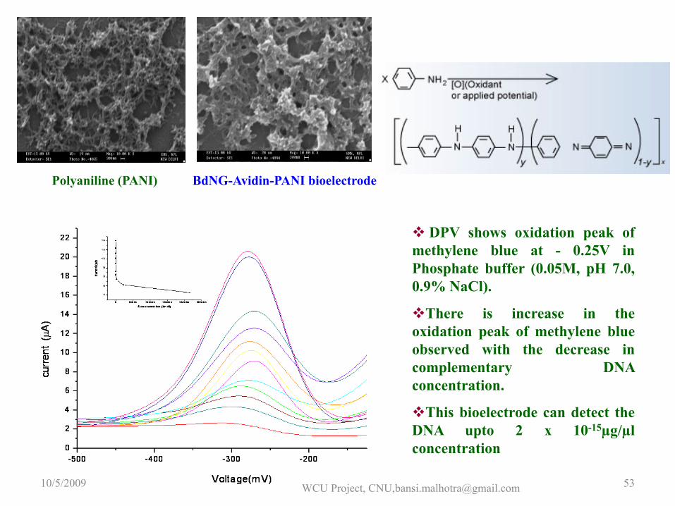
Polyaniline (PANI) BdNG-Avidin-PANI bioelectrode
DPV shows oxidation peak ofmethylene blue at - 0.25V inPhosphate buffer (0.05M, pH 7.0,0.9% NaCl).
There is increase in theoxidation peak of methylene blueobserved with the decrease incomplementary DNAconcentration.
This bioelectrode can detect theDNA upto 2 x 10-15µg/µlconcentration
10/5/2009 WCU Project, CNU,[email protected] 53
0 .8 0 .9 1 .0 1 .1 1 .2 1 .3 1 .4 1 .5 1 .6
2 .0 x 1 0 -6
4 .0 x 1 0 -6
6 .0 x 1 0 -6
8 .0 x 1 0 -6
1 .0 x 1 0 -5
1 .2 x 1 0 -5
1 .4 x 1 0 -5
1 .6 x 1 0 -5
1 .8 x 1 0 -5
2 .0 x 1 0 -5 B d E -av id in -P A N I H yb rid iza tio n w ith d E ' H yb rid iza tio n w ih t d E '1 H yb rid iza tio n w ith d E 'nc
I (µA
)
V (v o lts )
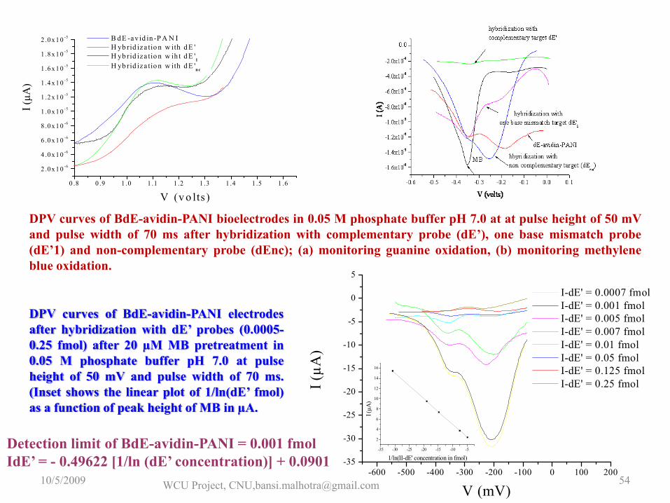
DPV curves of BdE-avidin-PANI bioelectrodes in 0.05 M phosphate buffer pH 7.0 at at pulse height of 50 mVand pulse width of 70 ms after hybridization with complementary probe (dE’), one base mismatch probe(dE’1) and non-complementary probe (dEnc); (a) monitoring guanine oxidation, (b) monitoring methyleneblue oxidation.
-600 -500 -400 -300 -200 -100 0 100 200-35
-30
-25
-20
-15
-10
-5
0
5
-35 -30 -25 -20 -15 -10 -5
2
4
6
8
10
12
14
16I (
µA)
1/ln(II-dE' concentration in fmol)
I (µA
)
V (mV)
I-dE' = 0.0007 fmol I-dE' = 0.001 fmol I-dE' = 0.005 fmol I-dE' = 0.007 fmol I-dE' = 0.01 fmol I-dE' = 0.05 fmol I-dE' = 0.125 fmol I-dE' = 0.25 fmol
DPV curves of BdE-avidin-PANI electrodesafter hybridization with dE’ probes (0.0005-0.25 fmol) after 20 µM MB pretreatment in0.05 M phosphate buffer pH 7.0 at pulseheight of 50 mV and pulse width of 70 ms.(Inset shows the linear plot of 1/ln(dE’ fmol)as a function of peak height of MB in µA.
Detection limit of BdE-avidin-PANI = 0.001 fmolIdE’ = - 0.49622 [1/ln (dE’ concentration)] + 0.0901
10/5/2009 WCU Project, CNU,[email protected] 54
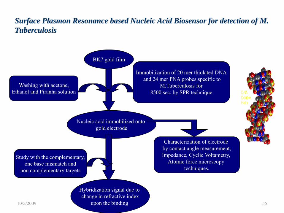
Surface Plasmon Resonance based Nucleic Acid Biosensor for detection of M. Tuberculosis
BK7 gold film
Nucleic acid immobilized onto gold electrode
Hybridization signal due to change in refractive index
upon the binding
Washing with acetone,Ethanol and Piranha solution
Immobilization of 20 mer thiolated DNAand 24 mer PNA probes specific to
M.Tuberculosis for 8500 sec. by SPR technique
Characterization of electrode by contact angle measurement,Impedance, Cyclic Voltametry,
Atomic force microscopy techniques.
Study with the complementary,one base mismatch and
non complementary targets
10/5/2009 55
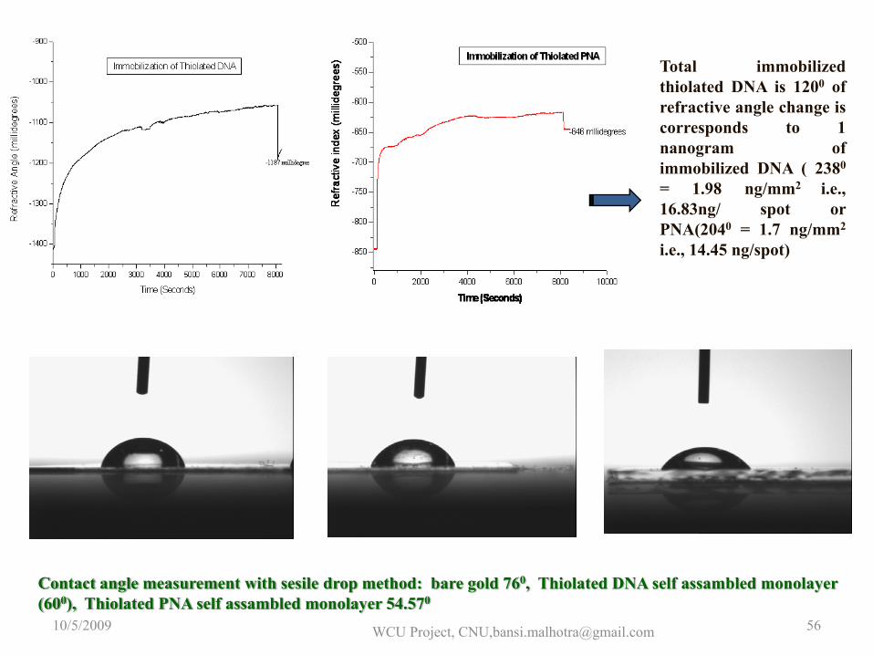
10/5/2009 WCU Project, CNU,[email protected] 56
Total immobilizedthiolated DNA is 1200 ofrefractive angle change iscorresponds to 1nanogram ofimmobilized DNA ( 2380
= 1.98 ng/mm2 i.e.,16.83ng/ spot orPNA(2040 = 1.7 ng/mm2
i.e., 14.45 ng/spot)
Contact angle measurement with sesile drop method: bare gold 760, Thiolated DNA self assambled monolayer (600), Thiolated PNA self assambled monolayer 54.570
0 200 400 600 800-800
-700
-600
-500
Ref
ract
ive
angl
e (m
illide
gree
s)
Time (Seconds)
One base mismatch non complementaryComplementary
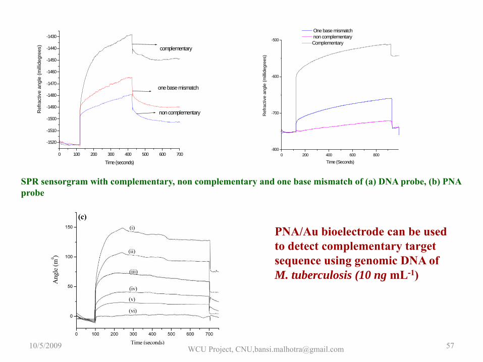
SPR sensorgram with complementary, non complementary and one base mismatch of (a) DNA probe, (b) PNA probe
0 100 200 300 400 500 600 700
-1520
-1510
-1500
-1490
-1480
-1470
-1460
-1450
-1440
-1430
one base mismatch
non complementary
complementary
Ref
ract
ive
angl
e (m
illide
gree
s)
Time (seconds)
PNA/Au bioelectrode can be used to detect complementary target sequence using genomic DNA of M. tuberculosis (10 ng mL-1)
10/5/2009 WCU Project, CNU,[email protected] 57
Immunosensor
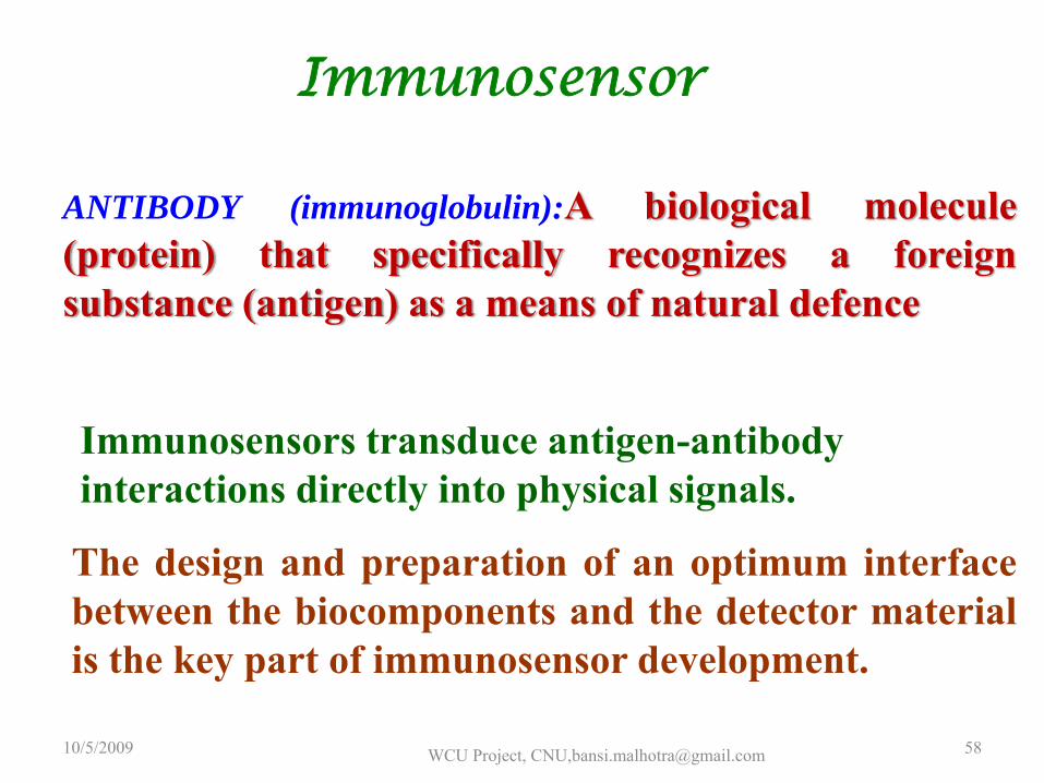
ANTIBODY (immunoglobulin):A biological molecule(protein) that specifically recognizes a foreignsubstance (antigen) as a means of natural defence
Immunosensors transduce antigen-antibody interactions directly into physical signals.
The design and preparation of an optimum interfacebetween the biocomponents and the detector materialis the key part of immunosensor development.
10/5/2009 WCU Project, CNU,[email protected] 58
Antibodies
10/5/2009 WCU Project, CNU,[email protected] 59
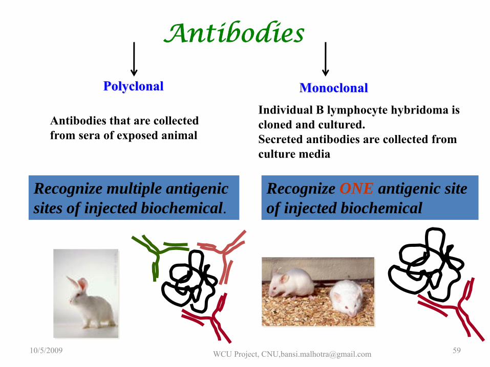
Polyclonal Monoclonal
Antibodies that are collected from sera of exposed animal
Recognize multiple antigenic sites of injected biochemical.
Individual B lymphocyte hybridoma is cloned and cultured. Secreted antibodies are collected from culture media
Recognize ONE antigenic site of injected biochemical
Fast , accurate and sensitive measurement are required,
Highest possible detection strength is required ,
Large numbers of samples are to be expected ,
Alternate of available expensive analytical methods.
Immunosensor becomes important when
10/5/2009 WCU Project, CNU,[email protected] 60
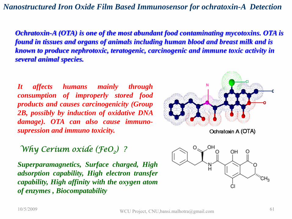
Nanostructured Iron Oxide Film Based Immunosensor for ochratoxin-A Detection
Ochratoxin-A (OTA) is one of the most abundant food contaminating mycotoxins. OTA is found in tissues and organs of animals including human blood and breast milk and is known to produce nephrotoxic, teratogenic, carcinogenic and immune toxic activity in several animal species.
It affects humans mainly throughconsumption of improperly stored foodproducts and causes carcinogenicity (Group2B, possibly by induction of oxidative DNAdamage). OTA can also cause immuno-supression and immuno toxicity.
Why Cerium oxide (FeO2) ?
Superparamagnetics, Surface charged, Highadsorption capability, High electron transfercapability, High affinity with the oxygen atomof enzymes , Biocompatability
10/5/2009 WCU Project, CNU,[email protected] 61
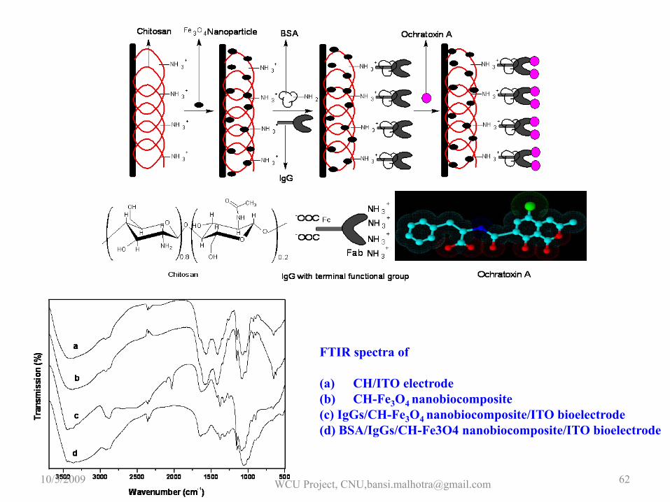
FTIR spectra of
(a) CH/ITO electrode(b) CH-Fe3O4 nanobiocomposite(c) IgGs/CH-Fe3O4 nanobiocomposite/ITO bioelectrode(d) BSA/IgGs/CH-Fe3O4 nanobiocomposite/ITO bioelectrode
10/5/2009 WCU Project, CNU,[email protected] 62
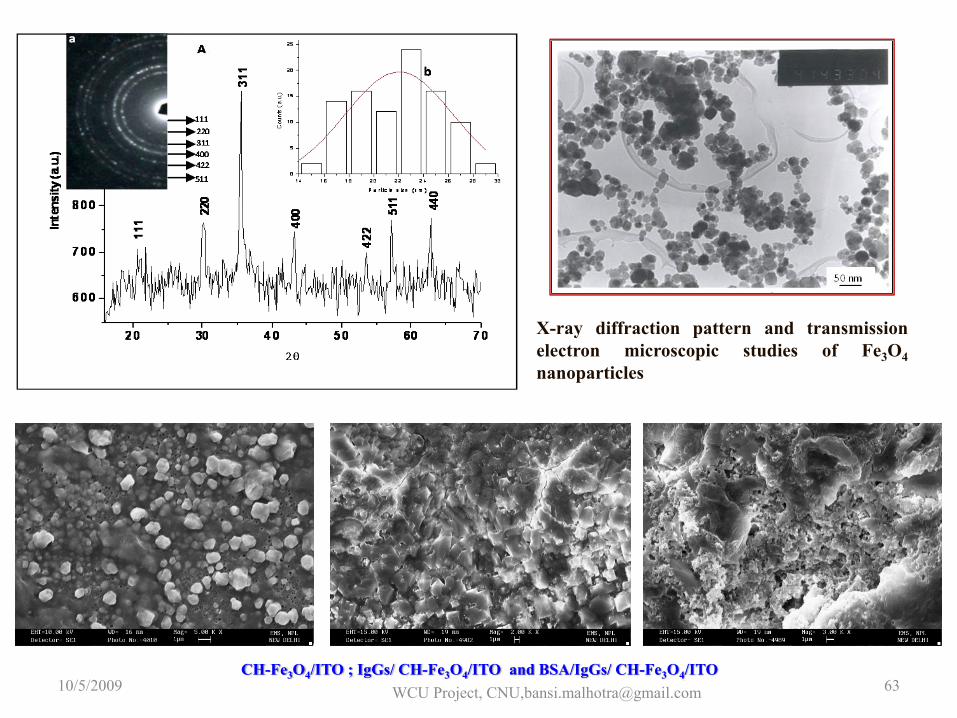
X-ray diffraction pattern and transmissionelectron microscopic studies of Fe3O4nanoparticles
CH-Fe3O4/ITO ; IgGs/ CH-Fe3O4/ITO and BSA/IgGs/ CH-Fe3O4/ITO 10/5/2009 WCU Project, CNU,[email protected] 63
-0.2 0.0 0.2 0.4 0.6 0.80.0
5.0x10⁻⁵
1.0x10⁻⁴
1.5x10⁻⁴
2.0x10⁻⁴
2.5x10⁻⁴
3.0x10⁻⁴
3.5x10⁻⁴
d
c
b a
Cur
rent
(A)
Potential (V)
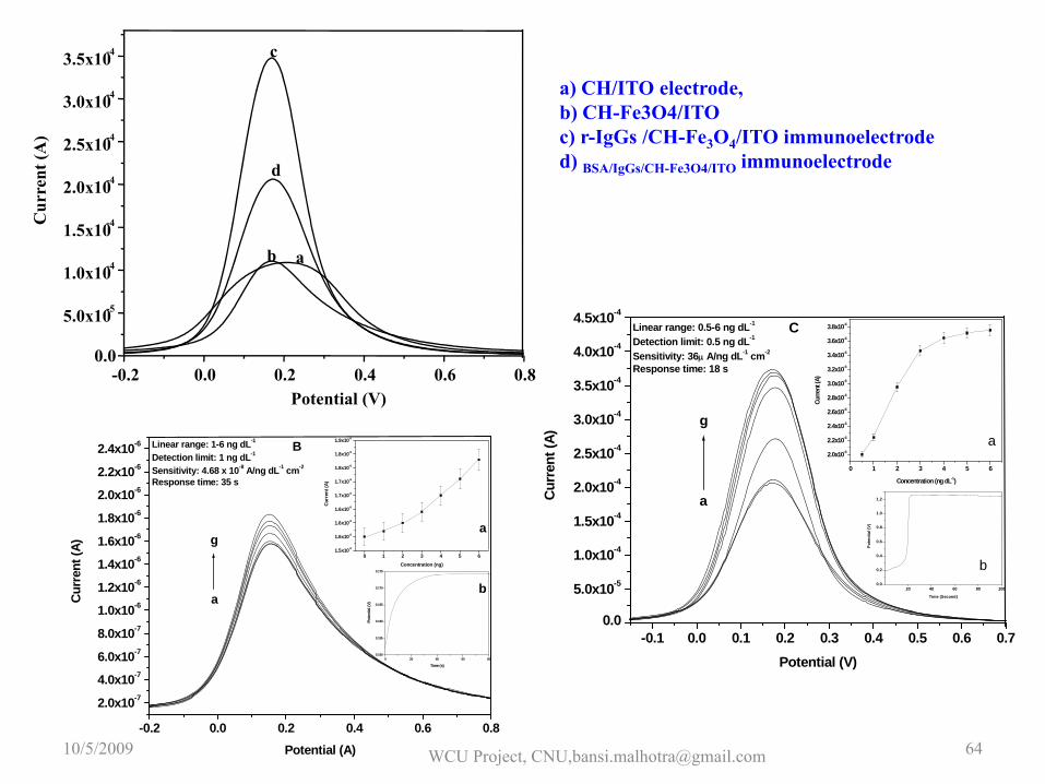
a) CH/ITO electrode, b) CH-Fe3O4/ITO c) r-IgGs /CH-Fe3O4/ITO immunoelectroded) BSA/IgGs/CH-Fe3O4/ITO immunoelectrode
-0.1 0.0 0.1 0.2 0.3 0.4 0.5 0.6 0.70.0
5.0x10⁻⁵
1.0x10⁻⁴
1.5x10⁻⁴
2.0x10⁻⁴
2.5x10⁻⁴
3.0x10⁻⁴
3.5x10⁻⁴
4.0x10⁻⁴
4.5x10⁻⁴
CLinear range: 0.5-6 ng dL-1
Detection limit: 0.5 ng dL-1
Sensitivity: 36μ A/ng dL-1 cm-2
Response time: 18 s
g
a
b
a
20 40 60 80 1000.0
0.2
0.4
0.6
0.8
1.0
1.2
Pote
ntia
l (V)
Time (Second)
0 1 2 3 4 5 6
2.0x10⁻⁴
2.2x10⁻⁴
2.4x10⁻⁴
2.6x10⁻⁴
2.8x10⁻⁴
3.0x10⁻⁴
3.2x10⁻⁴
3.4x10⁻⁴
3.6x10⁻⁴
3.8x10⁻⁴
Curre
nt (A
)
Concentration (ng dL-1)
Curr
ent (
A)
Potential (V)
-0.2 0.0 0.2 0.4 0.6 0.8
2.0x10⁻⁷
4.0x10⁻⁷
6.0x10⁻⁷
8.0x10⁻⁷
1.0x10⁻⁶
1.2x10⁻⁶
1.4x10⁻⁶
1.6x10⁻⁶
1.8x10⁻⁶
2.0x10⁻⁶
2.2x10⁻⁶
2.4x10⁻⁶
0 20 40 60 800.150
0.155
0.160
0.165
0.170
0.175
Pote
ntial
(V)
Time (s)
b
a
BLinear range: 1-6 ng dL-1
Detection limit: 1 ng dL-1
Sensitivity: 4.68 x 10⁻⁸ A/ng dL-1 cm-2
Response time: 35 s
0 1 2 3 4 5 61.5x10⁻⁶
1.6x10⁻⁶
1.6x10⁻⁶
1.6x10⁻⁶
1.7x10⁻⁶
1.7x10⁻⁶
1.8x10⁻⁶
1.8x10⁻⁶
1.9x10⁻⁶
Curr
ent (
A)
Concentration (ng)
g
aCurr
ent (
A)
Potential (A)10/5/2009 WCU Project, CNU,[email protected] 64
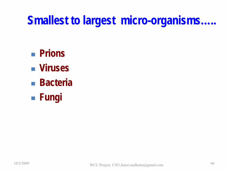
Smallest to largest micro-organisms…..
PrionsVirusesBacteriaFungi
10/5/2009 WCU Project, CNU,[email protected] 66
Many types of microbial sensors have been developed asanalytical tools since the first microbial sensor wasstudied by Karube et al. in 1977.
The microbial sensor consists of a transducer andmicrobe as a sensing element. The characteristics of themicrobial sensors are a complete contrast to those ofenzyme sensors or immunosensors, which are highlyspecific for the substrates of interest, although thespecificity of the microbial sensor has been improvedby genetic modification of the microbe used as thesensing element.
10/5/2009 WCU Project, CNU,[email protected] 67
•Microbial sensors have the advantages of tolerance tomeasuring conditions, a long lifetime, and cost effectiveperformance, and have the disadvantage of a longresponse time.
•Microbial sensors result from the combination of amicroorganism with a transducer capable of detecting themetabolite involved.
•Microorganisms possess enzymatic systems that effectbiological transformations. The immobilization of micro-organism on a transducer is first step in the constructionof a biosensor.10/5/2009 WCU Project, CNU,[email protected] 68
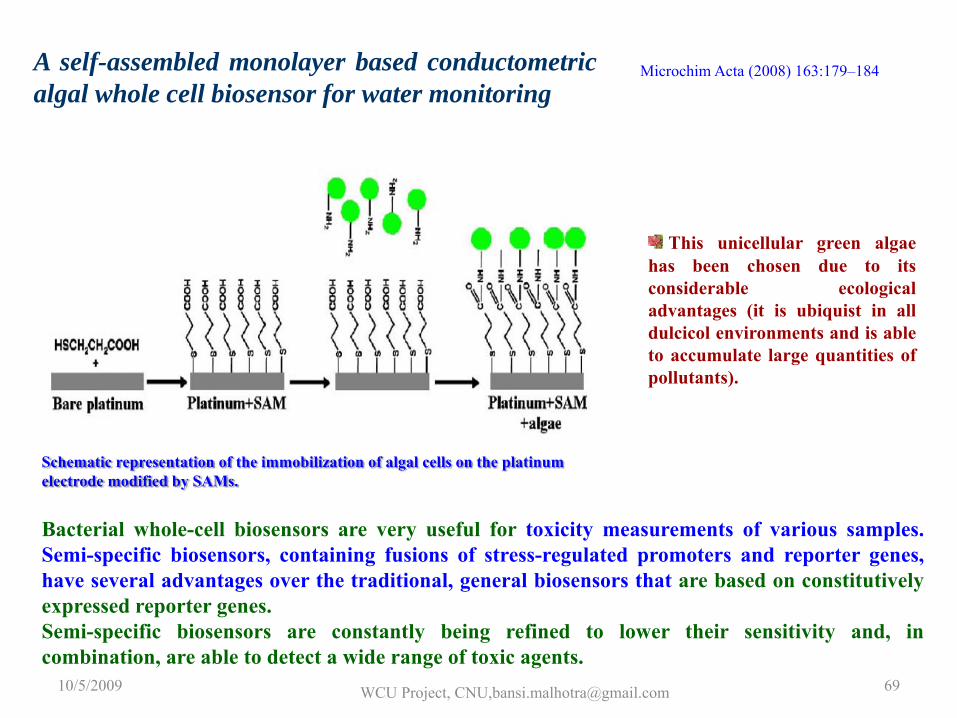
A self-assembled monolayer based conductometricalgal whole cell biosensor for water monitoring
Schematic representation of the immobilization of algal cells on the platinum electrode modified by SAMs.
This unicellular green algaehas been chosen due to itsconsiderable ecologicaladvantages (it is ubiquist in alldulcicol environments and is ableto accumulate large quantities ofpollutants).
Microchim Acta (2008) 163:179–184
Bacterial whole-cell biosensors are very useful for toxicity measurements of various samples.Semi-specific biosensors, containing fusions of stress-regulated promoters and reporter genes,have several advantages over the traditional, general biosensors that are based on constitutivelyexpressed reporter genes.Semi-specific biosensors are constantly being refined to lower their sensitivity and, incombination, are able to detect a wide range of toxic agents.
10/5/2009 WCU Project, CNU,[email protected] 69
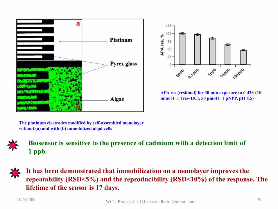
The platinum electrodes modified by self-assembled monolayerwithout (a) and with (b) immobilized algal cells
APA res (residual) for 30 min exposure to Cd2+ (10 mmol l−1 Tris–HCl, 50 μmol l−1 pNPP, pH 8.5)
Biosensor is sensitive to the presence of cadmium with a detection limit of 1 ppb.
It has been demonstrated that immobilization on a monolayer improves the repeatability (RSD<5%) and the reproducibility (RSD<10%) of the response. The lifetime of the sensor is 17 days.
10/5/2009 WCU Project, CNU,[email protected] 70
10/5/2009 WCU Project, CNU,[email protected] 71
Conclusions:
•Biocatalysis & Bioaffinity Sensors•Glucose,Urea,Cholesterol•DNA•Immunosensor•Whole Cell•Immunosensor ……
10/5/2009 WCU Project, CNU,[email protected] 72
Some literature for Studies ( Week 2):
•Prospects of conducting polymers in biosensors, B.D Malhotra, A. Chaubey and S. P. Singh, Analytica Chmica Acta , 578 (2006) 59–74.•Electrophoretically deposited conducting polymers for applications in organic electronics,Chetna Dhand and B.D.Malhotra,Organic Electronics in Sensors & Biotechology, J.Shinar & Ruth Shinar( Editors),McGraw-Hill),2008•Recent developments in urea biosensor, Gunjan Dhawan, G.Sumana and B.D.Malhotra, Biochemical Engineering Journal ,2009 ,44 , pp. 42-52.
•Electrocatalytic oxidation of hydrazine and hydroxylamine at gold nanoparticle—polypyrrole nanowiremodified glassy carbon electrode Jing Li, Xiangqin Lin, Sensors and Actuators B 126 (2007) 527–535•Application of Polyaniline as glucose biosensor, K. Ramanathan, S. Annapoorni and B. D. Malhotra,
Sensors & Acturators B, 21, 1994, 165 – 69.•Polythiophene gold nanoparticles composite film for application to glucose sensor, Pratibha Pandey, Sunil K. Arya , Zimple Matharu, S. P. Singh, Monika Datta and B. D. Malhotra, Journal of Applied Polymer Science , Vol. 110, 988–994 (2008),•Cholesterol biosensor based on cholesterol esterase, cholesterol oxidase and peroxidase Immobilized on conducting polyaniline films, Suman Singh, P. R. Solanki, M. K. Pandey and B. D. Malhotra, Sensors & Actuators B, 115,2006,pp534-541.
•Microchim Acta (2008) 163:179–184.•Fully integrated biocatalytic electrodes based on bioaffinity interactions, E Katz, V Heleg-Shabtai, A Bardea, I Willner, Biosensors and Bioelectronics, 1998

10/5/2009 WCU Project, CNU,[email protected] 73

10/5/2009 WCU Project, CNU,[email protected] 74

Michaelis‐Menton Equation
10/5/2009WCU Project,Chungnam National
University, Daejeon, Korea,[email protected]
75
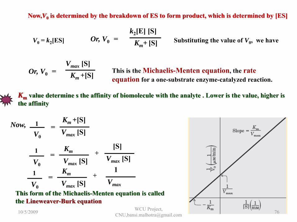
Now,V0 is determined by the breakdown of ES to form product, which is determined by [ES]
V0 = k2[ES] Substituting the value of V0, we haveOr, V0 = Km+ [S]k2[E] [S]
This is the Michaelis-Menten equation, the rate equation for a one-substrate enzyme-catalyzed reaction.
Km value determine s the affinity of biomolecule with the analyte . Lower is the value, higher is the affinity
Or, V0 = Km +[S]Vmax [S]
Now, Km +[S]
Vmax [S]V0
1 =
[S]
Vmax [S]Km
Vmax [S]V0
1 = +
Km
Vmax [S]V0
1 = +1
Vmax
This form of the Michaelis-Menten equation is calledthe Lineweaver-Burk equation10/5/2009 WCU Project,
CNU,[email protected] 76
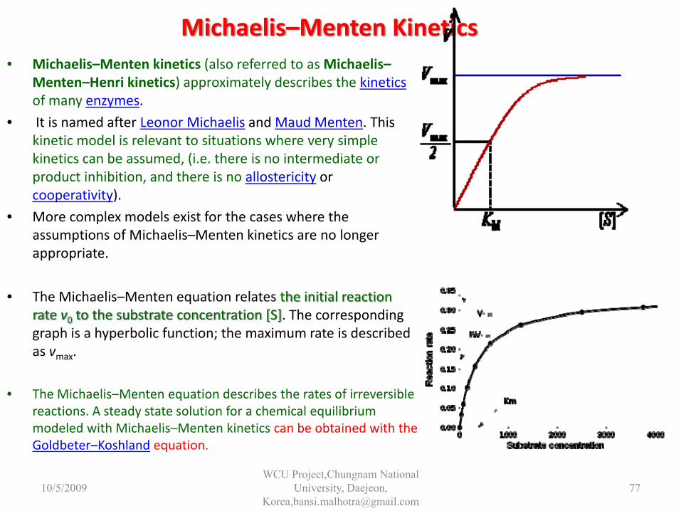
Michaelis–Menten Kinetics• Michaelis–Menten kinetics (also referred to as Michaelis–
Menten–Henri kinetics) approximately describes the kineticsof many enzymes.
• It is named after Leonor Michaelis and Maud Menten. This kinetic model is relevant to situations where very simple kinetics can be assumed, (i.e. there is no intermediate or product inhibition, and there is no allostericity or cooperativity).
• More complex models exist for the cases where the assumptions of Michaelis–Menten kinetics are no longer appropriate.
• The Michaelis–Menten equation relates the initial reaction rate v0 to the substrate concentration [S]. The corresponding graph is a hyperbolic function; the maximum rate is described as vmax.
• The Michaelis–Menten equation describes the rates of irreversible reactions. A steady state solution for a chemical equilibrium modeled with Michaelis–Menten kinetics can be obtained with the Goldbeter–Koshland equation.
10/5/2009WCU Project,Chungnam National
University, Daejeon, Korea,[email protected]
77