Embed Size (px)
Citation preview
Biochimica et Biophysica Acta 1843 (2014) 1497–1508
Contents lists available at ScienceDirect
Biochimica et Biophysica Acta
j ourna l homepage: www.e lsev ie r .com/ locate /bbamcr
Review
Structure and mechanism of Escherichia coli type I signal peptidase☆
Mark Paetzel ⁎Department of Molecular Biology and Biochemistry, Simon Fraser University, 8888 University Drive, Burnaby, British Columbia V5A 1S6, Canada
Abbreviations: SPase I, bacterial type I signal peptidas☆ This article is part of a Special Issue entitled: Protebacteria. Guest Editors: Anastassios Economou and Ross D⁎ Tel.: +1 778 782 4230; fax: +1 778 782 5583.
0167-4889/$ – see front matter © 2013 Elsevier B.V. All rhttp://dx.doi.org/10.1016/j.bbamcr.2013.12.003
a b s t r a c t
a r t i c l e i n f oArticle history:Received 25 August 2013Received in revised form 26 November 2013Accepted 4 December 2013Available online 11 December 2013
Keywords:Signal peptidaseLeader peptidaseSignal peptideLeader peptideProtein secretionPreprotein processing
Type I signal peptidase is the enzyme responsible for cleaving off the amino-terminal signal peptide from pro-teins that are secreted across the bacterial cytoplasmic membrane. It is an essential membrane bound enzymewhose serine/lysine catalytic dyad resides on the exo-cytoplasmic surface of the bacterialmembrane. This reviewdiscusses the progress that has been made in the structural and mechanistic characterization of Escherichia colitype I signal peptidase (SPase I) aswell as efforts to develop a novel class of antibiotics based on SPase I inhibition.This article is part of a Special Issue entitled: Protein trafficking and secretion in bacteria. Guest Editors:Anastassios Economou and Ross Dalbey.
© 2013 Elsevier B.V. All rights reserved.
1. Introduction
Protein secretion is an essential process in all cells. Important in-sights into protein secretion mechanisms have been gleaned fromstudies performed in bacteria, given that many of the fundamentalmechanistic elements of the process appear to have been conservedthroughout evolution. The hallmark of a secretory protein is theamino-terminal extension — the signal (or leader) peptide. The major-ity of bacterial proteins, whose final destination resides on the trans-side of the cytoplasmic membrane, are secreted post-translationallyvia the general Sec-system [1,2]. Protein secretion, catalyzed by theSec-system, can be thought of as occurring in three separate steps: 1)targeting to the membrane, 2) translocation across the membrane,and 3) release from the membrane. Very briefly, the molecular chaper-one SecB binds to themature region of a secretory preprotein, keeping itin a translocation competent (molten globular) state and helping toguide it to the translocase (SecYEG/SecA) at the membrane surface.SecA, an ATPase, appears to push the preprotein through the SecYEGchannel. Accessory proteins SecDFyajC may also play a role, but do notappear to be essential. The translocated preprotein is tethered tothe cytoplasmic membrane via its signal peptide until the signal pep-tide is cleaved off by type I signal peptidase (SPase I). Besides theSec-system, there are many more specialized bacterial secretion sys-tems and even other signal peptidases, such as the type II [3] andtype IV [4] signal peptidases that specialize in the processing ofpre-lipoproteins and pre-pilin proteins respectively. These enzymes
ein trafficking and secretion inalbey.
ights reserved.
(aspartic acid proteases) are neither structurally nor mechanisticallyrelated to SPase I (a serine protease).
This review will focus on what is currently known about the thirdstep of the general protein secretion process (SPase I catalyzed cleavageof secretory preproteins) in the model organism Escherichia coli.
2. The substrate–membrane bound secretory preproteins
The substrates for SPase I are secretory preproteins tethered to themembrane via their signal peptide (Fig. 1). SPase I catalyzes the cleav-age of the secretory preproteins to create two products, the releasedmature secretory protein and the membrane bound signal peptide.Early sequence analysis revealed the fundamental features of the signalpeptide located at the amino-terminus of every secretory protein: alength of approximately 20–30 residues, an amino-terminal regionwith a net positive charge (N-region), followed by a hydrophobic region(H-region), and then a protease recognition sequence (C-region)with apreference for small residues at the−3(P3) and −1(P1) positions rel-ative to the cleavage site (scissile bond). The specificity is sometimes re-ferred to as the “(−3,−1) rule” or the “Ala-X-Ala rule” [5–8] because ofits preference for alanine at the P1 and P3 positions. The average eu-karyotic signal peptide is ~23 residues in length while the averageGram-negative eubacterial signal peptide is ~25 residues in length,and the average Gram-positive eubacterial signal peptide is ~32 resi-dues in length [9,10]. It is believed that the variations in the signal pep-tide lengths may reflect the differences in the thickness of the lipidbilayers in which the signal peptides reside.
Modern genomic sequencing methods (UniProt [11,12]) along withcomputational signal sequence prediction programs (SignalP [13,14])have provided lists of potential SPase I substrates for a large numberof species whose genome has been sequenced. There is also a growing
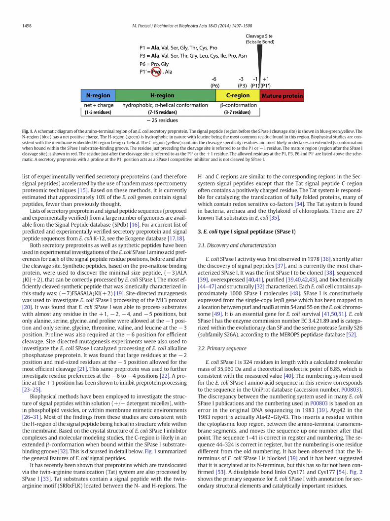
Fig. 1. A schematic diagram of the amino-terminal region of an E. coli secretory preprotein. The signal peptide (region before the SPase I cleavage site) is shown in blue/green/yellow. TheN-region (blue) has a net positive charge. The H-region (green) is hydrophobic in nature with leucine being the most common residue found in this region. Biophysical studies are con-sistent with themembrane embeddedH-region beingα-helical. The C-region (yellow) contains the cleavage specificity residues andmost likely undertakes an extended β-conformationwhen bound within the SPase I substrate-binding groove. The residue just preceding the cleavage site is referred to as the P1 or−1 residue. The mature region (region after the SPase Icleavage site) is shown in red. The residue just after the cleavage site is referred to as the P1′ or the +1 residue. The allowed residues at the P1, P3, P6 and P1′ are listed above the sche-matic. A secretory preprotein with a proline at the P1′ position acts as a SPase I competitive inhibitor and is not cleaved by SPase I.
1498 M. Paetzel / Biochimica et Biophysica Acta 1843 (2014) 1497–1508
list of experimentally verified secretory preproteins (and thereforesignal peptides) accelerated by the use of tandemmass spectrometryproteomic techniques [15]. Based on these methods, it is currentlyestimated that approximately 10% of the E. coli genes contain signalpeptides, fewer than previously thought.
Lists of secretory preprotein and signal peptide sequences (proposedand experimentally verified) from a large number of genomes are avail-able from the Signal Peptide database (SPdb) [16]. For a current list ofpredicted and experimentally verified secretory preprotein and signalpeptide sequences from E. coli K-12, see the Ecogene database [17,18].
Both secretory preproteins as well as synthetic peptides have beenused in experimental investigations of the E. coli SPase I amino acid pref-erences for each of the signal peptide residue positions, before and afterthe cleavage site. Synthetic peptides, based on the pre-maltose bindingprotein, were used to discover the minimal size peptide, (−3)ALA↓KI(+2), that can be correctly processed by E. coli SPase I. The most ef-ficiently cleaved synthetic peptide that was kinetically characterized inthis study was: (−7)FSASALA↓KI(+2) [19]. Site-directed mutagenesiswas used to investigate E. coli SPase I processing of the M13 procoat[20]. It was found that E. coli SPase I was able to process substrateswith almost any residue in the +1, −2, −4, and −5 positions, butonly alanine, serine, glycine, and proline were allowed at the−1 posi-tion and only serine, glycine, threonine, valine, and leucine at the −3position. Proline was also required at the −6 position for efficientcleavage. Site-directed mutagenesis experiments were also used toinvestigate the E. coli SPase I catalyzed processing of E. coli alkalinephosphatase preprotein. It was found that large residues at the −2position and mid-sized residues at the −5 position allowed for themost efficient cleavage [21]. This same preprotein was used to furtherinvestigate residue preferences at the −6 to −4 positions [22]. A pro-line at the+1 position has been shown to inhibit preprotein processing[23–25].
Biophysical methods have been employed to investigate the struc-ture of signal peptides within solution (+/− detergent micelles), with-in phospholipid vesicles, or within membrane mimetic environments[26–31]. Most of the findings from these studies are consistent withtheH-region of the signal peptide beinghelical in structurewhilewithinthe membrane. Based on the crystal structure of E. coli SPase I inhibitorcomplexes and molecular modeling studies, the C-region is likely in anextended β-conformation when bound within the SPase I substrate-binding groove [32]. This is discussed in detail below. Fig. 1 summarizedthe general features of E. coli signal peptides.
It has recently been shown that preproteins which are translocatedvia the twin-arginine translocation (Tat) system are also processed bySPase I [33]. Tat substrates contain a signal peptide with the twin-arginine motif (SRRxFLK) located between the N- and H-regions. The
H- and C-regions are similar to the corresponding regions in the Sec-system signal peptides except that the Tat signal peptide C-regionoften contains a positively charged residue. The Tat system is responsi-ble for catalyzing the translocation of fully folded proteins, many ofwhich contain redox sensitive co-factors [34]. The Tat system is foundin bacteria, archaea and the thylakoid of chloroplasts. There are 27known Tat substrates in E. coli [35].
3. E. coli type I signal peptidase (SPase I)
3.1. Discovery and characterization
E. coli SPase I activity was first observed in 1978 [36], shortly afterthe discovery of signal peptides [37], and is currently the most char-acterized SPase I. It was the first SPase I to be cloned [38], sequenced[39], overexpressed [40,41], purified [39,40,42,43], and biochemically[44–47] and structurally [32] characterized. Each E. coli cell contains ap-proximately 1000 SPase I molecules [48]. SPase I is constitutivelyexpressed from the single-copy lepB gene which has been mapped toa location betweenpurl andnadB atmin 54 and 55 on the E. coli chromo-some [49]. It is an essential gene for E. coli survival [41,50,51]. E. coliSPase I has the enzyme commission number EC 3.4.21.89 and is catego-rized within the evolutionary clan SF and the serine protease family S26(subfamily S26A), according to the MEROPS peptidase database [52].
3.2. Primary sequence
E. coli SPase I is 324 residues in length with a calculated molecularmass of 35,960 Da and a theoretical isoelectric point of 6.85, which isconsistent with the measured value [40]. The numbering system usedfor the E. coli SPase I amino acid sequence in this review correspondsto the sequence in the UniProt database (accession number, P00803).The discrepancy between the numbering system used in many E. coliSPase I publications and the numbering used in P00803 is based on anerror in the original DNA sequencing in 1983 [39]. Arg42 in the1983 report is actually Ala42–Gly43. This inserts a residue withinthe cytoplasmic loop region, between the amino-terminal transmem-brane segments, and moves the sequence up one number after thatpoint. The sequence 1–41 is correct in register and numbering. The se-quence 44–324 is correct in register, but the numbering is one residuedifferent from the old numbering. It has been observed that the N-terminus of E. coli SPase I is blocked [39] and it has been suggestedthat it is acetylated at its N-terminus, but this has so far not been con-firmed [53]. A disulphide bond links Cys171 and Cys177 [54]. Fig. 2shows the primary sequence for E. coli SPase I with annotation for sec-ondary structural elements and catalytically important residues.
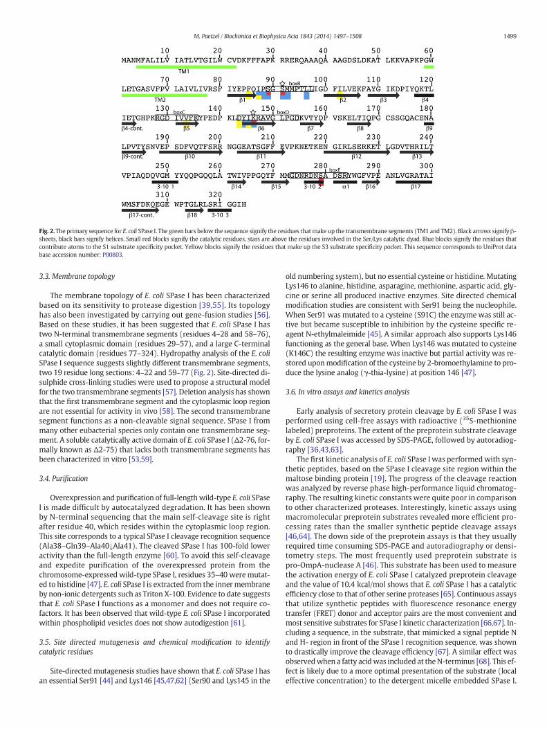
Fig. 2. The primary sequence for E. coli SPase I. The green bars below the sequence signify the residues thatmake up the transmembrane segments (TM1 and TM2). Black arrows signify β-sheets, black bars signify helices. Small red blocks signify the catalytic residues, stars are above the residues involved in the Ser/Lys catalytic dyad. Blue blocks signify the residues thatcontribute atoms to the S1 substrate specificity pocket. Yellow blocks signify the residues that make up the S3 substrate specificity pocket. This sequence corresponds to UniProt database accession number: P00803.
1499M. Paetzel / Biochimica et Biophysica Acta 1843 (2014) 1497–1508
3.3. Membrane topology
The membrane topology of E. coli SPase I has been characterizedbased on its sensitivity to protease digestion [39,55]. Its topologyhas also been investigated by carrying out gene-fusion studies [56].Based on these studies, it has been suggested that E. coli SPase I hastwo N-terminal transmembrane segments (residues 4–28 and 58–76),a small cytoplasmic domain (residues 29–57), and a large C-terminalcatalytic domain (residues 77–324). Hydropathy analysis of the E. coliSPase I sequence suggests slightly different transmembrane segments,two 19 residue long sections: 4–22 and 59–77 (Fig. 2). Site-directed di-sulphide cross-linking studies were used to propose a structural modelfor the two transmembrane segments [57]. Deletion analysis has shownthat the first transmembrane segment and the cytoplasmic loop regionare not essential for activity in vivo [58]. The second transmembranesegment functions as a non-cleavable signal sequence. SPase I frommany other eubacterial species only contain one transmembrane seg-ment. A soluble catalytically active domain of E. coli SPase I (Δ2-76, for-mally known as Δ2-75) that lacks both transmembrane segments hasbeen characterized in vitro [53,59].
3.4. Purification
Overexpression and purification of full-lengthwild-type E. coli SPaseI is made difficult by autocatalyzed degradation. It has been shownby N-terminal sequencing that the main self-cleavage site is rightafter residue 40, which resides within the cytoplasmic loop region.This site corresponds to a typical SPase I cleavage recognition sequence(Ala38–Gln39–Ala40↓Ala41). The cleaved SPase I has 100-fold loweractivity than the full-length enzyme [60]. To avoid this self-cleavageand expedite purification of the overexpressed protein from thechromosome-expressed wild-type SPase I, residues 35–40 weremutat-ed to histidine [47]. E. coli SPase I is extracted from the inner membraneby non-ionic detergents such as Triton X-100. Evidence to date suggeststhat E. coli SPase I functions as a monomer and does not require co-factors. It has been observed that wild-type E. coli SPase I incorporatedwithin phospholipid vesicles does not show autodigestion [61].
3.5. Site directed mutagenesis and chemical modification to identifycatalytic residues
Site-directedmutagenesis studies have shown that E. coli SPase I hasan essential Ser91 [44] and Lys146 [45,47,62] (Ser90 and Lys145 in the
old numbering system), but no essential cysteine or histidine. MutatingLys146 to alanine, histidine, asparagine, methionine, aspartic acid, gly-cine or serine all produced inactive enzymes. Site directed chemicalmodification studies are consistent with Ser91 being the nucleophile.When Ser91 was mutated to a cysteine (S91C) the enzymewas still ac-tive but became susceptible to inhibition by the cysteine specific re-agent N-ethylmaleimide [45]. A similar approach also supports Lys146functioning as the general base. When Lys146 was mutated to cysteine(K146C) the resulting enzyme was inactive but partial activity was re-stored uponmodification of the cysteine by 2-bromoethylamine to pro-duce the lysine analog (γ-thia-lysine) at position 146 [47].
3.6. In vitro assays and kinetics analysis
Early analysis of secretory protein cleavage by E. coli SPase I wasperformed using cell-free assays with radioactive (35S-methioninelabeled) preproteins. The extent of the preprotein substrate cleavageby E. coli SPase I was accessed by SDS-PAGE, followed by autoradiog-raphy [36,43,63].
The first kinetic analysis of E. coli SPase I was performed with syn-thetic peptides, based on the SPase I cleavage site region within themaltose binding protein [19]. The progress of the cleavage reactionwas analyzed by reverse phase high-performance liquid chromatog-raphy. The resulting kinetic constants were quite poor in comparisonto other characterized proteases. Interestingly, kinetic assays usingmacromolecular preprotein substrates revealed more efficient pro-cessing rates than the smaller synthetic peptide cleavage assays[46,64]. The down side of the preprotein assays is that they usuallyrequired time consuming SDS-PAGE and autoradiography or densi-tometry steps. The most frequently used preprotein substrate ispro-OmpA-nuclease A [46]. This substrate has been used to measurethe activation energy of E. coli SPase I catalyzed preprotein cleavageand the value of 10.4 kcal/mol shows that E. coli SPase I has a catalyticefficiency close to that of other serine proteases [65]. Continuous assaysthat utilize synthetic peptides with fluorescence resonance energytransfer (FRET) donor and acceptor pairs are the most convenient andmost sensitive substrates for SPase I kinetic characterization [66,67]. In-cluding a sequence, in the substrate, that mimicked a signal peptide Nand H- region in front of the SPase I recognition sequence, was shownto drastically improve the cleavage efficiency [67]. A similar effect wasobservedwhen a fatty acidwas included at theN-terminus [68]. This ef-fect is likely due to a more optimal presentation of the substrate (localeffective concentration) to the detergent micelle embedded SPase I.

1500 M. Paetzel / Biochimica et Biophysica Acta 1843 (2014) 1497–1508
Combinatorial libraries of synthetic peptides on beads have been usedto optimize fluorogenic peptide substrates [69,70]. See Table 1 for a de-scription of a selection of substrates used to characterize E. coli SPase I.
3.7. In vivo assays
A temperature sensitive strain of E. coli (IT41) has been used incomplementation assays to confirm the role of putative SPase Igene products from a number of different bacterial species [51].This mutant strain is normally only able to grow at permissive tem-peratures (28 to 32 °C), but when transformed with a plasmid con-taining a functional SPase I gene, IT41 is able to grow at a non-permissive temperature (42 °C). Briefly, IT41 cultures, transformedwith a plasmid +/− the gene for a functional SPase I, are grown atnon-permissive temperature and the optical density (540 nm) ofthe cultures is periodically measured (every 30 min) for 8 h.
The cause of the temperature sensitivity within the IT41 strainappears to be an amber mutation at nucleotide position 115 (C toT) within the lepB gene, which is within the codon for Gln39 and re-sults in a TAG amber termination codon [71].
An E. coli strainwas developed that has regulatable expression of thelepB gene [72] and can be used to test cellular inhibition of SPase I. Thestrain has a lepB gene within an L-arabinose inducible pBAD plasmidwhile the chromosomal copy of lepB has been removed.
3.8. Inhibitors
SPase I is a promising antibiotic target because, as mentioned above,it is an essential bacterial enzyme and its active site is located on theextra-cytoplasmic surface of the membrane and thus relatively accessi-ble to drugs. The fact that it has a different catalyticmechanismand olig-omeric nature from the functionally homologous enzyme found withinhuman cells suggests that there would likely be few side effects from aSPase I specific inhibitor. Therefore many industrial, as well academic,labs have been actively searching for compounds that inhibit SPase I.These compounds could potentially lead to a novel class of antibiotics.
The observation that E. coli SPase I was not inhibited by standardcommercially available protease inhibitors was one of the first cluesthat this enzyme may be mechanistically unique [53,73–75]. Othercharacteristics discovered about the inhibition of E. coli SPase I include:E. coli SPase I activity appears to decrease in the presence of sodiumchloride above 160 mM and magnesium chloride above 1 mM [73];E. coli SPase I shows product inhibition — the M13 procoat signal pep-tide has been demonstrated by in vitro assays to be a competitive inhib-itor [24]; and preproteins with a proline at the +1 (P1′) position areeffective competitive inhibitors of E. coli SPase I [23,25].
The first small molecule inhibitor of E. coli SPase I was reported in1994. It was shown that β-lactam compounds could inhibit E. coliSPase I in a pH and time-dependent manner [76]. The β-lactam (orpenem) class of compounds was subsequently investigated for their
Table 1E. coli SPase I in vitro activity assays.
Substrate Assay method
Preprotein: (Pro-OmpA-nuclease A) Outer membrane protein Asignal peptide from E. coli fused to nuclease A from Staphylococcusaureus.
SDS-PAGE gel-assay
Preprotein: (AS-b5) alkaline phosphatase signal peptide fused tofull-length mammalian cytochrome b5
SDS-PAGE gel-assay
Fluorometric peptide: Y(NO2)FSASALA↓KIK(Abz) FRET spectroscopyFluorometric peptide: K(5)-L(10)-Y(NO2)FSASALA↓KIK(Abz) FRET spectroscopyDecanoyl-LTPTAKA↓ASKIDD-OH HPLC–MSAc-WSASA↓LA↓KI-AMC Fluorescence (coupled
digest of KI-AMC produFSASALA↓KIEEG HPLCFSASALA↓KI HPLC
FRET: fluorescence resonance energy transfer; Abz: 2-aminobenzoyl; AMC: aminomethylcoum
SPase I inhibition potential [77–80]. The most effective penem com-pounds are the 5S stereoisomers. The compound allyl (5S,6S)-6-[(R)-acetoxyethyl]-penem-3-carboxylate has a IC50 of less than 1 μM(Fig. 6A). The stereochemistry of this inhibitor led to the proposal thatthe nucleophile of E. coli SPase I (Ser91) attacks the scissile bond of itssubstrate from the si-face rather than the more commonly seen re-face nucleophilic attack [77]. The crystal structure of the E. coli SPaseI–5S,6S penem complex would later confirm this hypothesis [32](Fig. 6). New routes to the synthesis of the 5S-penem SPase I inhibitorshave been reported and studies of structure–activity relationships havebegun to explore the binding specificity for these compounds that rep-resent a potential new class of antibiotic [81].
Arylomycins are lipohexapeptides that have been shown to have an-tibiotic properties [82,83] (Fig. 8A). These compounds were first isolat-ed from extracts of Streptomyces sp. Tu 6075. The hexapeptide has thesequence: D-MeSer, D-Ala, Gly, L-MeHpG, L-Ala, and L-Tyr. The aminoacid L-MeHpG is N-methyl-4-hydroxy-phenylglycine (a tyrosine miss-ing the Cβ carbon). The L-MeHpG is cross-linkedwith the C-terminal ty-rosine to form a three residue macrocycle via a 3,3-biaryl bridge. A fattyacid is attached to the N-terminus. Crystallographic and biophysicalanalysis of the mode of binding of arylomycin within the E. coli SPase Isubstrate-binding groove has revealed that the inhibitor binds in anon-covalent fashion. Specifics of the interactions are discussed below.Arylomycins have now been synthesized andwork is underway to opti-mize their SPase I inhibitory effectiveness and antibiotic propertiesagainst a broad range of bacterial species [84–90]. Recently arylomycincompounds have been identified from Streptomyces roseosporus [91].
A substrate based peptide aldehyde SPase I inhibitor has recent-ly been developed that has an IC50 value around 13 μM againstE. coli SPase I and is approximately 100-fold more effective againstSaccharomyces aureus SPase I (SpsB) [92].
Table 2 lists a number of E. coli SPase I inhibitors and their reportedIC50 values.
3.9. A soluble catalytically active domain
A catalytically active soluble form of E. coli SPase I (Δ2-76), whichlacks the two amino-terminal transmembrane segments, was first char-acterized in 1993 [53]. Electrospray ionizationmass-spectrometry anal-ysis revealed a mass of 27,952 a.m.u., 42 a.m.u. different from thetheoretical value based on sequence. This construct, like the full-length construct, is blocked to amino-terminal sequencing suggestingthat the N-terminus of E. coli SPase I (Δ2-76) is acylated. E. coli SPase I(Δ2-76) was further characterized in 1995 [59]. It was shown that thecatalytic efficiency of this construct is only 15-fold lower than full-length enzyme and that, for optimal activity, it requires detergent orE. coli phospholipids. The isoelectric point of this construct was mea-sured to be 5.6, in contrast to 6.9 for the full-length enzyme. An opti-mized large-scale refolding and purification procedure was developedthat allowed for the crystallization of E. coli SPase I (Δ2-76) [93].
kcat (s−1) Km (μM) kcat/Km (s−1 M−1) Ref.
8.7 16.5 5.3 × 10⁵ [46]
10.6 50 2.1 × 10⁵ [64]
0.0098 144 85 [66,67]1.5 0.6 2.5 × 10⁶ [67]418 988 4.2 × 10⁵ [68]
with leucine aminopeptidasect) or HPLC analysis
4.6 × 10⁻³ 78 59 [76]
1.25 × 10⁻⁴ 1400 8.9 × 10⁻² [19]3.2 × 10⁻² 800 40 [19]
arin; Y(NO2): 3-nitro-L-tyrosine; K(Abz), ε-(2-aminobenzoyl)-L-lysine.
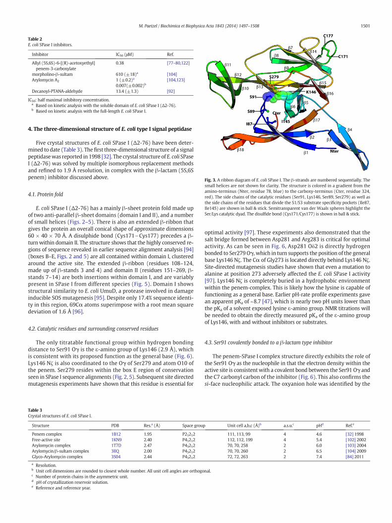
Table 2E. coli SPase I inhibitors.
Inhibitor IC50 (μM) Ref.
Allyl (5S,6S)-6-[(R)-acetoxyethyl]penem-3-carboxylate
0.38 [77–80,122]
morpholino-β-sultam 610 (±18)a [104]Arylomycin A2 1 (±0.2)a
0.007(±0.002)b[104,123]
Decanoyl-PTANA-aldehyde 13.4 (±1.3) [92]
IC50: half maximal inhibitory concentration.a Based on kinetic analysis with the soluble domain of E. coli SPase I (Δ2-76).b Based on kinetic analysis with the full-length E. coli SPase I.
Fig. 3. A ribbon diagram of E. coli SPase I. The β-strands are numbered sequentially. Thesmall helices are not shown for clarity. The structure is colored in a gradient from theamino-terminus (Nter, residue 78, blue) to the carboxy-terminus (Cter, residue 324,red). The side chains of the catalytic residues (Ser91, Lys146, Ser89, Ser279) as well asthe side chains of the residues that divide the S1/S3 substrate specificity pockets (Ile87,Ile145) are shown in ball & stick. Semitransparent van der Waals spheres highlight theSer/Lys catalytic dyad. The disulfide bond (Cys171/Cys177) is shown in ball & stick.
1501M. Paetzel / Biochimica et Biophysica Acta 1843 (2014) 1497–1508
4. The three-dimensional structure of E. coli type I signal peptidase
Five crystal structures of E. coli SPase I (Δ2-76) have been deter-mined to date (Table 3). Thefirst three-dimensional structure of a signalpeptidasewas reported in 1998 [32]. The crystal structure of E. coli SPaseI (Δ2-76) was solved by multiple isomorphous replacement methodsand refined to 1.9 Å resolution, in complex with the β-lactam (5S,6Spenem) inhibitor discussed above.
4.1. Protein fold
E. coli SPase I (Δ2-76) has a mainly β-sheet protein fold made upof two anti-parallel β-sheet domains (domain I and II), and a numberof small helices (Figs. 2–5). There is also an extended β-ribbon thatgives the protein an overall conical shape of approximate dimensions60 × 40 × 70 Å. A disulphide bond (Cys171\Cys177) precedes a β-turnwithin domain II. The structure shows that the highly conserved re-gions of sequence revealed in earlier sequence alignment analysis [94](boxes B–E, Figs. 2 and 5) are all contained within domain I, clusteredaround the active site. The extended β-ribbon (residues 108–124,made up of β-stands 3 and 4) and domain II (residues 151–269, β-stands 7–14) are both insertions within domain I, and are variablypresent in SPase I from different species (Fig. 5). Domain I showsstructural similarity to E. coli UmuD, a protease involved in damageinducible SOS mutagenesis [95]. Despite only 17.4% sequence identi-ty in this region, 69Cα atoms superimpose with a root mean squaredeviation of 1.6 Å [96].
4.2. Catalytic residues and surrounding conserved residues
The only titratable functional group within hydrogen bondingdistance to Ser91 Oγ is the ε-amino group of Lys146 (2.9 Å), whichis consistent with its proposed function as the general base (Fig. 6).Lys146 Nζ is also coordinated to the Oγ of Ser279 and atom O10 ofthe penem. Ser279 resides within the box E region of conservationseen in SPase I sequence alignments (Fig. 2, 5). Subsequent site directedmutagenesis experiments have shown that this residue is essential for
Table 3Crystal structures of E. coli SPase I.
Structure PDB Res.a (Å) Space group
Penem complex 1B12 1.95 P21212Free-active site 1KN9 2.40 P41212Arylomycin complex 1T7D 2.47 P43212Arylomycin/β-sultam complex 3IIQ 2.00 P43212Glyco-Arylomycin complex 3S04 2.44 P43212
a Resolution.b Unit cell dimensions are rounded to closest whole number. All unit cell angles are orthogoc Number of protein chains in the asymmetric unit.d pH of crystallization reservoir solution.e Reference and reference year.
optimal activity [97]. These experiments also demonstrated that thesalt bridge formed between Asp281 and Arg283 is critical for optimalactivity. As can be seen in Fig. 6, Asp281 Oδ2 is directly hydrogenbonded to Ser279Oγ, which in turn supports the position of the generalbase Lys146 Nζ. The Cα of Gly273 is located directly behind Lys146 Nζ.Site-directed mutagenesis studies have shown that even a mutation toalanine at position 273 adversely affected the E. coli SPase I activity[97]. Lys146 Nζ is completely buried in a hydrophobic environmentwithin the penem-complex. This is likely how the lysine is capable offunctioning as a general base. Earlier pH-rate profile experiments gavean apparent pKa of ~8.7 [47], which is nearly two pH units lower thanthe pKa of a solvent exposed lysine ε-amino group. NMR titrations willbe needed to obtain the directly measured pKa of the ε-amino groupof Lys146, with and without inhibitors or substrates.
4.3. Ser91 covalently bonded to a β-lactam type inhibitor
The penem-SPase I complex structure directly exhibits the role ofthe Ser91 Oγ as the nucleophile in that the electron density within theactive site is consistentwith a covalent bond between the Ser91 Oγ andthe C7 carbonyl carbon of the inhibitor (Fig. 6). This also confirms thesi-face nucleophilic attack. The oxyanion hole was identified by the
Unit cell a,b,c (Å)b a.s.u.c pHd Ref.e
111, 113, 99 4 4.6 [32] 1998112, 112, 199 4 5.4 [102] 200270, 70, 258 2 6.0 [103] 200470, 70, 260 2 6.5 [104] 200972, 72, 263 2 7.4 [84] 2011
nal.

Fig. 4. A Cα trace of E. coli SPase I. The figure is prepared in stereo. Every tenth residue is designated with a sphere and labeled. Side chains are shown for catalytic residues (Ser89, Ser91,Ser279, and Lys146) and the disulfide bond (Cys171/Cys177).
1502 M. Paetzel / Biochimica et Biophysica Acta 1843 (2014) 1497–1508
O8 carbonyl oxygen of the inhibitor pointing towards the NH ofSer91. The only other potential contributor to the oxyanion hole isthe hydroxyl group of Ser89, but the inhibitor has forced the χ1-angle of the Ser89 side chain into a position such that it is not avail-able to form the hydrogen bond to the would-be oxyanion. This maybe one of the means by which this inhibitor stabilizes its covalent at-tachment to the nucleophile. Subsequent site-directed mutagenesis ex-periments showed that mutating Ser89 to threonine results in anenzyme with almost wild-type activity, whereas mutating Ser89 to analanine or cysteine results in an enzyme with drastically lower activity[98]. The change in catalytic activity was found to bemostly due to a de-crease in the kcat while the Km did not change significantly. The calculat-ed differential free energy of transition stabilization provided by theSer89 hydroxyl groupwas found to be 5.2 kcal/mol. Another interestingobservation, within the active site of the structure, was that there were
Fig. 5. The conserved boxes of sequence seen within SPase I sequence alignments aremapped onto a Cα trace of the structure of E. coli SPase I. Boxes B (residues: 89–97), C (res-idues: 128–135), D (residues: 143–154) and E (residues: 273–283) are shown in red,green, blue and yellow respectively. Domain I is rendered in white but includes the con-served box regions shown in color. Domain II (residues: 155–263) and the β-ribbon (resi-dues: 108–124) are shown in black.
no suitably placed waters that could potentially function in thedeacylation step. Displacement of the deacylating water again mayhelp explain the inhibitory properties of this compound.
Fig. 6. Structure of a β-lactam (penem) type inhibitor co-crystallizedwith E. coli SPase I. A.The structure of the β-lactam-type inhibitor allyl (5S,6S)-6-[(R)-acetoxyethyl]- penem-3-carboxylate. B. Structure of the residues of the E. coli SPase I active site with the 5S,6S-penem covalently bound to the Ser91Oγ. Note that the bond between the carbonyl carbon(C7) and nitrogen (N4) within the penem is broken upon forming the acyl–enzyme esterbond to the Ser91Oγ. The carbon atoms of SPase I are in white. The carbon atoms of thepenem are in green. Nitrogen atoms are blue, oxygen atoms are red, and sulfur atomsare gold. C. Structure of the penem-SPase I complex with the SPase I rendered as solventaccessible surface. The C16 methyl group of the inhibitor is pointing into the S1 substratespecificity pocket. D. The si-face of a scissile bond.

1503M. Paetzel / Biochimica et Biophysica Acta 1843 (2014) 1497–1508
4.4. Substrate binding groove and the S1 and S3 specificity pockets
The structure explained a great deal about the SPase I substrate pref-erences. A methyl group on the penem inhibitor (C16), which wasshown to be essential for the effectiveness of the inhibitor, is pointinginto a shallow hydrophobic pocket on the E. coli SPase I surface — theS1 substrate specificity pocket [77] (Fig. 6C). The S1 pocket is made ofatoms from the residues: Met92, Ile145, Leu96 and Ile87 (Fig. 7). Theshallow hydrophobic S1 pocket is consistent with the strong preferencefor alanine at the P1 position of the SPase I preprotein substrates (Fig. 1).
Using the inhibitor as a guide, an extended poly-alanine β-strandwas modeled into the E. coli substrate-binding groove, which is con-structed on one side by the loop region following β-strand 1 thatleads to the nucleophile Ser91, and on the other side by the residuesfrom β-strand 6. Most proteases bind their substrates in an extendedβ-conformation [99]. This model allowed for the identification of theS3 substrate specificity binding pocket, which is made up of atomsfrom residues: Phe85, Ile87, Ile102, Val133, Ile145, and Asp143(Fig. 7). The residues Ile87 and Ile145 form a ridge between the S1and S3 pockets. The alternating up and down orientation of residueside chains within the extended β-strandmodel of the signal peptideC-region explains the Ala-X-Ala substrate preference of SPase I, inthat the P1 and P3 alanine residues are pointing into the shallow hy-drophobic pockets and the P2 residue is pointing out into the solventallowing for any side chain at this position. The structural informa-tion on the S1 and S3 binding pockets was used to design site-directed mutants to elucidate the residues that control secretorypreprotein cleavage fidelity [100]. It was found that mutatingIle145 to cysteine resulted in cleavage at multiple sites within thesubstrate and, if Ile145 and Ile87 were both mutated to alanine,SPase I was capable of cleaving after phenylalanine. It was also dis-covered that the double mutant I145C/I87C or I145C/I87T, whichmimics the residues at these positions within the mitochondrial homo-log (Imp1), was able to cleave substrates with an asparagine at the P1position, as preferred by Imp1. Later, the mutants I145C/I87T, I145C,and I145A were shown by combinatorial peptide library analysis tohave a relaxed substrate preference at the P3 position. The double mu-tantwas able to process substrates with arginine, glutamine, or tyrosineat the P3 position [101].
Fig. 7.A stereo ball & stick rendering of the empty substrate binding groove and active site of E. care in red. The proposed deacylating water is shown as a red sphere. The S1 and S3 substratebonds that involve the general base Lys146 Nζ are shown as dashed lines. Ile87 and Ile145 con
4.5. Structure of E. coli SPase I with a free substrate binding site
The crystal structure of E. coli SPase I in the absence of bound in-hibitor allowed for a structural comparison between the bound andunbound states of the active site [102]. This analysis revealed signif-icant main chain and side chain differences within the substratebinding groove and the active site that result in a smaller S1 pocketin the inhibitor free enzyme. In addition, the position of the Ser89side chain (OγH), in the absence of the penem inhibitor, is consistentwith its contribution to transition state oxyanion stabilization. A po-tential deacylating water was also identified (Fig. 7).
4.6. Structures of lipohexapeptide based inhibitors (arylomycins) bound toE. coli SPase I
The first crystal structure of arylomycin bound to E. coli SPase I re-vealed that the peptide based inhibitor binds non-covalently and ispositioned such that one of its C-terminal carboxylate oxygens iswithin hydrogen bonding distance to all of the functional groupswithin the catalytic center of the enzyme (Ser91 Oγ, Lys146Nζ, andSer89Oγ) (Fig. 8A, C) [103]. The inhibitor is therefore positioned sothat the macrocycle is closest to the active site, with the main chain ofthe peptide having parallel β-sheet type hydrogen bonding interactionswith both sides of the substrate-binding groove. All of the potential hy-drogen bonding donor and acceptors within the three residuemacrocycle are positioned to make hydrogen bonds with atoms in thebinding groove, whereas only two of the six potential hydrogen bond-ing donors and acceptors in the N-terminal three residues of the inhib-itor form hydrogen bonds. The side chain methyl group of thepenultimate alanine of the inhibitor sits within the S3 pocket. Weakelectron density was observed for the N-terminal fatty acid, suggestingit is dynamic in nature. The position of the N-terminal D-MeSer is locat-ed near the proposed membrane association surface. Both the crystalstructure and spectroscopic data are consistent with arylomycin bind-ing specifically to a single site. Fluorescence data are consistent with atwo-step binding mechanism — a rapid binding step followed by aslower adjustment to a final bound state. Binding parameters for this in-hibitor were also investigated using calorimetric methods.
oli SPase I. Carbon atoms are shown inwhite, nitrogen atoms are in blue and oxygen atomsspecificity pockets are highlighted with light green semi-transparent ovals. The hydrogentribute atoms to both pockets and make up the dividing point between the pockets.

Fig. 8. Crystal structures of arylomycin variants bound to E. coli SPase I. A. The structure of arylomycin. D-MeSer represents D-methyl serine, MeHpg stands for N-methyl-4-hydroxyphenyglycine. R1, R2, and R3 are defined below each structure. B. The morpholino-β-sultam derivative (BAL0019193). C. Arylomycin A2 (green carbon atoms) bound withinthe active site of E. coli SPase I (white carbon atoms). D. The ternary complex of Arylomycin A2 (green carbon atoms) and the morpholino-β-sultam derivative (yellow carbon atoms)bound within the active site of E. coli SPase I (white carbon atoms). E. The lipo-glycopeptide (glyco-arylomycin, green carbon atoms) bound within the active site of E. coli SPase I(white carbon atoms). The PDB accession code is listed above each structure. All nitrogen atoms are blue and oxygen atoms are red. The residues Ser91 and Pro84 are labeled.
Fig. 9. The proposedmembrane association surface of E. coli SPase I. To provide a perspectiveof the bilayer depth and the active site position relative to the proposed membrane associa-tion surface, a pair of phosphatidylethanolamine phospholipid molecules (van derWaals spheres — carbon, green; hydrogen, white; oxygen, red; nitrogen, blue) froma phospholipid bilayer structure simulation was rendered within the same file as the cat-alytic domain of E. coli SPase I. SPase I is shown as a black ribbonwith themolecular surfaceshown as a semi-transparent gray outline, side chains for residues Trp301, Trp311, Ser91and Lys146 are shown. Within the lipid bilayer, the distance from glycerol backbone toglycerol backbone is shown. The distance from the Ser91Oγ to the Trp301Cβ (a residueon the proposed membrane association surface) is shown.
1504 M. Paetzel / Biochimica et Biophysica Acta 1843 (2014) 1497–1508
The second structure of arylomycin bound to SPase I was in a ter-nary complex with another inhibitor, a morpholino-β-sultam deriv-ative (Fig. 8A, B, D) [104]. The structure and binding and inhibitionassays reveal that the compounds inhibit E. coli SPase I by bindingto non-overlapping subsites near the catalytic center. The β-sultamcompound binds in a noncovalent manner in close proximity toSPase I residues Ser89, Ser91, Lys146, Asn278, Ala280, and Glu308,as well as to a C-terminal carboxylate oxygen atom in arylomycin.There was clear electron density for the N-terminal fatty acid thatruns along the proposedmembrane association surface, near Trp301.
Themost recently reported crystal structure of an arylomycin–SPaseI complex was with a glycosylated form of arylomycin [84]. The struc-ture reveals that the deoxy-α-mannose attached to the MeHpg residueof the lipoglycopeptide is directed away from the active site into the sol-vent, suggesting that themodification may function to increase the sol-ubility of this natural product inhibitor (Fig. 8A, E).
Interestingly, it has been discovered that the presence of a SPase Ibinding site proline residue (at position 84, within β-strand 1 of E. coliSPase I) lends natural resistance to arylomycin's antibiotic activity inmany species of bacteria [89]. Binding assays revealed that the prolinemutation confers resistance by reducing the affinity of arylomycin tothe SPase I binding site. A proline at this position eliminates one poten-tial hydrogen bond donor on β-strand 1 of SPase I (Fig. 8).
4.7. Substrate recognition and the membrane association surface
Domain I of E. coli SPase I can be thought of as a β-barrel with onestrand missing. This missing β-strand makes up the substrate-bindinggroove. Based on the crystal structures of the inhibitor complexes, a
1505M. Paetzel / Biochimica et Biophysica Acta 1843 (2014) 1497–1508
model for signal peptide C-region recognition and binding can beproposed. During substrate recognition and binding, the C-regionof the signal peptide likely forms parallel β-sheet type hydrogenbonding interactions along β-strand 1 and the following loop thatleads to the nucleophile Ser91. The other side of the binding grooveis made up of residues from β-strand 6, which is also aligned in aparallel fashion to the signal peptide C-region. This side of thesubstrate-binding groove is fairly short and provides fewer potentialhydrogen-bond donors and acceptors. A structural model for this typeof interaction between the signal peptide of E. coli outermembrane pro-tein A (OmpA) and E. coli SPase I was presented previously [102]. Themodelwas guided by the E. coli SPase–penemcomplex crystal structure,
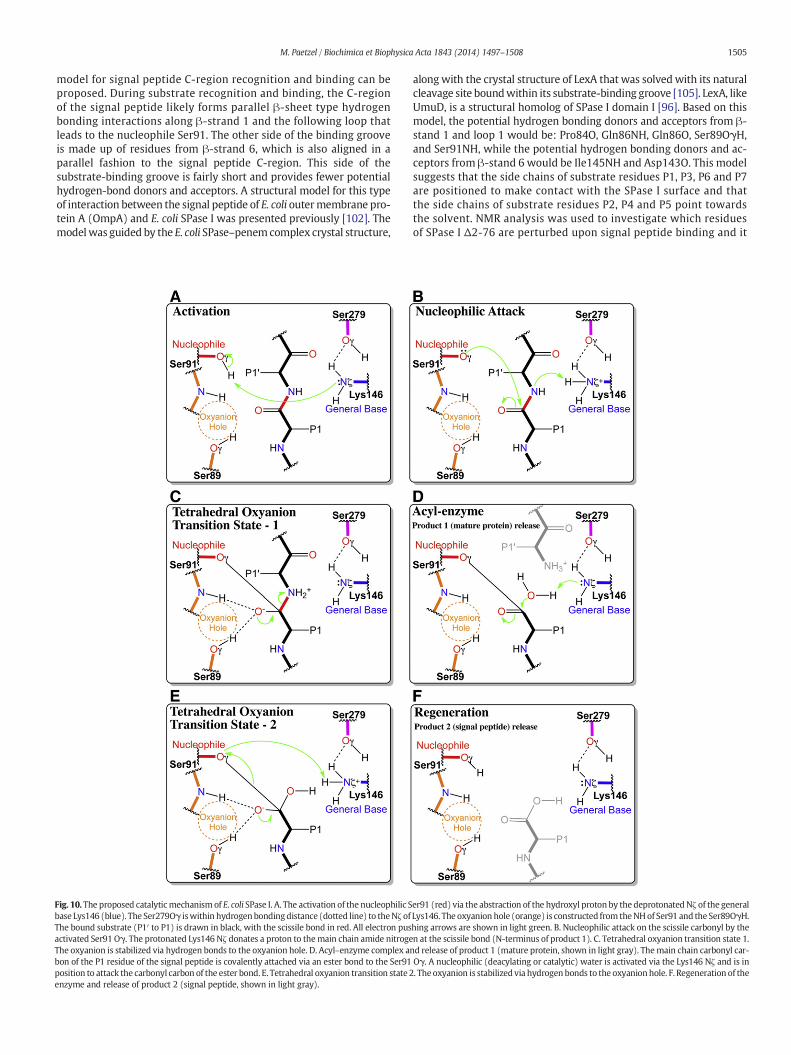
Fig. 10. The proposed catalyticmechanismof E. coli SPase I. A. The activation of the nucleophilic Sbase Lys146 (blue). The Ser279Oγ iswithin hydrogenbondingdistance (dotted line) to theNζ ofThe bound substrate (P1′ to P1) is drawn in black, with the scissile bond in red. All electron pusactivated Ser91 Oγ. The protonated Lys146 Nζ donates a proton to themain chain amide nitrogeThe oxyanion is stabilized via hydrogen bonds to the oxyanion hole. D. Acyl–enzyme complex anbon of the P1 residue of the signal peptide is covalently attached via an ester bond to the Ser91position to attack the carbonyl carbon of the ester bond. E. Tetrahedral oxyanion transition state 2enzyme and release of product 2 (signal peptide, shown in light gray).
alongwith the crystal structure of LexA that was solved with its naturalcleavage site boundwithin its substrate-binding groove [105]. LexA, likeUmuD, is a structural homolog of SPase I domain I [96]. Based on thismodel, the potential hydrogen bonding donors and acceptors from β-stand 1 and loop 1 would be: Pro84O, Gln86NH, Gln86O, Ser89OγH,and Ser91NH, while the potential hydrogen bonding donors and ac-ceptors from β-stand 6 would be Ile145NH and Asp143O. This modelsuggests that the side chains of substrate residues P1, P3, P6 and P7are positioned to make contact with the SPase I surface and thatthe side chains of substrate residues P2, P4 and P5 point towardsthe solvent. NMR analysis was used to investigate which residuesof SPase I Δ2-76 are perturbed upon signal peptide binding and it
er91 (red) via the abstraction of the hydroxyl proton by the deprotonatedNζ of the generalLys146. The oxyanion hole (orange) is constructed from theNHof Ser91 and the Ser89OγH.hing arrows are shown in light green. B. Nucleophilic attack on the scissile carbonyl by then at the scissile bond (N-terminus of product 1). C. Tetrahedral oxyanion transition state 1.d release of product 1 (mature protein, shown in light gray). Themain chain carbonyl car-Oγ. A nucleophilic (deacylating or catalytic) water is activated via the Lys146 Nζ and is in. The oxyanion is stabilized via hydrogen bonds to the oxyanionhole. F. Regeneration of the

1506 M. Paetzel / Biochimica et Biophysica Acta 1843 (2014) 1497–1508
was discovered that most of the perturbed residues map to the S1and S3 substrate binding pockets [106,107]. In addition, NMR hasbeen used to investigate the structure of the signal peptide uponbinding to SPase I Δ2-76 [108].
If the arylomycin lipopeptides are mimicking the signal peptide as-sociation contacts, then it is clear that sites far from the active site,such as Pro84, are important for substrate affinity. Since the full lengthE. coli SPase I is more active than the SPase I soluble domain, and fulllength substrates are cleaved more efficiently than synthetic peptidesubstrates that only contain the C-region residues, one could ask: arethere important contacts made between the SPase I transmembranesegment and the signal peptide H-region that dictate cleavage fidelity?Analysis of full length and soluble domains of both E. coli and Bacillussubtilis SPase I enzymes suggest there are not [109].
Studies have demonstrated that the accuracy of signal peptidecleavage by E. coli SPase I does not depend on the N-region, most ofthe H-region and also most of the mature region of preprotein sub-strates [110]. As mentioned earlier, site directed mutagenesis exper-iments show that the actual cleavage site fidelity is guidedsignificantly by the S1 and S3 substrate specificity pockets [100].
The relative position of the cleavage site and the SPase I substratebinding groove within or on the membrane surface likely providesan important contribution to the efficiency of the reaction giventhat peptide substrates with a hydrophobic H-region mimic se-quence, or a fatty acid attached to the N-terminus, are significantlymore effective substrates [67]. Therefore, it is important to knowwhat effect the membrane has on the secretory preprotein cleavagereaction and where the catalytic residues of SPase I are located,with respect to the preprotein cleavage site residues.
Purified E. coli SPase I incorporated within liposomes has beenused in a number of early investigations into SPase I activity[111,112] but most assays are performed with SPase I solubilizedwithin detergent micelles. Phospholipid vesicles with purifiedE. coli SPase I show that maximum catalytic activity is observedwith approximately 55% phosphotidylethanolamine, the most abun-dant phospholipid in the E. coli inner membrane. Phospholipids withnegatively charged head groups, found in abundance within theE. coli membrane, also enhance the catalytic activity [61]. Previousto this study, it was shown via membrane surface tension experi-ments and vesicle binding assays that phosphotidylethanolaminehelps facilitate insertion of the catalytic domain of E. coli SPase Iinto the membrane [113].
The crystal structure of E. coli SPase I revealed that a hydrophobicsurface runs along the full length of the enzyme, including the hydro-phobic substrate binding site [32]. This surface is likely involved inmembrane association. Located on this predicted membrane associa-tion surface are a number of aromatic residues. Trp301 has beenshown to be essential for optimal activity in E. coli SPase I [75,114]even though the crystal structure maps it to a position more than20 Å from the enzyme catalytic center (Fig. 9). It appears reasonablethat Trp301 and possibly Trp311 may help facilitate the insertion ofthe catalytic region of E. coli SPase I into themembrane. Sequence align-ments indicate that several conserved aromatic or hydrophobic resi-dues exist on the proposed membrane-association surface in bothGram-positive and Gram-negative bacterial type I SPases [115–117].Given the short length ofmost signal peptide H-regions, and the hydro-phobic nature of the proposed SPase I membrane association surface, itis very possible that the cleavage event occurs well within the lipidbilayer.
There is precedence for the hydrolysis of a peptide bond withinthe hydrophobic confines of the phospholipid bilayer. Rhomboidproteases utilize a Ser/His catalytic dyad to perform their catalysisin the lipid bilayer [118], and there are other intramembrane prote-ases that utilize aspartyl- and metallo-protease mechanisms [119].Interestingly, the remnant signal peptide left behind as a product ofSPase I catalysis is cleaved within its H-region by an enzyme called
signal peptide peptidase (SppA). SppA is likely an intramembraneprotease and also utilizes a Ser/Lys dyad mechanism [120,121].
5. Catalytic mechanism of E. coli SPase I
Based on kinetic analysis of site-directed mutants, chemical modifi-cation assays, as well as crystal structures (with andwithout inhibitors)a catalytic mechanism has been proposed for E. coli SPase I that is sum-marized in Fig. 10. Although the general components of the catalyticcenter (nucleophile: Ser91Oγ, general-base: Lys146Nζ, oxyanion hole:Ser91NH/Ser89OγH, and general-base positioning residue: Ser279Oγ)appear to be defined, manymechanistic details remain to be investigat-ed. For example, NMR titrations will help determine directly the pKa ofthe general base Lys146 ε-amino group. It will be interesting to see howthe pKa of this functional group changes in the presence and absence ofbound substrates and inhibitors, and also with the coordinating residueSer279 mutated to other residues, in order to see if the mechanism uti-lized by SPase I is more accurately classified as a Ser/Lys/Ser catalytictriad rather than a Ser/Lys dyad.
Acknowledgement
This work was supported in part by the Canadian Institute of HealthResearch and the National Science and Engineering Research Council ofCanada.
References
[1] K.E. Chatzi, M.F. Sardis, S. Karamanou, A. Economou, Breaking on through to theother side: protein export through the bacterial Sec system, Biochem. J. 449(2013) 25–37.
[2] A.J. Driessen, N. Nouwen, Protein translocation across the bacterial cytoplasmicmembrane, Annu. Rev. Biochem. 77 (2008) 643–667.
[3] S. Chimalapati, K. Sankaran, J.S. Brown, Chapter 62— signal peptidase II, Handbookof Proteolytic Enzymes, Academic Press, 2013, pp. 258–261.
[4] B. Dupuy, A.-E. Deghmane, M.-K. Taha, Chapter 63 — type IV prepilin peptidase,Handbook of Proteolytic Enzymes, Academic Press, 2013, pp. 261–265.
[5] G. von Heijne, The signal peptide, J. Membr. Biol. 115 (1990) 195–201.[6] G. von Heijne, Signal sequences. The limits of variation, J. Mol. Biol. 184 (1985)
99–105.[7] G. von Heijne, Patterns of amino acids near signal-sequence cleavage sites, Eur. J.
Biochem. 133 (1983) 17–21.[8] D. Perlman, H.O. Halvorson, A putative signal peptidase recognition site and se-
quence in eukaryotic and prokaryotic signal peptides, J. Mol. Biol. 167 (1983)391–409.
[9] H. Nielsen, J. Engelbrecht, S. Brunak, G. von Heijne, A neural network method foridentification of prokaryotic and eukaryotic signal peptides and prediction oftheir cleavage sites, Int. J. Neural Syst. 8 (1997) 581–599.
[10] H. Nielsen, J. Engelbrecht, S. Brunak, G. von Heijne, Identification of prokaryotic andeukaryotic signal peptides and prediction of their cleavage sites, Protein Eng. 10(1997) 1–6.
[11] C. UniProt, Update on activities at the Universal Protein Resource (UniProt) in2013, Nucleic Acids Res. 41 (2013) D43–D47.
[12] R. Apweiler, A. Bairoch, C.H. Wu, W.C. Barker, B. Boeckmann, S. Ferro, E. Gasteiger,H. Huang, R. Lopez, M. Magrane, M.J. Martin, D.A. Natale, C. O'Donovan, N.Redaschi, L.S. Yeh, UniProt: the Universal Protein knowledgebase, Nucleic AcidsRes. 32 (2004) D115–D119.
[13] J.D. Bendtsen, H. Nielsen, G. von Heijne, S. Brunak, Improved prediction of signalpeptides: SignalP 3.0, J. Mol. Biol. 340 (2004) 783–795.
[14] T.N. Petersen, S. Brunak, G. von Heijne, H. Nielsen, SignalP 4.0: discriminatingsignal peptides from transmembrane regions, Nat. Methods 8 (2011) 785–786.
[15] D.N. Ivankov, S.H. Payne, M.Y. Galperin, S. Bonissone, P.A. Pevzner, D. Frishman,How many signal peptides are there in bacteria? Environ. Microbiol. 15 (2013)983–990.
[16] K.H. Choo, T.W. Tan, S. Ranganathan, SPdb—a signal peptide database, BMCBioinforma. 6 (2005) 249.
[17] K.E. Rudd, EcoGene: a genome sequence database for Escherichia coli K-12, NucleicAcids Res. 28 (2000) 60–64.
[18] J. Zhou, K.E. Rudd, EcoGene 3.0, Nucleic Acids Res. 41 (2013) D613–D624.[19] I.K. Dev, P.H. Ray, P. Novak, Minimum substrate sequence for signal peptidase I of
Escherichia coli, J. Biol. Chem. 265 (1990) 20069–20072.[20] L.M. Shen, J.I. Lee, S.Y. Cheng, H. Jutte, A. Kuhn, R.E. Dalbey, Use of site-directed
mutagenesis to define the limits of sequence variation tolerated for processingof the M13 procoat protein by the Escherichia coli leader peptidase, Biochemis-try 30 (1991) 11775–11781.
[21] A.L. Karamyshev, Z.N. Karamysheva, A.V. Kajava, V.N. Ksenzenko, M.A.Nesmeyanova, Processing of Escherichia coli alkaline phosphatase: role of the

1507M. Paetzel / Biochimica et Biophysica Acta 1843 (2014) 1497–1508
primary structure of the signal peptide cleavage region, J. Mol. Biol. 277 (1998)859–870.
[22] A.V. Kajava, S.N. Zolov, K.I. Pyatkov, A.E. Kalinin, M.A. Nesmeyanova, Processing ofEscherichia coli alkaline phosphatase. Sequence requirements and possible confor-mations of the −6 to −4 region of the signal peptide, J. Biol. Chem. 277 (2002)50396–50402.
[23] G.A. Barkocy-Gallagher, P.J. Bassford Jr., Synthesis of precursor maltose-bindingprotein with proline in the +1 position of the cleavage site interferes with the ac-tivity of Escherichia coli signal peptidase I in vivo, J. Biol. Chem. 267 (1992)1231–1238.
[24] W. Wickner, K. Moore, N. Dibb, D. Geissert, M. Rice, Inhibition of purifiedEscherichia coli leader peptidase by the leader (signal) peptide of bacteriophageM13 procoat, J. Bacteriol. 169 (1987) 3821–3822.
[25] I. Nilsson, G. von Heijne, A signal peptide with a proline next to the cleavage siteinhibits leader peptidase when present in a sec-independent protein, FEBS Lett.299 (1992) 243–246.
[26] B. Bechinger, L.M. Gierasch, M. Montal, M. Zasloff, S.J. Opella, Orientations of helicalpeptides in membrane bilayers by solid state NMR spectroscopy, Solid State Nucl.Magn. Reson. 7 (1996) 185–191.
[27] M.B. Sankaram, D. Marsh, L.M. Gierasch, T.E. Thompson, Reorganization of lipiddomain structure in membranes by a transmembrane peptide: an ESR spinlabel study on the effect of the Escherichia coli outer membrane protein A signalpeptide on the fluid lipid domain connectivity in binary mixtures ofdimyristoyl phosphatidylcholine and distearoyl phosphatidylcholine, Biophys.J. 66 (1994) 1959–1968.
[28] Z. Wang, J.D. Jones, J. Rizo, L.M. Gierasch, Membrane-bound conformation of a sig-nal peptide: a transferred nuclear Overhauser effect analysis, Biochemistry 32(1993) 13991–13999.
[29] J. Rizo, F.J. Blanco, B. Kobe, M.D. Bruch, L.M. Gierasch, Conformational behavior ofEscherichia coli OmpA signal peptides in membrane mimetic environments, Bio-chemistry 32 (1993) 4881–4894.
[30] C.J. McKnight, S.J. Stradley, J.D. Jones, L.M. Gierasch, Conformational andmembrane-binding properties of a signal sequence are largely unaltered byits adjacent mature region, Proc. Natl. Acad. Sci. U. S. A. 88 (1991) 5799–5803.
[31] C.J. McKnight, M. Rafalski, L.M. Gierasch, Fluorescence analysis oftryptophan-containing variants of the LamB signal sequence upon insertion into alipid bilayer, Biochemistry 30 (1991) 6241–6246.
[32] M. Paetzel, R.E. Dalbey, N.C. Strynadka, Crystal structure of a bacterial signal pepti-dase in complex with a beta-lactam inhibitor, Nature 396 (1998) 186–190.
[33] I. Luke, J.I. Handford, T. Palmer, F. Sargent, Proteolytic processing of Escherichia colitwin-arginine signal peptides by LepB, Arch. Microbiol. 191 (2009) 919–925.
[34] T. Palmer, B.C. Berks, The twin-arginine translocation (Tat) protein export path-way, Nat. Rev. Microbiol. 10 (2012) 483–496.
[35] D. Tullman-Ercek, M.P. DeLisa, Y. Kawarasaki, P. Iranpour, B. Ribnicky, T. Palmer, G.Georgiou, Export pathway selectivity of Escherichia coli twin arginine translocationsignal peptides, J. Biol. Chem. 282 (2007) 8309–8316.
[36] C.N. Chang, G. Blobel, P. Model, Detection of prokaryotic signal peptidase in anEscherichia coli membrane fraction: endoproteolytic cleavage of nascent f1pre-coat protein, Proc. Natl. Acad. Sci. U. S. A. 75 (1978) 361–365.
[37] C. Milstein, G.G. Brownlee, T.M. Harrison, M.B. Mathews, A possible precursor ofimmunoglobulin light chains, Nat. New Biol. 239 (1972) 117–120.
[38] T. Date, W. Wickner, Isolation of the Escherichia coli leader peptidase gene and ef-fects of leader peptidase overproduction in vivo, Proc. Natl. Acad. Sci. U. S. A. 78(1981) 6106–6110.
[39] P.B. Wolfe, W. Wickner, J.M. Goodman, Sequence of the leader peptidase gene ofEscherichia coli and the orientation of leader peptidase in the bacterial envelope,J. Biol. Chem. 258 (1983) 12073–12080.
[40] P.B. Wolfe, P. Silver, W. Wickner, The isolation of homogeneous leader peptidasefrom a strain of Escherichia coli which overproduces the enzyme, J. Biol. Chem.257 (1982) 7898–7902.
[41] R.E. Dalbey, W. Wickner, Leader peptidase catalyzes the release of exported pro-teins from the outer surface of the Escherichia coli plasma membrane, J. Biol.Chem. 260 (1985) 15925–15931.
[42] W.R. Tschantz, R.E. Dalbey, Bacterial leader peptidase 1, Methods Enzymol. 244(1994) 285–301.
[43] C. Zwizinski, W. Wickner, Purification and characterization of leader (signal) pep-tidase from Escherichia coli, J. Biol. Chem. 255 (1980) 7973–7977.
[44] M. Sung, R.E. Dalbey, Identification of potential active-site residues in theEscherichia coli leader peptidase, J. Biol. Chem. 267 (1992) 13154–13159.
[45] W.R. Tschantz, M. Sung, V.M. Delgado-Partin, R.E. Dalbey, A serine and a lysine res-idue implicated in the catalytic mechanism of the Escherichia coli leader peptidase,J. Biol. Chem. 268 (1993) 27349–27354.
[46] S. Chatterjee, D. Suciu, R.E. Dalbey, P.C. Kahn, M. Inouye, Determination of Km andkcat for signal peptidase I using a full length secretory precursor,pro-OmpA-nuclease A, J. Mol. Biol. 245 (1995) 311–314.
[47] M. Paetzel, N.C. Strynadka, W.R. Tschantz, R. Casareno, P.R. Bullinger, R.E. Dalbey,Use of site-directed chemical modification to study an essential lysine inEscherichia coli leader peptidase, J. Biol. Chem. 272 (1997) 9994–10003.
[48] W. van Klompenburg, P. Whitley, R. Diemel, G. von Heijne, B. de Kruijff, A quanti-tative assay to determine the amount of signal peptidase I in E. coli and the orien-tation of membrane vesicles, Mol. Membr. Biol. 12 (1995) 349–353.
[49] P. Silver, W. Wickner, Genetic mapping of the Escherichia coli leader (signal) pep-tidase gene (lep): a new approach for determining the map position of a clonedgene, J. Bacteriol. 154 (1983) 569–572.
[50] T. Date, Demonstration by a novel genetic technique that leader peptidase is an es-sential enzyme of Escherichia coli, J. Bacteriol. 154 (1983) 76–83.
[51] T. Inada, D.L. Court, K. Ito, Y. Nakamura, Conditionally lethal amber mutations inthe leader peptidase gene of Escherichia coli, J. Bacteriol. 171 (1989) 585–587.
[52] N.D. Rawlings, A.J. Barrett, A. Bateman, MEROPS: the peptidase database, NucleicAcids Res. 38 (2010) D227–D233.
[53] D.W. Kuo, H.K. Chan, C.J. Wilson, P.R. Griffin, H. Williams, W.B. Knight, Escherichiacoli leader peptidase: production of an active form lacking a requirement for deter-gent and development of peptide substrates, Arch. Biochem. Biophys. 303 (1993)274–280.
[54] P. Whitley, G. von Heijne, The DsbA–DsbB system affects the formation of disulfidebonds in periplasmic but not in intramembraneous protein domains, FEBS Lett.332 (1993) 49–51.
[55] K.E. Moore, S. Miura, A small hydrophobic domain anchors leader peptidase to thecytoplasmic membrane of Escherichia coli, J. Biol. Chem. 262 (1987) 8806–8813.
[56] J.L. San Millan, D. Boyd, R. Dalbey, W. Wickner, J. Beckwith, Use of phoA fusions tostudy the topology of the Escherichia coli inner membrane protein leader pepti-dase, J. Bacteriol. 171 (1989) 5536–5541.
[57] P. Whitley, L. Nilsson, G. von Heijne, Three-dimensional model for the membranedomain of Escherichia coli leader peptidase based on disulfide mapping, Biochem-istry 32 (1993) 8534–8539.
[58] N. Bilgin, J.I. Lee, H.Y. Zhu, R. Dalbey, G. von Heijne, Mapping of catalytically impor-tant domains in Escherichia coli leader peptidase, EMBO J. 9 (1990) 2717–2722.
[59] W.R. Tschantz, M. Paetzel, G. Cao, D. Suciu, M. Inouye, R.E. Dalbey, Characterizationof a soluble, catalytically active form of Escherichia coli leader peptidase: require-ment of detergent or phospholipid for optimal activity, Biochemistry 34 (1995)3935–3941.
[60] T.L. Talarico, I.K. Dev, P.J. Bassford Jr., P.H. Ray, Inter-molecular degradation of sig-nal peptidase I in vitro, Biochem. Biophys. Res. Commun. 181 (1991) 650–656.
[61] Y. Wang, R. Bruckner, R.L. Stein, Regulation of signal peptidase by phospholipids inmembrane: characterization of phospholipid bilayer incorporated Escherichia colisignal peptidase, Biochemistry 43 (2004) 265–270.
[62] M.T. Black, Evidence that the catalytic activity of prokaryote leader peptidase de-pends upon the operation of a serine-lysine catalytic dyad, J. Bacteriol. 175(1993) 4957–4961.
[63] P. Ray, I. Dev, C. MacGregor, P. Bassford Jr., Signal peptidases, Curr. Top. Microbiol.Immunol. 125 (1986) 75–102.
[64] J. Gallagher, N.N. Kaderbhai, M.A. Kaderbhai, Kinetic constants of signal peptidase Iusing cytochrome b5 as a precursor substrate, Biochim. Biophys. Acta 1550 (2001)1–5.
[65] D. Suciu, S. Chatterjee, M. Inouye, Catalytic efficiency of signal peptidase I ofEscherichia coli is comparable to that of members of the serine protease family,Protein Eng. 10 (1997) 1057–1060.
[66] W. Zhong, S.J. Benkovic, Development of an internally quenched fluorescent sub-strate for Escherichia coli leader peptidase, Anal. Biochem. 255 (1998) 66–73.
[67] R.L. Stein, M.D. Barbosa, R. Bruckner, Kinetic and mechanistic studies of signal pep-tidase I from Escherichia coli, Biochemistry 39 (2000) 7973–7983.
[68] G. Bruton, A. Huxley, P. O'Hanlon, B. Orlek, D. Eggleston, J. Humphries, S. Readshaw,A.West, S. Ashman, M. Brown, K. Moore, A. Pope, K. O'Dwyer, L. Wang, Lipopeptidesubstrates for SpsB, the Staphylococcus aureus type I signal peptidase: design, con-formation and conversion to alpha-ketoamide inhibitors, Eur. J. Med. Chem. 38(2003) 351–356.
[69] O.D. Ekici, J. Zhu, I.Y. Wah Chung, M. Paetzel, R.E. Dalbey, D. Pei, Profiling the sub-strate specificity of viral protease VP4 by a FRET-based peptide library approach,Biochemistry 48 (2009) 5753–5759.
[70] G. Rosse, E. Kueng, M.G. Page, V. Schauer-Vukasinovic, T. Giller, H.W. Lahm, P.Hunziker, D. Schlatter, Rapid identification of substrates for novel proteasesusing a combinatorial peptide library, J. Comb. Chem. 2 (2000) 461–466.
[71] K.M. Cregg, I. Wilding, M.T. Black, Molecular cloning and expression of the spsBgene encoding an essential type I signal peptidase from Staphylococcus aureus, J.Bacteriol. 178 (1996) 5712–5718.
[72] M.D. Barbosa, S. Lin, J.A. Markwalder, J.A. Mills, J.A. DeVito, C.A. Teleha, V. Garlapati,C. Liu, A. Thompson, G.L. Trainor, M.G. Kurilla, D.L. Pompliano, Regulated expres-sion of the Escherichia coli lepB gene as a tool for cellular testing of antimicrobialcompounds that inhibit signal peptidase I in vitro, Antimicrob. Agents Chemother.46 (2002) 3549–3554.
[73] C. Zwizinski, T. Date, W. Wickner, Leader peptidase is found in both the inner andouter membranes of Escherichia coli, J. Biol. Chem. 256 (1981) 3593–3597.
[74] M.T. Black, J.G. Munn, A.E. Allsop, On the catalytic mechanism of prokaryotic leaderpeptidase 1, Biochem. J. 282 (1992) 539–543.
[75] Y.T. Kim, T. Muramatsu, K. Takahashi, Leader peptidase from Escherichia coli: over-expression, characterization, and inactivation by modification of tryptophan resi-dues 300 and 310 with N-bromosuccinimide, J. Biochem. 117 (1995) 535–544.
[76] D. Kuo, J. Weidner, P. Griffin, S.K. Shah, W.B. Knight, Determination of the ki-netic parameters of Escherichia coli leader peptidase activity using a contin-uous assay: the pH dependence and time-dependent inhibition bybeta-lactams are consistent with a novel serine protease mechanism, Bio-chemistry 33 (1994) 8347–8354.
[77] M.T. Black, G. Bruton, Inhibitors of bacterial signal peptidases, Curr. Pharm. Des. 4(1998) 133–154.
[78] A. Allsop, G. Brooks, P.D. Edwards, A.C. Kaura, R. Southgate, Inhibitors of bacterialsignal peptidase: a series of 6-(substituted oxyethyl)penems, J. Antibiot. (Tokyo)49 (1996) 921–928.
[79] C.R. Perry, M.J. Ashby, S.A. Elsmere, Penems as research tools to investigate the ac-tivity of E.coli leader peptidase, Biochem. Soc. Trans. 23 (1995) 548S.
[80] A.E. Allsop, G. Brooks, G. Bruton, S. Coulton, P.D. Edwards, I.K. Hatton, A.C. Kaura,S.D. McLean, N.D. Pearson, T.C. Smale, R. Southgate, Penem inhibitors of bacterialsignal peptidase, Bioorg. Med. Chem. Lett. 5 (1995) 443–448.
1508 M. Paetzel / Biochimica et Biophysica Acta 1843 (2014) 1497–1508
[81] D.A. Harris, M.E. Powers, F.E. Romesberg, Synthesis and biological evaluation ofpenem inhibitors of bacterial signal peptidase, Bioorg. Med. Chem. Lett. 19(2009) 3787–3790.
[82] J. Schimana, K. Gebhardt, A. Holtzel, D.G. Schmid, R. Sussmuth, J. Muller, R. Pukall,H.P. Fiedler, Arylomycins A and B, new biaryl-bridged lipopeptide antibiotics pro-duced by Streptomyces sp. Tu 6075. I. Taxonomy, fermentation, isolation andbiological activities, J. Antibiot. (Tokyo) 55 (2002) 565–570.
[83] A. Holtzel, D.G. Schmid, G.J. Nicholson, S. Stevanovic, J. Schimana, K. Gebhardt, H.P.Fiedler, G. Jung, Arylomycins A and B, new biaryl-bridged lipopeptide antibioticsproduced by Streptomyces sp. Tu 6075. II. Structure elucidation, J. Antibiot.(Tokyo) 55 (2002) 571–577.
[84] J. Liu, C. Luo, P.A. Smith, J.K. Chin, M.G. Page, M. Paetzel, F.E. Romesberg, Synthesisand characterization of the arylomycin lipoglycopeptide antibiotics and the crys-tallographic analysis of their complex with signal peptidase, J. Am. Chem. Soc.133 (2011) 17869–17877.
[85] T.C. Roberts, M.A. Schallenberger, J. Liu, P.A. Smith, F.E. Romesberg, Initial efforts to-ward the optimization of arylomycins for antibiotic activity, J. Med. Chem. 54(2011) 4954–4963.
[86] T.C. Roberts, P.A. Smith, R.T. Cirz, F.E. Romesberg, Structural and initial biologicalanalysis of synthetic arylomycin A2, J. Am. Chem. Soc. 129 (2007) 15830–15838.
[87] T.C. Roberts, P.A. Smith, F.E. Romesberg, Synthesis and biological characterizationof arylomycin B antibiotics, J. Nat. Prod. 74 (2011) 956–961.
[88] P.A. Smith, M.E. Powers, T.C. Roberts, F.E. Romesberg, In vitro activities of arylomycinnatural-product antibiotics against Staphylococcus epidermidis and other coagulase-negative staphylococci, Antimicrob. Agents Chemother. 55 (2011) 1130–1134.
[89] P.A. Smith, T.C. Roberts, F.E. Romesberg, Broad-spectrum antibiotic activity of thearylomycin natural products is masked by natural target mutations, Chem. Biol.17 (2010) 1223–1231.
[90] P.A. Smith, F.E. Romesberg, Mechanism of action of the arylomycin antibiotics andeffects of signal peptidase I inhibition, Antimicrob. Agents Chemother. 56 (2012)5054–5060.
[91] W.T. Liu, R.D. Kersten, Y.L. Yang, B.S. Moore, P.C. Dorrestein, Imagingmass spectrome-try and genomemining via short sequence tagging identified the anti-infective agentarylomycin in Streptomyces roseosporus, J. Am. Chem. Soc. 133 (2011) 18010–18013.
[92] P. Buzder-Lantos, K. Bockstael, J. Anne, P. Herdewijn, Substrate based peptide alde-hyde inhibits bacterial type I signal peptidase, Bioorg. Med. Chem. Lett. 19 (2009)2880–2883.
[93] M. Paetzel, M. Chernaia, N. Strynadka, W. Tschantz, G. Cao, R.E. Dalbey, M.N. James,Crystallization of a soluble, catalytically active form of Escherichia coli leader pepti-dase, Proteins 23 (1995) 122–125.
[94] R.E. Dalbey, M.O. Lively, S. Bron, J.M. van Dijl, The chemistry and enzymology of thetype I signal peptidases, Protein Sci. 6 (1997) 1129–1138.
[95] M. Paetzel, R. Woodgate, Chapter 773 — UmuD and UmuD′ proteins, Handbook ofProteolytic Enzymes, Academic Press, 2013, pp. 3487–3492.
[96] M. Paetzel, N.C. Strynadka, Common protein architecture and binding sites in pro-teases utilizing a Ser/Lys dyad mechanism, Protein Sci. 8 (1999) 2533–2536.
[97] P.A. Klenotic, J.L. Carlos, J.C. Samuelson, T.A. Schuenemann, W.R. Tschantz, M.Paetzel, N.C. Strynadka, R.E. Dalbey, The role of the conserved box E residues inthe active site of the Escherichia coli type I signal peptidase, J. Biol. Chem. 275(2000) 6490–6498.
[98] J.L. Carlos, P.A. Klenotic, M. Paetzel, N.C. Strynadka, R.E. Dalbey, Mutational evi-dence of transition state stabilization by serine 88 in Escherichia coli type I signalpeptidase, Biochemistry 39 (2000) 7276–7283.
[99] J.D. Tyndall, T. Nall, D.P. Fairlie, Proteases universally recognize beta strands in theiractive sites, Chem. Rev. 105 (2005) 973–999.
[100] A. Karla, M.O. Lively, M. Paetzel, R. Dalbey, The identification of residues that con-trol signal peptidase cleavage fidelity and substrate specificity, J. Biol. Chem. 280(2005) 6731–6741.
[101] O.D. Ekici, A. Karla, M. Paetzel, M.O. Lively, D. Pei, R.E. Dalbey, Altered−3 substratespecificity of Escherichia coli signal peptidase 1 mutants as revealed by screening acombinatorial peptide library, J. Biol. Chem. 282 (2007) 417–425.
[102] M. Paetzel, R.E. Dalbey, N.C. Strynadka, Crystal structure of a bacterial signal pepti-dase apoenzyme: implications for signal peptide binding and the Ser–Lys dyadmechanism, J. Biol. Chem. 277 (2002) 9512–9519.
[103] M. Paetzel, J.J. Goodall, M. Kania, R.E. Dalbey, M.G. Page, Crystallographic andbiophysical analysis of a bacterial signal peptidase in complex with alipopeptide-based inhibitor, J. Biol. Chem. 279 (2004) 30781–30790.
[104] C. Luo, P. Roussel, J. Dreier, M.G. Page, M. Paetzel, Crystallographic analysis of bac-terial signal peptidase in ternary complex with arylomycin A2 and a beta-sultaminhibitor, Biochemistry 48 (2009) 8976–8984.
[105] Y. Luo, R.A. Pfuetzner, S. Mosimann, M. Paetzel, E.A. Frey, M. Cherney, B. Kim, J.W.Little, N.C. Strynadka, Crystal structure of LexA: a conformational switch for regu-lation of self-cleavage, Cell 106 (2001) 585–594.
[106] M. Musial-Siwek, D.A. Kendall, P.L. Yeagle, Solution NMR of signal peptidase, amembrane protein, Biochim. Biophys. Acta 1778 (2008) 937–944.
[107] M. Musial-Siwek, P.L. Yeagle, D.A. Kendall, A small subset of signal peptidase resi-dues are perturbed by signal peptide binding, Chem. Biol. Drug Des. 72 (2008)140–146.
[108] P. De Bona, L. Deshmukh, V. Gorbatyuk, O. Vinogradova, D.A. Kendall, Structuralstudies of a signal peptide in complex with signal peptidase I cytoplasmic domain:the stabilizing effect of membrane-mimetics on the acquired fold, Proteins 80(2012) 807–817.
[109] J.L. Carlos,M. Paetzel, G. Brubaker, A. Karla, C.M.Ashwell,M.O. Lively, G. Cao, P. Bullinger,R.E. Dalbey, The role of the membrane-spanning domain of type I signal peptidases insubstrate cleavage site selection, J. Biol. Chem. 275 (2000) 38813–38822.
[110] R. Dierstein,W.Wickner, Requirements for substrate recognition by bacterial lead-er peptidase, EMBO J. 5 (1986) 427–431.
[111] C. Watts, P. Silver, W. Wickner, Membrane assembly from purified components. II.Assembly of M13 procoat into liposomes reconstituted with purified leader pepti-dase, Cell 25 (1981) 347–353.
[112] Y. Ohno-Iwashita, P. Wolfe, K. Ito, W. Wickner, Processing of preproteins by lipo-somes bearing leader peptidase, Biochemistry 23 (1984) 6178–6184.
[113] W. van Klompenburg, M. Paetzel, J.M. de Jong, R.E. Dalbey, R.A. Demel, G. vonHeijne, B. de Kruijff, Phosphatidylethanolamine mediates insertion of thecatalytic domain of leader peptidase in membranes, FEBS Lett. 431 (1998)75–79.
[114] Y.T. Kim, T. Muramatsu, K. Takahashi, Identification of Trp300 as an important res-idue for Escherichia coli leader peptidase activity, Eur. J. Biochem. 234 (1995)358–362.
[115] J.L. Carlos, M. Paetzel, P.A. Klenotic, N.C.J. Strynadka, R.E. Dalbey, Bacterial type I sig-nal peptidases, in: E.D. Rose, S.S. David (Eds.), The Enzymes, volume 22, AcademicPress, 2002, pp. 27–55.
[116] M. Paetzel, R.E. Dalbey, N.C. Strynadka, The structure and mechanism of bacterialtype I signal peptidases. A novel antibiotic target, Pharmacol. Ther. 87 (2000)27–49.
[117] M. Paetzel, A. Karla, N.C. Strynadka, R.E. Dalbey, Signal peptidases, Chem. Rev. 102(2002) 4549–4580.
[118] M. Freeman, Rhomboid proteases and their biological functions, Annu. Rev. Genet.42 (2008) 191–210.
[119] E. Erez, D. Fass, E. Bibi, How intramembrane proteases bury hydrolytic reactions inthe membrane, Nature 459 (2009) 371–378.
[120] A.C. Kim, D.C. Oliver, M. Paetzel, Crystal structure of a bacterial signal peptide pep-tidase, J. Mol. Biol. 376 (2008) 352–366.
[121] P. Wang, E. Shim, B. Cravatt, R. Jacobsen, J. Schoeniger, A.C. Kim, M. Paetzel, R.E.Dalbey, Escherichia coli signal peptide peptidase A is a serine-lysine proteasewith a lysine recruited to the nonconserved amino-terminal domain in the S49protease family, Biochemistry 47 (2008) 6361–6369.
[122] A.C. Barbrook, J.C. Packer, C.J. Howe, Inhibition by penem of processing peptidasesfrom cyanobacteria and chloroplast thylakoids, FEBS Lett. 398 (1996) 198–200.
[123] K. Bockstael, N. Geukens, C.V. Rao, P. Herdewijn, J. Anne, A. Van Aerschot, An easyand fast method for the evaluation of Staphylococcus epidermidis type I signal pep-tidase inhibitors, J. Microbiol. Methods 78 (2009) 231–237.