Embed Size (px)
Citation preview
Biochimica et Biophysica Acta 1777 (2008) 1079–1091
Contents lists available at ScienceDirect
Biochimica et Biophysica Acta
j ourna l homepage: www.e lsev ie r.com/ locate /bbab io
Review
Regulatory interactions in the dimeric cytochrome bc1 complex: The advantagesof being a twin
Raul Covian, Bernard L. Trumpower ⁎Department of Biochemistry, Dartmouth Medical School, 7200 Vail, Hanover, New Hampshire 03755, USA
⁎ Corresponding author. Tel.: +1 603 650 1621; fax: +E-mail address: [email protected] (B.L. Tr
0005-2728/$ – see front matter © 2008 Elsevier B.V. Aldoi:10.1016/j.bbabio.2008.04.022
A B S T R A C T
A R T I C L E I N F OArticle history:
The dimeric cytochrome b Received 14 January 2008Received in revised form 10 April 2008Accepted 12 April 2008Available online 22 April 2008Keywords:bc1 complexElectron transferQuinoneStigmatellinAntimycin
c1 complex catalyzes the oxidation–reduction of quinol and quinone at siteslocated in opposite sides of the membrane in which it resides. We review the kinetics of electron transfer andinhibitor binding that reveal functional interactions between the quinol oxidation site at center P andquinone reduction site at center N in opposite monomers in conjunction with electron equilibration betweenthe cytochrome b subunits of the dimer. A model for the mechanism of the bc1 complex has emerged fromthese studies in which binding of ligands that mimic semiquinone at center N regulates half-of-the-sitesreactivity at center P and binding of ligands that mimic catalytically competent binding of ubiquinol at centerP regulates half-of-the-sites reactivity at center N. An additional feature of this model is that inhibition ofquinol oxidation at the quinone reduction site is avoided by allowing catalysis in only one monomer at atime, which maximizes the number of redox acceptor centers available in cytochrome b for electrons comingfrom quinol oxidation reactions at center P and minimizes the leakage of electrons that would result in thegeneration of damaging oxygen radicals.
© 2008 Elsevier B.V. All rights reserved.
1. Introduction
The cytochrome bc1 complex is present in the inner membrane ofmost mitochondria and heterotrophic bacteria. The functional core ofthis respiratory enzyme is comprised of the highly hydrophobicdiheme cytochrome b associated with the trans-membrane regions ofthe Rieske iron–sulfur protein and cytochrome c1. One of the mostsurprising features revealed by the atomic-resolution structures of thebc1 complex [1–5] is that this enzyme can only function as a dimer(Fig. 1). The inclination of the trans-membrane helix of the Rieskeprotein results in the interaction of its extramembrane domain withthe quinol oxidation site (center P) of cytochrome b in the oppositemonomer.
In the protonmotive Q cycle mechanism [[6], reviewed in Ref. [7]],one of the two electrons coming from the oxidation of quinol at centerP, located close to the positive side of the membrane, is transferred tocytochrome c via the Rieske protein and cytochrome c1. The otherelectron from quinol moves from the bL to the bH heme across themembrane dielectric and reduces quinone bound at center N to form astable, tightly bound semiquinone [8–10]. A further quinol oxidationevent at center P donates the electron needed to reduce semiquinoneto quinol at center N. The deprotonation of quinol at center P and theprotonation of quinone at center N results in the net movement ofprotons across the membrane, which together with the movement of
1 603 650 1128.umpower).
l rights reserved.
the negative charge of the electron from bL to bH, contributes to theformation of a protonmotive gradient.
Having two sites catalyzing the same net reaction in oppositedirections (oxido-reduction of quinol/quinone) introduces the poten-tial for significant inhibition of quinol oxidation at center P by thereversibility of reactions at center N. The lack of oxidized acceptors incytochrome b for the second electron coming from center P wouldresult in detrimental reactions, such as the one-electron reduction ofoxygen to form superoxide [11]. Therefore, mechanisms that avoid theaccumulation of electrons in cytochrome b are expected to exist in thebc1 complex. In this article, we review the experimental evidenceshowing how the dimeric structure of the bc1 complex allows theregulation of center P and center N in a manner that maximizes theavailability of electron acceptors in cytochrome b in order to avoidelectron leakage out of center P.
2. Half-of-the-sites activity at center P
2.1. Inactivation of one center P in the presence of antimycin
One possible mechanism that can be envisioned in order todecrease spurious electron transfer to oxygen in a dimeric bc1complex is to avoid simultaneous quinol oxidation at both center Psites. Evidence for the existence of this mechanism was first providedin pre-steady-state assays in which the semi-purified bovine complexwas reduced with duroquinol, a more hydrophilic analog of ubiquinol[12]. It was reported that with antimycin bound at both center N sitesonly half of the iron–sulfur clusters and of the c1 hemes were reduced
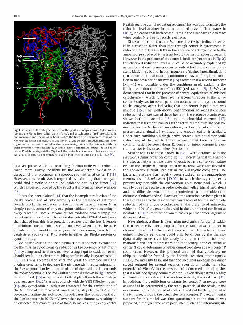
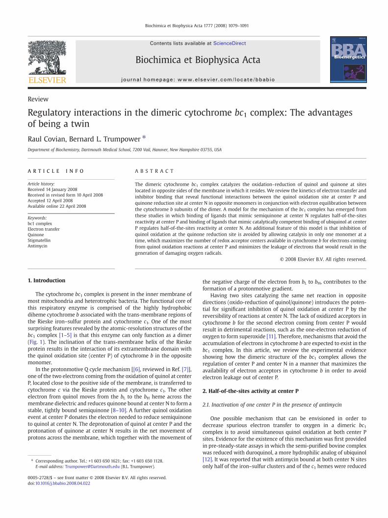
Fig. 1. Structure of the catalytic subunits of the yeast bc1 complex dimer. Cytochrome b(green), the Rieske iron–sulfur protein (blue), and cytochrome c1 (red) are colored inone monomer and shown as ribbons. Notice the tilted trans-membrane helix of theRieske protein that is imbedded in one monomer and connects through a flexible linkerregion to the extrinsic iron–sulfur cluster containing domain that interacts with theother monomer. Redox centers (c1, bL and bH hemes, and the FeS cluster), as well as thecenter P inhibitor stigmatellin (Stg) and the center N ubiquinone (Ubi) are shown asball-and-stick models. The structure is taken from Protein Data Bank code 1EZV [4].
1080 R. Covian, B.L. Trumpower / Biochimica et Biophysica Acta 1777 (2008) 1079–1091
in a fast phase, while the remaining fraction underwent reductionmuch more slowly, possibly by the one-electron oxidation ofduroquinol that accompanies superoxide formation at center P [11].However, this result was interpreted as indicating that antimycincould bind directly to one quinol oxidation site in the dimer [12],which has been disproved by the structural information now available[13].
It has also been claimed [14] that the incomplete reduction of theRieske protein and of cytochrome c1 in the presence of antimycin(which blocks the oxidation of the bH heme through center N) issimply a consequence of only one quinol oxidation event occurring inevery center P. Since a second quinol oxidation would imply thereduction of heme bL (which has a redox potential 120–150 mV lowerthan that of bH), this interpretation assumes that the less favorableequilibrium constant for a second turnover when the bH heme isalready reduced would allow only one electron coming from the firstcatalysis at each center P to reside in either the Rieske protein orcytochrome c1.
We have excluded the “one turnover per monomer” explanationfor the missing cytochrome c1 reduction in the presence of antimycin[14] by using conditions inwhich even a single quinol oxidation eventshould result in an electron residing preferentially in cytochrome c1[15]. This was accomplished with the yeast bc1 complex by usingalkaline conditions to decrease the pH-dependent redox potential ofthe Rieske protein, or by mutation of one of the residues that controlsthe redox potential of the iron–sulfur cluster. As shown in Fig. 2 wheredata from Ref. [15] is reproduced, both at pH 8.8 with the wild-typeyeast enzyme (Fig. 2A), or at neutral pH with the Y185F Rieske mutant(Fig. 2B), cytochrome c1 reduction (corrected for the contribution ofthe bH heme at the measured wavelengths) stays below 50% in thepresence of antimycin (red traces). In both cases, the redox potential ofthe Rieske protein is 60–70 mV lower than cytochrome c1, resulting inan expected reduction of ~80% of the c1 heme, assuming every center
P catalyzed one quinol oxidation reaction. This was approximately thereduction level attained in the uninhibited enzyme (blue traces inFig. 2), indicating that both center P sites in the dimer are able to reactwhen center N is free to recycle electrons.
Since quinol can reduce the bH heme directly by binding to centerN in a reaction faster than that through center P, cytochrome c1reduction did not reach 100% in the absence of antimycin due to theamount of pre-reduced bH present before the first turnover at center P.However, in the presence of the center N inhibitor (red traces in Fig. 2),the observed reduction level in c1 could be accurately explained byassuming that one turnover occurred only at half of the center P sites(solid black line), but not in bothmonomers (dashed line). Simulationsthat included the calculated equilibrium constants for quinol oxida-tion in the presence of antimycin [15] showed that a second turnover(Keq ~1) was possible under the conditions used, explaining thefurther reduction of c1 from 40% to 50% (red traces in Fig. 2). We alsodemonstrated that in the presence of several equivalents of oxidizedcytochrome c, which further favor a second turnover at the activecenter P, only two turnovers per dimer occur when antimycin is boundto the enzyme, again indicating that one center P per dimer wasinactive [15]. The well-known phenomenon of oxidant-inducedreduction of at least part of the bL hemes in the presence of antimycin,shown both in bacterial [16] and mitochondrial enzymes [17],indicates that further turnovers at the active center P site are possibleeven when the bH hemes are reduced, as long as cytochrome c ispresent and maintained oxidized, and enough quinol is available.Under such conditions, a single active center P site per dimer couldreduce any of the two bL hemes provided that there is electroncommunication between them. Evidence for inter-monomeric elec-tron transfer is discussed below (Section 4).
Similar results to those shown in Fig. 2 were obtained with theParacoccus denitrificans bc1 complex [18], indicating that this half-of-the-sites activity is not exclusive to yeast, but is a conserved featureeven in the simpler bc1 complexes from bacteria, which are devoid ofthe non-redox subunits present in the eukaryotic complexes. Thebacterial enzyme has mostly been studied in chromatophoremembranes of Rhodobacter [19,20], in which the bc1 complex ispresent together with the endogenous ubiquinone pool (which isusually poised at a particular redox potential with artificial mediators)and the diffusible cytochrome c2 (equivalent to the soluble cyto-chrome c of mitochondria). However, little attention has been given inthese studies as to the reasons that could account for the incompletereduction of the c-type cytochromes in the presence of antimycin,which is ~30% of the extent observed in the uninhibited condition atneutral pH [14], except for the “one turnover per monomer” argumentdiscussed above.
Nevertheless, a dimeric alternating mechanism for quinol oxida-tion at center P has been proposed for the bacterial bc1 complex inchromatophores [21]. This model proposed that the oxidation of onequinol molecule per dimer could only be driven by the thermo-dynamically more favorable catalysis at center P in the othermonomer, and that the presence of either semiquinone or quinol atcenter N could determine whether quinol oxidation at each center Pcould occur. However, this proposal assumed that absolutely noubiquinol could be formed by the bacterial reaction center upon asingle, low intensity flash, and that one ubiquinol molecule per dimerstayed reduced for several seconds even at an oxidizing redoxpotential of 250 mV in the presence of redox mediators (implyingthat it remained tightly bound to center P), even though it was readilyoxidized upon activation of the reaction center by the weak flash [21].In addition, the equilibrium constants for center P turnovers wereassumed to be determined by the redox potential of the semiquinoneor quinone molecules bound at center N, and not by the potential ofthe bH heme, which is the actual electron acceptor. The experimentalsupport for this model was thus questionable at the time it wasproposed, although some of its postulates, such as an alternating site
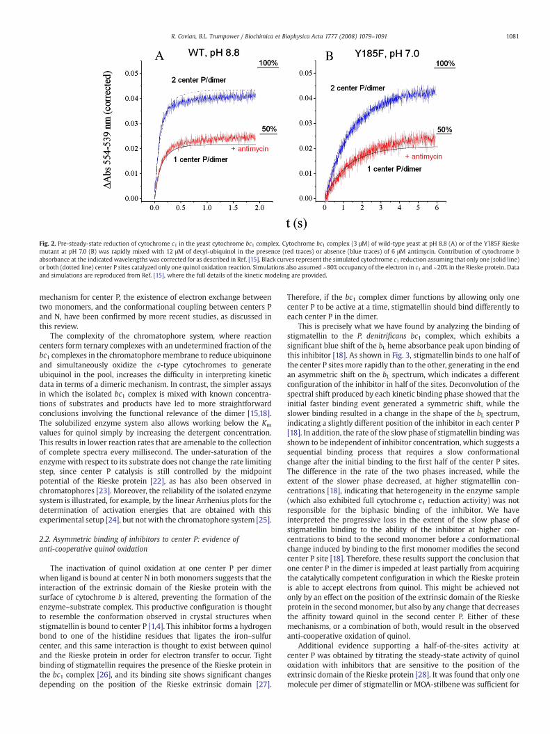
Fig. 2. Pre-steady-state reduction of cytochrome c1 in the yeast cytochrome bc1 complex. Cytochrome bc1 complex (3 μM) of wild-type yeast at pH 8.8 (A) or of the Y185F Rieskemutant at pH 7.0 (B) was rapidly mixed with 12 μM of decyl-ubiquinol in the presence (red traces) or absence (blue traces) of 6 μM antimycin. Contribution of cytochrome babsorbance at the indicated wavelengths was corrected for as described in Ref. [15]. Black curves represent the simulated cytochrome c1 reduction assuming that only one (solid line)or both (dotted line) center P sites catalyzed only one quinol oxidation reaction. Simulations also assumed ~80% occupancy of the electron in c1 and ~20% in the Rieske protein. Dataand simulations are reproduced from Ref. [15], where the full details of the kinetic modeling are provided.
1081R. Covian, B.L. Trumpower / Biochimica et Biophysica Acta 1777 (2008) 1079–1091
mechanism for center P, the existence of electron exchange betweentwo monomers, and the conformational coupling between centers Pand N, have been confirmed by more recent studies, as discussed inthis review.
The complexity of the chromatophore system, where reactioncenters form ternary complexes with an undetermined fraction of thebc1 complexes in the chromatophore membrane to reduce ubiquinoneand simultaneously oxidize the c-type cytochromes to generateubiquinol in the pool, increases the difficulty in interpreting kineticdata in terms of a dimeric mechanism. In contrast, the simpler assaysin which the isolated bc1 complex is mixed with known concentra-tions of substrates and products have led to more straightforwardconclusions involving the functional relevance of the dimer [15,18].The solubilized enzyme system also allows working below the Km
values for quinol simply by increasing the detergent concentration.This results in lower reaction rates that are amenable to the collectionof complete spectra every millisecond. The under-saturation of theenzyme with respect to its substrate does not change the rate limitingstep, since center P catalysis is still controlled by the midpointpotential of the Rieske protein [22], as has also been observed inchromatophores [23]. Moreover, the reliability of the isolated enzymesystem is illustrated, for example, by the linear Arrhenius plots for thedetermination of activation energies that are obtained with thisexperimental setup [24], but not with the chromatophore system [25].
2.2. Asymmetric binding of inhibitors to center P: evidence ofanti-cooperative quinol oxidation
The inactivation of quinol oxidation at one center P per dimerwhen ligand is bound at center N in both monomers suggests that theinteraction of the extrinsic domain of the Rieske protein with thesurface of cytochrome b is altered, preventing the formation of theenzyme–substrate complex. This productive configuration is thoughtto resemble the conformation observed in crystal structures whenstigmatellin is bound to center P [1,4]. This inhibitor forms a hydrogenbond to one of the histidine residues that ligates the iron–sulfurcenter, and this same interaction is thought to exist between quinoland the Rieske protein in order for electron transfer to occur. Tightbinding of stigmatellin requires the presence of the Rieske protein inthe bc1 complex [26], and its binding site shows significant changesdepending on the position of the Rieske extrinsic domain [27].
Therefore, if the bc1 complex dimer functions by allowing only onecenter P to be active at a time, stigmatellin should bind differently toeach center P in the dimer.
This is precisely what we have found by analyzing the binding ofstigmatellin to the P. denitrificans bc1 complex, which exhibits asignificant blue shift of the bL heme absorbance peak upon binding ofthis inhibitor [18]. As shown in Fig. 3, stigmatellin binds to one half ofthe center P sites more rapidly than to the other, generating in the endan asymmetric shift on the bL spectrum, which indicates a differentconfiguration of the inhibitor in half of the sites. Deconvolution of thespectral shift produced by each kinetic binding phase showed that theinitial faster binding event generated a symmetric shift, while theslower binding resulted in a change in the shape of the bL spectrum,indicating a slightly different position of the inhibitor in each center P[18]. In addition, the rate of the slow phase of stigmatellin binding wasshown to be independent of inhibitor concentration, which suggests asequential binding process that requires a slow conformationalchange after the initial binding to the first half of the center P sites.The difference in the rate of the two phases increased, while theextent of the slower phase decreased, at higher stigmatellin con-centrations [18], indicating that heterogeneity in the enzyme sample(which also exhibited full cytochrome c1 reduction activity) was notresponsible for the biphasic binding of the inhibitor. We haveinterpreted the progressive loss in the extent of the slow phase ofstigmatellin binding to the ability of the inhibitor at higher con-centrations to bind to the second monomer before a conformationalchange induced by binding to the first monomer modifies the secondcenter P site [18]. Therefore, these results support the conclusion thatone center P in the dimer is impeded at least partially from acquiringthe catalytically competent configuration in which the Rieske proteinis able to accept electrons from quinol. This might be achieved notonly by an effect on the position of the extrinsic domain of the Rieskeprotein in the secondmonomer, but also by any change that decreasesthe affinity toward quinol in the second center P. Either of thesemechanisms, or a combination of both, would result in the observedanti-cooperative oxidation of quinol.
Additional evidence supporting a half-of-the-sites activity atcenter P was obtained by titrating the steady-state activity of quinoloxidation with inhibitors that are sensitive to the position of theextrinsic domain of the Rieske protein [28]. It was found that only onemolecule per dimer of stigmatellin or MOA-stilbene was sufficient for
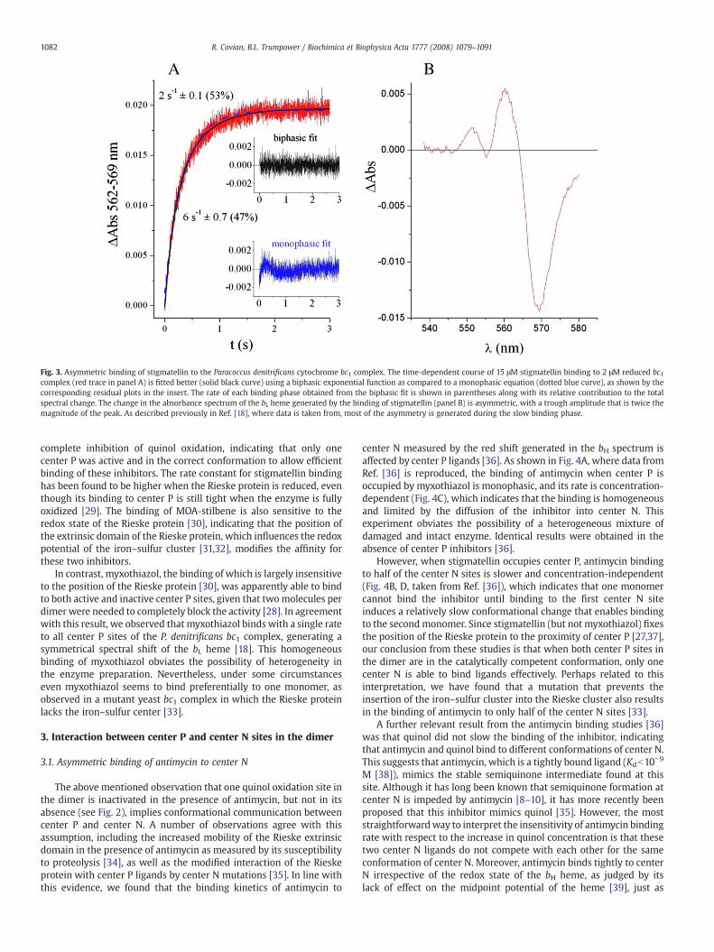
Fig. 3. Asymmetric binding of stigmatellin to the Paracoccus denitrificans cytochrome bc1 complex. The time-dependent course of 15 μM stigmatellin binding to 2 μM reduced bc1complex (red trace in panel A) is fitted better (solid black curve) using a biphasic exponential function as compared to a monophasic equation (dotted blue curve), as shown by thecorresponding residual plots in the insert. The rate of each binding phase obtained from the biphasic fit is shown in parentheses along with its relative contribution to the totalspectral change. The change in the absorbance spectrum of the bL heme generated by the binding of stigmatellin (panel B) is asymmetric, with a trough amplitude that is twice themagnitude of the peak. As described previously in Ref. [18], where data is taken from, most of the asymmetry is generated during the slow binding phase.
1082 R. Covian, B.L. Trumpower / Biochimica et Biophysica Acta 1777 (2008) 1079–1091
complete inhibition of quinol oxidation, indicating that only onecenter P was active and in the correct conformation to allow efficientbinding of these inhibitors. The rate constant for stigmatellin bindinghas been found to be higher when the Rieske protein is reduced, eventhough its binding to center P is still tight when the enzyme is fullyoxidized [29]. The binding of MOA-stilbene is also sensitive to theredox state of the Rieske protein [30], indicating that the position ofthe extrinsic domain of the Rieske protein, which influences the redoxpotential of the iron–sulfur cluster [31,32], modifies the affinity forthese two inhibitors.
In contrast, myxothiazol, the binding of which is largely insensitiveto the position of the Rieske protein [30], was apparently able to bindto both active and inactive center P sites, given that twomolecules perdimer were needed to completely block the activity [28]. In agreementwith this result, we observed that myxothiazol binds with a single rateto all center P sites of the P. denitrificans bc1 complex, generating asymmetrical spectral shift of the bL heme [18]. This homogeneousbinding of myxothiazol obviates the possibility of heterogeneity inthe enzyme preparation. Nevertheless, under some circumstanceseven myxothiazol seems to bind preferentially to one monomer, asobserved in a mutant yeast bc1 complex in which the Rieske proteinlacks the iron–sulfur center [33].
3. Interaction between center P and center N sites in the dimer
3.1. Asymmetric binding of antimycin to center N
The above mentioned observation that one quinol oxidation site inthe dimer is inactivated in the presence of antimycin, but not in itsabsence (see Fig. 2), implies conformational communication betweencenter P and center N. A number of observations agree with thisassumption, including the increased mobility of the Rieske extrinsicdomain in the presence of antimycin as measured by its susceptibilityto proteolysis [34], as well as the modified interaction of the Rieskeprotein with center P ligands by center N mutations [35]. In line withthis evidence, we found that the binding kinetics of antimycin to
center N measured by the red shift generated in the bH spectrum isaffected by center P ligands [36]. As shown in Fig. 4A, where data fromRef. [36] is reproduced, the binding of antimycin when center P isoccupied by myxothiazol is monophasic, and its rate is concentration-dependent (Fig. 4C), which indicates that the binding is homogeneousand limited by the diffusion of the inhibitor into center N. Thisexperiment obviates the possibility of a heterogeneous mixture ofdamaged and intact enzyme. Identical results were obtained in theabsence of center P inhibitors [36].
However, when stigmatellin occupies center P, antimycin bindingto half of the center N sites is slower and concentration-independent(Fig. 4B, D, taken from Ref. [36]), which indicates that one monomercannot bind the inhibitor until binding to the first center N siteinduces a relatively slow conformational change that enables bindingto the secondmonomer. Since stigmatellin (but not myxothiazol) fixesthe position of the Rieske protein to the proximity of center P [27,37],our conclusion from these studies is that when both center P sites inthe dimer are in the catalytically competent conformation, only onecenter N is able to bind ligands effectively. Perhaps related to thisinterpretation, we have found that a mutation that prevents theinsertion of the iron–sulfur cluster into the Rieske cluster also resultsin the binding of antimycin to only half of the center N sites [33].
A further relevant result from the antimycin binding studies [36]was that quinol did not slow the binding of the inhibitor, indicatingthat antimycin and quinol bind to different conformations of center N.This suggests that antimycin, which is a tightly bound ligand (Kdb10−9
M [38]), mimics the stable semiquinone intermediate found at thissite. Although it has long been known that semiquinone formation atcenter N is impeded by antimycin [8–10], it has more recently beenproposed that this inhibitor mimics quinol [35]. However, the moststraightforwardway to interpret the insensitivity of antimycin bindingrate with respect to the increase in quinol concentration is that thesetwo center N ligands do not compete with each other for the sameconformation of center N. Moreover, antimycin binds tightly to centerN irrespective of the redox state of the bH heme, as judged by itslack of effect on the midpoint potential of the heme [39], just as
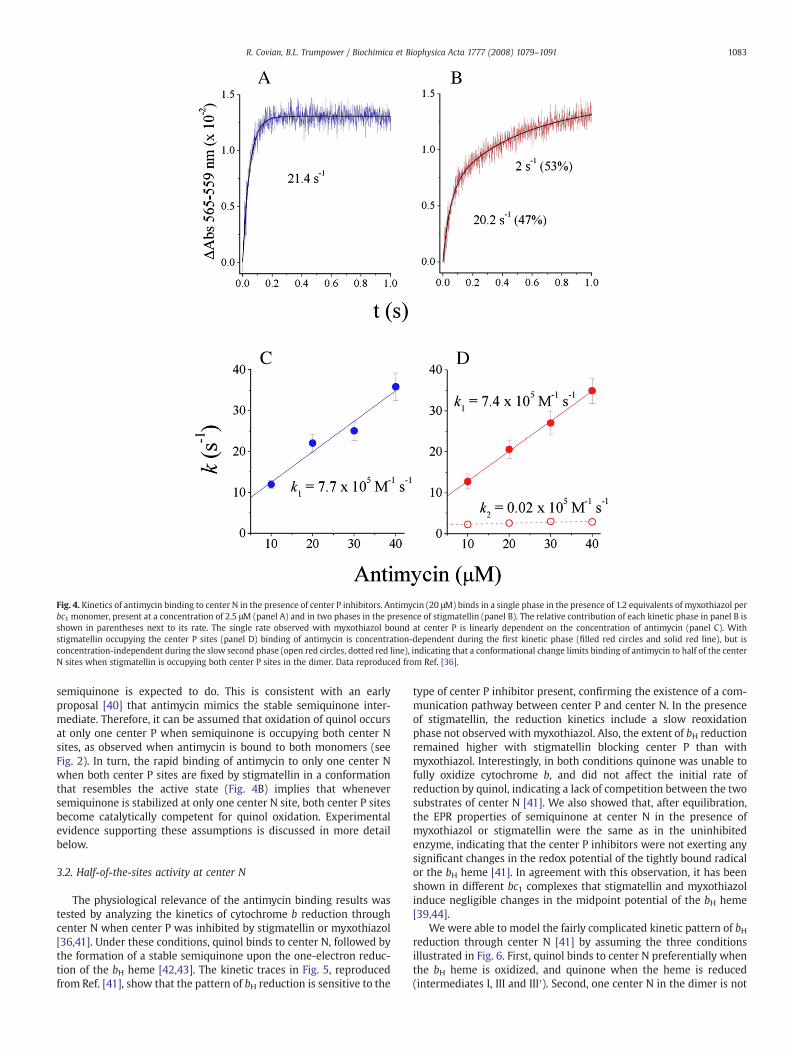
Fig. 4. Kinetics of antimycin binding to center N in the presence of center P inhibitors. Antimycin (20 μM) binds in a single phase in the presence of 1.2 equivalents of myxothiazol perbc1 monomer, present at a concentration of 2.5 μM (panel A) and in two phases in the presence of stigmatellin (panel B). The relative contribution of each kinetic phase in panel B isshown in parentheses next to its rate. The single rate observed with myxothiazol bound at center P is linearly dependent on the concentration of antimycin (panel C). Withstigmatellin occupying the center P sites (panel D) binding of antimycin is concentration-dependent during the first kinetic phase (filled red circles and solid red line), but isconcentration-independent during the slow second phase (open red circles, dotted red line), indicating that a conformational change limits binding of antimycin to half of the centerN sites when stigmatellin is occupying both center P sites in the dimer. Data reproduced from Ref. [36].
1083R. Covian, B.L. Trumpower / Biochimica et Biophysica Acta 1777 (2008) 1079–1091
semiquinone is expected to do. This is consistent with an earlyproposal [40] that antimycin mimics the stable semiquinone inter-mediate. Therefore, it can be assumed that oxidation of quinol occursat only one center P when semiquinone is occupying both center Nsites, as observed when antimycin is bound to both monomers (seeFig. 2). In turn, the rapid binding of antimycin to only one center Nwhen both center P sites are fixed by stigmatellin in a conformationthat resembles the active state (Fig. 4B) implies that wheneversemiquinone is stabilized at only one center N site, both center P sitesbecome catalytically competent for quinol oxidation. Experimentalevidence supporting these assumptions is discussed in more detailbelow.
3.2. Half-of-the-sites activity at center N
The physiological relevance of the antimycin binding results wastested by analyzing the kinetics of cytochrome b reduction throughcenter N when center P was inhibited by stigmatellin or myxothiazol[36,41]. Under these conditions, quinol binds to center N, followed bythe formation of a stable semiquinone upon the one-electron reduc-tion of the bH heme [42,43]. The kinetic traces in Fig. 5, reproducedfrom Ref. [41], show that the pattern of bH reduction is sensitive to the
type of center P inhibitor present, confirming the existence of a com-munication pathway between center P and center N. In the presenceof stigmatellin, the reduction kinetics include a slow reoxidationphase not observed with myxothiazol. Also, the extent of bH reductionremained higher with stigmatellin blocking center P than withmyxothiazol. Interestingly, in both conditions quinone was unable tofully oxidize cytochrome b, and did not affect the initial rate ofreduction by quinol, indicating a lack of competition between the twosubstrates of center N [41]. We also showed that, after equilibration,the EPR properties of semiquinone at center N in the presence ofmyxothiazol or stigmatellin were the same as in the uninhibitedenzyme, indicating that the center P inhibitors were not exerting anysignificant changes in the redox potential of the tightly bound radicalor the bH heme [41]. In agreement with this observation, it has beenshown in different bc1 complexes that stigmatellin and myxothiazolinduce negligible changes in the midpoint potential of the bH heme[39,44].
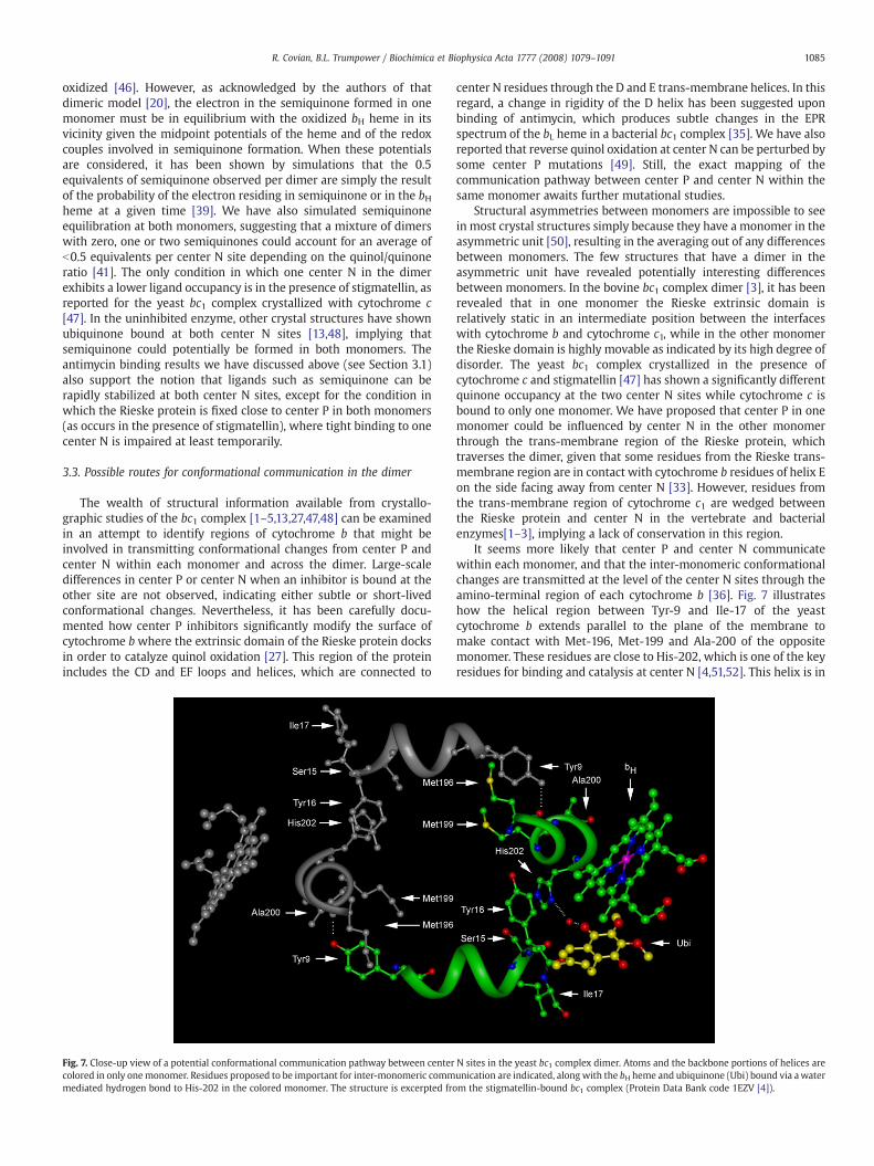
We were able to model the fairly complicated kinetic pattern of bHreduction through center N [41] by assuming the three conditionsillustrated in Fig. 6. First, quinol binds to center N preferentially whenthe bH heme is oxidized, and quinone when the heme is reduced(intermediates I, III and III′). Second, one center N in the dimer is not
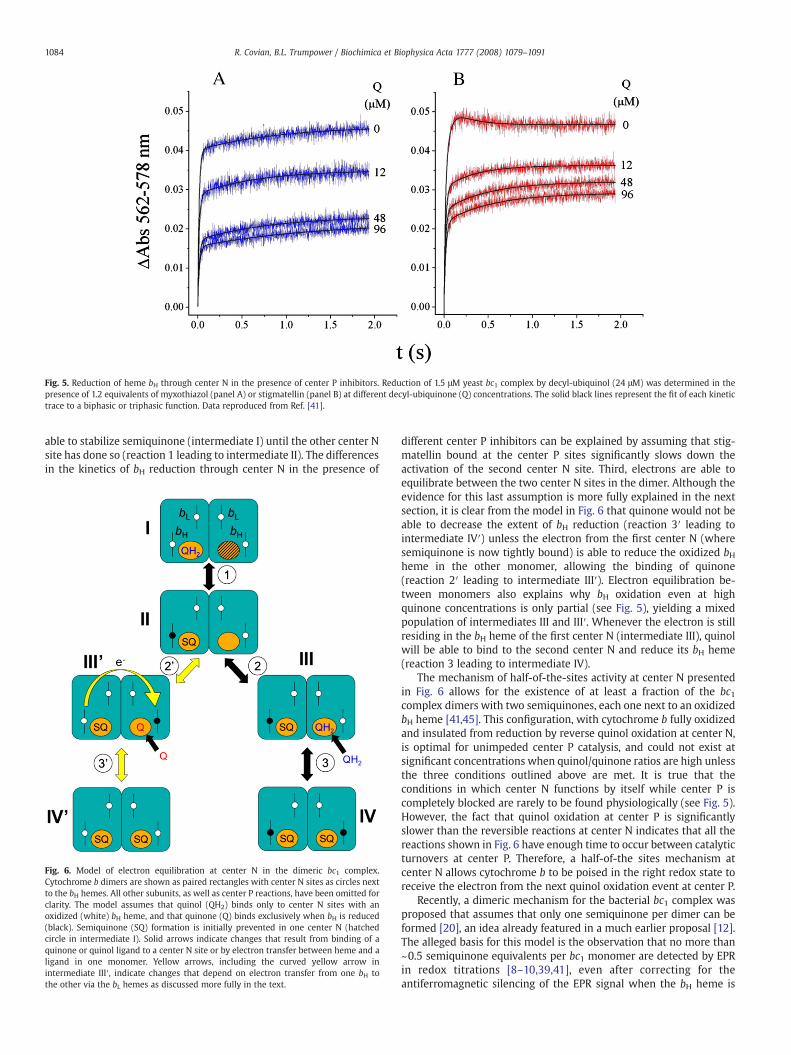
Fig. 5. Reduction of heme bH through center N in the presence of center P inhibitors. Reduction of 1.5 μM yeast bc1 complex by decyl-ubiquinol (24 μM) was determined in thepresence of 1.2 equivalents of myxothiazol (panel A) or stigmatellin (panel B) at different decyl-ubiquinone (Q) concentrations. The solid black lines represent the fit of each kinetictrace to a biphasic or triphasic function. Data reproduced from Ref. [41].
1084 R. Covian, B.L. Trumpower / Biochimica et Biophysica Acta 1777 (2008) 1079–1091
able to stabilize semiquinone (intermediate I) until the other center Nsite has done so (reaction 1 leading to intermediate II). The differencesin the kinetics of bH reduction through center N in the presence of
Fig. 6. Model of electron equilibration at center N in the dimeric bc1 complex.Cytochrome b dimers are shown as paired rectangles with center N sites as circles nextto the bH hemes. All other subunits, as well as center P reactions, have been omitted forclarity. The model assumes that quinol (QH2) binds only to center N sites with anoxidized (white) bH heme, and that quinone (Q) binds exclusively when bH is reduced(black). Semiquinone (SQ) formation is initially prevented in one center N (hatchedcircle in intermediate I). Solid arrows indicate changes that result from binding of aquinone or quinol ligand to a center N site or by electron transfer between heme and aligand in one monomer. Yellow arrows, including the curved yellow arrow inintermediate III′, indicate changes that depend on electron transfer from one bH tothe other via the bL hemes as discussed more fully in the text.
different center P inhibitors can be explained by assuming that stig-matellin bound at the center P sites significantly slows down theactivation of the second center N site. Third, electrons are able toequilibrate between the two center N sites in the dimer. Although theevidence for this last assumption is more fully explained in the nextsection, it is clear from the model in Fig. 6 that quinone would not beable to decrease the extent of bH reduction (reaction 3′ leading tointermediate IV′) unless the electron from the first center N (wheresemiquinone is now tightly bound) is able to reduce the oxidized bHheme in the other monomer, allowing the binding of quinone(reaction 2′ leading to intermediate III′). Electron equilibration be-tween monomers also explains why bH oxidation even at highquinone concentrations is only partial (see Fig. 5), yielding a mixedpopulation of intermediates III and III′. Whenever the electron is stillresiding in the bH heme of the first center N (intermediate III), quinolwill be able to bind to the second center N and reduce its bH heme(reaction 3 leading to intermediate IV).
The mechanism of half-of-the-sites activity at center N presentedin Fig. 6 allows for the existence of at least a fraction of the bc1complex dimers with two semiquinones, each one next to an oxidizedbH heme [41,45]. This configuration, with cytochrome b fully oxidizedand insulated from reduction by reverse quinol oxidation at center N,is optimal for unimpeded center P catalysis, and could not exist atsignificant concentrations when quinol/quinone ratios are high unlessthe three conditions outlined above are met. It is true that theconditions in which center N functions by itself while center P iscompletely blocked are rarely to be found physiologically (see Fig. 5).However, the fact that quinol oxidation at center P is significantlyslower than the reversible reactions at center N indicates that all thereactions shown in Fig. 6 have enough time to occur between catalyticturnovers at center P. Therefore, a half-of-the sites mechanism atcenter N allows cytochrome b to be poised in the right redox state toreceive the electron from the next quinol oxidation event at center P.
Recently, a dimeric mechanism for the bacterial bc1 complex wasproposed that assumes that only one semiquinone per dimer can beformed [20], an idea already featured in a much earlier proposal [12].The alleged basis for this model is the observation that no more than~0.5 semiquinone equivalents per bc1 monomer are detected by EPRin redox titrations [8–10,39,41], even after correcting for theantiferromagnetic silencing of the EPR signal when the bH heme is
1085R. Covian, B.L. Trumpower / Biochimica et Biophysica Acta 1777 (2008) 1079–1091
oxidized [46]. However, as acknowledged by the authors of thatdimeric model [20], the electron in the semiquinone formed in onemonomer must be in equilibrium with the oxidized bH heme in itsvicinity given the midpoint potentials of the heme and of the redoxcouples involved in semiquinone formation. When these potentialsare considered, it has been shown by simulations that the 0.5equivalents of semiquinone observed per dimer are simply the resultof the probability of the electron residing in semiquinone or in the bHheme at a given time [39]. We have also simulated semiquinoneequilibration at both monomers, suggesting that a mixture of dimerswith zero, one or two semiquinones could account for an average ofb0.5 equivalents per center N site depending on the quinol/quinoneratio [41]. The only condition in which one center N in the dimerexhibits a lower ligand occupancy is in the presence of stigmatellin, asreported for the yeast bc1 complex crystallized with cytochrome c[47]. In the uninhibited enzyme, other crystal structures have shownubiquinone bound at both center N sites [13,48], implying thatsemiquinone could potentially be formed in both monomers. Theantimycin binding results we have discussed above (see Section 3.1)also support the notion that ligands such as semiquinone can berapidly stabilized at both center N sites, except for the condition inwhich the Rieske protein is fixed close to center P in both monomers(as occurs in the presence of stigmatellin), where tight binding to onecenter N is impaired at least temporarily.
3.3. Possible routes for conformational communication in the dimer
The wealth of structural information available from crystallo-graphic studies of the bc1 complex [1–5,13,27,47,48] can be examinedin an attempt to identify regions of cytochrome b that might beinvolved in transmitting conformational changes from center P andcenter N within each monomer and across the dimer. Large-scaledifferences in center P or center N when an inhibitor is bound at theother site are not observed, indicating either subtle or short-livedconformational changes. Nevertheless, it has been carefully docu-mented how center P inhibitors significantly modify the surface ofcytochrome bwhere the extrinsic domain of the Rieske protein docksin order to catalyze quinol oxidation [27]. This region of the proteinincludes the CD and EF loops and helices, which are connected to
Fig. 7. Close-up view of a potential conformational communication pathway between centercolored in only onemonomer. Residues proposed to be important for inter-monomeric commmediated hydrogen bond to His-202 in the colored monomer. The structure is excerpted fro
center N residues through the D and E trans-membrane helices. In thisregard, a change in rigidity of the D helix has been suggested uponbinding of antimycin, which produces subtle changes in the EPRspectrum of the bL heme in a bacterial bc1 complex [35]. We have alsoreported that reverse quinol oxidation at center N can be perturbed bysome center P mutations [49]. Still, the exact mapping of thecommunication pathway between center P and center N within thesame monomer awaits further mutational studies.
Structural asymmetries between monomers are impossible to seein most crystal structures simply because they have a monomer in theasymmetric unit [50], resulting in the averaging out of any differencesbetween monomers. The few structures that have a dimer in theasymmetric unit have revealed potentially interesting differencesbetween monomers. In the bovine bc1 complex dimer [3], it has beenrevealed that in one monomer the Rieske extrinsic domain isrelatively static in an intermediate position between the interfaceswith cytochrome b and cytochrome c1, while in the other monomerthe Rieske domain is highly movable as indicated by its high degree ofdisorder. The yeast bc1 complex crystallized in the presence ofcytochrome c and stigmatellin [47] has shown a significantly differentquinone occupancy at the two center N sites while cytochrome c isbound to only one monomer. We have proposed that center P in onemonomer could be influenced by center N in the other monomerthrough the trans-membrane region of the Rieske protein, whichtraverses the dimer, given that some residues from the Rieske trans-membrane region are in contact with cytochrome b residues of helix Eon the side facing away from center N [33]. However, residues fromthe trans-membrane region of cytochrome c1 are wedged betweenthe Rieske protein and center N in the vertebrate and bacterialenzymes[1–3], implying a lack of conservation in this region.
It seems more likely that center P and center N communicatewithin each monomer, and that the inter-monomeric conformationalchanges are transmitted at the level of the center N sites through theamino-terminal region of each cytochrome b [36]. Fig. 7 illustrateshow the helical region between Tyr-9 and Ile-17 of the yeastcytochrome b extends parallel to the plane of the membrane tomake contact with Met-196, Met-199 and Ala-200 of the oppositemonomer. These residues are close to His-202, which is one of the keyresidues for binding and catalysis at center N [4,51,52]. This helix is in
N sites in the yeast bc1 complex dimer. Atoms and the backbone portions of helices areunication are indicated, along with the bH heme and ubiquinone (Ubi) bound via awaterm the stigmatellin-bound bc1 complex (Protein Data Bank code 1EZV [4]).
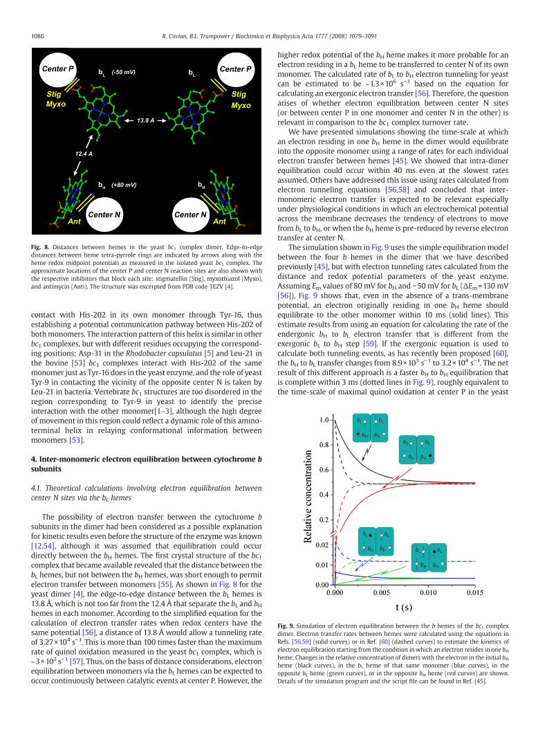
Fig. 8. Distances between hemes in the yeast bc1 complex dimer. Edge-to-edgedistances between heme tetra-pyrrole rings are indicated by arrows along with theheme redox midpoint potentials as measured in the isolated yeast bc1 complex. Theapproximate locations of the center P and center N reaction sites are also shown withthe respective inhibitors that block each site: stigmatellin (Stig), myxothiazol (Myxo),and antimycin (Anti). The structure was excerpted from PDB code 1EZV [4].
Fig. 9. Simulation of electron equilibration between the b hemes of the bc1 complexdimer. Electron transfer rates between hemes were calculated using the equations inRefs. [56,59] (solid curves) or in Ref. [60] (dashed curves) to estimate the kinetics ofelectron equilibration starting from the condition inwhich an electron resides in one bHheme. Changes in the relative concentration of dimers with the electron in the initial bHheme (black curves), in the bL heme of that same monomer (blue curves), in theopposite bL heme (green curves), or in the opposite bH heme (red curves) are shown.Details of the simulation program and the script file can be found in Ref. [45].
1086 R. Covian, B.L. Trumpower / Biochimica et Biophysica Acta 1777 (2008) 1079–1091
contact with His-202 in its own monomer through Tyr-16, thusestablishing a potential communication pathway between His-202 ofbothmonomers. The interaction pattern of this helix is similar in otherbc1 complexes, but with different residues occupying the correspond-ing positions; Asp-31 in the Rhodobacter capsulatus [5] and Leu-21 inthe bovine [53] bc1 complexes interact with His-202 of the samemonomer just as Tyr-16 does in the yeast enzyme, and the role of yeastTyr-9 in contacting the vicinity of the opposite center N is taken byLeu-21 in bacteria. Vertebrate bc1 structures are too disordered in theregion corresponding to Tyr-9 in yeast to identify the preciseinteraction with the other monomer[1–3], although the high degreeof movement in this region could reflect a dynamic role of this amino-terminal helix in relaying conformational information betweenmonomers [53].
4. Inter-monomeric electron equilibration between cytochrome bsubunits
4.1. Theoretical calculations involving electron equilibration betweencenter N sites via the bL hemes
The possibility of electron transfer between the cytochrome bsubunits in the dimer had been considered as a possible explanationfor kinetic results even before the structure of the enzymewas known[12,54], although it was assumed that equilibration could occurdirectly between the bH hemes. The first crystal structure of the bc1complex that became available revealed that the distance between thebL hemes, but not between the bH hemes, was short enough to permitelectron transfer between monomers [55]. As shown in Fig. 8 for theyeast dimer [4], the edge-to-edge distance between the bL hemes is13.8 Å, which is not too far from the 12.4 Å that separate the bL and bHhemes in each monomer. According to the simplified equation for thecalculation of electron transfer rates when redox centers have thesame potential [56], a distance of 13.8 Å would allow a tunneling rateof 3.27×10⁴ s−1. This is more than 100 times faster than the maximumrate of quinol oxidation measured in the yeast bc1 complex, which is~3×10² s−1 [57]. Thus, on the basis of distance considerations, electronequilibration between monomers via the bL hemes can be expected tooccur continuously between catalytic events at center P. However, the
higher redox potential of the bH heme makes it more probable for anelectron residing in a bL heme to be transferred to center N of its ownmonomer. The calculated rate of bL to bH electron tunneling for yeastcan be estimated to be ~1.3×10⁶ s−1 based on the equation forcalculating an exergonic electron transfer [56]. Therefore, the questionarises of whether electron equilibration between center N sites(or between center P in one monomer and center N in the other) isrelevant in comparison to the bc1 complex turnover rate.
We have presented simulations showing the time-scale at whichan electron residing in one bH heme in the dimer would equilibrateinto the opposite monomer using a range of rates for each individualelectron transfer between hemes [45]. We showed that intra-dimerequilibration could occur within 40 ms even at the slowest ratesassumed. Others have addressed this issue using rates calculated fromelectron tunneling equations [56,58] and concluded that inter-monomeric electron transfer is expected to be relevant especiallyunder physiological conditions in which an electrochemical potentialacross the membrane decreases the tendency of electrons to movefrom bL to bH, or when the bH heme is pre-reduced by reverse electrontransfer at center N.
The simulation shown in Fig. 9 uses the simple equilibrationmodelbetween the four b hemes in the dimer that we have describedpreviously [45], but with electron tunneling rates calculated from thedistance and redox potential parameters of the yeast enzyme.Assuming Em values of 80 mV for bH and −50 mV for bL (ΔEm=130 mV[56]), Fig. 9 shows that, even in the absence of a trans-membranepotential, an electron originally residing in one bH heme shouldequilibrate to the other monomer within 10 ms (solid lines). Thisestimate results from using an equation for calculating the rate of theendergonic bH to bL electron transfer that is different from theexergonic bL to bH step [59]. If the exergonic equation is used tocalculate both tunneling events, as has recently been proposed [60],the bH to bL transfer changes from 8.9×10³ s−1 to 3.2×10⁴ s−1. The netresult of this different approach is a faster bH to bH equilibration thatis complete within 3 ms (dotted lines in Fig. 9), roughly equivalent tothe time-scale of maximal quinol oxidation at center P in the yeast
1087R. Covian, B.L. Trumpower / Biochimica et Biophysica Acta 1777 (2008) 1079–1091
enzyme (~300 s−1). It is noteworthy that the Keq for the one-electronsharing between bH and bL resulting from the two calculations isquite different (40.6 compared to 146.1 using the lower and higher bHto bL transfer rates, respectively). Still, equilibration between the fourb hemes in the dimer would be completed within 10 ms in bothscenarios.
Using data obtained with the bacterial bc1 complex in chromato-phores [61], it has been shown that the Keq for electron transferbetween the two b hemes in a monomer is much lower (10–25) thanwould be predicted from the Boltzmann distribution of one electronbetween two redox centers separated by 130–140 mV (Keq ~200),implying Coulombic interactions between the hemes. This seems to besupported experimentally by the increase of 70–80 mV in the redoxpotential of the bL heme when either one of the histidines thatcoordinates the bH heme is mutated, resulting in the absence of bH inthe enzyme [62]. However, it should be pointed out that, on purelytheoretical grounds, it is impossible for the equilibration of oneelectron between the bH and bL hemes to be influenced by anyelectrostatic (Coulombic) effect of the ferro-bH heme on the ferri-bLheme. This is because movement of the electron from the reduced bHheme to the oxidized bL heme would eliminate the putative source ofthe Coulombic effect in the bH heme as it becomes oxidized. Therefore,the fact that the redox potential of the bL heme is in reality only 50–60 mV lower than that of the bH heme implies a Keq ~10 for thedistribution of a single electron between the b hemes in a monomer.Obviously, this value would allow even faster equilibration betweenthe two bH hemes in the dimer than what is shown in Fig. 9, since theelectron would reside a longer period of time in the bL hemes.Nevertheless, the simulations we show in Fig. 9 using tunneling ratecalculations indicate that, even at significantly higher Keq values ofN100 resulting from a slower bH to bL electron transfer, fast electronequilibration via the bL hemes in the dimer would still be expected.
4.2. Experimental evidence for fast inter-monomeric electron transfer
The crystal structure of the bc1 complex shows that the spacebetween the bL hemes is occupied by three aromatic residues, one ofwhich (Phe-195 in Rhodobacter sphaeroides) has been proposed to berelevant for electron equilibration between monomers [63]. Mutationof this residue to alanine increased the rate of superoxide generationby the bc1 complex three-fold, while decreasing the catalytic activityby ~20%. However, it was not directly shown that this mutation
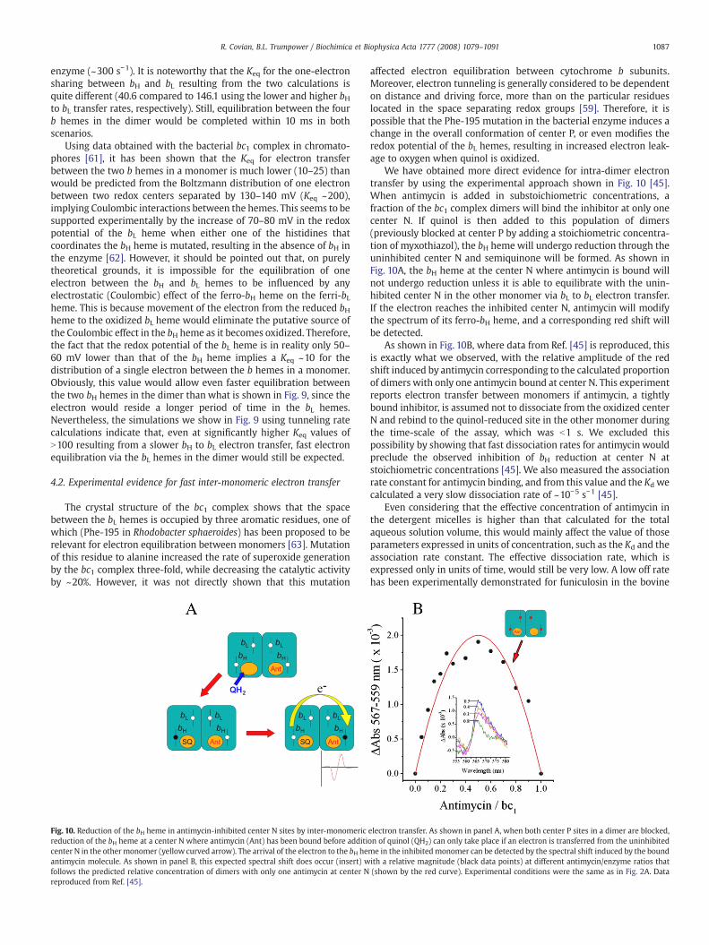
Fig. 10. Reduction of the bH heme in antimycin-inhibited center N sites by inter-monomericreduction of the bH heme at a center N where antimycin (Ant) has been bound before additicenter N in the othermonomer (yellow curved arrow). The arrival of the electron to the bH heantimycin molecule. As shown in panel B, this expected spectral shift does occur (insert) wfollows the predicted relative concentration of dimers with only one antimycin at center Nreproduced from Ref. [45].
affected electron equilibration between cytochrome b subunits.Moreover, electron tunneling is generally considered to be dependenton distance and driving force, more than on the particular residueslocated in the space separating redox groups [59]. Therefore, it ispossible that the Phe-195 mutation in the bacterial enzyme induces achange in the overall conformation of center P, or even modifies theredox potential of the bL hemes, resulting in increased electron leak-age to oxygen when quinol is oxidized.
We have obtained more direct evidence for intra-dimer electrontransfer by using the experimental approach shown in Fig. 10 [45].When antimycin is added in substoichiometric concentrations, afraction of the bc1 complex dimers will bind the inhibitor at only onecenter N. If quinol is then added to this population of dimers(previously blocked at center P by adding a stoichiometric concentra-tion of myxothiazol), the bH heme will undergo reduction through theuninhibited center N and semiquinone will be formed. As shown inFig. 10A, the bH heme at the center N where antimycin is bound willnot undergo reduction unless it is able to equilibrate with the unin-hibited center N in the other monomer via bL to bL electron transfer.If the electron reaches the inhibited center N, antimycin will modifythe spectrum of its ferro-bH heme, and a corresponding red shift willbe detected.
As shown in Fig. 10B, where data from Ref. [45] is reproduced, thisis exactly what we observed, with the relative amplitude of the redshift induced by antimycin corresponding to the calculated proportionof dimers with only one antimycin bound at center N. This experimentreports electron transfer between monomers if antimycin, a tightlybound inhibitor, is assumed not to dissociate from the oxidized centerN and rebind to the quinol-reduced site in the other monomer duringthe time-scale of the assay, which was b1 s. We excluded thispossibility by showing that fast dissociation rates for antimycin wouldpreclude the observed inhibition of bH reduction at center N atstoichiometric concentrations [45]. We also measured the associationrate constant for antimycin binding, and from this value and the Kd wecalculated a very slow dissociation rate of ~10−5 s−1 [45].
Even considering that the effective concentration of antimycin inthe detergent micelles is higher than that calculated for the totalaqueous solution volume, this would mainly affect the value of thoseparameters expressed in units of concentration, such as the Kd and theassociation rate constant. The effective dissociation rate, which isexpressed only in units of time, would still be very low. A low off ratehas been experimentally demonstrated for funiculosin in the bovine
electron transfer. As shown in panel A, when both center P sites in a dimer are blocked,on of quinol (QH2) can only take place if an electron is transferred from the uninhibitedme in the inhibitedmonomer can be detected by the spectral shift induced by the boundith a relative magnitude (black data points) at different antimycin/enzyme ratios that(shown by the red curve). Experimental conditions were the same as in Fig. 2A. Data
1088 R. Covian, B.L. Trumpower / Biochimica et Biophysica Acta 1777 (2008) 1079–1091
bc1 complex [64] and for ilicicolin in the yeast enzyme [65]. Whenthese inhibitors are bound to center N, they dissociate on a time-scaleof tens of minutes asmeasured by the change in spectral shift of the bHheme upon addition of antimycin. Funiculosin and ilicicolin have Kd
values 1–3 orders of magnitude higher than antimycin, implying thatthe dissociation rate constant for antimycin is even lower, at least aslow as our estimated value of 10−5 s−1. The argument that antimycinmight dissociate from center N into the lipid or detergent phase muchfaster than into the aqueous volume in which Kd determinations aremade [54] is also disproved by the extremely slow exchange betweenfuniculosin or ilicicolin and antimycin when these are mixed togetherin the bc1 phospholipid/detergent micelle [64,65], since these are allhighly hydrophobic inhibitors.
For antimycin to bind to the quinol-reduced center N site andinduce a spectral shift in the absence of inter-monomeric electrontransfer, semiquinone would need to vacate the site where it wasformed on a millisecond time-scale. This contradicts the strongstabilization of semiquinone at center N, which behaves as a tightlybound ligand. Therefore, the result shown in Fig. 10 is direct evidencefor fast electron equilibration between the center N sites in the dimerby means of bL to bL electron tunneling. Additional evidence wasobtained from the non-linear inhibition of the extent of cytochrome breduction by antimycin [45], and as mentioned previously (see Figs. 5and 6), from the kinetic modeling of the partial inhibition by quinone[41].
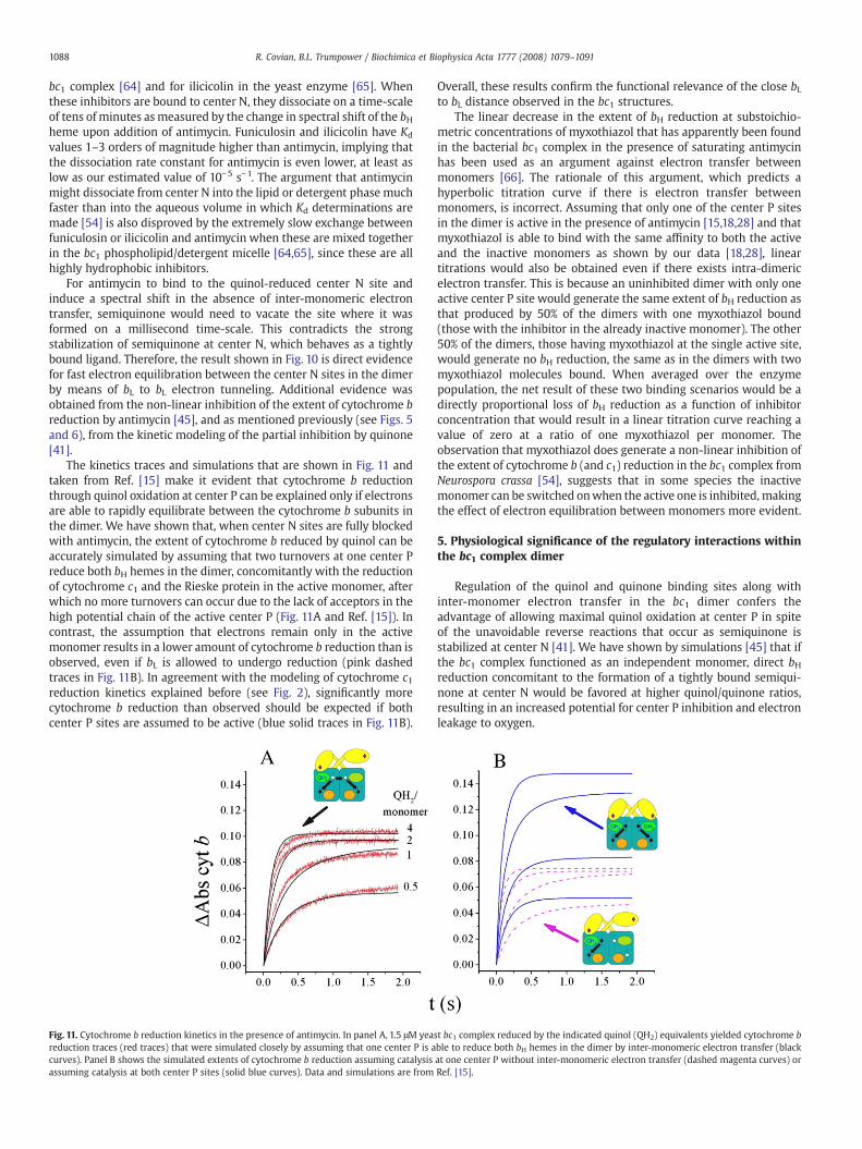
The kinetics traces and simulations that are shown in Fig. 11 andtaken from Ref. [15] make it evident that cytochrome b reductionthrough quinol oxidation at center P can be explained only if electronsare able to rapidly equilibrate between the cytochrome b subunits inthe dimer. We have shown that, when center N sites are fully blockedwith antimycin, the extent of cytochrome b reduced by quinol can beaccurately simulated by assuming that two turnovers at one center Preduce both bH hemes in the dimer, concomitantly with the reductionof cytochrome c1 and the Rieske protein in the active monomer, afterwhich no more turnovers can occur due to the lack of acceptors in thehigh potential chain of the active center P (Fig. 11A and Ref. [15]). Incontrast, the assumption that electrons remain only in the activemonomer results in a lower amount of cytochrome b reduction than isobserved, even if bL is allowed to undergo reduction (pink dashedtraces in Fig. 11B). In agreement with the modeling of cytochrome c1reduction kinetics explained before (see Fig. 2), significantly morecytochrome b reduction than observed should be expected if bothcenter P sites are assumed to be active (blue solid traces in Fig. 11B).
Fig. 11. Cytochrome b reduction kinetics in the presence of antimycin. In panel A, 1.5 μM yeareduction traces (red traces) that were simulated closely by assuming that one center P is acurves). Panel B shows the simulated extents of cytochrome b reduction assuming catalysisassuming catalysis at both center P sites (solid blue curves). Data and simulations are from
Overall, these results confirm the functional relevance of the close bLto bL distance observed in the bc1 structures.
The linear decrease in the extent of bH reduction at substoichio-metric concentrations of myxothiazol that has apparently been foundin the bacterial bc1 complex in the presence of saturating antimycinhas been used as an argument against electron transfer betweenmonomers [66]. The rationale of this argument, which predicts ahyperbolic titration curve if there is electron transfer betweenmonomers, is incorrect. Assuming that only one of the center P sitesin the dimer is active in the presence of antimycin [15,18,28] and thatmyxothiazol is able to bind with the same affinity to both the activeand the inactive monomers as shown by our data [18,28], lineartitrations would also be obtained even if there exists intra-dimericelectron transfer. This is because an uninhibited dimer with only oneactive center P site would generate the same extent of bH reduction asthat produced by 50% of the dimers with one myxothiazol bound(those with the inhibitor in the already inactive monomer). The other50% of the dimers, those having myxothiazol at the single active site,would generate no bH reduction, the same as in the dimers with twomyxothiazol molecules bound. When averaged over the enzymepopulation, the net result of these two binding scenarios would be adirectly proportional loss of bH reduction as a function of inhibitorconcentration that would result in a linear titration curve reaching avalue of zero at a ratio of one myxothiazol per monomer. Theobservation that myxothiazol does generate a non-linear inhibition ofthe extent of cytochrome b (and c1) reduction in the bc1 complex fromNeurospora crassa [54], suggests that in some species the inactivemonomer can be switched onwhen the active one is inhibited, makingthe effect of electron equilibration between monomers more evident.
5. Physiological significance of the regulatory interactions withinthe bc1 complex dimer
Regulation of the quinol and quinone binding sites along withinter-monomer electron transfer in the bc1 dimer confers theadvantage of allowing maximal quinol oxidation at center P in spiteof the unavoidable reverse reactions that occur as semiquinone isstabilized at center N [41]. We have shown by simulations [45] that ifthe bc1 complex functioned as an independent monomer, direct bHreduction concomitant to the formation of a tightly bound semiqui-none at center N would be favored at higher quinol/quinone ratios,resulting in an increased potential for center P inhibition and electronleakage to oxygen.
st bc1 complex reduced by the indicated quinol (QH2) equivalents yielded cytochrome bble to reduce both bH hemes in the dimer by inter-monomeric electron transfer (blackat one center P without inter-monomeric electron transfer (dashed magenta curves) orRef. [15].
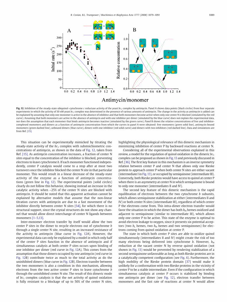
Fig. 12. Inhibition of the steady-state ubiquinol–cytochrome c reductase activity of the yeast bc1 complex by antimycin. Panel A shows data points (black circles) from four separateexperiments in which the activity of 50 nM yeast bc1 complex was determined in the presence of various amounts of antimycin. The change in the activity as antimycin is added canbe explained by assuming that only onemonomer is active in the absence of inhibitor and that bothmonomers become activewhen only one center N is blocked (simulated by the redcurve). Assuming that both monomers are active in the absence of antimycin and with one inhibitor per dimer (simulated by the blue curve) does not explain the experimental data,nor does the assumption that each monomer that binds antimycin becomes inactive (simulated by the green curve). Panel B shows the relative concentrations of free and inhibitor-complexed monomers and dimers as a function of antimycin concentration from which the curves in panel A were obtained: free monomers (green solid line), antimycin-boundmonomers (green dashed line), unbound dimers (blue curve), dimers with one inhibitor (red solid curve) and dimers with two inhibitors (red dashed line). Data and simulations arefrom Ref. [15].
1089R. Covian, B.L. Trumpower / Biochimica et Biophysica Acta 1777 (2008) 1079–1091
This situation can be experimentally mimicked by titrating thesteady-state activity of the bc1 complex with substoichiometric con-centrations of antimycin, as shown in the data of Fig. 12, taken fromRef. [15]. As antimycin concentration increases, a fraction of center Nsites equal to the concentration of the inhibitor is blocked, preventingelectrons to leave cytochrome b. If eachmonomer functioned indepen-dently, center P catalysis would come to a stop after at most twoturnovers once the inhibitor blocked the center N site in that particularmonomer. This would result in a linear decrease of the steady-stateactivity of the enzyme as a function of antimycin concentra-tions (green line in Fig. 12). The experimental points (solid circles)clearly do not follow this behavior, showing instead an increase in thecatalytic activity when ~25% of the center N sites are blocked withantimycin. It should be noted that this apparent activation cannot beexplained by alternative models that assume that the non-lineartitration curves with antimycin are due to a fast movement of theinhibitor directly between center N sites [54], for which there is nostructural support, since the crystal structures do not show any chan-nel that would allow direct interchange of center N ligands betweenmonomers [1–5,13].
Inter-monomer electron transfer by itself would allow the twocenter P sites in the dimer to recycle electrons out of cytochrome bthrough a single center N site, resulting in an increased resistance ofthe activity to antimycin (blue curve in Fig. 12A). However, theexperimental data can only be explained by amodel inwhich only halfof the center P sites function in the absence of antimycin and ifsimultaneous catalysis at both center P sites occurs upon binding ofone inhibitor per dimer (red curve in Fig. 12A). This comes from theassumption that those dimers occupied by one antimycin (red curve inFig. 12B) contribute twice as much to the total activity as do theuninhibited dimers (blue curve in Fig. 12B). Electron transfer betweenthe two monomers is also a condition in this mechanism, allowingelectrons from the two active center P sites to leave cytochrome bthrough the uninhibited center N site. The result of this dimeric modeof bc1 complex catalysis is that the net activity of quinol oxidationis fully resistant to a blockage of up to 50% of the center N sites,
highlighting the physiological relevance of this dimeric mechanism inminimizing inhibition of center P by backward reactions at center N.
Considering all of the experimental observations explained in thisreview, a model for the regulation of quinol oxidation in the dimeric bc1complex can beproposed as shown in Fig.13 andpreviously discussed inRef. [36]. The first key feature in thismechanism is an inverse symmetryrelation between center P and center N that allows only one Rieskeprotein to approach center P when both center N sites are either vacant(intermediate I in Fig.13), or occupied by semiquinone (intermediate III).Conversely, both Rieske proteinswould have access to quinol at center Pwhen there is an asymmetry at center N inwhich semiquinone is boundto only one monomer (intermediates II and IV).
The second key feature of this dimeric mechanism is the rapidequilibration of electrons between the two cytochrome b subunits,which allows semiquinone stabilization at either (intermediates II andIV) or both center N sites (intermediate III), regardless of which centerP the electrons come from. This intra-dimer electron transfer wouldfavor the situation inwhich the dimer has both bH hemes oxidized andadjacent to semiquinone (similar to intermediate III), which allowsonly one center P to be active. This state of the enzyme is optimal toavoid electron leakage to oxygen, since it presents six potential accep-tors (two bL hemes, two bH hemes and two semiquinones) for elec-trons coming from quinol oxidation at center P.
The state in which both center P sites are able to oxidize quinolsimultaneously (intermediates II and IV) might create the risk of toomany electrons being delivered into cytochrome b. However, bHreduction at the vacant center N by reverse quinol oxidation (notshown in Fig. 13) would be prevented by rendering stabilization of asecond semiquinone unfavorable as long as both Rieske proteins are ina catalytically competent configuration (see Fig. 6). Furthermore, thehigh mobility of the Rieske protein domain [37] would make itunlikely for a conformation with two Rieske proteins in the vicinity ofcenter P to be a stable intermediate. Even if the configuration inwhichsimultaneous catalysis at center P occurs is stabilized by bindingone antimycin per dimer (see Fig. 12), electron transfer betweenmonomers and the fast rate of reactions at center N would allow
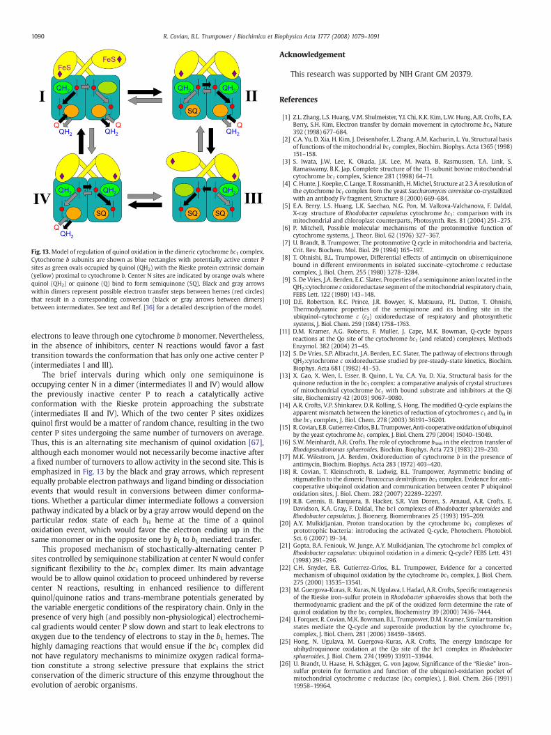
Fig. 13.Model of regulation of quinol oxidation in the dimeric cytochrome bc1 complex.Cytochrome b subunits are shown as blue rectangles with potentially active center Psites as green ovals occupied by quinol (QH2) with the Rieske protein extrinsic domain(yellow) proximal to cytochrome b. Center N sites are indicated by orange ovals wherequinol (QH2) or quinone (Q) bind to form semiquinone (SQ). Black and gray arrowswithin dimers represent possible electron transfer steps between hemes (red circles)that result in a corresponding conversion (black or gray arrows between dimers)between intermediates. See text and Ref. [36] for a detailed description of the model.
1090 R. Covian, B.L. Trumpower / Biochimica et Biophysica Acta 1777 (2008) 1079–1091
electrons to leave through one cytochrome b monomer. Nevertheless,in the absence of inhibitors, center N reactions would favor a fasttransition towards the conformation that has only one active center P(intermediates I and III).
The brief intervals during which only one semiquinone isoccupying center N in a dimer (intermediates II and IV) would allowthe previously inactive center P to reach a catalytically activeconformation with the Rieske protein approaching the substrate(intermediates II and IV). Which of the two center P sites oxidizesquinol first would be a matter of random chance, resulting in the twocenter P sites undergoing the same number of turnovers on average.Thus, this is an alternating site mechanism of quinol oxidation [67],although each monomer would not necessarily become inactive aftera fixed number of turnovers to allow activity in the second site. This isemphasized in Fig. 13 by the black and gray arrows, which representequally probable electron pathways and ligand binding or dissociationevents that would result in conversions between dimer conforma-tions. Whether a particular dimer intermediate follows a conversionpathway indicated by a black or by a gray arrow would depend on theparticular redox state of each bH heme at the time of a quinoloxidation event, which would favor the electron ending up in thesame monomer or in the opposite one by bL to bL mediated transfer.
This proposed mechanism of stochastically-alternating center Psites controlled by semiquinone stabilization at center N would confersignificant flexibility to the bc1 complex dimer. Its main advantagewould be to allow quinol oxidation to proceed unhindered by reversecenter N reactions, resulting in enhanced resilience to differentquinol/quinone ratios and trans-membrane potentials generated bythe variable energetic conditions of the respiratory chain. Only in thepresence of very high (and possibly non-physiological) electrochemi-cal gradients would center P slow down and start to leak electrons tooxygen due to the tendency of electrons to stay in the bL hemes. Thehighly damaging reactions that would ensue if the bc1 complex didnot have regulatory mechanisms to minimize oxygen radical forma-tion constitute a strong selective pressure that explains the strictconservation of the dimeric structure of this enzyme throughout theevolution of aerobic organisms.
Acknowledgement
This research was supported by NIH Grant GM 20379.
References
[1] Z.L. Zhang, L.S. Huang, V.M. Shulmeister, Y.I. Chi, K.K. Kim, L.W. Hung, A.R. Crofts, E.A.Berry, S.H. Kim, Electron transfer by domain movement in cytochrome bc1, Nature392 (1998) 677–684.
[2] C.A. Yu, D. Xia, H. Kim, J. Deisenhofer, L. Zhang, A.M. Kachurin, L. Yu, Structural basisof functions of the mitochondrial bc1 complex, Biochim. Biophys. Acta 1365 (1998)151–158.
[3] S. Iwata, J.W. Lee, K. Okada, J.K. Lee, M. Iwata, B. Rasmussen, T.A. Link, S.Ramaswamy, B.K. Jap, Complete structure of the 11-subunit bovine mitochondrialcytochrome bc1 complex, Science 281 (1998) 64–71.
[4] C. Hunte, J. Koepke, C. Lange, T. Rossmanith, H. Michel, Structure at 2.3 Å resolution ofthe cytochrome bc1 complex from the yeast Saccharomyces cerevisiae co-crystallizedwith an antibody Fv fragment, Structure 8 (2000) 669–684.
[5] E.A. Berry, L.S. Huang, L.K. Saechao, N.G. Pon, M. Valkova-Valchanova, F. Daldal,X-ray structure of Rhodobacter capsulatus cytochrome bc1: comparison with itsmitochondrial and chloroplast counterparts, Photosynth. Res. 81 (2004) 251–275.
[6] P. Mitchell, Possible molecular mechanisms of the protonmotive function ofcytochrome systems, J. Theor. Biol. 62 (1976) 327–367.
[7] U. Brandt, B. Trumpower, The protonmotive Q cycle in mitochondria and bacteria,Crit. Rev. Biochem. Mol. Biol. 29 (1994) 165–197.
[8] T. Ohnishi, B.L. Trumpower, Differential effects of antimycin on ubisemiquinonebound in different environments in isolated succinate–cytochrome c reductasecomplex, J. Biol. Chem. 255 (1980) 3278–3284.
[9] S. De Vries, J.A. Berden, E.C. Slater, Properties of a semiquinone anion located in theQH2:cytochrome c oxidoreductase segment of themitochondrial respiratory chain,FEBS Lett. 122 (1980) 143–148.
[10] D.E. Robertson, R.C. Prince, J.R. Bowyer, K. Matsuura, P.L. Dutton, T. Ohnishi,Thermodynamic properties of the semiquinone and its binding site in theubiquinol–cytochrome c (c2) oxidoreductase of respiratory and photosyntheticsystems, J. Biol. Chem. 259 (1984) 1758–1763.
[11] D.M. Kramer, A.G. Roberts, F. Muller, J. Cape, M.K. Bowman, Q-cycle bypassreactions at the Qo site of the cytochrome bc1 (and related) complexes, MethodsEnzymol. 382 (2004) 21–45.
[12] S. De Vries, S.P. Albracht, J.A. Berden, E.C. Slater, The pathway of electrons throughQH2:cytochrome c oxidoreductase studied by pre-steady-state kinetics, Biochim.Biophys. Acta 681 (1982) 41–53.
[13] X. Gao, X. Wen, L. Esser, B. Quinn, L. Yu, C.A. Yu, D. Xia, Structural basis for thequinone reduction in the bc1 complex: a comparative analysis of crystal structuresof mitochondrial cytochrome bc1 with bound substrate and inhibitors at the Qisite, Biochemistry 42 (2003) 9067–9080.
[14] A.R. Crofts, V.P. Shinkarev, D.R. Kolling, S. Hong, The modified Q-cycle explains theapparent mismatch between the kinetics of reduction of cytochromes c1 and bH inthe bc1 complex, J. Biol. Chem. 278 (2003) 36191–36201.
[15] R. Covian, E.B.Gutierrez-Cirlos, B.L. Trumpower,Anti-cooperativeoxidationof ubiquinolby the yeast cytochrome bc1 complex, J. Biol. Chem. 279 (2004) 15040–15049.
[16] S.W. Meinhardt, A.R. Crofts, The role of cytochrome b566 in the electron transfer ofRhodopseudomonas sphaeroides, Biochim. Biophys. Acta 723 (1983) 219–230.
[17] M.K. Wikstrom, J.A. Berden, Oxidoreduction of cytochrome b in the presence ofantimycin, Biochim. Biophys. Acta 283 (1972) 403–420.
[18] R. Covian, T. Kleinschroth, B. Ludwig, B.L. Trumpower, Asymmetric binding ofstigmatellin to the dimeric Paracoccus denitrificans bc1 complex. Evidence for anti-cooperative ubiquinol oxidation and communication between center P ubiquinoloxidation sites, J. Biol. Chem. 282 (2007) 22289–22297.
[19] R.B. Gennis, B. Barquera, B. Hacker, S.R. Van Doren, S. Arnaud, A.R. Crofts, E.Davidson, K.A. Gray, F. Daldal, The bc1 complexes of Rhodobacter sphaeroides andRhodobacter capsulatus, J. Bioenerg. Biomembranes 25 (1993) 195–209.
[20] A.Y. Mulkidjanian, Proton translocation by the cytochrome bc1 complexes ofprototrophic bacteria: introducing the activated Q-cycle, Photochem. Photobiol.Sci. 6 (2007) 19–34.
[21] Gopta, B.A. Feniouk, W. Junge, A.Y. Mulkidjanian, The cytochrome bc1 complex ofRhodobacter capsulatus: ubiquinol oxidation in a dimeric Q-cycle? FEBS Lett. 431(1998) 291–296.
[22] C.H. Snyder, E.B. Gutierrez-Cirlos, B.L. Trumpower, Evidence for a concertedmechanism of ubiquinol oxidation by the cytochrome bc1 complex, J. Biol. Chem.275 (2000) 13535–13541.
[23] M. Guergova-Kuras, R. Kuras, N. Ugulava, I. Hadad, A.R. Crofts, Specificmutagenesisof the Rieske iron–sulfur protein in Rhodobacter sphaeroides shows that both thethermodynamic gradient and the pK of the oxidized form determine the rate ofquinol oxidation by the bc1 complex, Biochemistry 39 (2000) 7436–7444.
[24] I. Forquer, R. Covian,M.K. Bowman, B.L. Trumpower, D.M. Kramer, Similar transitionstates mediate the Q-cycle and superoxide production by the cytochrome bc1complex, J. Biol. Chem. 281 (2006) 38459–38465.
[25] Hong, N. Ugulava, M. Guergova-Kuras, A.R. Crofts, The energy landscape forubihydroquinone oxidation at the Qo site of the bc1 complex in Rhodobactersphaeroides, J. Biol. Chem. 274 (1999) 33931–33944.
[26] U. Brandt, U. Haase, H. Schägger, G. von Jagow, Significance of the “Rieske” iron–sulfur protein for formation and function of the ubiquinol-oxidation pocket ofmitochondrial cytochrome c reductase (bc1 complex), J. Biol. Chem. 266 (1991)19958–19964.
1091R. Covian, B.L. Trumpower / Biochimica et Biophysica Acta 1777 (2008) 1079–1091
[27] L. Esser, B. Quinn, Y.F. Li, M. Zhang, M. Elberry, L. Yu, C.A. Yu, D. Xia, Crystallographicstudies of quinol oxidation site inhibitors: amodified classification of inhibitors forthe cytochrome bc1 complex, J. Mol. Biol. 341 (2004) 281–302.
[28] E.B. Gutierrez-Cirlos, B.L. Trumpower, Inhibitory analogs of ubiquinol act anti-cooperatively on the yeast cytochrome bc1 complex. Evidence for an alternating, half-of-the-sites mechanism of ubiquinol oxidation, J. Biol. Chem. 277 (2002) 1195–1202.
[29] R. Covian, J.P. Pardo, R. Moreno-Sanchez, Tight binding of inhibitors to bovine bc1complex is independent of the Rieske protein redox state, J. Biol. Chem. 277 (2002)48449–48455.
[30] U. Brandt, G. von Jagow, Analysis of inhibitor binding to the mitochondrialcytochrome c reductase by fluorescence quench titration. Evidence for a ‘catalyticswitch’ at the Qo center, Eur. J. Biochem. 195 (1991) 163–170.
[31] E. Darrouzet, M. Valkova-Valchanova, F. Daldal, The [2Fe–2S] cluster Em as anindicator of the iron–sulfur subunit position in the ubihydroquinone oxidation siteof the cytochrome bc1 complex, J. Biol. Chem. 277 (2002) 3464–3470.
[32] J.W. Cooley, A.G. Roberts, M.K. Bowman, D.M. Kramer, F. Daldal, The raisedmidpoint potential of the [2Fe2S] cluster of cytochrome bc1 is mediated by boththe Qo site occupants and the head domain position of the Fe–S protein subunit,Biochemistry 43 (2004) 2217–2227.
[33] E.B. Gutierrez-Cirlos, T. Merbitz-Zahradnik, B.L. Trumpower, Failure to insert theiron–sulfur cluster into the Rieske iron–sulfur protein impairs both center N andcenter P of the cytochrome bc1 complex, J. Biol. Chem. 277 (2002) 50703–50709.
[34] M. Valkova-Valchanova, E. Darrouzet, C.R. Moomaw, C.A. Slaughter, F. Daldal,Proteolytic cleavage of the Fe–S subunit hinge region of Rhodobacter capsulatus bc1complex: effects of inhibitors and mutations, Biochemistry 39 (2000) 15484–15492.
[35] J.W. Cooley, T. Ohnishi, F. Daldal, Binding dynamics at the quinone reduction (Qi)site influence the equilibrium interactions of the iron–sulfur protein andhydroquinone oxidation (Qo) site of the cytochrome bc1 complex, Biochemistry44 (2005) 10520–10532.
[36] R. Covian, B.L. Trumpower, Regulatory interactions between ubiquinol oxidationand ubiquinone reduction sites in the dimeric cytochrome bc1 complex, J. Biol.Chem. 281 (2006) 30925–30932.
[37] A.R. Crofts, S. Hong, Z. Zhang, E.A. Berry, Physicochemical aspects of the movementof the Rieske iron–sulfur protein during quinol oxidation by the bc1 complex frommitochondria and photosynthetic bacteria, Biochemistry 38 (1999) 15827–15839.
[38] G. Von Jagow, T.A. Link, Use of specific inhibitors of the mitochondrial bc1 complex,Methods Enzymol. 126 (1986) 253–271.
[39] P.R. Rich, A.E. Jeal, S.A. Madgwick, S.J. Moody, Inhibitor effects on redox-linkedprotonations of the b haems of the mitochondrial bc1 complex, Biochim. Biophys.Acta 1018 (1990) 29–40.
[40] V.A. Kostyrko, L.S. Iaguzhinskii, Two sites of ubiquinone binding in mitochondrialsuccinate oxidase, Biokimiia 44 (1979) 1884–1890.
[41] R. Covian, K. Zwicker, F.A. Rotsaert, B.L. Trumpower, Asymmetric and redox-specificbinding of quinone and quinol at center N of the dimeric yeast cytochrome bc1complex. Consequences for semiquinone stabilization, J. Biol. Chem. 282 (2007)24198–24208.
[42] DeVries, S.P. Albracht, J.A. Berden, C.A.Marres, E.C. Slater, The effect ofpH, ubiquinonedepletion and myxothiazol on the reduction kinetics of the prosthetic groups ofubiquinol:cytochrome c oxidoreductase, Biochim. Biophys. Acta 723 (1983) 91–103.
[43] Glaser, S.W. Meinhardt, A.R. Crofts, Reduction of cytochrome b-561 through theantimycin-sensitive site of the ubiquinol–cytochrome c2 oxidoreductase complexof Rhodopseudomonas sphaeroides, FEBS Lett. 178 (1984) 336–342.
[44] A.L. Tsai, R. Kauten, G. Palmer, The interaction of yeast Complex III with somerespiratory inhibitors, Biochim. Biophys. Acta 806 (1985) 418–426.
[45] R. Covian, B.L. Trumpower, Rapid electron transfer between monomers when thecytochrome bc1 complex dimer is reduced through center N, J. Biol. Chem. 280(2005) 22732–22740.
[46] F.F. De la Rosa, G. Palmer, Reductive titration of CoQ-depleted Complex III frombaker's yeast. Evidence for an exchange-coupled complex between QHP and low-spin ferricytochrome b, FEBS Lett. 163 (1983) 140–143.
[47] C. Lange, C. Hunte, Crystal structure of the yeast cytochrome bc1 complex with itsbound substrate cytochrome c, Proc. Natl. Acad. Sci. U. S. A. 99 (2002) 2800–2805.
[48] L. Esser, M. Elberry, F. Zhou, C.A. Yu, L. Yu, D. Xia, Inhibitor-complexed structuresfrom the photosynthetic bacterium Rhodobacter sphaeroides, J. Biol. Chem. 283(2008) 2846–2857.
[49] T. Wenz, R. Covian, P. Hellwig, F. Macmillan, B. Meunier, B.L. Trumpower, C. Hunte,Mutational analysis of cytochrome b at the ubiquinol oxidation site of yeastcomplex III, J. Biol. Chem. 282 (2007) 3977–3988.
[50] E.A. Berry, M. Guergova-Kuras, L. Huang, A.R. Crofts, Structure and function ofcytochrome bc complexes, Annu. Rev. Biochem. 69 (2000) 1005–1075.
[51] K.A. Gray, P.L. Dutton, F. Daldal, Requirement of histidine 217 for ubiquinonereductase activity (Qi site) in the cytochrome bc1 complex, Biochemistry 33 (1994)723–733.
[52] D.R. Kolling, R.I. Samoilova, J.T. Holland, E.A. Berry, S.A. Dikanov, A.R. Crofts,Exploration of ligands to the Qi site semiquinone in the bc1 complex using high-resolution EPR, J. Biol. Chem. 278 (2003) 39747–39754.
[53] L.S. Huang, B. Cobessi, E.Y. Tung, E.A. Berry, Binding of the respiratory chaininhibitor antimycin to the mitochondrial bc1 complex: a new crystal structurereveals an altered intramolecular hydrogen-bonding pattern, J. Mol. Biol. 351(2005) 573–597.
[54] G. Bechmann, H. Weiss, P.R. Rich, Non-linear inhibition curves for tight-bindinginhibitors of dimeric ubiquinol–cytochrome c oxidoreductases. Evidence for rapidinhibitor mobility, Eur. J. Biochem. 208 (1992) 315–325.
[55] D. Xia, C.A. Yu, H. Kim, J.Z. Xian, A.M. Kachurin, L. Zhang, L. Yu, J. Deisenhofer,Crystal structure of the cytochrome bc1 complex from bovine heart mitochondria,Science 277 (1997) 60–66.
[56] C.C. Moser, T.A. Farid, S.E. Chobot, P.L. Dutton, Electron tunneling chains ofmitochondria, Biochim. Biophys. Acta 1757 (2006) 1096–1109.
[57] P.O. Ljungdahl, J.D. Pennoyer, B.L. Trumpower, Purification of cytochrome bc1complexes from phylogenically diverse species by a single method, MethodsEnzymol. 126 (1986) 181–191.
[58] V.P. Shinkarev, C.A. Wraight, Intermonomer electron transfer in the bc1 complexdimer is controlled by the energized state and by impaired electron transferbetween low and high potential hemes, FEBS Lett. 581 (2007) 1535–1541.
[59] C.C. Page, C.C. Moser, X.X. Chen, P.L. Dutton, Natural engineering principles ofelectron tunnelling in biological oxidation–reduction, Nature 402 (1999) 47–52.
[60] A.R. Crofts, S. Rose, Marcus treatment of endergonic reactions: a commentary,Biochim. Biophys. Acta 1767 (2002) 1228–1232.
[61] V.P. Shinkarev, A.R. Crofts, C.A. Wraight, The electric field generated byphotosynthetic reaction center induces rapid reversed electron transfer in thebc1 complex, Biochemistry 40 (2001) 12584–12590.
[62] C.H. Yun, A.R. Crofts, R.B. Gennis, Assignment of the histidine axial ligands to thecytochrome bH and cytochrome bL components of the bcl complex from Rhodo-bacter sphaeroides by site-directed mutagenesis, Biochemistry 30 (1991)6747–6754.
[63] X. Gong, L. Yu, D. Xia, C.A. Yu, Evidence for electron equilibrium between the twohemes bL in the dimeric cytochrome bc1 complex, J. Biol. Chem. 280 (2005)9251–9257.
[64] Y. Kamensky, A.A. Konstantinov, W.S. Kunz, S. Surkov, Effects of bc1-site electrontransfer inhibitors on the absorption spectra of mitochondrial cytochromes b,FEBS Lett. 181 (1985) 95–99.
[65] E.B. Gutierrez-Cirlos, T. Merbitz-Zahradnik, B.L. Trumpower, Inhibition of the yeastcytochrome bc1 complex by ilicicolin H, a novel inhibitor that acts at the Qn site ofthe bc1 complex, J. Biol. Chem. 279 (2004) 8708–8714.
[66] A.R. Crofts, E.A. Berry, Structure and function of the cytochrome bc1 complex ofmitochondria and photosynthetic bacteria, Curr. Opin. Struct. Biol. 8 (1998)501–509.
[67] B.L. Trumpower, A concerted, alternating sites mechanism of ubiquinol oxidationby the dimeric cytochrome bc1 complex, Biochim. Biophys. Acta 1555 (2002)166–173.


![Biochimica et Biophysica Acta - immed.org considerations/09.07.2017 updates/Membrane... · G.L. Nicolson, M.E. Ash / Biochimica et Biophysica Acta 1859 (2017) 1704–1724 1705 [8]](https://img.pdfslide.net/doc/110x75/5c684f1e09d3f2f5638b5509/biochimica-et-biophysica-acta-immed-considerations09072017-updatesmembrane.jpg)