Embed Size (px)
Citation preview

Biology
11
…IS LIFE

2
3
UNIT 1
INTRODUCING BIOLOGY AS A SCIENCE
Reading, discussion and Laboratory work are all essential parts of any biology program. The exercises in this manual have been designed to give you both experience and understanding of the main principles and concepts of modern biology. Throughout, we have made special efforts to encourage cooperative activities of the imagination, accurate and logical reasoning, and careful observations. We do not believe that routine memorization of unrelated facts is a sound approach to biology, or to any other science. However, you will be asked to come to grips with detailed information when that information is required to document, illustrate, or explain a basic biological principle. Our emphasis is on individual discovery of concepts and principles and how they relate to the various structures and their functions presented in the course. Life is examined from an evolutionary perspective taking into account the progression of increasing complexity as we examine the “tree of life”. It is our hope that the approach we have taken in preparing this material will increase your awareness of science as a method of investigating the world around you. It is also our hope that you gain an appreciation for the connectedness that all life shares whether we discuss the vastness of the biosphere or the details of molecular genetics.

4
5
MOUNT BAKER SECONDARY SCHOOL
TEACHER: Mrs. D. Dupley Room 200
MATERIALS REQUIRED: Three-ring binder with a constant supply of Loose-leaf paper, ruler, pencil, pen, calculator, a good eraser, and your BRAIN.
TEXT: Biology – Miller/Levine GENERAL RESPONSIBILITIES:
Show up on time at the start of class and after break
Come to class prepared. (do your readings, bring a pencil, calculator etc.)
Respect the needs and learning rights of others.
Do not bring phones, iPods or headphones to class.
Hand in assignments and labs on the due date. ATTENDANCE AND LATES: Attendance is critical for your success in this course. Any absences must be accompanied by a note or a phone call to my voice-mail 426-5241 loc. 722 from your parents. Your teacher will follow up after three absences. After every 3 unexcused lates, a detention will be served at lunch.
HOMEWORK WEBSITE: A homework website is available to you and your parents to check due dates daily for all assignments. Visit (http://mbss.sd5.bc.ca/), select Students found on the top menu, then select Class Web Pages and Ms. Dupley.)
6
ASSIGNMENTS, LABS AND EXAMS: Any assignment, lab or activity is expected to be handed in on the due date. In Biology, once work is graded, returned and answers are discussed, a grade can not be assigned to late work, but will be assessed as the content will be evaluated later on with unit exams. Examinations and quizzes will be given on all units studied in class. There are no rewrites. Short quizzes are written almost every week. Students are expected to write all exams on the day they are provided. A student that misses an exam will be provided an opportunity to demonstrate their learning later in the semester (providing a phone call has been received from your parents regarding illness). Personal holidays taken during instructional time will be the responsibility of the student to catch up independently on the information missed. ASSESSMENT AND GRADING: Activities and labs comprise about 15% of the mark for the course Quizzes and tests comprise about 60% of the mark for the course The resulting mark will then be added to the final exam worth 25% I have read and understood this outline Student Signature ___________________________________ Parent Signature ___________________________________
7
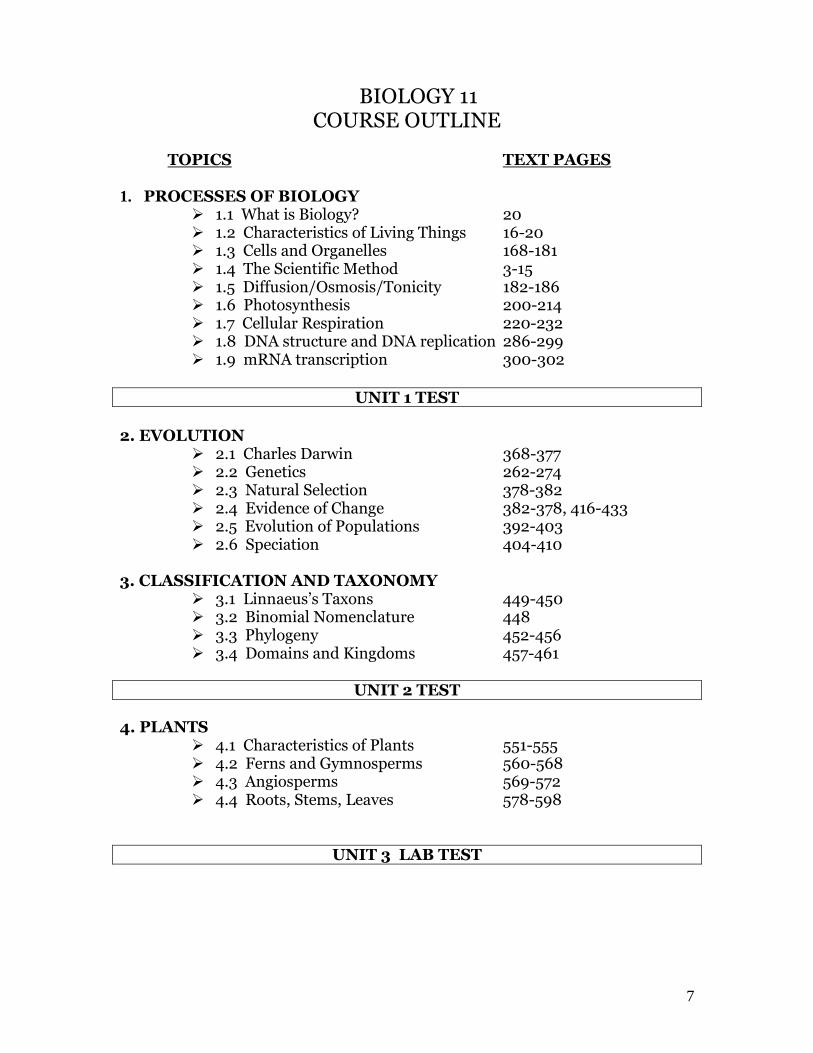
BIOLOGY 11 COURSE OUTLINE
TOPICS TEXT PAGES 1. PROCESSES OF BIOLOGY
1.1 What is Biology? 20 1.2 Characteristics of Living Things 16-20 1.3 Cells and Organelles 168-181 1.4 The Scientific Method 3-15 1.5 Diffusion/Osmosis/Tonicity 182-186 1.6 Photosynthesis 200-214 1.7 Cellular Respiration 220-232 1.8 DNA structure and DNA replication 286-299 1.9 mRNA transcription 300-302
UNIT 1 TEST
2. EVOLUTION
2.1 Charles Darwin 368-377 2.2 Genetics 262-274 2.3 Natural Selection 378-382 2.4 Evidence of Change 382-378, 416-433 2.5 Evolution of Populations 392-403 2.6 Speciation 404-410
3. CLASSIFICATION AND TAXONOMY 3.1 Linnaeus’s Taxons 449-450 3.2 Binomial Nomenclature 448 3.3 Phylogeny 452-456 3.4 Domains and Kingdoms 457-461
UNIT 2 TEST
4. PLANTS
4.1 Characteristics of Plants 551-555 4.2 Ferns and Gymnosperms 560-568 4.3 Angiosperms 569-572 4.4 Roots, Stems, Leaves 578-598
UNIT 3 LAB TEST
8
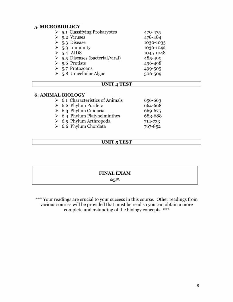
5. MICROBIOLOGY 5.1 Classifying Prokaryotes 470-475 5.2 Viruses 478-484 5.3 Disease 1030-1035 5.3 Immunity 1036-1042 5.4 AIDS 1045-1048 5.5 Diseases (bacterial/viral) 485-490 5.6 Protists 496-498 5.7 Protozoans 499-505 5.8 Unicellular Algae 506-509
UNIT 4 TEST
6. ANIMAL BIOLOGY
6.1 Characteristics of Animals 656-663 6.2 Phylum Porifera 664-668 6.3 Phylum Cnidaria 669-675 6.4 Phylum Platyhelminthes 683-688 6.5 Phylum Arthropoda 714-733 6.6 Phylum Chordata 767-852
UNIT 5 TEST
FINAL EXAM
25%
*** Your readings are crucial to your success in this course. Other readings from
various sources will be provided that must be read so you can obtain a more complete understanding of the biology concepts. ***
9
BIOLOGICAL TERMINOLOGY Write what you think is the meaning of the following words in one sentence:
1. antibody_______________________________________________
2. chemotherapy___________________________________________
3. extracellular_____________________________________________
4. photosynthesis___________________________________________
5. spermatogenesis__________________________________________
6. taxonomy______________________________________________
7. zoology________________________________________________
8. interphase______________________________________________
9. exoskeleton_____________________________________________
10. endocytosis_____________________________________________
11. osteopathology___________________________________________
12. cardiograph_____________________________________________
10
BIOLOGICAL TERMINOLOGY Re-write what you think is the meaning of the following words using the Greek and Latin roots from page 11:
1. antibody_______________________________________________
2. chemotherapy___________________________________________
3. extracellular_____________________________________________
4. photosynthesis___________________________________________
5. spermatogenesis__________________________________________
6. taxonomy______________________________________________
7. zoology________________________________________________
8. interphase______________________________________________
9. exoskeleton_____________________________________________
10. endocytosis_____________________________________________
11. osteopathology___________________________________________
12. cardiograph_____________________________________________

11
GREEK AND LATIN ROOTS a-, an- without deficiency atrophy, abiogenesis ab- away from abnormal ad- to, near adductor aero- air aerobic ameb- change ameba amphi- on both sides amphibian angio- case, capsule angiosperm anth flower anther anthropo- man anthropology anti- against antigen, antibiotic atmo- breath atmosphere auto- self autoimmune bi-,bis- two, double binomial, bicuspid bio-,be- life biology, microbe bot- graze, feed botany bry- grow embryo, bryophyte calori- heat calorimeter, calorie capilli- hair capillary cardi- heart cardiovascular carni- flesh carnivore celli- cell cellular cerebro- brain cerebrospinal chemo- chemical chemoreceptor chrom- colour chromosome cnido- stinging cnidarian, cnidoblast cocco- berry, sphere streptococcus coelo- hollow coelenterate cuti- skin subcutaneous cyto- cell cytoplasm, cytologist de- down, away from dehydration, decomposer demi- people epidemic di-, two, double dihybrid, dichotomous diplo- double diploid dorso- back dorsal e-,ex-,ec- out of exhale eco- house ecology ecto outside ectoderm en- in enzyme, endemic endo- within endosperm exo outside exoskeleton extra- beyond extracellular folli- bag follicle ganglio- knot ganglion gastro- stomach gastric, gastrula geno-, gen- production genotype, genetic gono- seed gonad geo- earth geology, geotropism grapho- write chromatograph gymno- naked gymnosperm hemo- blood hemoglobin haplo- single haploid hepato liver hepatitis hetero- other heterotroph homo- man Homo sapiens hydro water hydrolysis hyper- too much hypertension hypo too little hypotonic in- without invertebrate, inorganic infra- below infrared inter- between interphase intra- within intracellular iso- equal isotonic, isogamete it is- inflamed gastroenteritis latero- side bilateral loco- place locus lyso- dissolve lysosome, lysis meta- with, beyond metamorphosis micro- little micronucleus morpho- form morphology nephro- kidney nephron neuro- nerve neuron -nomy law taxonomy noto- back notochord osteo- bone osteon, osteoblast ovi- egg oviduct, ovary para- alongside parasite phago- eat phagocyte photo- light photosynthesis phyllo leaf chlorophyll phylo tribe phylum phyto plant phytoplankton plasmo- molded plasma,protoplasm poro- passage poriferan -plasm chloroplast pro- before prophase -plast pulmo- lung pulmonary radio- spoke, wheel, ray radial symmetry reni- kidneys adrenal rhizo- root rhizome semi- half, partly semipermeable somato- body somatic spermato- seed spermatozoa spira- breath transpiration, respiration stasis- balance homeostasis syn-, sym-, with symbiosis, synthesis taxo- arrangement taxonomy therpia- nurse, care therapy therm- warm hypothermia tropho- nourishment atrophy, autotrophic ultra- beyond ultraviolet vacci- cow vaccine vaso- vessel vasodilate ventri- front ventral, ventricle -vore eat herbivore zoo-, zoon animal zoology, zooplankton zygo- yoke zygote, zygomatic zygomato-

12
13
OBJECTIVE SHEET PROCESSES OF BIOLOGY 1. Identify various branches of biology.
2. List 8 characteristics of living things and show the levels of life’s organization.
3. Demonstrate an ability to work with SI and common derived units for volume, mass, length, temperature and time.
4. Determine the actual size of an organism viewed under the microscope and
calculate the magnification of a drawing. 5. Demonstrate the ability to make a wet-mount slide and focus the microscope
on all three powers. 6. Identify three essential parts of the cell theory and list the differences between prokaryotic and eukaryotic cells. 7. Label the following parts of plant and animal cells and state the functions of the parts: cell wall, cell membrane, cytoplasm, mitochondrion, nucleus, lysosome, vacuole, and chloroplast. 8. List four differences between a plant and an animal cell. _______________________________________________________ 9. Differentiate between the terms science and technology. 10. Identify, define and formulate a formal hypothesis. 11. Demonstrate appropriate procedures for testing, confirming or rejecting a hypothesis. 12. Identify valid steps in an experiment and criticize or defend an entire
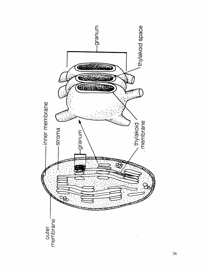
experiment in terms of its validity of methods. 13. List two rules that may be used to assess the validity of an experiment. 14. Define the following terms: solute, solution, solvent, osmosis, diffusion, hypertonic, isotonic, hypotonic, and osmotic pressure. 15. Solve problems involving concentrations of differences between cells and solutions. _______________________________________________________ 16. Diagram the internal structure of a chloroplast and a mitochondrion, including the following structures: granum, grana, stroma, cristae, matrix.
Define the following terms: ATP, reduction and oxidation.
14
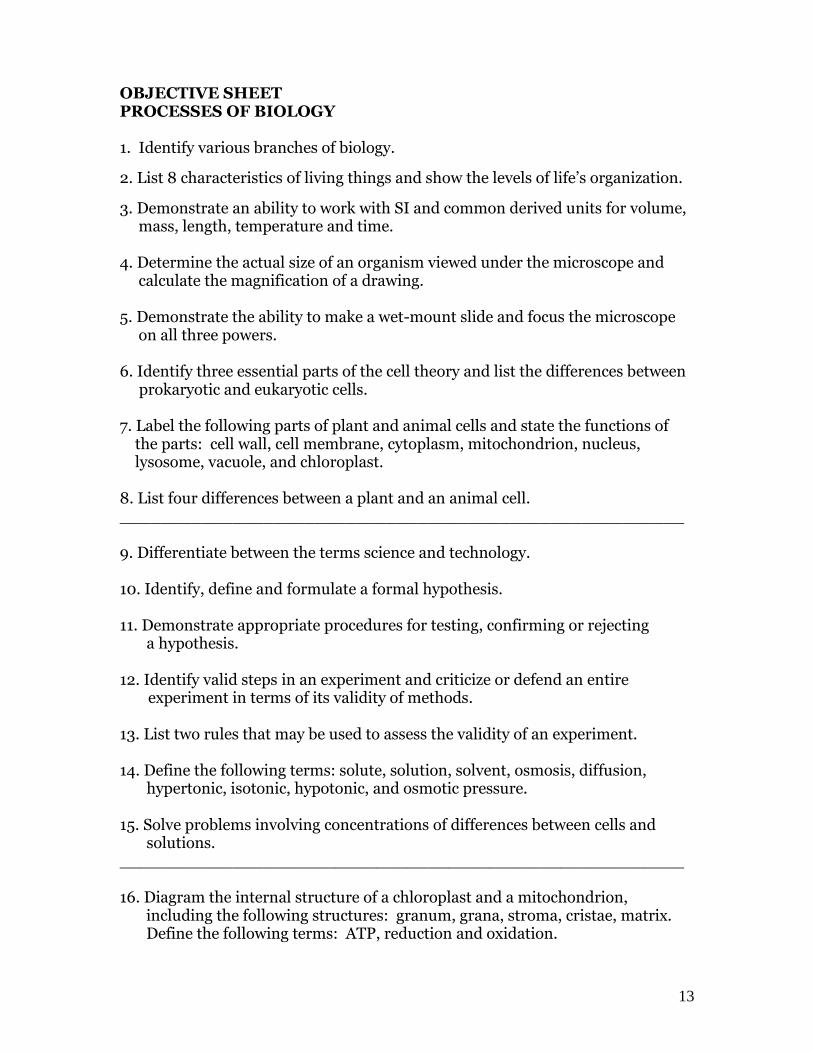
17. Explain the process of photosynthesis by filling in the chart below: Reactants Products Location Light Rx
Light Independent Rx (Calvin Cycle)
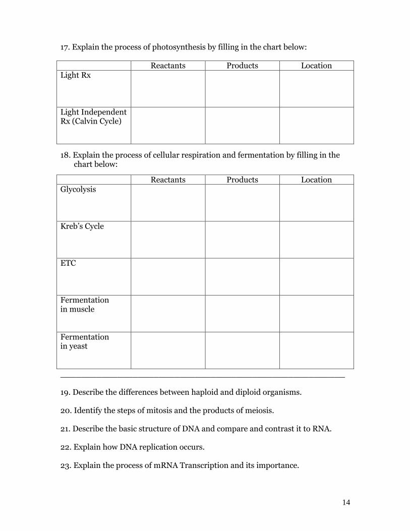
18. Explain the process of cellular respiration and fermentation by filling in the chart below:
Reactants Products Location Glycolysis
Kreb’s Cycle
ETC
Fermentation in muscle
Fermentation in yeast
_______________________________________________________ 19. Describe the differences between haploid and diploid organisms. 20. Identify the steps of mitosis and the products of meiosis. 21. Describe the basic structure of DNA and compare and contrast it to RNA. 22. Explain how DNA replication occurs. 23. Explain the process of mRNA Transcription and its importance.
15
THE CHARACTERISTICS OF LIVING THINGS
Living things are made up of cells All living organisms are made up of cells, which are the building block of all living things. Many living things are made up of single cells and are called unicellular. Most of the familiar organisms are multicellular organisms.
Living things reproduce During reproduction, a copy of its hereditary information (genes) is passed on. Unicellular organisms reproduce asexually by dividing into two cells with identical genetic structure as the original. Multicellular organisms may reproduce asexually. Each parent contributes ½ of the total number of genes to an offspring, which then has characteristics of both parents and may not resemble either one exactly.
Living things use DNA as a universal genetic code The inheritance of traits from parent to offspring is carried on a molecule called deoxyribonucleic acid, or DNA. This molecule determines the traits of every organism on Earth.
Living things grow and develop Growth is recognized by an increase in size and often the number of cells. Human development includes all changes that take place between conception and death.
Living things obtain and use materials and energy All organisms take in selected materials that they need from their environment to perform life activities. The sum of all the chemical reactions that occur in an organism is called the organism’s metabolism. Nutrient molecules from the environment must be used to maintain energy needs.
Living things respond to their environment Organisms detect and respond to stimuli from the environment. This response allows the organism to prevent damage or to promote growth and development. The more complex the organism is the more sophisticated the response can be towards a stimulus.
Homeostasis means “staying the same” The internal environment of an organism stays relatively constant compared to external environments; for example, body temperature fluctuates only slightly during a day. Homeostasis is the process of maintaining environments within certain levels.
Taken as a population, all living things evolve over time Individuals have adaptations and modifications that make that organism suited for its way of life. For example, hollow bones in birds reduce weight and permit flight. Adaptations are selected by evolution, the process where characteristics of species change over time. When new variations arise that allow certain members of the species to capture more resources, survive in higher numbers and have more offspring than poorly adapted members. Evolution explains the diversity and unity of life on Earth.
16
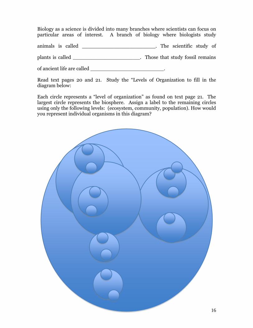
Biology as a science is divided into many branches where scientists can focus on particular areas of interest. A branch of biology where biologists study animals is called _______________________. The scientific study of plants is called _____________________. Those that study fossil remains of ancient life are called _______________________. Read text pages 20 and 21. Study the “Levels of Organization to fill in the diagram below: Each circle represents a “level of organization” as found on text page 21. The largest circle represents the biosphere. Assign a label to the remaining circles using only the following levels: (ecosystem, community, population). How would you represent individual organisms in this diagram?

17
THE AMAZING MICROSCOPE
Microscopes are so common today in a regular classroom that most students know a little about them. Even if you have never really used a microscope, everyone has looked at a bug or a blade of grass at some point in their life even if it was just with a magnifying glass. The microscope has proven to be one of the greatest accomplishments of humankind over the last five centuries as new and more powerful microscopes have been developed with stunning views of objects and life that have enabled scientists to tackle questions and problems that continue to help and amaze us every day. The microscope, in its simplest form, dates back to the 17th century when a Dutch lens maker, Anton Von Leuwenhoek, ground and polished a tiny bead of glass until it magnified whatever he looked at. To his great astonishment and awe he found that a drop of stagnant water was teeming with life never before visible to the human eye. For greater convenience, he fashioned a crude microscope of metal in which he inserted and secured this bead of glass.
Since that pioneering discovery of a revolutionary new use of optical lenses, there have been vast improvements and advances in magnifying lenses and microscopes. An Englishman, Robert Hooke, made the first compound microscope. This type is what you use today, however some can magnify as much as 1800 times. Such a microscope contains many lenses which, when combined, really increase magnification. Early in this century, it was discovered that ultraviolet light could be used instead of light visible to the human eye, to obtain even higher magnification, as much as 4,000 times the life size of an object. This light cannot be seen by the human eye but can be photographed with a UV microscope. In very recent years, engineers have developed a Transmission Electron Microscope (TEMS) or more commonly called an Electron microscope which can produce a magnification of over 20,000 times. Scanning Electron Microscopes (SEMS) provide three-dimensional pictures that provide surface details of objects. The most amazing microscope today is the Scanning Tunneling Microscope which has allowed scientists to see individual atoms at a magnification of 24 million times!! A Nobel prize was awarded in 1986 to the Swiss co-inventors Heinrich Rohre and Gerd Binning. The benefits to molecular biologists, to physicians, and microchip designers, as well as electrochemists and geneticists are invaluable. There is no doubt that the microscope is a great invention, which leaves us with one question – ………………… How small can we go?
18
USE AND CARE OF THE LABOMED COMPOUND MICROSCOPE The microscope has undoubtedly been an important tool in a wide range of biological discoveries. It has, more than any other instrument, become a symbol of the activities of biologists. For many of you, this exercise will be an introduction to a new dimension. Indeed, for some individuals it has stimulated a curiosity that has led to a lifetime of work in biology. There are several kinds of microscopes at Mount Baker. You will use dissecting microscopes to allow you to see three dimensions and surface features of specimens. You will become most familiar with the compound microscope in class. You will observe many prepared slides and even make some of your own in class. 1. Read text pages 25 and 26 on Microscopes. Light microscopes produce images by focusing _______________________ while electron microscopes focus beams of ____________________. There are two main types of electron microscopes. These include ___________ which shine a beam of electrons through a specimen and ____________ which scan a narrow beam of electrons back and forth across the surface. The end result of a TEMS image shows __________________________________ while a SEMS image shows ____________________________________. 2. Obtain a LABOMED binocular compound microscope from the cupboard and place it on your table. You will be assigned this microscope for the entire course. Take good care of it by knowing how to clean and operate the lenses properly, how to carry the microscope properly, how to operate the microscope to allow you to see specimens and how to properly store the microscope. Go to the front desk and print your name and your partner’s name on the “MICROSCOPES” sign-up sheet to indicate your microscope number that you will be responsible for and use throughout the course. NOTIFY me immediately if you notice any problem with your microscope.
19
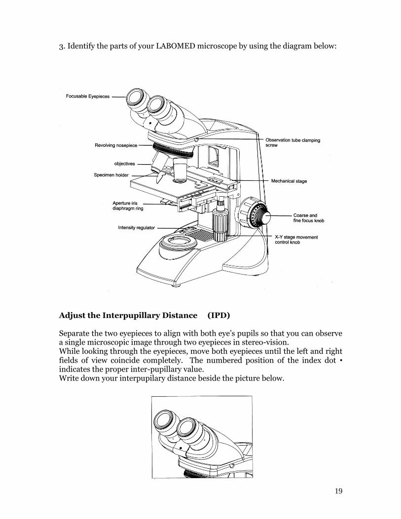
3. Identify the parts of your LABOMED microscope by using the diagram below:
Adjust the Interpupillary Distance (IPD) Separate the two eyepieces to align with both eye’s pupils so that you can observe a single microscopic image through two eyepieces in stereo-vision. While looking through the eyepieces, move both eyepieces until the left and right fields of view coincide completely. The numbered position of the index dot • indicates the proper inter-pupillary value. Write down your interpupilary distance beside the picture below.
20
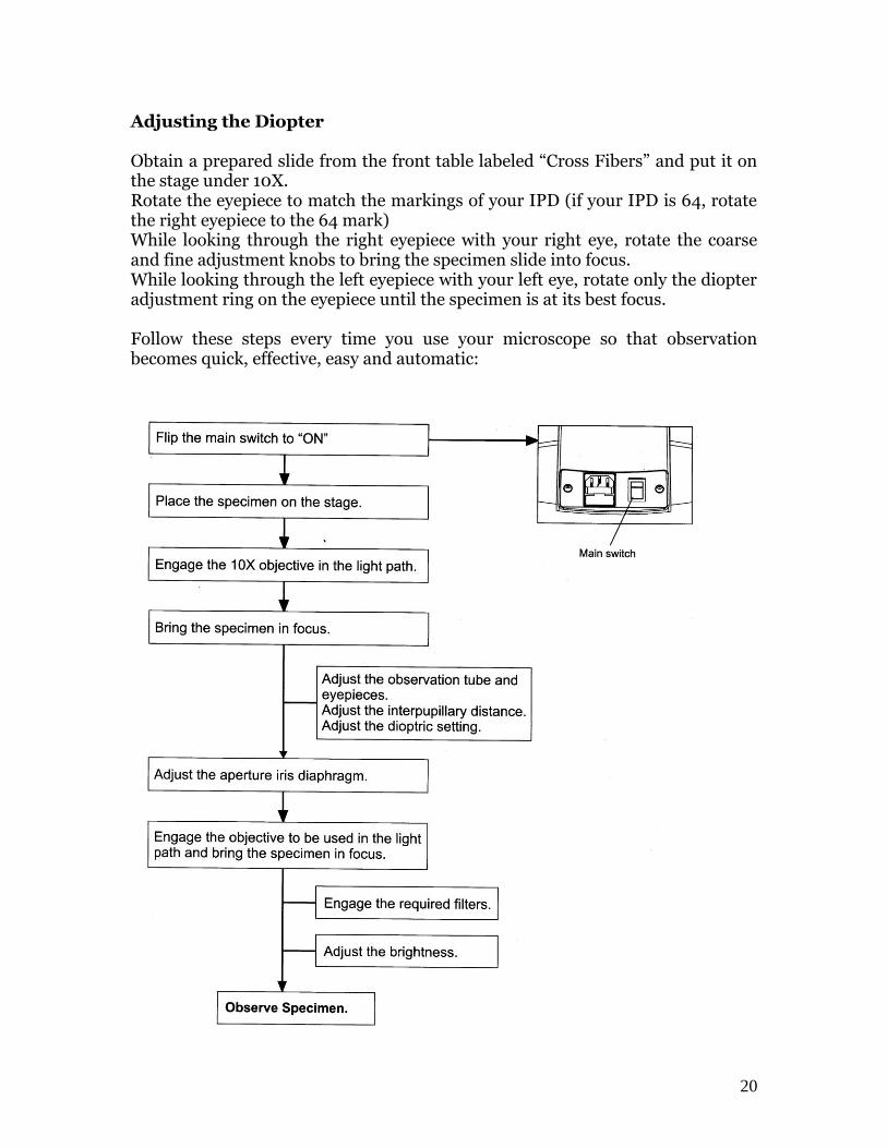
Adjusting the Diopter Obtain a prepared slide from the front table labeled “Cross Fibers” and put it on the stage under 10X. Rotate the eyepiece to match the markings of your IPD (if your IPD is 64, rotate the right eyepiece to the 64 mark) While looking through the right eyepiece with your right eye, rotate the coarse and fine adjustment knobs to bring the specimen slide into focus. While looking through the left eyepiece with your left eye, rotate only the diopter adjustment ring on the eyepiece until the specimen is at its best focus. Follow these steps every time you use your microscope so that observation becomes quick, effective, easy and automatic:
21
Use these 7 steps every time you use a microscope in this class. As we progress in the course, you will refer to these pages when the time comes to prepare Wet-Mount slides, observe Gram Stains or attempt to follow a living Paramecium or Euglena.
1. When using prepared slides, always find any specimen on the low power objective (4x red band) and centre it. If you want to observe the specimen under high power (40X blue band), first centre under medium power (10x yellow band) then switch to high power.
2. The iris diaphragm is very often the most important part of the
microscope that allows you to see detail. Adjusting the iris diaphragm changes the contrast of the light and a lot of specimens that may be “drowned-out” by too much light all of a sudden come into sharp detail.
3. Keep the stage dry and clean. Very often a wet glass slide prevents the
mechanical stage from moving the slide around. 4. Clean lenses with lens paper only. Always clean in a circular motion as the
lens glass is soft and easy to scratch.
5. Some objects being have depth to them. Constantly adjust your fine focus so that an area that is blurred now can come into focus later.
6. You can leave your eyeglasses on or take them off when looking through
the microscope. Try to keep both eyes open and avoid squinting to prevent eye fatigue.
7. Before storing the microscope, clean the lenses with lens paper and place
the low power objective (4x red band) down for the next person to use.

22
23
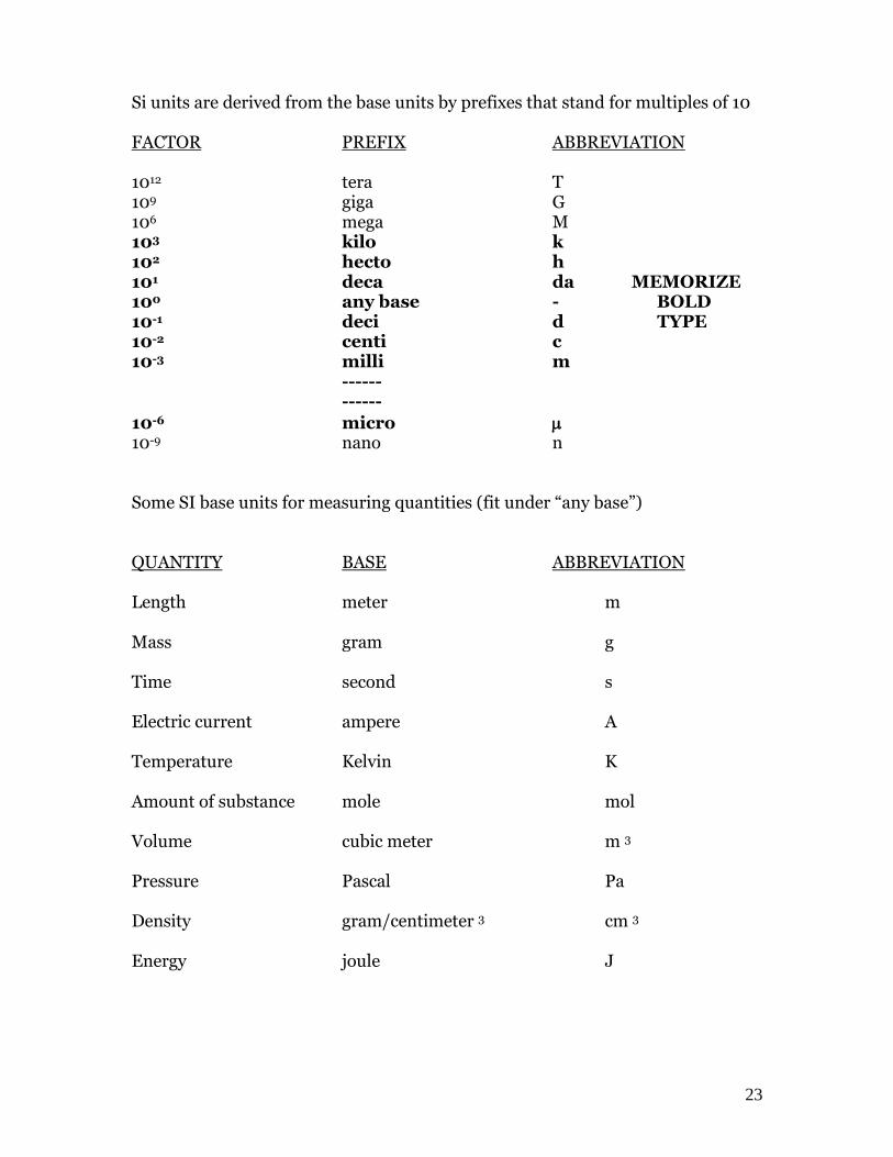
Si units are derived from the base units by prefixes that stand for multiples of 10 FACTOR PREFIX ABBREVIATION 1012 tera T 109 giga G 106 mega M 103 kilo k 102 hecto h 101 deca da MEMORIZE 100 any base - BOLD 10-1 deci d TYPE 10-2 centi c 10-3 milli m ------ ------
10-6 micro 10-9 nano n Some SI base units for measuring quantities (fit under “any base”) QUANTITY BASE ABBREVIATION Length meter m Mass gram g Time second s Electric current ampere A Temperature Kelvin K Amount of substance mole mol Volume cubic meter m 3 Pressure Pascal Pa Density gram/centimeter 3 cm 3
Energy joule J
24
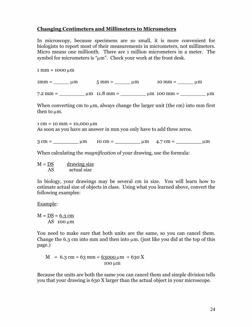
Changing Centimeters and Millimeters to Micrometers
In microscopy, because specimens are so small, it is more convenient for biologists to report most of their measurements in micrometers, not millimeters. Micro means one millionth. There are 1 million micrometers in a meter. The
symbol for micrometers is “m”. Check your work at the front desk.
1 mm = 1000 m
1mm = _____ m 5 mm = _____ m 10 mm = _____ m
7.2 mm = ________ m 11.8 mm = ________ m 100 mm = ________ m
When converting cm to m, always change the larger unit (the cm) into mm first
then to m.
1 cm = 10 mm = 10,000 m As soon as you have an answer in mm you only have to add three zeros.
3 cm = ________ m 10 cm = ________ m 4.7 cm = ________ m
When calculating the magnification of your drawing, use the formula: M = DS drawing size AS actual size
In biology, your drawings may be several cm in size. You will learn how to estimate actual size of objects in class. Using what you learned above, convert the following examples: Example:
M = DS = 6.3 cm
AS 100 m
You need to make sure that both units are the same, so you can cancel them.
Change the 6.3 cm into mm and then into m. (just like you did at the top of this page.)
M = 6.3 cm = 63 mm = 63000 m = 630 X
100 m
Because the units are both the same you can cancel them and simple division tells you that your drawing is 630 X larger than the actual object in your microscope.
25
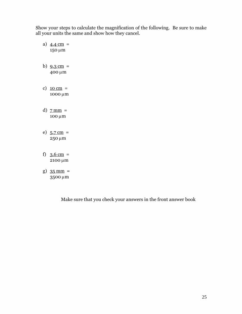
Show your steps to calculate the magnification of the following. Be sure to make all your units the same and show how they cancel.
a) 4.4 cm =
150 m
b) 9.3 cm =
400 m
c) 10 cm =
1000 m
d) 7 mm =
100 m
e) 5.7 cm =
250 m
f) 3.6 cm =
2100 m
g) 35 mm =
3500 m
Make sure that you check your answers in the front answer book

26
27
HANDING IN LAB REPORTS AND GROUP ASSIGNMENTS BIOLOGY 11
1. Every student is required to make their own individual copy of the lab or group assignment to be handed in. Your full name goes on the top right corner along with your block.
2. You may discuss answers/questions/conclusions with your lab partners.
This is why you have a lab group. Your group must come to consensus on all the answers/questions/conclusions for your lab report.
3. On the due date for your lab, you will be given time to look over each
other’s lab report and sign your name below theirs. (all labs will probably be the same anyway, just check to see if they are missing anything) Never let anyone sign your name for you, sign your own name. This is NOT the time to finish your lab if you are not done. Your partner(s) will not sign your lab nor let you sign theirs if it is incomplete.
4. Place the labs on the desk ready for the teacher to choose one of the labs or
bring all the signed copies to the front desk for the teacher to choose.
5. If a lab partner did not finish their lab, do not sign theirs and do not let them sign yours. Let them get the mark they deserve as they have to hand-in their own unfinished lab.
6. If a lab partner is not present on the due date, their name cannot be
included on the lab chosen because I do not have the option of choosing the lab of the person who is absent. (for all I know, they may not have finished the lab report unless they have previously left their lab report with you)
7. You always have the option of handing in your own lab report. If you wish
to work on all questions and conclusions by yourself, you cannot have the benefit of asking your lab partners for answers. Even though you may be performing the lab procedures with other people, you will do all questions and conclusions by yourself. Do not share/discuss or get answers from the lab group that you performed the lab with. You will not sign anyone’s lab nor will they be allowed to sign your lab upon collection.
8. Never send your lab home with someone.

28
29
Microscope Use The ability to distinguish between two points is called resolution. In Biology 11 class at Mt. Baker, you will use the microscope quite often. As you work, you will develop skill in using the compound light microscope. These skills are important for you to retain for the duration of the course. An important aspect of working with microscopes is the ability to draw (IN PENCIL) what is observed and to determine the actual size of the object being observed. Actual size of objects can be estimated if you know the field diameter of the microscope that you are working on. The field diameter for the various microscope magnifications:
Low power 4 x 4.4 mm = _____ m
Medium power 10 x 1.8 mm = _____ m
High power 40 x 0.44 mm = _____ m
After drawing what you see in the microscope you must always calculate the magnification of the drawing. (How many times larger is the drawing than the actual size of the object?) Use this formula. It is all you need for microscope use:
AS = FD M = DS NAF AS
1. Obtain a microscope and carefully place it on the table. Clear any books or papers so you can work. Familiarize yourself with all the parts again and follow the 7 guidelines when always using your LABOMED microscope.
2. Obtain the following slides: Clonorchis sinensis, and the Spirogyra. Make drawings of the slides below. (USING PENCIL). While looking under the
microscope, estimate the actual size of the specimen in m and calculate the magnification of each drawing. Find Clonorchis under low power (red band) and then Spirogyra under high power (blue band).
Clonorchis sinensis

30
Spirogyra
3. Carefully remove a hair from your head and pass it to your lab partner. Your partner will make a slide of your hair by taping the two ends of the hair to a glass slide. Observe the diameter of the hair under high power. Have your partner secretly estimate the diameter of your hair as accurate as they can to
the nearest m. It’s now your turn to estimate. After you are ready, compare estimations. REMOVE THE TAPE FROM THE GLASS SLIDE. You must now make a slide of your partners’ hair and repeat the process. Replace the glass slide in the cupboard after you remove the tape. Write the name of the
person with the “fattest” hair and the hair’s diameter (in m) on the white board. You do not need to draw their hair.
What is the formula for calculating magnification? _________ What is the advantage of adjusting the iris diaphragm? _____________________________________________________ State two guidelines that you must always do when you want to find and focus an object under high power. _____________________________________________________ _____________________________________________________

31
Approximately 500 anthrax bacteria can fit across your low power field of view in your microscope. What is the approximate size of one bacterium? Show your work. _____________________________________________________ _____________________________________________________ _____________________________________________________ A student wants you to explain to them how to estimate the size of the object that they are looking at in the microscope. How would you explain this? _____________________________________________________ _____________________________________________________ _____________________________________________________ _____________________________________________________
32
Cell Theory Reference text pgs. 168-174
Life on our planet is ‘cellular’. This means that the cell is the basic building block of all living things on Earth. A fundamental concept of biology is the CELL THEORY. The cell theory states three things:
1. ___________________________________________________
2. ___________________________________________________
3. ___________________________________________________ Prokaryotes and Eukaryotes Cells fall into two broad categories, depending on whether they contain a nucleus. The nucleus is a large membrane-enclosed structure that contains the cell’s genetic material in the form of DNA. The nucleus controls many of the cell’s chemical activities. _______________________ are cells that contain nuclei. Cells that do not contain nuclei or membrane-bound organelles are ____________________. List the activities and characteristics of prokaryotes: _______________________________________________________ _______________________________________________________ _______________________________________________________ Give an example of a Prokaryote: ________________________________ List the activities and characteristics of eukaryotes: _______________________________________________________ _______________________________________________________ _______________________________________________________ Give an example of a eukaryote: _________________________________
33
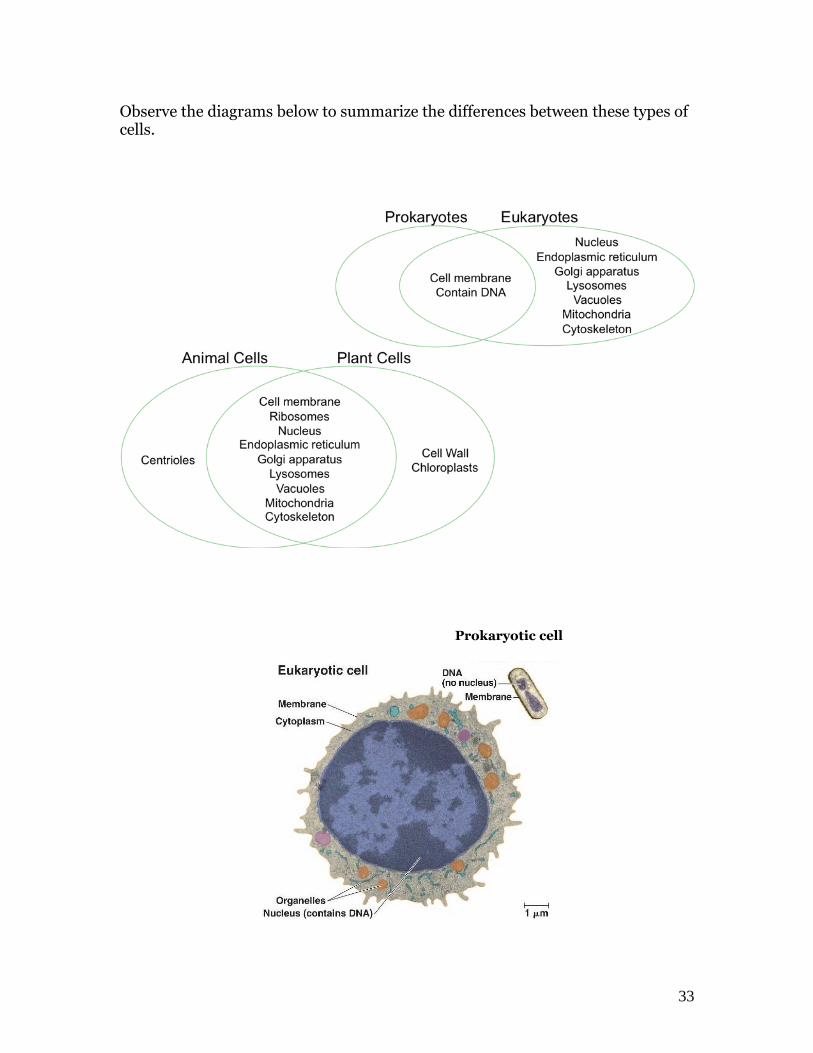
Observe the diagrams below to summarize the differences between these types of cells. Prokaryotic cell

34
35
Cell Organelles The Structure of Cell
You are required to demonstrate that you can compare and contrast the structure of plant and animal cells.
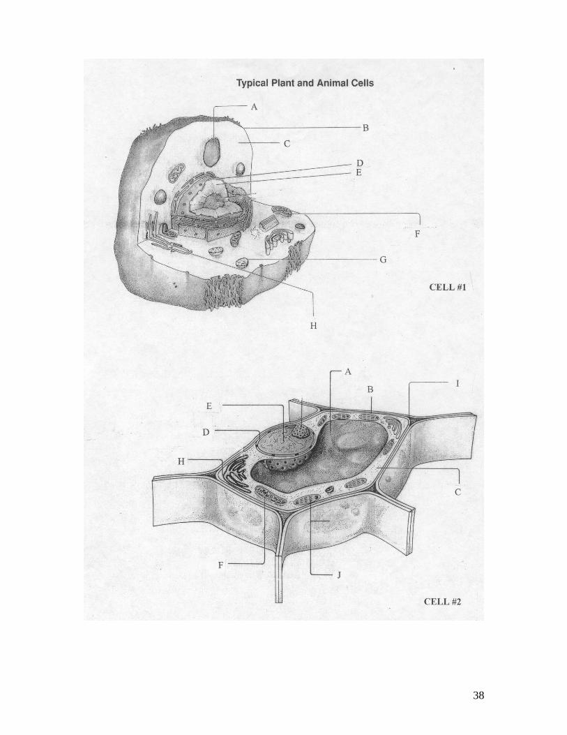
You need to learn the function of the following cell parts as listed in your objective sheet: Cell wall, cell membrane, cytoplasm, mitochondria, nucleus, lysosome, vacuole and chloroplast. Make sure you can recognize these structures in diagrams.
You will demonstrate that you can make a “Wet-mount slide” with appropriate biological stains.
Onion Cell
1. Prepare a wet mount slide of an onion skin cell by snapping a piece of onion in half and placing the thinnest possible section on a slide. Add a drop of water and a drop of iodine stain. Use a cover slip.
2. Place a drop of clear water beside the cover slip and draw water under with paper towel on the opposite side.
3. Focus on Medium (yellow band 10 X). Diagram any 4 cells below to show how they fit together. Label one cell to include the following: cell wall, cell membrane, nucleus and cytoplasm. Include the magnification of your drawing. Remember to show all your work.

36
Epithelial Cell
1. Place a drop of water in the center of a clean glass slide.
2. Take a toothpick and gently scrape the inside of your cheek to remove epithelial cells. Touch the toothpick to the drop of water and gently smear in the cells. THROW THE TOOTHPICK INTO THE GARBAGE NOW!!
3. Add a cover slip and place a drop of methylene blue biological stain on the glass slide by the corner of the cover slip.
4. Draw off the stain with a paper towel from the opposite side.
5. Diagram 1 epithelial cell in the space below under high power (blue band 40 X). Include the following labels: cytoplasm, nucleus, mitochondria and cell membrane.
37
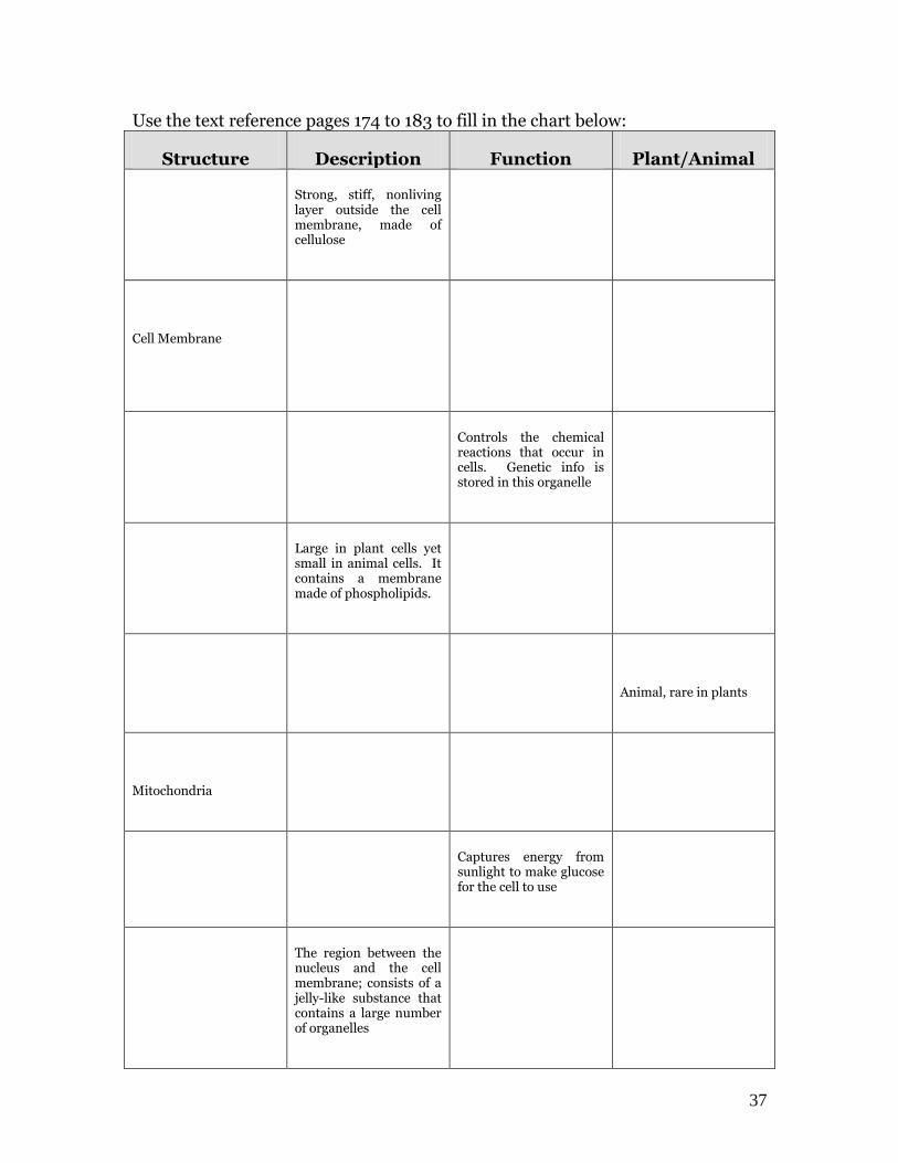
Use the text reference pages 174 to 183 to fill in the chart below:
Structure Description Function Plant/Animal
Strong, stiff, nonliving layer outside the cell membrane, made of cellulose
Cell Membrane
Controls the chemical reactions that occur in cells. Genetic info is stored in this organelle
Large in plant cells yet small in animal cells. It contains a membrane made of phospholipids.
Animal, rare in plants
Mitochondria
Captures energy from sunlight to make glucose for the cell to use
The region between the nucleus and the cell membrane; consists of a jelly-like substance that contains a large number of organelles
38

39
Can You Identify These Cell Organelles? 1. I’m a real ‘powerhouse’. 6. I’m a series of tubes that’s plain to see, found throughout the cell I break down food I transport proteins to release energy. and other things as well. What am I? ________________ What am I? _______________ 2. I’m strong and stiff 7. I’m full of holes, getting through me is not tough flexible and thin. I’m found only in plants I control what gets out but I guess that’s enough as well as what gets in. What am I? ________________ What am I? ______________ 3. My name means ‘colored bodies’ 8. Proteins are made here and I contain DNA. even though I’m quite small. I pass on traits to new cells You can find me in the cytoplasm in a systematic way. or attached to the E.R.’s wall.
What am I? ________________ What am I? ______________ 4. I’m the ‘brain’ of the cell 9. I’ve been called a ‘storage tank’ or so they say by those with little taste. I regulate activities I’m a sac filled with water, from day to day food, enzymes, or waste. What am I? ________________ What am I? ______________ 5. Found only in plant cells 10. Since I contain many enzymes I’m as green as can be I can digest an injured cell I make food for the plant and break down big molecule using the sun’s energy into smaller ones as well What am I? ________________ What am I? ____________

40

41
The Nature of Science Although scientists and their particular science may differ in their focuses, scientists- biologists, chemists, physicists, geologists, and so forth- study the natural world and share scientific methods that usually include all of the following key features: observation, hypothesis development, and testing a hypothesis. A hypothesis is a testable statement or tentative explanation for an observation made by a scientist. They test their hypotheses in a variety of ways, producing results that are open to verification by other experts. If other scientists repeatedly verify the results over a long period of time, a theory may be developed. A theory comes from a well-supported and well-tested hypothesis. Unfortunately, many people misuse the meaning of the word theory. To the average Canadian, the word theory just means “ I have an idea”, when in science, a theory is only developed after extensive testing confirms an idea (for now) over many years. Because a hallmark of science is the testable hypothesis, science does not try to explain teleological - philosophical or religious questions such as “What is the meaning of life?” or “Does life exist after death?” within the framework of the scientific inquiry. This is not to say that these questions are not important; it is to say that they cannot be tested using scientific methods. Mistakes occur in science all the time. History is full of old theories that have been proven wrong or erroneous in many ways. This suggests that scientific theories are “falsifiable.” Within this ability to prove methods and theories as false lies sciences’ greatest strength – self-correction. Whether a mistake is made honestly or dishonestly, in time it will be “flushed out” of the system by the lack of verification by other experts. Science as a way of knowing allows us to avoid dogmatism, which is when a person bases a conclusion on authority rather than logic and evidence. Scientific progress is the cumulative growth of a system of knowledge over time, in which useful features are retained and non-useful features are abandoned, based on the rejection or confirmation of testable knowledge. Technology Allows us to apply the knowledge of science to everyday life so that we may be more productive.

42
43
Designing an Experiment and Writing Formal Hypotheses When thinking like a scientist, the scientific method must be used:
1. Ask a question 2. Form a hypothesis 3. Set up a controlled experiment 4. Record and Analyze your results 5. Draw conclusions 6. Have your hypothesis tested by others.
The Hypothesis A hypothesis is a tentative, testable statement that proposes a possible explanation to some natural phenomena or event. It often contains a prediction as to the outcome. Parts of a Hypothesis The test of the hypothesis is the experiment. It is a test of how 2 variables might be related. One is the dependent variable. This is the one that the experimenter observes or measures the results that occur. The other is the independent variable and it is the one that the experimenter controls. Formalized Hypotheses These contain the words ‘if’ and ‘then’. If skin cancer is related to ultraviolet light, then people with a high exposure to UV light will have a higher frequency of skin cancer. If leaf color change is related to temperature, then exposing plants to low temperatures will result in changes in leaf color. NOTE: that not all ‘if-then’ statements are hypotheses. If I buy lottery tickets, then I will get rich. This is a simple prediction. In a hypothesis, a tentative relationship is stated. For example: If the frequency of winning is related to the frequency of buying “then” is followed by a prediction of what will happen if you increase or decrease the frequency of buying tickets. The ultimate value of a formalized hypothesis is that it forces us to think about what results we should look for in an experiment. Look at the underlined ‘variables’ from the formalized hypotheses. Fill in the chart.
Dependent Variable Independent Variable
44
The following statements are not particularly useful because they contain the word ‘may’ which does not suggest how you would go about confirming it.
1. Chocolate may cause pimples. 2. Plant growth may be affected by the color of the light. 3. Bacterial growth may be affected by temperature.
Re-write these as formal hypotheses. Always use this format: “If dependent variable is related to independent variable, then, prediction”. 1. ______________________________________________________ ______________________________________________________ 2. ______________________________________________________
_____________________________________________________________________
3. _____________________________________________________________________
_____________________________________________________________________
The Controlled Experiment This is when an experiment is set up in duplicate, with a single variable being tested. The control group experiences all the steps in the experiment except for the one being tested. The outcome of an experiment allows for the support or rejection of the hypothesis that was just tested. A constantly supported hypothesis is raised to the level of a THEORY. Theories must always be falsifiable.
45
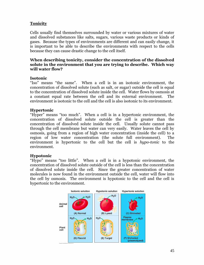
Tonicity Cells usually find themselves surrounded by water or various mixtures of water and dissolved substances like salts, sugars, various waste products or kinds of gases. Because the types of environments are different and can easily change, it is important to be able to describe the environments with respect to the cells because they can cause drastic change to the cell itself. When describing tonicity, consider the concentration of the dissolved solute in the environment that you are trying to describe. Which way will water flow? Isotonic “Iso” means “the same”. When a cell is in an isotonic environment, the concentration of dissolved solute (such as salt, or sugar) outside the cell is equal to the concentration of dissolved solute inside the cell. Water flows by osmosis at a constant equal rate between the cell and its external environment. The environment is isotonic to the cell and the cell is also isotonic to its environment. Hypertonic “Hyper” means “too much”. When a cell is in a hypertonic environment, the concentration of dissolved solute outside the cell is greater than the concentration of dissolved solute inside the cell. Usually solute cannot pass through the cell membrane but water can very easily. Water leaves the cell by osmosis, going from a region of high water concentration (inside the cell) to a region of low water concentration (the solute full environment). The environment is hypertonic to the cell but the cell is hypo-tonic to the environment. Hypotonic “Hypo” means “too little”. When a cell is in a hypotonic environment, the concentration of dissolved solute outside of the cell is less than the concentration of dissolved solute inside the cell. Since the greater concentration of water molecules is now found in the environment outside the cell, water will flow into the cell by osmosis. The environment is hypotonic to the cell and the cell is hypertonic to the environment.
46
Osmosis and Tonicity Practice Text reference pages 182-186 1. Define diffusion in a complete sentence. ____________________________________________________________ ____________________________________________________________ How is osmosis different? ___________________________________________________
2. Define osmotic pressure as explained in class.
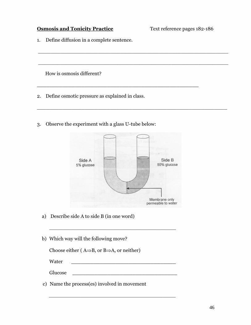
____________________________________________________________ 3. Observe the experiment with a glass U-tube below:
a) Describe side A to side B (in one word) ________________________________________
b) Which way will the following move?
Choose either ( AB, or BA, or neither) Water _________________________________ Glucose _________________________________
c) Name the process(es) involved in movement
________________________________________
47
d) What happens to the concentration of the glucose in side B as the experiment runs? ____________________________________________________________ e) Which side of the U-tube has the highest osmotic pressure at the start of the experiment? What happens to the OP of side B as the experiment runs? What happens to the OP of side A as the experiment runs?
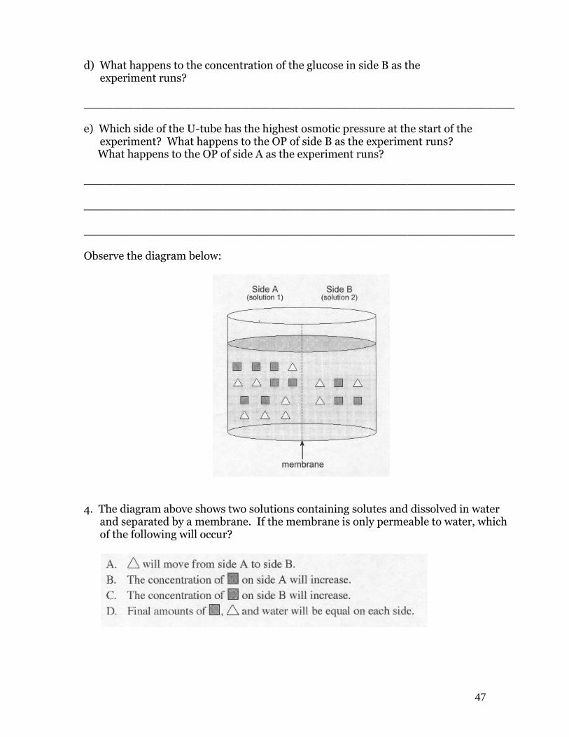
____________________________________________________________ ____________________________________________________________ ____________________________________________________________ Observe the diagram below:
4. The diagram above shows two solutions containing solutes and dissolved in water and separated by a membrane. If the membrane is only permeable to water, which of the following will occur?
48
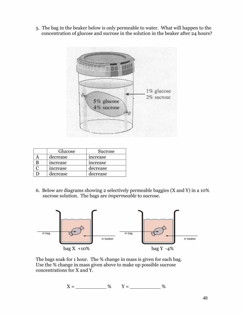
5. The bag in the beaker below is only permeable to water. What will happen to the concentration of glucose and sucrose in the solution in the beaker after 24 hours?
Glucose Sucrose A decrease increase B increase increase C increase decrease D decrease decrease
6. Below are diagrams showing 2 selectively permeable baggies (X and Y) in a 10% sucrose solution. The bags are impermeable to sucrose.
bag X +10% bag Y -4% The bags soak for 1 hour. The % change in mass is given for each bag. Use the % change in mass given above to make up possible sucrose concentrations for X and Y. X = __________ % Y = __________ %
49
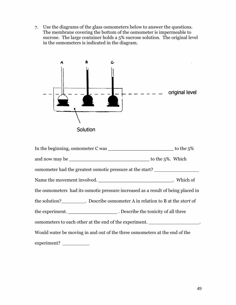
7. Use the diagrams of the glass osmometers below to answer the questions. The membrane covering the bottom of the osmometer is impermeable to sucrose. The large container holds a 5% sucrose solution. The original level in the osmometers is indicated in the diagram.
In the beginning, osmometer C was ______________________ to the 5% and now may be ___________________________ to the 5%. Which osmometer had the greatest osmotic pressure at the start? _______________ Name the movement involved. _________________________. Which of the osmometers had its osmotic pressure increased as a result of being placed in the solution?________. Describe osmometer A in relation to B at the start of the experiment. . Describe the tonicity of all three osmometers to each other at the end of the experiment. _________________. Would water be moving in and out of the three osmometers at the end of the experiment? _________

50

51
Lab You Design It Cell Membranes and Osmosis
Background Information Cell membranes are semi permeable. A dialysis membrane is a synthetic material that can be used to simulate the way a cell membrane works. Before you proceed with this lab, look up the definitions for osmosis and diffusion and write them under your “Observations” section of your formal lab write-up. Include the chemical formulas for water and sucrose.
Purpose:
Your challenge is to devise an experimental procedure to determine the identity of three unknown solutions. Materials Available: Dialysis tubing, beakers, funnels, electronic balance, string (this list is only a list of what is available, you may use other materials not provided, check with me) Procedural Information: You will be given three jugs of solution labeled X, Y, and Z. One jug will be a
5% sucrose solution, another a 10% sucrose solution, and the 3rd jug will be pure water. You will not know which solution is in which jar.
The only known you will be given is a 4th jug with a 5% sucrose solution labeled on it.
You will be given some dialysis tubing, which will allow you to separate solutions by making a “baggy” out of the tubing by tying off the end with string.
Review your handout on the “Scientific Method” to help your group solve this problem. You must include and identify a “control” in your experiment
52
Your lab report must include the following: (20 marks) Review lab manual page 43 Purpose Hypothesis
Write a formal hypothesis of what you are testing. (2) Materials Procedure
Outline clearly the steps that you will follow. Include diagrams.(3) Observations
Devise a data table to record your results. If you are finding the mass, you must include the % change in mass. This is equal to the change in mass/initial mass X 100%.(3)
Questions Answer in complete sentence answers.
Conclusions Identify each solution and explain how you arrive at this conclusion. Which solution is which? (3)
Questions: 1. Which material, water or sucrose was moving through the membrane?
Explain why you would know whether water or sucrose is moving through the membrane by using your results from your experiment. (2)
2. What was the control in your experiment? What was the variable? (2)
3. List some possible experimental errors that could have caused your group
to get inaccurate results. (2)
4. Describe a second way in which you could have set up the bags and beakers and still be able to come to the same conclusion.(3)
53
Photosynthesis
Life is powered by the sun. The energy that gives a cheetah its incredible speed or a bird
its power to fly does not originate within the planet, but travels 150 million kilometers
from the sun. The energy of sunlight, however, becomes energy for life only after it has
been transformed into chemical energy by the process of photosynthesis.
The cheetah for example, gets its food by eating the antelope, which in turn ate grasses
and other plants that had photosynthesized. Virtually all organisms rely on
photosynthesis for supplies of energy-rich food. For this reason, photosynthesis might be
considered the most important set of chemical reactions on Earth.
This is the summary equation for Photosynthesis that you must know:
6 CO2 + 6 H2O C6H12O6 + 6 O2
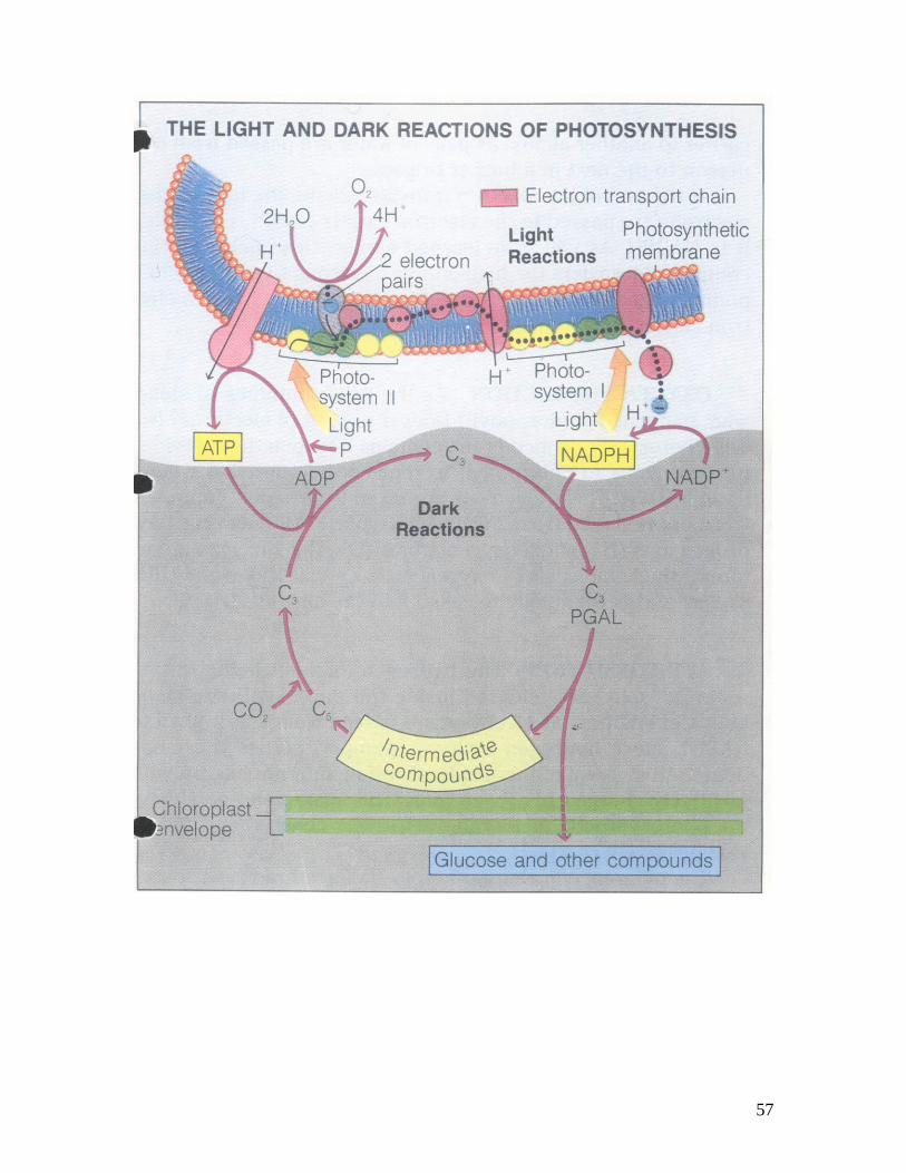
Botanists – biologists who specialize in the study of plant life – group the reactions of
photosynthesis into two general stages: the light reactions and the light independent
reactions.
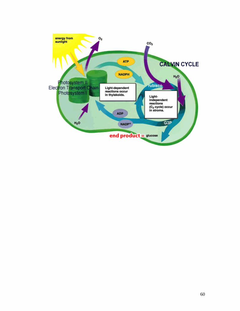
LIGHT REACTIONS The light reactions occur in the thylakoid membranes of the grana within the chloroplast of a cell. The main purpose of the light reactions is to make two energy storing molecules: ATP and NADPH.
There are two major clusters of light capturing pigments in plant chloroplasts: chlorophyll and carotenids. Plants are typically green because chlorophyll, which is the predominant plant pigment, does not absorb light of green wavelengths. Instead, this light is reflected to our eyes which is why we see the green color in leaves.
Capturing light and converting it into useable chemical energy for plants requires an ordered biological structure. Chlorophyll is embedded in the thylakoid membranes of chloroplasts in clusters called photosystems. When sunlight strikes the thylakoid membrane, energy is absorbed simultaneously by photosystems 1 and 2 in the membrane. Electrons from chlorophyll are “boosted” with energy from the sunlight and each photo-excited electron is then passed to an “open” electron acceptor molecule, which in turn transfers electrons to an electron transport chain. The electrons lost from photosystem 1 are replenished by electrons from the breakdown of water molecules. The overall consequence is to remove low energy electrons from water, boost them to a higher energy level, and transfer the energized electrons to NADP+. So, NADP+ becomes ‘reduced’ to become an energy-storing compound called NADPH
54
The splitting of water by the chloroplast does a lot more than just provide electrons for the photosystems to make NADPH. When water gets split, the reaction generates two other products: Oxygen gas is released into the atmosphere and H+ begin to accumulate inside the lumen of the thylakoid. The accumulation of H+ ions on one side of the thylakoid membrane provides a ‘concentration gradient’ of protons across the membrane, which provides a source of energy to cause phosphorous to join with ADP to form a molecule of ATP. Every molecule of water can produce about four molecules of ATP in this way.
So the light reactions make two very important energy-storing molecules called NADPH and ATP. These molecules are used by the chloroplast to forge the fuel used by almost all organisms on Earth. That fuel is glucose.
LIGHT INDEPENDENT REACTIONS
Since both ATP and NADPH are forms of chemical energy, why do photosynthesizers go on to manufacture a different chemical energy – energy-rich sugars such as glucose? The answer is that neither ATP nor NADPH can be stored or moved from one part of the plant to another, but sugars can. This is critical for multicellular plants to transport sugars to areas such as roots or stems that don’t photosynthesize or to store sugars for use at various points of the growing season.
In the light independent reactions, glucose is made in the stroma of the chloroplast. Glucose contains six carbon atoms in it so the plant must take CO2 from the air to supply carbon for glucose production.
CO2 from the air joins a 5-carbon compound called ‘Rubisco’ in the stroma of the chloroplast to form a short-lived 6-carbon compound which gets transformed into another energy rich 3-carbon compound called PGAL by the powers of ATP and NADPH. It is the PGAL that ultimately gives rise to one glucose molecule.
PGAL can also make other important products for the plant when needed.
It must be remembered that the making of glucose does not provide the plant cell with the energy it needs. Just like in you, the glucose has to be broken down to make many molecules of ATP using cellular respiration.
55
56
57

58

59
Check Your Understanding - Photosynthesis
After reading the text reference pages 201-214, attempt the following:
Try to answer the following questions from memory once you have studied the light and light independent reactions in class: 1. From memory, write out the balanced chemical equation for photosynthesis.
____________________________________________________
2. Which reactant in the basic equation is used in the light reactions?
______________________
3. Which product in the basic equation is obtained directly from water?
______________________
4. What other substance is obtained from water during the light reactions? ______________________
5. Which reactant in the basic equation is used in the dark reactions?
______________________
6. What product is carbon dioxide eventually converted into? ______________________
7. What must be added to carbon dioxide from the light reactions to complete the formation of a carbohydrate (glucose) molecule?
______________________
8. What substances must be made in the light reactions to allow a plant cell to
make glucose in complete darkness?
_________________________ _________________________
9. If molecules of atoms and all atoms have equal numbers of protons and electrons, how can a molecule of chlorophyll continue to exist in the photosynthetic membrane if chlorophyll molecules continually lose high-energy electrons to the electron transport chain? (Hint: look at the reactants in the basic equation)
_____________________________________________________
60

61
62
Cellular Respiration
As you go along doing your daily activities your body requires energy. It’s a
strange concept, energy. What exactly is it that our body needs? Where does
energy come from? Is it a type of electricity in our body? If so, how is it made
and distributed? Is energy a molecule flowing in our veins being delivered to all
our cells? If so, can we put it in a bottle and obtain energy whenever we need it?
These questions and many more are asked by curious biologists as we try to
figure out how life works.
We know that every living organism on earth uses the same series of methods for
obtaining energy to fuel their cells and thus their bodies. We share an
evolutionary “chemical unity” in obtaining energy with all living things on Earth.
From the lowly yeast cell to a garden weed to a human being; we all contain
similar genes that allow us to obtain energy in the same way. We accomplish this
by shuffling energy from one compound to another by virtually the same set of
reactions that arose at a very early stage of evolution. The starting point for most
of these reactions is glucose.
Glucose is an energy-rich molecule, but it cannot directly power biological
activities. In other words a glucose molecule is like you carrying around a $500
bill and trying to use it to purchase a lunch from the Safeway Deli: Both the cell
and the lunch purchaser require smaller denominations. Therefore, cells convert
energy-rich molecules such as glucose, to other compounds containing more
usable quantities of energy, most often a very important molecule called ATP.
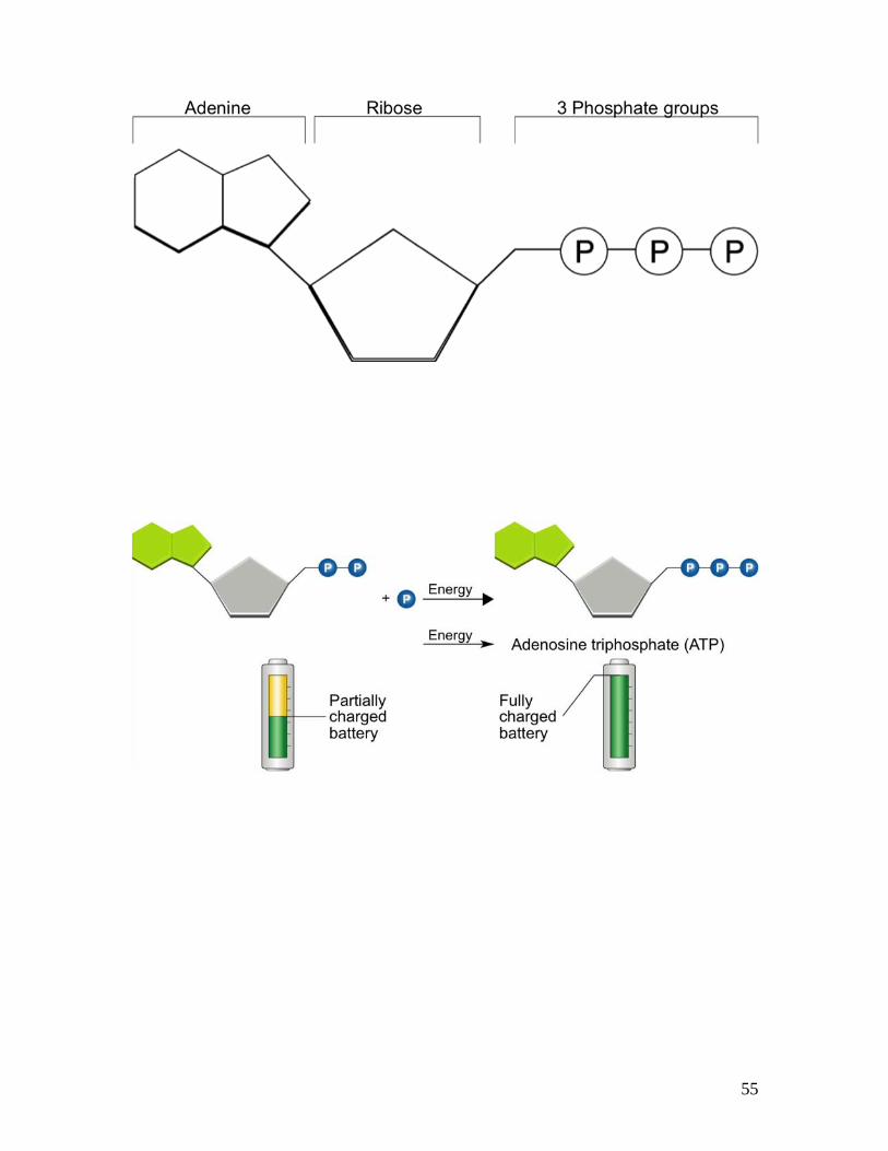
ATP stands for adenosine triphosphate. A molecule of ATP as its name
indicates is made of adenosine bonded to three phosphate groups. There is a lot
of energy stored in the bonds that hold the phosphate groups to the molecule.
(See manual page 18)
When energy is required for some process in the cell (such as making cell
membrane), the cell will hydrolyze (break) the bond between the second and
third phosphate molecules on ATP. This releases the energy. Energy is stored
in the high-energy chemical bonds of these molecules. What remains is
a molecule of adenosine diphosphate (ADP) and a loose free-floating molecule of
phosphate.
Glucose is the primary source of “cellular food”, meaning that it is the molecule
that a cell can break down to make ATP. Glycogen is another larger molecule in
your body made of many glucose molecules bonded together. In other words, the
purpose of glycogen is to store energy. When it comes to energy, think of it like
this:
ATP is like cash Glucose is like a debit card Glycogen is like a bank
63
When a cell needs ATP, it goes to its glycogen storage and takes out a glucose
molecule. It cashes the glucose molecule in order to get ATP. The glucose is
cashed through an important process we will learn called Cellular Respiration.
Electron Carrier Molecules
This is the summary equation for Cellular Respiration that you must know:
C6H12O6 + 6 O2 <---> 6 CO2 + 6 H2O + ATP
There are three steps involved in Cellular Respiration. These include Glycolysis,
Kreb’s Citric Acid Cycle, and the Electron Transport Chain.
Cellular respiration is essentially the breakdown of glucose to release energy in
the form of ATP. Useable energy for the body is ATP. However, the energy
released from the breakdown of glucose is not all in the form of ATP. High
energy electrons are being stripped away from the glucose bit by bit by special
molecules in the cell called “electron carriers” and they eventually carry these
high energy electrons over to molecular oxygen (that we breathe). This makes
water and a lot of ATP.
The two most common electron carriers in the body (and the ones that will be
used during cellular respiration) are NAD+ and FAD+.
These electron carriers are found in an organelle of the cell called a
mitochondrion. When an “empty” electron carrier accepts a pair of electrons we
say it has become reduced. When it gives those electrons up later on, we say it
has become oxidized. What typically happens to glucose as it enters a
mitochondrion is:
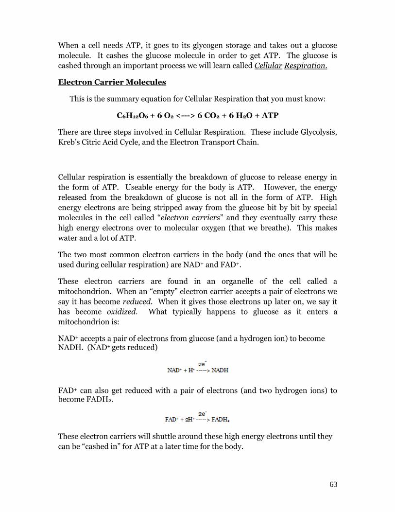
NAD+ accepts a pair of electrons from glucose (and a hydrogen ion) to become NADH. (NAD+ gets reduced)
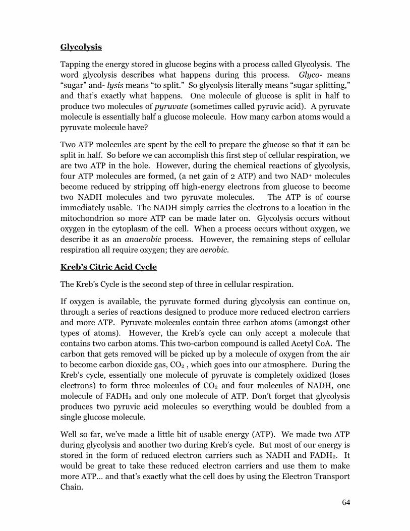
FAD+ can also get reduced with a pair of electrons (and two hydrogen ions) to become FADH2.
These electron carriers will shuttle around these high energy electrons until they
can be “cashed in” for ATP at a later time for the body.
64
Glycolysis
Tapping the energy stored in glucose begins with a process called Glycolysis. The
word glycolysis describes what happens during this process. Glyco- means
“sugar” and- lysis means “to split.” So glycolysis literally means “sugar splitting,”
and that’s exactly what happens. One molecule of glucose is split in half to
produce two molecules of pyruvate (sometimes called pyruvic acid). A pyruvate
molecule is essentially half a glucose molecule. How many carbon atoms would a
pyruvate molecule have?
Two ATP molecules are spent by the cell to prepare the glucose so that it can be
split in half. So before we can accomplish this first step of cellular respiration, we
are two ATP in the hole. However, during the chemical reactions of glycolysis,
four ATP molecules are formed, (a net gain of 2 ATP) and two NAD+ molecules
become reduced by stripping off high-energy electrons from glucose to become
two NADH molecules and two pyruvate molecules. The ATP is of course
immediately usable. The NADH simply carries the electrons to a location in the
mitochondrion so more ATP can be made later on. Glycolysis occurs without
oxygen in the cytoplasm of the cell. When a process occurs without oxygen, we
describe it as an anaerobic process. However, the remaining steps of cellular
respiration all require oxygen; they are aerobic.
Kreb’s Citric Acid Cycle
The Kreb’s Cycle is the second step of three in cellular respiration.
If oxygen is available, the pyruvate formed during glycolysis can continue on,
through a series of reactions designed to produce more reduced electron carriers
and more ATP. Pyruvate molecules contain three carbon atoms (amongst other
types of atoms). However, the Kreb’s cycle can only accept a molecule that
contains two carbon atoms. This two-carbon compound is called Acetyl CoA. The
carbon that gets removed will be picked up by a molecule of oxygen from the air
to become carbon dioxide gas, CO2 , which goes into our atmosphere. During the
Kreb’s cycle, essentially one molecule of pyruvate is completely oxidized (loses
electrons) to form three molecules of CO2 and four molecules of NADH, one
molecule of FADH2 and only one molecule of ATP. Don’t forget that glycolysis
produces two pyruvic acid molecules so everything would be doubled from a
single glucose molecule.
Well so far, we’ve made a little bit of usable energy (ATP). We made two ATP
during glycolysis and another two during Kreb’s cycle. But most of our energy is
stored in the form of reduced electron carriers such as NADH and FADH2. It
would be great to take these reduced electron carriers and use them to make
more ATP… and that’s exactly what the cell does by using the Electron Transport
Chain.
65
The Electron Transport Chain
The electron transport chain is the third step in cellular respiration and it is a
system in the mitochondrial membrane that allows the energy of the electrons
carried in the NADH and FADH2 to be used in the formation of ATP. If we don’t
oxidize the electron carriers back to their “empty” states we can’t keep running
glycolysis or the Kreb’s cycle. We only have a finite amount of electron carriers
and we have emptied them so that they can accept electrons from other glucose
molecules.
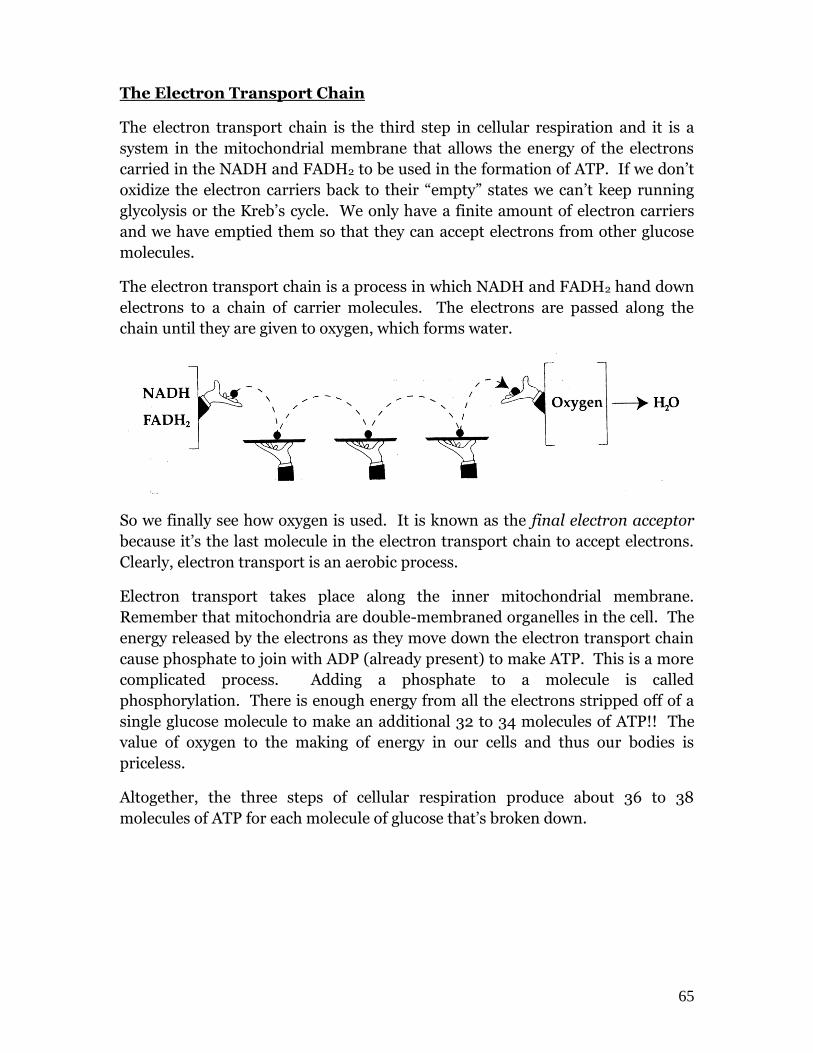
The electron transport chain is a process in which NADH and FADH2 hand down
electrons to a chain of carrier molecules. The electrons are passed along the
chain until they are given to oxygen, which forms water.
So we finally see how oxygen is used. It is known as the final electron acceptor
because it’s the last molecule in the electron transport chain to accept electrons.
Clearly, electron transport is an aerobic process.
Electron transport takes place along the inner mitochondrial membrane.
Remember that mitochondria are double-membraned organelles in the cell. The
energy released by the electrons as they move down the electron transport chain
cause phosphate to join with ADP (already present) to make ATP. This is a more
complicated process. Adding a phosphate to a molecule is called
phosphorylation. There is enough energy from all the electrons stripped off of a
single glucose molecule to make an additional 32 to 34 molecules of ATP!! The
value of oxygen to the making of energy in our cells and thus our bodies is
priceless.
Altogether, the three steps of cellular respiration produce about 36 to 38
molecules of ATP for each molecule of glucose that’s broken down.

66
Fermentation
If oxygen is not available, the electron carriers can’t be oxidized back to “empty”
so Kreb’s and glycolysis shut down as there is no free NAD+ or FAD+. Glycolysis
has its own way of freeing up NAD+ that doesn’t rely on oxygen. Regenerating
free NAD+ in the absence of oxygen is called Fermentation.
At the end of our anaerobic process called glycolysis we have: 2 ATP, 2 NADH
and 2 molecules of pyruvic acid.
The ATP is, of course, usable energy. The NADH can’t be used to make more ATP
because the electron transport chain is shut down in the absence of oxygen. This
means that pyruvate cannot be used either as it can’t enter the Kreb’s cycle
because Kreb’s is shut down as well without oxygen. So, because these two
substances are not being used, why not take the electrons off the NADH and
donate them to pyruvate? That would generate free NAD+ that could be re-used
to run glycolysis again, and that’s exactly what happens.
Pyruvate gets converted to ethanol (alcohol) and CO2 gas in yeast cells running
fermentation. In human muscle cells, pyruvate is reduced to lactic acid.
Three problems with fermentation:
1. The end products, (alcohol or lactic acid) are toxic. Yeast cells die at about
12% alcohol concentration. Our muscle cells hurt in the presence of lactic acid
and can stop contracting if levels get too high.
2. The only ATP you get is the two net ATP from glycolysis. This might be
enough for a unicellular yeast cell to survive but human muscle cells require all
36 ATP per glucose if we are to survive. Your muscles will only work for a short
time. This is why humans, other animals, and plants absolutely must have
oxygen in order to survive.
3. Organisms that rely on fermentation do not have the same ability to evolve as
much as organisms that use oxygen. They simply do not have the means to make
the energy needed to adapt and perform more complex processes such as ours.

67
Check Your Understanding - Cellular Respiration
After reading the reference text pages 221-229, attempt the following:
1. The general chemical equation for Cellular Respiration is:
______________________________________________________ 2. There are 4 main processes that summarize cellular respiration. These are (in order): Process Location A._______________________ _______________________ B._______________________ _______________________ C._______________________ _______________________ D. ______________________ _______________________ The reactant from the general equation used in part A is ____________________ The reactant from the equation used in part C is ____________________ The product from the equation made in part B is ____________________ The product from the equation made in part C is ____________________ The energy found in ATP is found in the __________________________________ of the three phosphate groups. 3. In glycolysis, a molecule of glucose is eventually
___________________________ by a molecule of NAD+. The NAD+ gets ___________________________ to a molecule of NADH. Glucose is broken into two molecules of __________________________

68
4. During the Kreb’s cycle, pyruvic acid loses a _______________________
atom before it can enter the cycle. This process requires oxygen to occur so the process is called an _______________________________ process. 5. Glycolysis and Kreb’s only make a total of 4 ATP for every pyruvate molecule, so most of the ATP (another 34) has to be made in the folded membranes called the __________________________________which is found in the cristae of the mitochondria. The diagram below has labels. Identify where each of the following processes occurs:
Glycolysis, Fermentation, ETC, Kreb’s Citric Acid Cycle
69
Cell Energy Review
From the list, identify 6 different processes as headings on the back side of this sheet.
Group the like ones together --- you may use the terms more than once.
light reactions lactic acid PGAL
fermentation ATP muscle
Kreb’s cycle NADPH bacteria
oxygen debt cytoplasm yeast
pyruvic acid ethyl alcohol free NAD
Calvin cycle anaerobic respiration matrix
NADH cristae oxygen
2 ATP 34 ATP stroma
ETC carbon dioxide water
FADH2 grana glycolysis
glucose sun mitochondrion

70
71
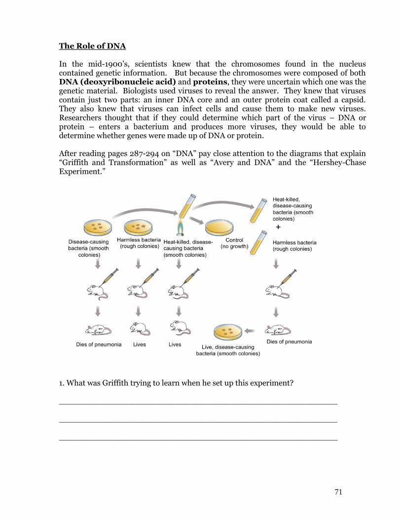
The Role of DNA In the mid-1900’s, scientists knew that the chromosomes found in the nucleus contained genetic information. But because the chromosomes were composed of both DNA (deoxyribonucleic acid) and proteins, they were uncertain which one was the genetic material. Biologists used viruses to reveal the answer. They knew that viruses contain just two parts: an inner DNA core and an outer protein coat called a capsid. They also knew that viruses can infect cells and cause them to make new viruses. Researchers thought that if they could determine which part of the virus – DNA or protein – enters a bacterium and produces more viruses, they would be able to determine whether genes were made up of DNA or protein. After reading pages 287-294 on “DNA” pay close attention to the diagrams that explain “Griffith and Transformation” as well as “Avery and DNA” and the “Hershey-Chase Experiment.”
1. What was Griffith trying to learn when he set up this experiment?
_______________________________________________________ _______________________________________________________ _______________________________________________________
72
2. How did Griffith show that the disease-causing bacteria were killed by the heat?
_______________________________________________________ _______________________________________________________ _______________________________________________________ 3. What result was Griffith expecting when he injected the mixture of live
harmless bacteria and heat-killed disease causing bacteria? _______________________________________________________ 4. Why do you think Griffith called this process “Transformation”? _______________________________________________________ _______________________________________________________
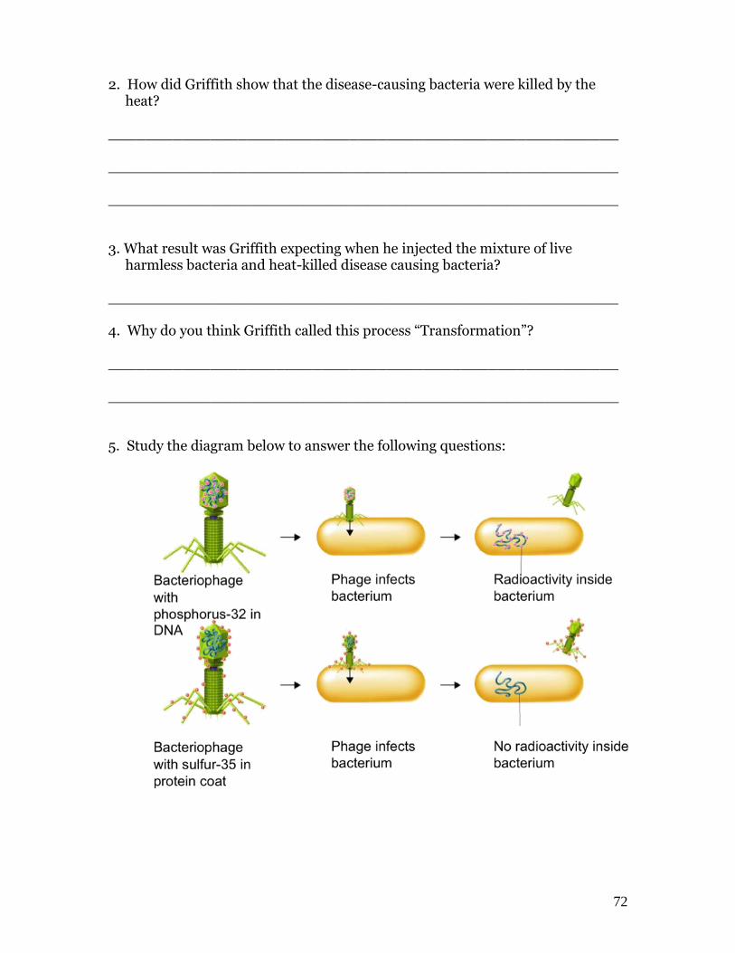
5. Study the diagram below to answer the following questions:
73
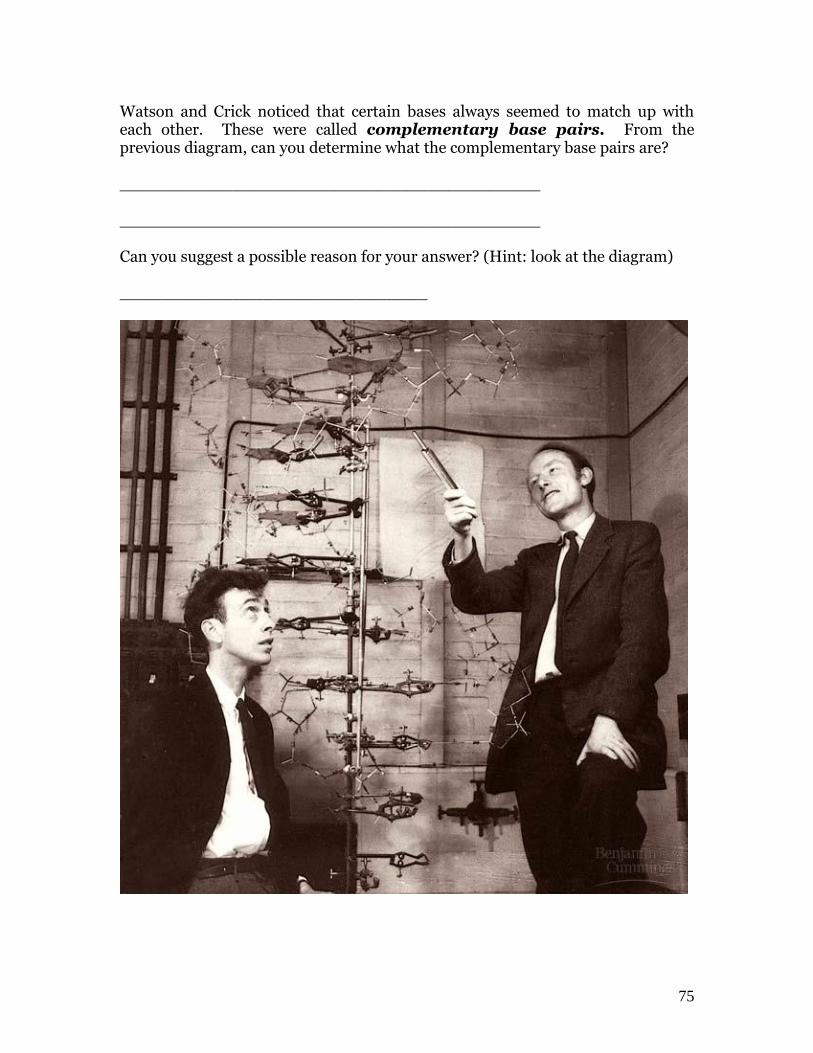
How would the radioactive markers of phosphorus 32 and sulfur 35 help Hershey-Chase figure out if DNA or protein was injected into bacteria cells by the bacteriophage virus? _______________________________________________________ _______________________________________________________ _______________________________________________________ _______________________________________________________ _______________________________________________________ DNA Structure The structure of DNA was determined by James Watson and Francis Crick in the early 1950’s. DNA is a long molecule made up of units called nucleotides. A nucleotide is a building block of DNA. Each DNA molecule in each cell of your body has close to 3 billion pairs of nucleotides. In the space below, draw and label a nucleotide. Include in your labels, a phosphate group, deoxyribose, and one of the four nitrogenous bases.

74
There are four possible bases: two are purines with a double ring, and two are pyrimidines with a single ring. The names of the bases and their shapes are indicated below.
A molecule of DNA would take the shape of a double helix as shown above. The nucleotides would join together and the molecule would begin to twist.
75
Watson and Crick noticed that certain bases always seemed to match up with each other. These were called complementary base pairs. From the previous diagram, can you determine what the complementary base pairs are? _________________________________________ _________________________________________ Can you suggest a possible reason for your answer? (Hint: look at the diagram) ______________________________

76

77
Nucleic Acids The Structure of DNA In humans, DNA is a large molecule made up of about 6 billion smaller molecules called nucleotides. Each nucleotide is composed of molecules that you can review on lab manual pages 73 and 74. After reading text reference pages 295-299 begin the following exercise: You are going to build a portion of a section of DNA using paper models. From the envelopes provided at the front of the room, go get the following nucleotides: 8 cytosine, 8 guanine, 4 thymine and 4 adenine. Arrange the nucleotides below in a vertical line on your desk: Cytosine Thymine These represent the “left side” of the DNA molecule. The Guanine molecule will look like half a ladder. Adenine Guanine Cytosine 1. Name the 2 molecules that alternate to form the “hand-rail” in our “ladder” _______________________________________________________ 2. Name the molecule to which each base is attached. _______________________________________________________ 3. Name the molecules that form the “half-rungs” of the “ladder” _______________________________________________________ Now use the remaining paper nucleotides that you have to match the left side of the DNA on your desk. You will now form a “full ladder” with 6 pairs of nucleotides. From now on, you can us “A” to represent adenine, “C” to represent cytosine, etc. 4. Name the complementary base pairs in your model. _______________________________________________________ 5. If the left side of your DNA is the letters A T T C G G C T, what will be the right side? _______________________________________________________
78
DNA Replication As cells grow, they reach their size limit and they begin to divide. A copy of the DNA is required for each new cell. When a cell makes a copy of its DNA we call the process DNA Replication. You are going to use your paper model of DNA to see how this occurs in your cells. Your DNA molecule has 2 sides to it. Your cells use a special chemical called an enzyme that splits the DNA in half. The enzyme is called helicase and it splits the double helix in half. On your table, split the DNA molecule you made down the middle into a left and right side. Move them far enough away from each other so that you can add new complementary nucleotides to each side. New nucleotide molecules are added by an enzyme called DNA polymerase. This enzyme not only adds new complementary nucleotides to each open side of the DNA molecule, it also “proof-reads” to make sure that there are no mistakes made in the copy. 6. How many DNA “ladders” do you have now? _______ 7. How do these ladders compare to each other? _______________________________________________________ 8. How do these ladders compare to the original DNA ladder? _______________________________________________________ 9. If complementary base pairing did not exist as a property of DNA, would you
expect your two DNA “ladders” to be identical? ________ Explain the value of complementary base pairing in DNA. _______________________________________________________ _______________________________________________________ _______________________________________________________ 10. Once a cell nucleus has completed DNA Replication, what do you think the
cell will do now that it has two copies of DNA? _______________________________________________________
79
After reading the reference pgs. 300-302 begin the exercise: mRNA Transcription. The role of DNA is to control the chemical activities that go on in a cell. This is why we call the nucleus the “brain” of the cell. To control the cytoplasm, DNA must get its message out of the nucleus. The problem is that DNA is too large a molecule to pass through the nuclear membrane. This forces the cell to make a new molecule—a messenger molecule-- a molecule that carries the “message” of the DNA to the rest of the cytoplasm. A molecule called messenger RNA made in the nucleus carries the DNA’s instructions out to the cytoplasm. The making of a molecule of mRNA in the nucleus is called Transcription. A section of DNA that performs a certain job in the cell is called a gene. Each gene acts by building a different molecule of mRNA. Open up one of your DNA helixes. Keep the “right side” and put the other DNA nucleotides back into their correct envelope. Go get the following mRNA nucleotides from the other envelopes that say RNA: 2 guanine, 1 adenine, 2 cytosine, and 1 uracil. NOTE that the mRNA nucleotides contain the sugar ribose instead of deoxyribose. Match up the mRNA nucleotides with the right side of the DNA molecule. Real cells use an enzyme called Transcriptase to add the new complementary mRNA nucleotides to the DNA. When complete, separate the mRNA molecule from the DNA. This is the mRNA molecule that would now leave the nucleus and carry a portion of the DNA’s message to the cytoplasm. 11. What is the difference between ribose and deoxyribose? _______________________________________________________ 12. If DNA has the bases A T T C G G C A, what will be the order of the
mRNA bases? ____________________________________________________ 13. List three structural differences between DNA and mRNA. _______________________________________________________ _______________________________________________________ _______________________________________________________

80
14. How is it ensured that the same mRNA molecule always is formed from the same section of DNA?
_______________________________________________________ _______________________________________________________ 15. Use the following terms to help you explain how Transcription occurs:
helicase, complementary base pairs, transcriptase
_______________________________________________________ _______________________________________________________ _______________________________________________________ _______________________________________________________ _______________________________________________________ 16. Write out the full names for DNA and RNA correctly. _______________________________________________________ _______________________________________________________
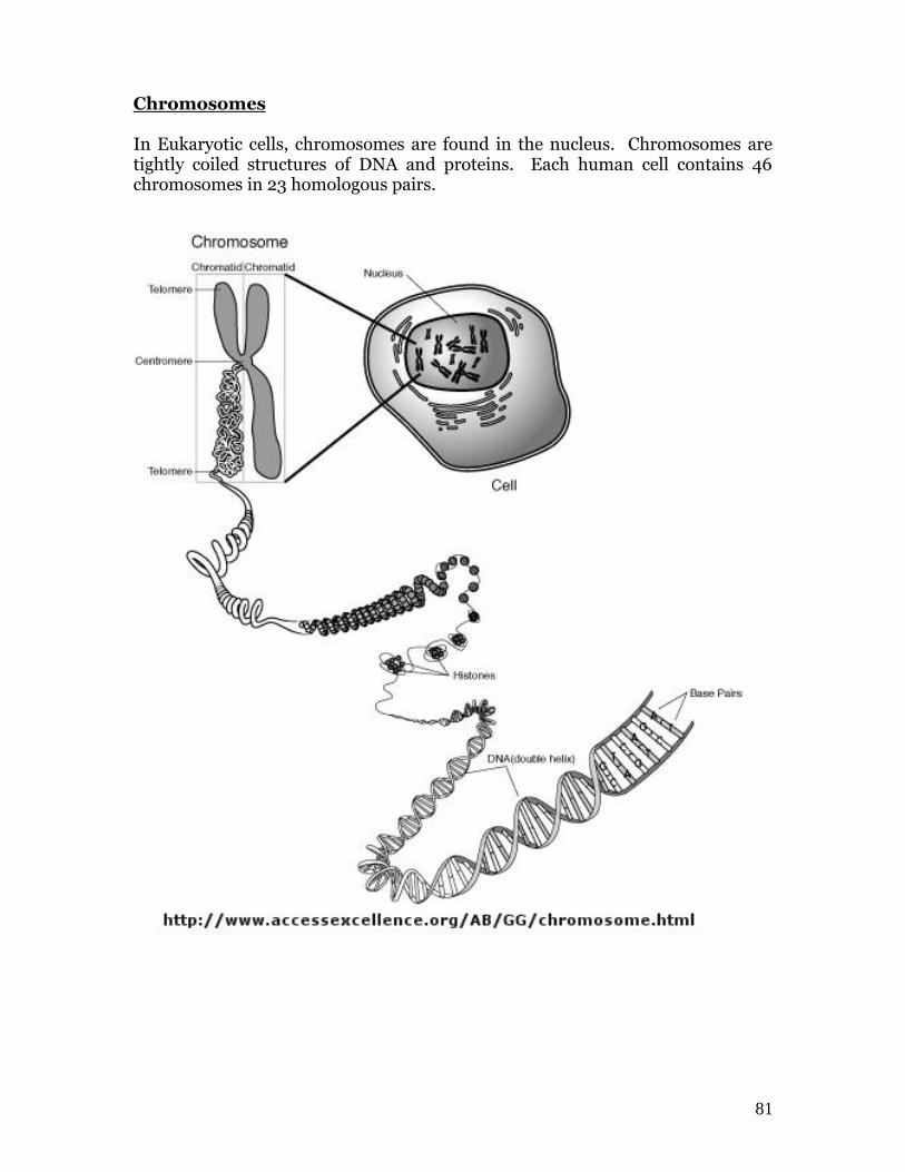
81
Chromosomes In Eukaryotic cells, chromosomes are found in the nucleus. Chromosomes are tightly coiled structures of DNA and proteins. Each human cell contains 46 chromosomes in 23 homologous pairs.
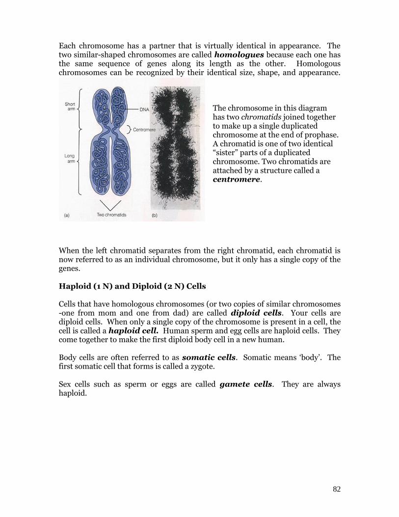
82
Each chromosome has a partner that is virtually identical in appearance. The two similar-shaped chromosomes are called homologues because each one has the same sequence of genes along its length as the other. Homologous chromosomes can be recognized by their identical size, shape, and appearance.
The chromosome in this diagram has two chromatids joined together to make up a single duplicated chromosome at the end of prophase. A chromatid is one of two identical “sister” parts of a duplicated chromosome. Two chromatids are attached by a structure called a centromere.
When the left chromatid separates from the right chromatid, each chromatid is now referred to as an individual chromosome, but it only has a single copy of the genes. Haploid (1 N) and Diploid (2 N) Cells Cells that have homologous chromosomes (or two copies of similar chromosomes -one from mom and one from dad) are called diploid cells. Your cells are diploid cells. When only a single copy of the chromosome is present in a cell, the cell is called a haploid cell. Human sperm and egg cells are haploid cells. They come together to make the first diploid body cell in a new human. Body cells are often referred to as somatic cells. Somatic means ‘body’. The first somatic cell that forms is called a zygote. Sex cells such as sperm or eggs are called gamete cells. They are always haploid.
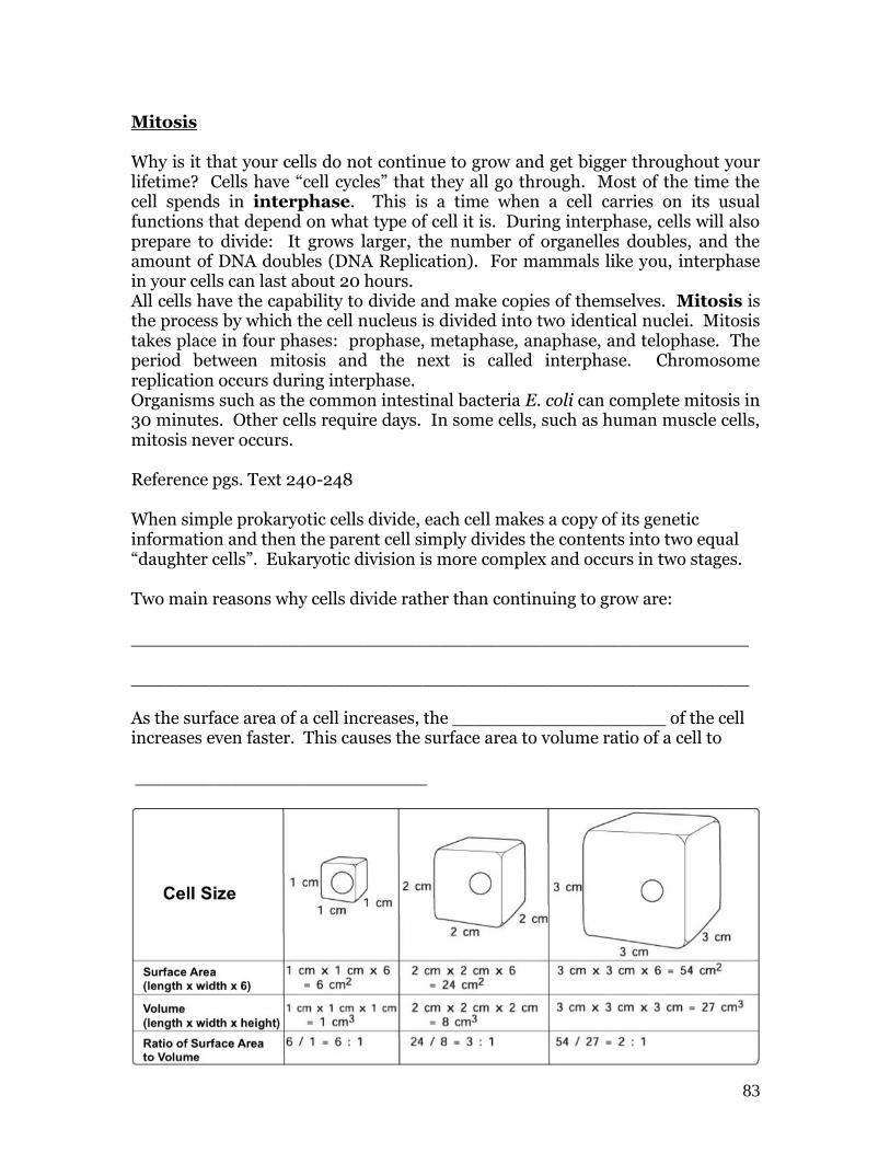
83
Mitosis
Why is it that your cells do not continue to grow and get bigger throughout your lifetime? Cells have “cell cycles” that they all go through. Most of the time the cell spends in interphase. This is a time when a cell carries on its usual functions that depend on what type of cell it is. During interphase, cells will also prepare to divide: It grows larger, the number of organelles doubles, and the amount of DNA doubles (DNA Replication). For mammals like you, interphase in your cells can last about 20 hours. All cells have the capability to divide and make copies of themselves. Mitosis is the process by which the cell nucleus is divided into two identical nuclei. Mitosis takes place in four phases: prophase, metaphase, anaphase, and telophase. The period between mitosis and the next is called interphase. Chromosome replication occurs during interphase. Organisms such as the common intestinal bacteria E. coli can complete mitosis in 30 minutes. Other cells require days. In some cells, such as human muscle cells, mitosis never occurs. Reference pgs. Text 240-248 When simple prokaryotic cells divide, each cell makes a copy of its genetic information and then the parent cell simply divides the contents into two equal “daughter cells”. Eukaryotic division is more complex and occurs in two stages. Two main reasons why cells divide rather than continuing to grow are: _______________________________________________________ _______________________________________________________ As the surface area of a cell increases, the ___________________ of the cell increases even faster. This causes the surface area to volume ratio of a cell to __________________________

84
The first stage is called mitosis and includes the division of the ___________________________. The second stage is called _________________________ and is a
separation of the cells ______________________. Because mitosis
produces cells that are genetically identical, we classify mitosis as a form of
______________________ reproduction.
Each of the cells above is either in interphase or in mitosis. Can you see the
chromosomes that are going through metaphase, Anaphase and Prophase?
85
Lab Activity
Time to Split! Determine the Time Needed For Mitosis
You will be performing some extensive microscope work in this lab as you learn to recognize cells that are in various phases of mitosis. You need to be proficient and patient with your microscope as you adjust light contrast and focus to identify the phases. You will be working with both plant and animal cells so it’s a good idea to trade off duties with your lab partners as you work. As always, review manual page 27. 1. Review the phases of plant and animal cell mitosis by reviewing pages 246 - 248 from your textbook. 2. From the front of the room, obtain 2 prepared slides: The Allium Onion root
and the Whitefish slide. Obtain a microscope for your table. 3. Observe the prepared onion root tip slide under the 4 X red band objective.
Focus on the cells just above the tip of the root. The tip of the root contains cells that make up the protective cap, which protects the delicate dividing cells just above them. Switch to 10 X yellow band and then finally 40 x blue band and count the total number of cells in the field of view. Record your total in Data Table 1.
4. Without changing the field of view, count the number of cells in each phase of
mitosis: prophase, metaphase, anaphase, and telophase. Record in Data Table 1.
5. To determine the approximate proportion of time a cell spends in each phase
of mitosis, you need to divide the number of cells in each phase the total number of cells in the field of view
Dividing these two numbers will always give you a decimal. You need to convert this decimal to a percent, so simply multiply this number by 100 to get your percent and record this in Data Table 1.
6. Put this slide back on the tray and repeat steps 3-5 using the Whitefish animal mitosis slide. Record your information in Data Table 2. You may need to adjust the iris diaphragm of your microscope.
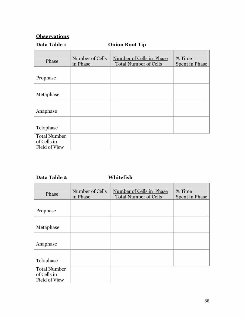
86
Observations
Data Table 1 Onion Root Tip
Phase
Number of Cells in Phase
Number of Cells in Phase Total Number of Cells
% Time Spent in Phase
Prophase
Metaphase
Anaphase
Telophase
Total Number of Cells in Field of View
Data Table 2 Whitefish
Phase
Number of Cells in Phase
Number of Cells in Phase Total Number of Cells
% Time Spent in Phase
Prophase
Metaphase
Anaphase
Telophase
Total Number of Cells in Field of View
87
Conclusions:
1. In which phase of plant cell and animal cell mitosis is the most and least time
spent? 2. Based on your group’s results, what is the total time the plant cells and animal
cells spend undergoing mitosis? 3. What are plant cells doing when they are not undergoing mitosis? 4. Determine the percentages of time spent in each phase of mitosis for the
onion root tip using the total number of cells undergoing mitosis instead of the total number of cells in the field of view.
Percentage = Number of cells in phase Number of cells undergoing mitosis Give a percentage for prophase, metaphase, anaphase and telophase. Your lab activity must include Data Tables 1 and 2 as well as Conclusions 1-4. After completing the questions 5- 7 come check your answers at the front desk. Use the following information about mitosis to answer question 5 Before mitosis After mitosis The number of chromosomes in the parent cell
The number of chromosomes in a daughter cell.
5. Which statement is correct about the relative number of chromosomes? a. The number of chromosomes are equal. b. The number of chromosomes in the parent cell is greater. c. The number of chromosomes in a daughter cell is greater. d. The relationship cannot be determined from the information given.
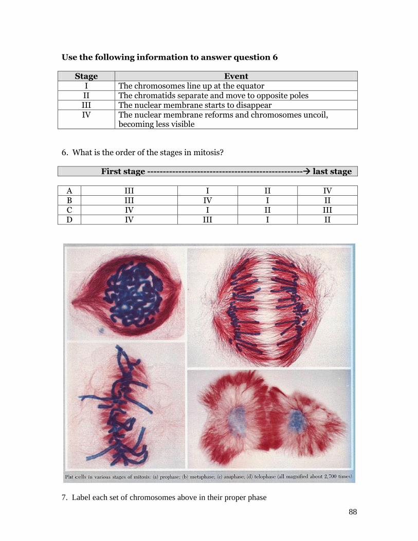
88
Use the following information to answer question 6
Stage Event I The chromosomes line up at the equator II The chromatids separate and move to opposite poles III The nuclear membrane starts to disappear IV The nuclear membrane reforms and chromosomes uncoil,
becoming less visible 6. What is the order of the stages in mitosis? First stage -------------------------------------------------- last stage
A III I II IV B III IV I II C IV I II III D IV III I II
7. Label each set of chromosomes above in their proper phase