Embed Size (px)
Citation preview

4/12/2010
1
Block 11 - Theme: Male sex hormones
fPhysiology of primary and secondary male sex organs

4/12/2010
2
Anatomical organisation
• Primary sex organ/gonad:Testes– Testes
• Duct system:– For the conveyance of sperm and semen
• Accessory sex organs:– Seminal vesicles, prostate gland, urethra and
penis• External genitalia:
– Scrotum, penis
Urinary bladder
Seminal vesicle
Ureter
Ductusdeferens Penis
Epididymis
Bulbourethral gland Ejaculatory duct
Prostate gland
Glanspenis
Testis
Epididymis
Urethra

4/12/2010
3
Testis• Paired ovoid structures about 5 by 2.5 cm located in the
scrotum.• Each testis is enclosed by 3 coverings: tunica vasculosa,
albuginea and vaginalis• Parenchyma: each lobule of testis contains 1-4 coiled
tubules known as seminiferous tubules, length of each is between 30-70 cm. Tubules form single, double or triple arches.T d f h l b l t b l it ith ith• Towards apex of each lobule, tubules unite with with a straight narrow tubule which join and form a network of channels called rete testis
• See fig next slide
Epididymis
Ductusdeferens
Seminiferoustubules
Testis

4/12/2010
4
• From rete testis, 8-15 tubules called vas efferens arise. Vas efferens form head of epididymis and then converge to form ductepididymis and then converge to form duct of epididymis (4m).
• Duct of epi turns sharply at caudal pole of testis and passes as vas deferens.
• In between seminiferous tubules in the testis, there are some home secreting cells called interstitial cells of Leydig
Pathway for passage of sperm
Seminiferous tubules↓
Rete testis↓↓
Vas efferens↓
Head of epidydimis↓
Duct of epidydimis↓
Vas deferensVas deferens↓
Ampulla↓
Ejaculatory duct↓
Urethra
4/12/2010
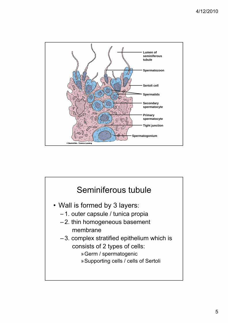
5
Lumen ofseminiferoustubule
Spermatozoon
Sertoli cell
Spermatids
Secondaryspermatocyte
Primaryspermatocyte
Tight junction
Spermatogonium
Seminiferous tubule
• Wall is formed by 3 layers:– 1. outer capsule / tunica propia– 2. thin homogeneous basement
membrane– 3. complex stratified epithelium which is
consists of 2 types of cells:consists of 2 types of cells: »Germ / spermatogenic»Supporting cells / cells of Sertoli

4/12/2010
6
Germ cell• Lie between Sertoli cells and arranged in an
orderly manner in 4-8 layersorderly manner in 4 8 layers.• In children, testis not fully developed. Therefore,
only primitive germ cells called spermatogonia are present.
• With onset of sexual maturity, spermatogenic cells are represented in all stages of diff ti ti i f i h t ldifferentiation, viz., from periphery to lumen: spermatogonium→ primary spermatocyte →secondary spermatocyte → spermatid.
Sertoli cells• Large and tall irregular columnar cells extending
from basement membrane to lumen offrom basement membrane to lumen of seminiferous tubule
• Germ cells are attached to Sertoli cells by means of cytoplasmic connections.
• Adjacent Sertoli cells near basement membrane are attached with one another by tight junctions
d f bl d t ti b iand form a blood-testis barrier• They support germ cells so also called
sustentacular cells

4/12/2010
7
Functions of Sertoli cells
• Support and nourish germ cells until spermatozoa are released from them.p
• Provide hormonal and other substances necessary for spermatogenesis
• Convert androgens into estrogen. Enzyme aromatase present in Sertoli cells is responsible for conversion.
• Secrete ABP (androgen binding protein)( g g p )• Secrete inhibin• Secrete Mullerian regression factor (MRF) in
fetal testes, MRF also called MIS.
Functions of testis
• Performs 2 functions:1 Gametogenic1. Gametogenic2. Endocrine
• Gametogenic function = Spermatogenesis:is the process by which the male gametes called spermatozoa / sperm are formed from primitive germ cells / spermatogonia in testis

4/12/2010
8
Stages of spermatogenesis• Occurs in 4 stages:
1. Proliferation
2. Growth
3 M t ti3. Maturation
4. transformation
Stages
Mitoticproliferation
Primaryt t
SpermatogoniumOne daughter cell remainsat the outer edge of theseminiferous tubule tomaintain the germ cell line
One daughter cell movestoward the lumen to produce spermatozoa
Chromosomesin each cell46 (diploid number;single strands)
46 (diploid number;single strands)
Spermatogonia
Meiosis
Spermatids
Secondaryspermatocyte
spermatocyte
First meioticdivision
Second meioticdivision
46 (diploid number;doubled strands)
23(haploid number;single strands)
23(haploid number;
Packaging Spermatozoa 23(haploid number;doubled strands)
(single strands)

4/12/2010
9
Role of Sertoli cells in spermatogenesis
• Support and nourish germ cells• Provide hormonal and other substances
necessary for spermatogenesis• Secrete androgen binding protein which is
essential for testosterone activity, particularly on spermatogenesisparticularly on spermatogenesis
• Release the sperms into lumen of semi-niferous tubules
Role of hormones in spermatogenesis
• FSH• Testosterone• Oestrogen• LH• GH• Inhibin• Activin

4/12/2010
10
Figure 20.9Page 762
Hypothalamus
Gonadotropin-releasing hormone
Anterior pituitary
FSH-secretingcells
LH-secretingcells
FSH LH
Testes
Sertoli LeydigSe tocell
Leydigcell
Spermatogenesis
Inhibin Testosterone
Other factors affecting spermatogenesis
• ↑ in temperature prevents t i ( ll t ispermatogenesis. (normally, temp in
scrotum is 2 0C less than body temp. Low temp is essential for spermatogenesis)
• Infectious diseases e g mumps• Infectious diseases e.g. mumps

4/12/2010
11
Endocrine function of testes• Male sex hormones = androgens
• Testis secretes 3 androgens:1. Testosterone2. Dihydrotestosterone3. Androstenedione
• Sertoli cells secrete inhibin, which inhibits secretion of FSH from pituitary, but does not possess any androgenic action
Androgens• Source:
– Testis – interstitial cells of LeydigAdrenal cortex zona reticularis– Adrenal cortex – zona reticularis
• Chemistry:– Steroid hormones synthesised from
cholesterol• Synthesis:
– See diagram• Transport:
– ⅔ by a β globulin, ⅓ by albumin

4/12/2010
12
Functions of testosterone
In fetal life
• 3 functions in fetus:
1. Sex differentiation of fetus – Mullerain duct gives rise to female acc organs and Wolffian duct gives rise to male acc sex organs
1. Development of accessory sex organs
1. Descent of testes

4/12/2010
13
In adult life
1. On sex organs
• ↑ size of penis, scrotum and testes• Necessary for spermatogenesis
2. On secondary sexual characters
• Muscular growth• Bone growth• Changes in skin• Hair distribution• Change in voice• BMR• BMR• Electrolyte and water balance• blood

4/12/2010
14
Mode of action of testosterone
• It is converted into dihydrotestosterone t t ll ftarget cells of acc sex organs
• In brain it is converted into oestrogen• Dihydrotestosterone combines with
receptor proteinsHormone receptor complex migrates to• Hormone receptor complex migrates to nucleus, binds with a nuclear protein and induces DNA-RNA transcription
Regulation of testosterone secretion
• In fetus:– Secretion from testis is stimulated by HCG
HCG stimulates development of Leydig cells and– HCG stimulates development of Leydig cells and promotes testosterone secretion
• In adults:– LH or ICSH stimulates Leydig cells and quantity of
testosterone secreted is directly proportional to amount of LH
– Secretion of LH from ant pit is stimulated by LHRH from hypothalamusfrom hypothalamus
• Feedback control:– Testosterone regulates its own secretion by negative
feedback, see diag

4/12/2010
15
Seminal vesicles
• Structure:– Paired glands situated on either side of
prostate gland– Lined by complex folded mucus membrane– Mucus membrane is formed by pseudo
striated columnar epithelium– Secretions are added to semen via ampulla of
vas deferens
• Properties:– Secretion from seminal vesicles is mucoid
and viscous– It is neutral/slightly alkaline in reaction– Forms 60% of semen
• Composition:S t i t t b t– Secrete many important substances
– Fig 77.1 for products of seminal vesicle secretion

4/12/2010
16
FIGURE 77-1: Composition of semen
Functions of seminal vesicle secretion
• Nutrition to sperms:– Fructose and other nutritive substances are utilized uctose a d ot e ut t e substa ces a e ut ed
by sperm after being ejaculated into female genital tract
• Clotting of sperm:– Fibrinogen from secretions is converted into
coagulum as soon as semen is ejaculated• On fertilization:
PG h f tili ti f b i i th– PG enhances fertilization of ovum by increasing the receptive capacity of cervical mucosa for sperms and causes reverse peristaltic movement of uterus and fallopian tubes.
4/12/2010
17
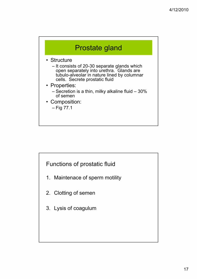
Prostate gland• Structure
– It consists of 20-30 separate glands which– It consists of 20-30 separate glands which open separately into urethra. Glands are tubulo-alveolar in nature lined by columnar cells. Secrete prostatic fluid
• Properties:– Secretion is a thin, milky alkaline fluid – 30%
of semenof semen• Composition:
– Fig 77.1
Functions of prostatic fluid
1. Maintenace of sperm motility
2. Clotting of semen
3. Lysis of coagulum

4/12/2010
18
Semen
• Nature of semen:Semen is a white/grey fluid that contains– Semen is a white/grey fluid that contains spermatozoa/sperms. It is collection of fluids from testes, seminal vesicles, prostate gland and bulbourethral glands.
• Properties of semen:– 2-6 ml/ejaculation. It is alkaline with pH of 7.5
• Composition:– Contains 10% sperm and 90% of fluid part which is
called seminal plasma, see fig 77.1
Sperm• Total count is about 100 to 150 million/ml of
semensemen• Sterility occurs when sperm count falls below 20
million/ml• After ejaculation survival time is 24-48 hours at a
temp equivalent to body temp.• Rate of motility of sperm in female tract is y p
3mm/min. Sperm reach fallopian tube in 30-60 min after sexual intercourse. Uterine contractions facilitate movement of sperms

4/12/2010
19
Structure of spermatozoon / sperm
• 60 μ long. Consists of 4 parts:1 Head:1. Head:
Formed by condensed nucleus, thin cytoplasm and cell membrane. Acrosome – hyaluronidase and proteolytic enzymes
2. Neck3. Body:
Axial filament is surrounded by a closely wound y yspiral fialment consisting of mitochondria
4. Tail:Chief or main piece and terminal or end piece
Acrosome
Mit h d i
Microtubules
Mitochondria
Nucleus
Head Midpiece Tail

4/12/2010
20
Plasma membrane Mitochondria
Acrosome
CentrioleNucleus
Qualities of semen - minimum required qualities for fertility are:1. Volume per ejaculation at least 2 ml2 Sperm count at least 2 million2. Sperm count – at least 2 million3. No. of sperm in each ejaculation – 40 million4. 75% of sperm per ejaculation must be alive5. 50% of sperm must be motile6. 30% must have normal shape and structure7 S ith h d d f t < 35%7. Sperms with head defect < 35%8. Sperm with midpiece defect < 20%9. Sperm with tail defect must be < 20%


![FBC Haem Lecture 1SA13 ClickUP 1.ppt [Read-Only]](https://img.pdfslide.net/doc/110x75/61689282d394e9041f70b88d/fbc-haem-lecture-1sa13-clickup-1ppt-read-only.jpg)