Embed Size (px)
Citation preview

National Capodistrian
UniversityOf Athens
Bone and Inflammation
George E. Fragoulis, MD
Metsovo 2012
PathophysiologyDept
School of Medicine

Outline
Physiology of bone turn-over
RANK-RANKL-OPG System
Wnt System
Pathophysiology of Bone in Inflammation
Inflammation and bone destruction
Role of specific cytokines / Th17
Disease Paradigms
RA
Spondyloarthropathies
Psoriatic Arhritis
Systemic Lupus Erythematosus

RANK
RANK
RANK-RANKL-OPG System
Osteoclasts (OCL) are multinucleated cells formed by fusion of mononuclear precursors in the monocyte/macrophage lineage
Osteoclastogenesis is directed by Osteoblasts (OBL) & bone stromal cells
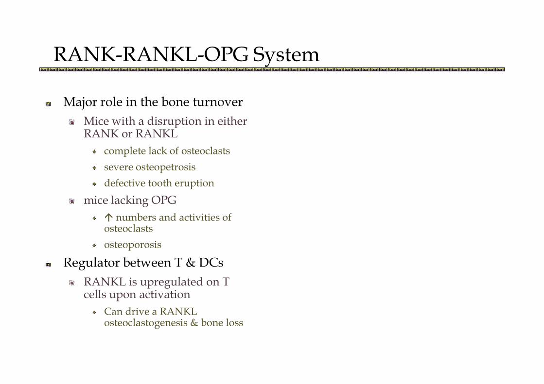
RANK-RANKL-OPG System
Major role in the bone turnover
Mice with a disruption in either RANK or RANKL
complete lack of osteoclasts
severe osteopetrosis
defective tooth eruption
mice lacking OPG
� numbers and activities of osteoclasts
osteoporosis
Regulator between T & DCs
RANKL is upregulated on Tcells upon activation
Can drive a RANKL osteoclastogenesis & bone loss

RANK-RANKL-OPG System
Pathway
Binding of RANKL to RANK
signaling cascades that control lineage commitmentcommitmentand activation of osteoclasts

RANK-RANKL-OPG System
Pathway
Negative & Positive Regulators

Wnt Pathway
Bone formation
Mechanism
OBL differentiation & activity
� growth rate of OBL & inhibiting their apoptosis
Inhibiting osteoclastogenesis
Wnt proteinsWnt proteins
Secreted growth factors proteins (regulate key procedures)
4 signaling pathways
Wnt/b-catenin pathway is the most important
Inhibitors
DKK-1: binds LRP5/6 + Kremen (LRP’s internalized)
SOST, produced by osteocytes: binds LRP5

Wnt Pathway
DKK-1 experimental models (mice)
�: osteopenia, limb deformities, hairlessness, � No of OBL
�: � No of OBL, � bone formation rate
LRP5
LRP5 -/- mice
� bone mass
Mice with gain of function mutation (G171V)Mice with gain of function mutation (G171V)
High bone mass phenotype
SFRPs (Secreted frizzled-related protein)
able to bind Wnt proteins and prevents them from binding the Fz receptors
SFRP1 -/- mice
High trabecular bone density
In co-cultures of murine osteoblasts with spleen cells
Anti-sFRP-1: � osteoclast formation
Recombinant sFRP-1: inhibited osteoclast formation
Bind and inactivate RANKL
thus SFRPs not only act blocking WNt but also via RANKL!

Possible interplay between
RANK/RANKL/OPG & Wnt Systems
B-catenin regulate OPG expression in OBL
Cocultures of mouse mononuclear spleen cells & OBL
Treated with Treated with
Wnt3a (activation) or Lithium (indirect activation)
Inhibition of formation of OCL
� RANKL (mRNA, protein)

Inflammation and bone destruction
IL-4IL-12IL-18IFN-β
Promote bone resorption
Inhibit osteoclastogenesis� OPG/RANKL
IFN-βTGF-b
TNFaIL-1IL-6IL-7OSMPTHrP

Inflammation and bone destruction
TNF-a
Inhibits differentiation of mature OBL
Stimulates Dkk-1
� expression of RANKL
Stromal cells
T lymphocytes
B lymphocytes
endothelial cells
� RANK
� OCL precursor populationInduces apoptosis of OBL, OBL
precursors, chondrocytes
� OCL precursor population
mobilize CD11bhigh osteoclast from the bone marrow into the circulation
� OCL differentiation and activation
Along with IL-1 and RANKL
� M-CSF production
stromal cells
Mice deficient in TNFa : Some of them develop bone erosions (Indicating that there are TNF independent pathways leading to erosions)
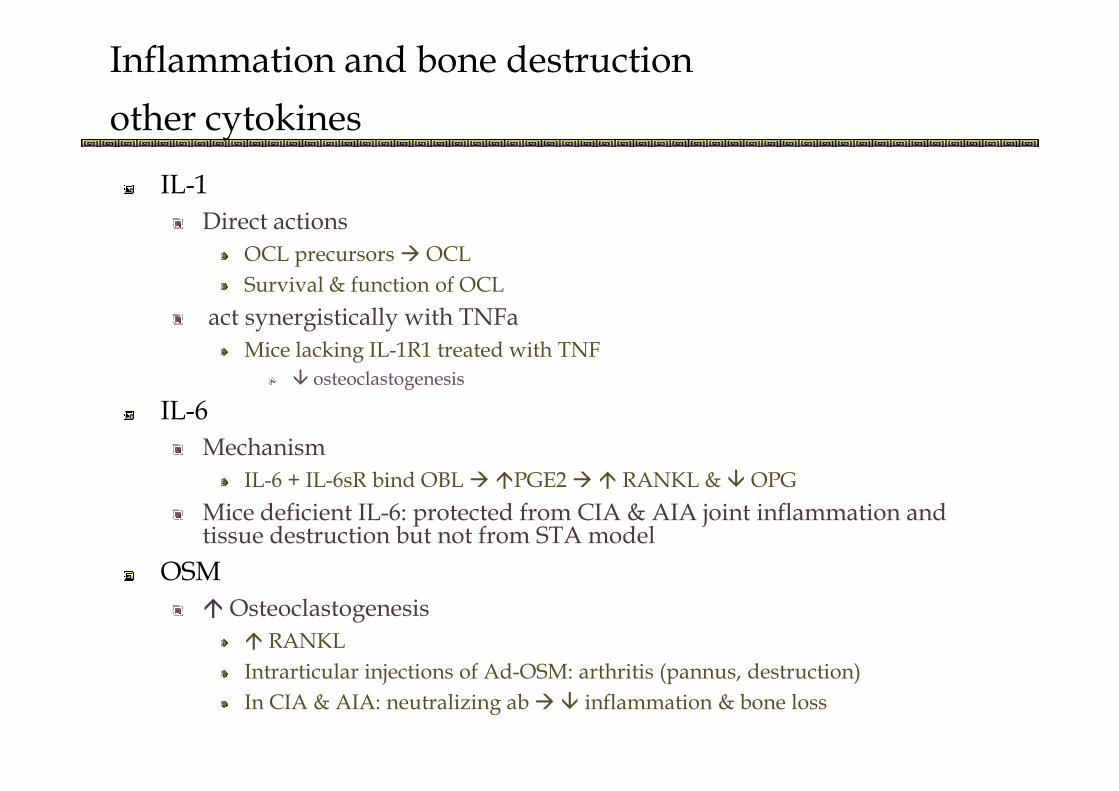
Inflammation and bone destruction
other cytokines
IL-1
Direct actions
OCL precursors � OCL
Survival & function of OCL
act synergistically with TNFa
Mice lacking IL-1R1 treated with TNF
� osteoclastogenesis
IL-6IL-6
Mechanism
IL-6 + IL-6sR bind OBL � �PGE2 � � RANKL & � OPG
Mice deficient IL-6: protected from CIA & AIA joint inflammation and tissue destruction but not from STA model
OSM
� Osteoclastogenesis
� RANKL
Intrarticular injections of Ad-OSM: arthritis (pannus, destruction)
In CIA & AIA: neutralizing ab �� inflammation & bone loss

Inflammation and bone destruction
other cytokines
IL-7
Stimulating differentiation of OCL precursors � OCL
� RANKL in T cells
PTHrP
Induced by TNFa and IL-1
Acts synergistically with TNFa and IL-1Acts synergistically with TNFa and IL-1
� RANKL in OBL
� OPG
Induces secretion of IL-6
CIA: � in joint
SCW model: block
� No of OCL
� bone erosions
Inflammation: unaltered

Inflammation and bone destruction
Uncoupling inflammation from bone resorption
OCL is the major player
Inflammatory arthritis can be present in mouse models lacking OCLs (i.e cfos -/- / hTNFtg, RANKL KO) despite hTNFtg, RANKL KO) despite the complete loss of bone erosions
No altering in cellular populations
MMPs
No influence on TNF

Inflammation and bone destruction
Uncoupling inflammation from bone resorption
Tx with OPG Fc in TNFtg mice
� OCL No
� focal erosion
Ongoing inflammation
tgTNF mice treated with anti-Dkk-1: inhibition of bone erosions without altering inflammation attributed to � OCLwithout altering inflammation attributed to � OCL
Thus: TNF and other inflammatory cytokines enhance bone erosion but can’t lead alone to bone erosions

IL-17 : a novel regulator
A combination of TGF-b, IL-6, IL-1 and IL-23 is required for Th17 differentiation and IL-17 production
Th17 cells
Express � RANKL
Cartilage breakdown
Synergistic action with TNFa
OsteoclastogenesisOsteoclastogenesis
Cocultures of OCL precursors with either RANKL, MCSF or OBL
Osteoclastogenesis was � in the presence of IL-17
Probably via � RANKL in OBL
AIA/CIA: In vivo blockade
� joint destruction
� IL-6
�bone erosions
�VEGF

Disease Paradigms
Inflammatory conditions
Bone loss (e.g Rheumatoid arthritis)
Bone loss & bone formation (e.g Spondylarthritis)
Type of bone loss
Generalized (osteopenia)Generalized (osteopenia)
Focal (bone erosion)
Local (periarticular osteopenia)
Type of bone formation
Sclerosis
Osteophyte formation
Calcification of soft tissues

Rheumatoid Arthritis
Proinflammatory mediators in RA joints � production of OCL precursors � in bone/pannus � differentiating to OCL (under RANKL)
Total hip BMD correlated with erosion scoresTotal hip BMD correlated with erosion scores
Stronger among patients with early RA
After adjustment disappeared
Relationship between focal erosions and generalized osteoporosis is complicated and modified by many factors

Rheumatoid Arthritis
Synovial tissue
IL-17
� spnt of synovial tissue
� IL-6, IL-1, TNFa
By synoviocytes
Collagen destruction
Inhibition of collagen synthesis
� RANKL
OBL
T cells
Fibroblasts
� OPG
IL-7Inhibition of collagen synthesis
� MMP-1
� RANKL
Synoviocytes
OBL
� OPG
OBL
IL-7
MF
Fibroblasts
Endothelial cells
PTHrP
Synovial lining cells
Fibrolast like cells

Rheumatoid Arthritis
Serum
� Dkk-1
Synovial fluid
� IL-17
� OPG
� IL-6 & sIL-6R� Dkk-1
Association with radiographic progression
After anti-TNF Tx: �
� IL-6 & sIL-6R
� IL-6 & sIL-6R
Correlation with radiographic damage
� OSM
� IL-7
� PTHrP
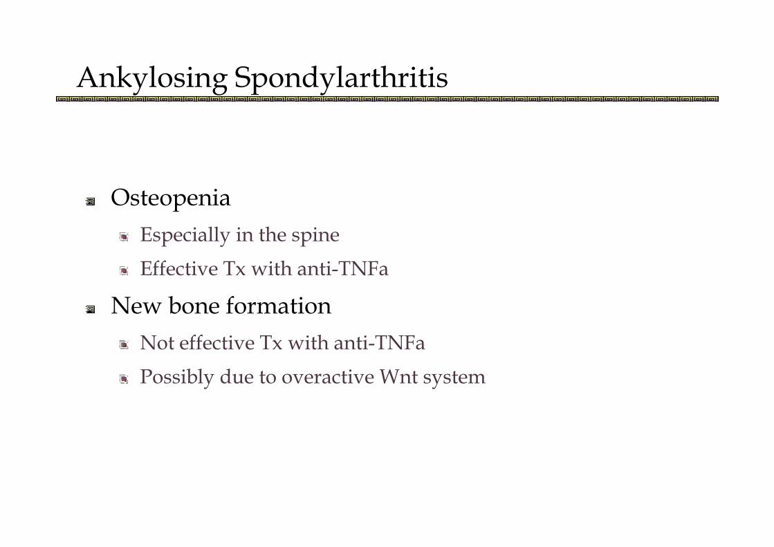
Ankylosing Spondylarthritis
Osteopenia
Especially in the spine
Effective Tx with anti-TNFa Effective Tx with anti-TNFa
New bone formation
Not effective Tx with anti-TNFa
Possibly due to overactive Wnt system

Ankylosing Spondylarthritis
Osteopenia
Associated with
� OPG� OPG
� bone absorption markers (indicating role for RANKL)

Ankylosing Spondylarthritis
New bone formation
Dkk-1 serum levels
� in AS vs. Healthy, RA, PsA
However: � Dkk-1 binding to LRP6
No association with inflammation markers or disease activity
� in AS after anti-TNF Tx
Trying to balance the new bone formation resulting from
resolution of inflammation (“TNF brake hypothesis”)

Ankylosing Spondylarthritis
Synovial tissue
� TNFa
� Bone Morphogenetic Protein (BMP) -2 and -6� Bone Morphogenetic Protein (BMP) -2 and -6
Thought responsible for bone formation
Also found in RA
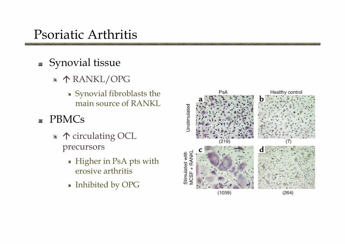
Psoriatic Arthritis
Synovial tissue
� RANKL/OPG
Synovial fibroblasts the main source of RANKL
PBMCsPBMCs
� circulating OCL precursors
Higher in PsA pts with erosive arthritis
Inhibited by OPG

SLE
Only 4-6% display erosive changes in Rx
Histological
Mild synovial hyperplasia
Microvascular changes
Perivascular inflammation
One possible explanation
� IFNa in SLE skews myelomonocyte precursors to mDC instead of OCL
IFNar1-/- mice: � OCL precursosrs & osteopenia
Does not explain the osteopenia in SLE

Take home messages
Bone remodelling is a complex procedure
RANK/RANKL/OPG – Osteoclastogenesis
Inflammatory cytokinesInflammatory cytokines
RA
Wnt system – Bone formation
Dkk-1 is the major player
AS