Embed Size (px)
Citation preview
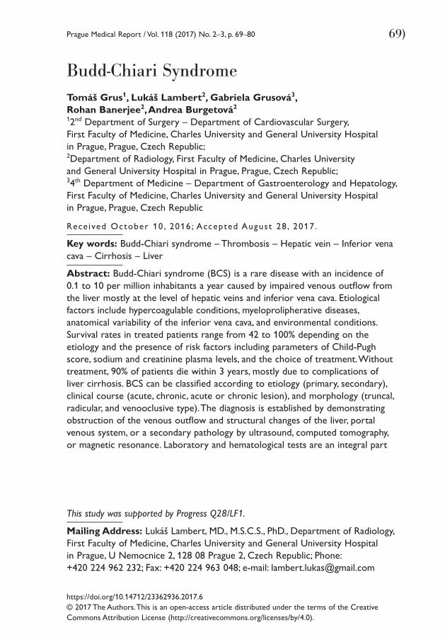
Budd-Chiari Syndrome
Prague Medical Report / Vol. 118 (2017) No. 2–3, p. 69–80 69)
Budd-Chiari SyndromeTomáš Grus1, Lukáš Lambert2, Gabriela Grusová3, Rohan Banerjee2, Andrea Burgetová2
12nd Department of Surgery – Department of Cardiovascular Surgery, First Faculty of Medicine, Charles University and General University Hospital in Prague, Prague, Czech Republic;2Department of Radiology, First Faculty of Medicine, Charles University and General University Hospital in Prague, Prague, Czech Republic;34th Department of Medicine – Department of Gastroenterology and Hepatology, First Faculty of Medicine, Charles University and General University Hospital in Prague, Prague, Czech Republic
Rece ived October 10 , 2016 ; Accepted August 28 , 2017 .
Key words: Budd-Chiari syndrome – Thrombosis – Hepatic vein – Inferior vena cava – Cirrhosis – Liver
Abstract: Budd-Chiari syndrome (BCS) is a rare disease with an incidence of 0.1 to 10 per million inhabitants a year caused by impaired venous outflow from the liver mostly at the level of hepatic veins and inferior vena cava. Etiological factors include hypercoagulable conditions, myeloprolipherative diseases, anatomical variability of the inferior vena cava, and environmental conditions. Survival rates in treated patients range from 42 to 100% depending on the etiology and the presence of risk factors including parameters of Child-Pugh score, sodium and creatinine plasma levels, and the choice of treatment. Without treatment, 90% of patients die within 3 years, mostly due to complications of liver cirrhosis. BCS can be classified according to etiology (primary, secondary), clinical course (acute, chronic, acute or chronic lesion), and morphology (truncal, radicular, and venooclusive type). The diagnosis is established by demonstrating obstruction of the venous outflow and structural changes of the liver, portal venous system, or a secondary pathology by ultrasound, computed tomography, or magnetic resonance. Laboratory and hematological tests are an integral part
https://doi.org/10.14712/23362936.2017.6© 2017 The Authors. This is an open-access article distributed under the terms of the Creative Commons Attribution License (http://creativecommons.org/licenses/by/4.0).
This study was supported by Progress Q28/LF1.
Mailing Address: Lukáš Lambert, MD., M.S.C.S., PhD., Department of Radiology, First Faculty of Medicine, Charles University and General University Hospital in Prague, U Nemocnice 2, 128 08 Prague 2, Czech Republic; Phone: +420 224 962 232; Fax: +420 224 963 048; e-mail: [email protected]
Grus T.; Lambert L.; Grusová G.; Banerjee R.; Burgetová A.
70) Prague Medical Report / Vol. 118 (2017) No. 2–3, p. 69–80
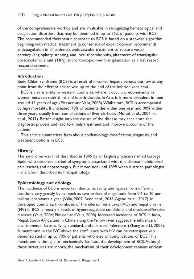
of the comprehensive workup and are invaluable in recognizing hematological and coagulation disorders that may be identified in up to 75% of patients with BCS. The recommended therapeutic approach to BCS is based on a stepwise algorithm beginning with medical treatment (a consensus of expert opinion recommends anticoagulation in all patients), endovascular treatment to restore vessel patency (angioplasty, stenting, and local thrombolysis), placement of transjugular portosystemic shunt (TIPS), and orthotopic liver transplantation as a last resort rescue treatment.
IntroductionBudd-Chiari syndrome (BCS) is a result of impaired hepatic venous outflow at any point from the efferent acinar vein up to the end of the inferior vena cava.
BCS is a rare entity in western countries, where it occurs predominantly in women between their third and fourth decade. In Asia, it is more prevalent in men around 45 years of age (Plessier and Valla, 2008). Whilst rare, BCS is accompanied by high mortality. If untreated, 70% of patients die within one year and 90% within three years, usually from complications of liver cirrhosis (Murad et al., 2004; Fu et al., 2011). Better insight into the nature of the disease may accelerate the diagnostic process and lead to timely treatment and improve outcome of the patient.
This article summarizes facts about epidemiology, classification, diagnosis, and treatment options in BCS.
HistoryThe syndrome was first described in 1845 by an English physician named George Budd, who observed a triad of symptoms associated with this disease – abdominal pain, ascites, and hepatomegaly. But it was not until 1899 when Austrian pathologist Hans Chiari described its histopathology.
Epidemiology and etiologyThe incidence of BCS is uncertain due to its rarity and figures from different locations vary greatly by as much as two orders of magnitude from 0.1 to 10 per million inhabitants a year (Valla, 2009; Renc et al., 2013; Ageno et al., 2017). In developed countries, thrombosis of the inferior vena cava (IVC) and hepatic veins (HV) in BCS is mostly a result of hypercoagulable conditions and myeloproliferative diseases (Valla, 2004; Plessier and Valla, 2008). Increased incidence of BCS in India, Nepal, South Africa, and in China along the Yellow river suggest the influence of environmental factors, living standard, and microbial infections (Zhang and Li, 2007). A membrane in the IVC above the confluence with HV can be retrospectively demonstrated in up to 70% of patients who died of complications of BCS. This membrane is thought to mechanically facilitate the development of BCS. Although these structures are inborn, the mechanism of their development remains unclear.
Budd-Chiari Syndrome
Prague Medical Report / Vol. 118 (2017) No. 2–3, p. 69–80 71)
In Western countries, BCS is more common in females and occurs more commonly at the level of the hepatic veins. In Asia, occlusion of the IVC is more common and its incidence is comparable between the genders.
PrognosisFive-year survival rates range from 42 to 100% depending on the etiology and the presence of risk factors including parameters of Child-Pugh score, sodium and creatinine plasma levels, and the choice of treatment (Valla, 2006). Murad et al. (2004) derived a prognostic formula that distinguished three classes based on the calculated score:
score = 1.27 × encephalopathy + 1.04 × ascites + 0.72 × INR + 0.004 × bilirubinwhere ascites and encephalopathy are set to 1 when present, INR (International
Normalized Ratio) higher than 2.3 also set to 1 and bilirubin used as a continuous variable in µmol/l. Class I represent scores between 0 and 1.1, class II between 1.1 and 1.5, and class III scores 1.5 and higher with 5-year survival rates of 89, 74, and 42% respectively.
As shown by Rautou and associates (2009), other prognostic scores including the Child-Pugh score, model for end-stage liver disease (MELD), Rotterdam index and New Clichy have their prognostic value too.
Best survival rates, irrespective of the prognostic score, have been reported in cohorts where venous drainage of the liver was restored by interventional methods (Eapen et al., 2006). The most common cause of death in patients treated for BCS is liver failure, postoperative multiorgan failure and sepsis (Murad et al., 2004).
ClassificationThere are several classifications of BCS (Senzolo et al., 2005). A classification suggested by prof. Wang included eight types based on anatomical location and the extent of the outflow obstruction (Wang and Jones, 1996). Less complex classifications used at present divide BCS according to the velocity of its development to fulminant, acute, subacute and chronic and by etiology as primary or secondary BCS (Plessier and Valla, 2008). Some authors do not consider cases with obstruction of small liver veins below 300 µm in diameter as BCS.
Etiology: primary and secondary BCSPrimary BCS occurs in patients with primary hematological disorders or hypercoagulable conditions. Up to half of patients with BCS are diagnosed with myeloproliferative disorders, polycythemia vera, essential thrombocytemia, or, in particular, primary myelofibrosis (Zhang and Li, 2007). The most common primary hypercoagulable conditions in these patients are mutations of factor V Leiden, factor II (prothrombin), JAK-2 tyrosine kinase genes and antiphospholipid syndrome (Colaizzo et al., 2008). The role of hyperhomocysteinemia and primary deficiency
Grus T.; Lambert L.; Grusová G.; Banerjee R.; Burgetová A.
72) Prague Medical Report / Vol. 118 (2017) No. 2–3, p. 69–80
of protein C or S or antithrombin III is difficult to establish due to the associated liver disease. BCS occurs in up to 30% of patients with primary nocturnal hemoglobinuria, which is 5% of all diagnosed patients in developed countries. Other risk factors include pregnancy, hypereosinophilic syndrome, ulcerative colitis, oral contraception, and several underlying conditions may act in synergy. Therefore, comprehensive hematological examination is indicated in every patient with excluded secondary etiology of BCS.
Secondary BCS describes any disease that causes BCS by invasion or compression of the IVC or HV with their consequent thrombosis. This includes focal liver lesions (hepatocellular carcinoma, abscess, cyst, …), renal or adrenal adenocarcinoma, blunt abdominal trauma, and rarely primary sarcomas of IVC or myxoma of the right atrium (Mukund and Gamanagatti, 2011).
Clinical: type I – acute, type II – chronic, type III – acute or chronic lesion with the worst prognosis (Langlet et al., 2003).
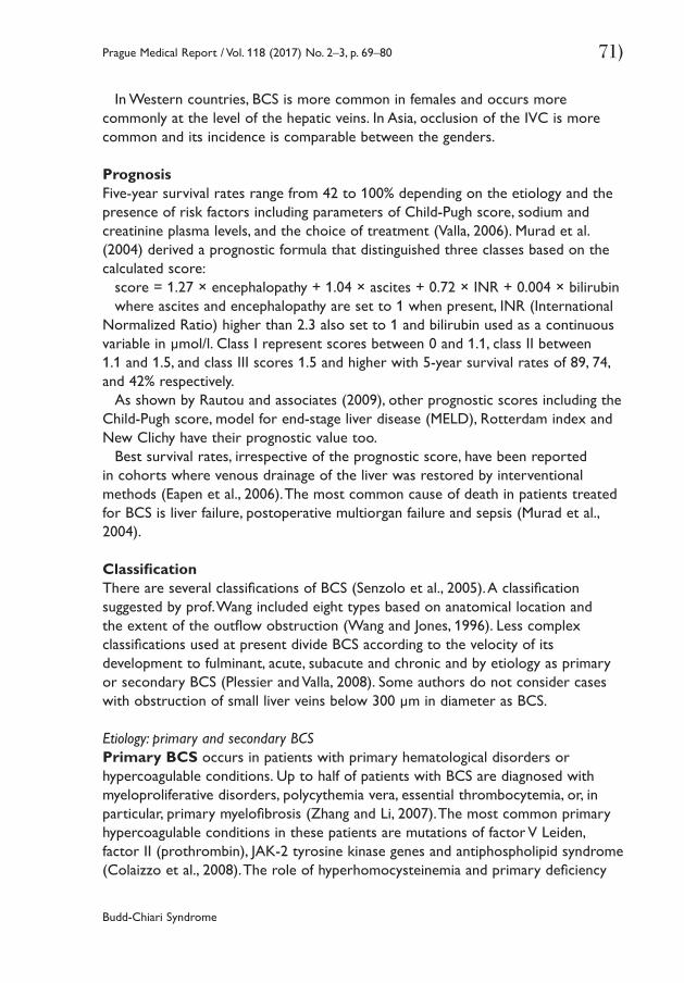
Morphology: according to the location of the obstruction – truncal type (I) with obstruction of the IVC (±HV), radicular type (II) with obstruction of HV, venoocclusive type (III) with obstruction of small centrilobular veins (Figure 1) (Lambert, 2016).
PathophysiologyBetween plates of hepatocytes and the sinusoids there is a narrow space of Disse. Microvillous projections from the hepatocytes into this space are responsible for metabolism and transport of molecules between blood and the hepatocyte. Impaired blood outflow results in increased intrasinusoidal pressure, dilation of sinusoids, and extravasation of erythrocytes that accumulate in the space of Disse. In early stages, congestion affects the perivenular zone but gradually extends towards the periportal zone (Valla, 2003). Persistent reduction of hepatic perfusion
Figure 1 – Schematic drawing of the types of Budd-Chiari syndrome according to the location of the obstruction – truncal type (I) with obstruction of the IVC (±HV), radicular type (II) with obstruction of HV, venoocclusive type (III) with obstruction of small centrilobular veins.
Budd-Chiari Syndrome
Prague Medical Report / Vol. 118 (2017) No. 2–3, p. 69–80 73)
results in ischemic injury to the hepatocytes followed by fibrosis and nodular regenerative changes, resulting in liver cirrhosis. However, large regenerative nodules that develop to maintain perfusion of the liver may in turn lead to compression of adjacent intrahepatic veins (Cazals-Hatem et al., 2003).
Clinical presentationBCS may cause a range of symptoms that present with various severities. Although some patients may be virtually asymptomatic, most of them develop some symptoms that are a result of liver failure (portal hypertension with variceal bleeding, encephalopathy, ascites, weakness, fatigue) or extension of secondary pathology and metastatic disease. Clinical presentation depends on the extent of hepatic venous outflow obstruction and velocity of its development. The most common symptoms are summarized in Table 1.
DiagnosisImaging is the mainstay of diagnosis of BCS, which is established by demonstrating obstruction of IVC, HV and structural changes of the liver, portal venous system, or a secondary pathology.
Ultrasound (US) is a first-line imaging method with high sensitivity and specificity of up to 85% (Bolondi et al., 1991). Venous outflow obstruction is demonstrated by absent flow and echogenic luminal content. Blood flow in a stenosis becomes accelerated with spectral broadening indicating turbulence. US can detect structural changes of the liver parenchyma (macronodular cirrhosis, focal liver lesions), hypertrophy of the caudate lobe (which is typical for BCS), ascites, collateral blood flow, or direct invasion and compression of the IVC by a tumoral mass.
Contrast-enhanced computed tomography (CT) is performed in portal venous phase in order to achieve good contrast filling in the portal, mesenteric,
Table 1 – The most common symptoms in patients with Budd-Chiari syndrome
Okuda et al. (1998)
Hadengue et al. (1994)
Murad et al. (2004)
Singh et al. (2000)
Number of patientsLocation of obstruction
Abdominal painHepatomegalyAscitesLeg swellingJaundice
157IVC
235531326
88HV
7288833333
237HV
–7684–50
81HV (75%)IVC (25%)
3261816233
IVC – inferior vena cava; HV – hepatic veins
Grus T.; Lambert L.; Grusová G.; Banerjee R.; Burgetová A.
74) Prague Medical Report / Vol. 118 (2017) No. 2–3, p. 69–80
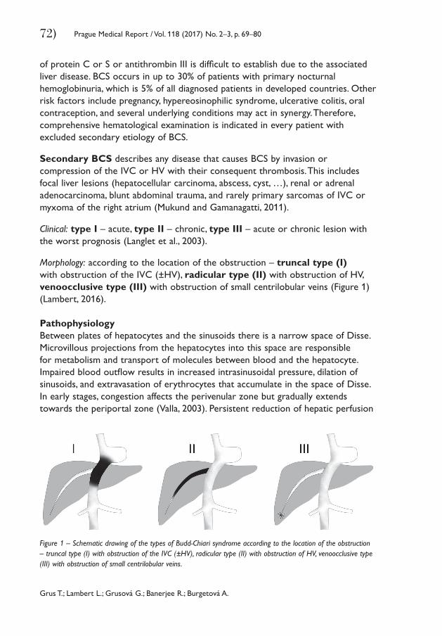
and hepatic veins and in inferior vena cava for optimal detection of their pathology. Structural changes of the liver are similar to findings in US. Inhomogeneous enhancement of the liver parenchyma results in mottled appearance with relative sparing of the caudate lobe and perivenous parenchyma that have separate venous drainage (Figure 2). These changes are more obvious on magnetic resonance (MR), which provides images with far better tissue contrast (Zhou et al., 2014).
Invasive imaging methods that can be used to depict obstruction of the venous outflow include angiography of the inferior vena cava and hepatic veins (cavography). They are commonly used together with interventional procedures to restore patency of the vessels.
Liver biopsy is indicated in patients with suspected venoocclusive type (III) of BCS of unknown etiology, when macroscopic obstruction of the venous outflow has been excluded by imaging.
Gastroscopy is warranted in patients with liver cirrhosis to exclude esophageal or fundal varices and perform their ligation in order to decrease the risk of variceal bleeding. Portal hypertensive gastropathy refers to macroscopic changes of the gastric mucosa with prominent vessels that result in mosaic appearance.
Laboratory and hematological tests are an integral part of comprehensive workup of patients with BCS and are invaluable in hematological and coagulation disorders that may be recognized in up to 75% of patients (Singh et al., 2000). In 25% of patients, at least two underlying conditions can be identified (Aydinli and Bayraktar, 2007). Primary BCS requires laboratory test for factor V Leiden and factor II (prothrombin) mutations, for the presence of antiphospholipid antibodies
Figure 2 – Computed tomography (CT) in portal venous phase of a 32-years-old female patient with Budd-Chiari syndrome, who developed liver cirrhosis, portal hypertension, and splenomegaly showing macronodular regeneration in the liver parenchyma that has mottled appearance (asterisks), an enlarged caudate lobe (two-headed arrow), dilated splenic vein with conspicuous collateral veins (arrows), and enlarged spleen (arrowheads) in coronal (1) and axial (2) plane. This patient received a transjugular portosystemic shunt (TIPS) in order to reduce porto-systemic gradient and the risk of variceal bleeding.
Budd-Chiari Syndrome
Prague Medical Report / Vol. 118 (2017) No. 2–3, p. 69–80 75)
and plasma levels of homocysteine, protein C, protein S, and antithrombin III (EASL, 2016). The deficiency of antithrombin III or protein C and S can also be the result of liver dysfunction. Myeloproliferative disorders are common in BCS and their diagnosis is based on bone marrow biopsy. Mutations of the JAK-2 kinase gene can be demonstrated in the majority of patients, including those with latent forms (Boissinot et al., 2006). Indeed, standard biochemical and haematological blood analysis including electrolytes, proteins (including serum electrophoresis), liver and kidney function tests, complete blood count, and coagulation tests are required as well.
TreatmentThe recommended therapeutic approach to BCS is based on a stepwise algorithm beginning with medical treatment, endovascular treatment to restore vessel patency (angioplasty, stenting, and local thrombolysis), placement of transjugular portosystemic shunt (TIPS), and orthotopic liver transplantation as a rescue treatment.
Although no prospective randomized trials on anticoagulation therapy in BCS have been conducted so far, a consensus of expert opinions recommended anticoagulation in all patients (Janssen et al., 2003). The risk of bleeding complications in patients with BCS is comparable to patients with anticoagulation therapy for other indications (Plessier and Valla, 2008). Its main goal is to prevent progression of the thrombosis. As soon as the diagnosis of BCS has been established, anticoagulation with low molecular weight heparin (LMWH) with a target value of AntiXa between 0.5 and 0.8 IU/ml has to be initiated without delay (DeLeve et al., 2009). Before switching from LMWH to oral anticoagulants, contraindications such as liver cirrhosis, portal hypertension, and esophageal varices have to be excluded and the diagnostic workup (screening for coagulation disorders, or liver biopsy if indicated) has to be completed (DeLeve et al., 2009). The recommended targeted INR (International Normalized Ratio) for treatment of patients with BCS with vitamin K antagonists is between 2.5 and 3 (Senzolo et al., 2005; Mancuso, 2011). Regular INR testing is necessary to ensure that the target INR range is maintained. In patients with primary BCS, prolonged, possibly even lifelong anticoagulation therapy is recommended. There are limited data about the use of direct factor Xa inhibitors (e.g. rivaroxaban) in patients with BCS.
In general, thrombolysis in BCS, whether systemic or intraarterial into thehepatic artery, is ineffective, unless it is administered locally (HV, IVC, TIPS) very early in case of acute thrombosis of HV or TIPS followed by endovascular intervention. In such patients, no major complications have been reported (Sharma et al., 2004).
Treatment of consequent portal hypertension and its complications is symptomatic and treatment recommendations are based on their management in patients with liver cirrhosis: diuretic therapy (spironolactone, furosemide) or paracentesis for ascites, endoscopic ligation of esophageal varices, administration
Grus T.; Lambert L.; Grusová G.; Banerjee R.; Burgetová A.
76) Prague Medical Report / Vol. 118 (2017) No. 2–3, p. 69–80
of proton pump inhibitors and beta-blockers to reduce the risk of bleeding from esophageal varices in selected patients (EASL, 2016).
Medical therapy alone may not be sufficient in patients who have persistent abnormal liver function tests, intractable ascites, coagulopathy, encephalopathy, abdominal discomfort, hepatorenal syndrome, gastrointestinal bleeding or ongoing necrosis shown on liver biopsy (Menon et al., 2004). These patients require timely relief of hepatic obstruction, decompression of portal hypertension, or orthotopic liver transplantation as the last resort rescue treatment.
The increasing use of interventional procedures in the treatment of BCS is guided by the type of venous occlusion (Wang et al., 2005). It consists in aspiration thrombectomy followed by predilatation with a small diameter catheter, which decreases the risk of pulmonary embolism and allows introduction of a thrombolytic catheter with subsequent administration of thrombolytic agent.
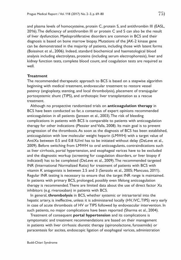
In patients with thrombosed IVC, endovascular treatment is indicated in patients with subtypes I and II, and in patients with acute IVC thrombosis of subtype III (Figure 3). In patients with subtype III (i.e. that extends beyond HV) subacute to chronic thrombosis, conservative treatment or, ultimately, placement of TIPS are preferred (Pelage et al., 2003; Ruihua et al., 2013). Recanalization of the occluded IVC results in rapid clinical improvement, and has good mid-term results (Han et al., 2013).
Patients with occluded HV and patent IVC are suitable for angioplasty and stenting if the HV has a straight course and is of sufficient diameter (≥7 mm). The HV is usually accessed through the internal jugular vein. If this is not successful or feasible, percutaneous transhepatic approach or access through the femoral vein is another option. In acute thrombosis, thrombolytic therapy alone has good results as well (Mukund and Gamanagatti, 2011).
In short stenosis, interventional treatment with stenting warrants excellent long-term patency – 97% in IVC and 91% in HV, but angioplasty alone has a high rate
Figure 3 – Schematic drawing of types of occlusion of inferior vena cava (IVC) and their relationship to the hepatic veins (HV) with regard to planning of interventional procedures. Location of a membrane in the IVC, found in up to 70% of patients with Budd-Chiari syndrome, is denoted by an arrowhead. In type I (left) and II (middle), the thrombosis does not involve confluence with the hepatic veins.
Budd-Chiari Syndrome
Prague Medical Report / Vol. 118 (2017) No. 2–3, p. 69–80 77)
of restenosis. If restenosis occurs after stenting, it is usually due to inadequate anticoagulant therapy that has to be maintained for at least 6 months after the intervention (Mukund and Gamanagatti, 2011). In general, 5-year survival in patients treated by endovascular recanalization is 86% in patients with intermediate disease severity and 77% with severe BCS (Eapen et al., 2006).
In some patients, who developed liver cirrhosis, placement of transjugular portosystemic shunt (TIPS) is indicated to decrease porto-systemic gradient in order to reduce the risk of variceal bleeding. The procedure should be planned in collaboration with a surgeon who performs liver transplantations (Menon et al., 2004). Introduction of TIPS requires endovascular access using an internal jugular approach to the ostium of the hepatic veins. The portal vein is punctured through the remnant of the hepatic vein, if present, or directly from the inferior vena cava. Introduction of TIPS has a 5-year survival without the need for liver transplantation of 78% (Garcia-Pagán et al., 2008). The use of a TIPS stent covered with polytetrafluorethylene (ePTFE) or TIPS stentgraft improves long-term patency rates compared to bare stents and therefore became a standard (Hernández-Guerra et al., 2004; Šafka et al., 2005; Renc et al., 2013; Dulíček et al., 2016). Surgical construction of a portosystemic shunt is rarely required and some cases can be solved by combination of endovascular and surgical therapy (hybrid procedures).
Orthotopic liver transplantation is the last resort rescue treatment if conservative and interventional therapy does not prevent development of liver cirrhosis and progressive liver failure in chronic BCS. Its indication should be carefully evaluated. However, in patients presenting with fulminant hepatic failure, it is an urgent indication. Patients after liver transplantation require life-long immunosuppressive therapy. Although liver transplantation can cure a majority of the hereditary thrombophilias, in most of the patients with BCS, multiple etiologic factors may be present and long-term or life-long anticoagulation therapy may also be indicated. Five-year survival of BCS patients after orthotopic liver transplantation is around 90% (Hefaiedh et al., 2013).
ConclusionBudd-Chiari syndrome is a result of impaired hepatic venous outflow at any point from the efferent acinar vein up to the end of the inferior vena cava. Congestion in the liver sinusoids results in portal hypertension, liver fibrosis or cirrhosis, and their complications. Imaging is the mainstay of diagnosis of BCS. Laboratory and haematological tests are invaluable in recognizing hematological and coagulation disorders that may be identified in up to 75% of the patients. The recommended therapeutic approach to BCS is based on a stepwise algorithm beginning with medical treatment. A consensus of expert opinion recommends anticoagulation in all patients. In selected patients, it can be combined with local thrombolysis, angioplasty and stenting. Restoring outflow vessel patency by endovascular
Grus T.; Lambert L.; Grusová G.; Banerjee R.; Burgetová A.
78) Prague Medical Report / Vol. 118 (2017) No. 2–3, p. 69–80
treatment results in improved survival. In patients with progressive liver failure and portal hypertension, placement of TIPS may decrease the risk of variceal bleeding and relieve symptoms. Orthotopic liver transplantation is the last resort with good long-term results.
ReferencesAgeno, W., Dentali, F., Pomero, F., Fenoglio, L., Squizzato, A., Pagani, G., Re, R., Bonzini, M. (2017) Incidence
rates and case fatality rates of portal vein thrombosis and Budd-Chiari syndrome. Thromb. Haemost. 117, 794–800.
Aydinli, M., Bayraktar, Y. (2007) Budd-Chiari syndrome: Etiology, pathogenesis and diagnosis. World J. Gastroenterol. 13, 2693–2696.
Boissinot, M., Lippert, E., Girodon, F., Dobo, I., Fouassier, M., Masliah, C., Praloran, V., Hermouet, S. (2006) Latent myeloproliferative disorder revealed by the JAK2-V617F mutation and endogenous megakaryocytic colonies in patients with splanchnic vein thrombosis. Blood 108, 3223–3224.
Bolondi, L., Gaiani, S., Li Bassi, S., Zironi, G., Bonino, F., Brunetto, M., Barbara, L. (1991) Diagnosis of Budd-Chiari syndrome by pulsed Doppler ultrasound. Gastroenterology 100, 1324–1331.
Cazals-Hatem, D., Vilgrain, V., Genin, P., Denninger, M.-H., Durand, F., Belghiti, J., Valla, D., Degott, C. (2003) Arterial and portal circulation and parenchymal changes in Budd-Chiari syndrome: a study in 17 explanted livers. Hepatology 37, 510–519.
Colaizzo, D., Amitrano, L., Tiscia, G. L., Iannaccone, L., Gallone, A., Grandone, E., Guardascione, M. A., Margaglione, M. (2008) Occurrence of the JAK2 V617F mutation in the Budd-Chiari syndrome. Blood Coagul. Fibrinolysis 19, 459–462.
DeLeve, L. D., Valla, D.-C., Garcia-Tsao, G., American Association for the Study Liver Diseases (2009) Vascular disorders of the liver. Hepatology 49, 1729–1764.
Dulíček, P., Hůlek, P., Krajina, A., Renc, O., Šafka, V., Fejfar, T., Sadílek, P., Beránek, M., Michiels, J. J., Žák, P. (2016) Diagnosis, etiology and management of the Budd-Chiari syndrome: A bloodcoagulation and hepatological study on the course of the disease treated with TIPS. Int. Angiol. 35, 90–97.
Eapen, C. E., Velissaris, D., Heydtmann, M., Gunson, B., Olliff, S., Elias, E. (2006) Favourable medium term outcome following hepatic vein recanalisation and/or transjugular intrahepatic portosystemic shunt for Budd Chiari syndrome. Gut 55, 878–884.
EASL (2016) EASL Clinical Practice Guidelines: Vascular diseases of the liver. J. Hepatol. 64, 179–202.Fu, Y., Sun, Y.-L., Ma, X.-X., Xu, P.-Q., Feng, L.-S., Tang, Z., Guan, S., Wang, Z.-W., Luo, C.-H. (2011) Necessity
and indications of invasive treatment for Budd-Chiari syndrome. Hepatobiliary Pancreat. Dis. Int. 10, 254–260.
Garcia-Pagán, J. C., Heydtmann, M., Raffa, S., Plessier, A., Murad, S., Fabris, F., Vizzini, G., Abraldes, J. G., Olliff, S., Nicolini, A., Luca, A., Primignani, M., Janssen, H. L. A., Valla, D., Elias, E., Bosch, J. (2008) TIPS for Budd-Chiari syndrome: Long-term results and prognostics factors in 124 patients. Gastroenterology 135, 808–815.
Hadengue, A., Poliquin, M., Vilgrain, V., Belghiti, J., Degott, C., Erlinger, S., Benhamou, J. P. (1994) The changing scene of hepatic vein thrombosis: recognition of asymptomatic cases. Gastroenterology 106, 1042–1047.
Han, G., Qi, X., Zhang, W., He, C., Yin, Z., Wang, J., Xia, J., Xu, K., Guo, W., Niu, J., Wu, K., Fan, D. (2013) Percutaneous recanalization for Budd-Chiari syndrome: An 11-year retrospective study on patency and survival in 177 Chinese patients from a single center. Radiology 266, 657–667.
Hefaiedh, R., Cheikh, M., Marsaoui, L., Ennaifer, R., Romdhane, H., Nejma, H. B. (2013) The Budd-Chiari syndrome. Tunis. Med. 91, 376–381.
Budd-Chiari Syndrome
Prague Medical Report / Vol. 118 (2017) No. 2–3, p. 69–80 79)
Hernández-Guerra, M., Turnes, J., Rubinstein, P., Olliff, S., Elias, E., Bosch, J., García-Pagán, J. C. (2004) PTFE-covered stents improve TIPS patency in Budd-Chiari syndrome. Hepatology 40, 1197–1202.
Janssen, H. L. A., Garcia-Pagan, J.-C., Elias, E., Mentha, G., Hadengue, A., Valla, D.-C. (2003) Budd-Chiari syndrome: a review by an expert panel. J. Hepatol. 38, 364–371.
Lambert, L. (2016) Types of Budd-Chiari syndrome. In: Classifications, Online Calculators, and Tables in Radiology. Available at: http://radclass.mudr.org/content/types-budd-chiari-syndrome (accessed September 30, 2016)
Langlet, P., Escolano, S., Valla, D., Coste-Zeitoun, D., Denie, C., Mallet, A., Levy, V.-G., Franco, D., Vinel, J.-P., Belghiti, J., Lebrec, D., Hay, J.-M., Zeitoun, G. (2003) Clinicopathological forms and prognostic index in Budd-Chiari syndrome. J. Hepatol. 39, 496–501.
Mancuso, A. (2011) Budd-Chiari syndrome management: lights and shadows. World J. Hepatol. 3, 262–264.Menon, K. V. N., Shah, V., Kamath, P. S. (2004) The Budd-Chiari syndrome. N. Engl. J. Med. 350, 578–585.Mukund, A., Gamanagatti, S. (2011) Imaging and interventions in Budd-Chiari syndrome. World J. Radiol.
3, 169–177.Murad, S. D., Valla, D.-C., de Groen, P. C., Zeitoun, G., Hopmans, J. A. M., Haagsma, E. B., van Hoek, B.,
Hansen, B. E., Rosendaal, F. R., Janssen, H. L. A. (2004) Determinants of survival and the effect of portosystemic shunting in patients with Budd-Chiari syndrome. Hepatology 39, 500–508.
Okuda, K., Kage, M., Shrestha, S. M. (1998) Proposal of a new nomenclature for Budd-Chiari syndrome: Hepatic vein thrombosis versus thrombosis of the inferior vena cava at its hepatic portion. Hepatology 28, 1191–1198.
Pelage, J.-P., Denys, A., Valla, D., Sibert, A., Sauvanet, A., Belghiti, J., Menu, Y. (2003) Budd-Chiari syndrome due to prothrombotic disorder: mid-term patency and efficacy of endovascular stents. Eur. Radiol. 13, 286–293.
Plessier, A., Valla, D.-C. (2008) Budd-Chiari syndrome. Semin. Liver Dis. 28, 259–269.Rautou, P.-E., Moucari, R., Escolano, S., Cazals-Hatem, D., Denié, C., Chagneau-Derrode, C., Charpignon, C.,
de Lédinghen, V., Grenouillet-Delacre, M., Habersetzer, F., Nousbaum, J.-B., Denninger, M.-H., Valla, D. C., Plessier, A. (2009) Prognostic indices for Budd-Chiari syndrome: Valid for clinical studies but insufficient for individual management. Am. J. Gastroenterol. 104, 1140–1146.
Renc, O., Krajina, A., Hůlek, P., Lojík, M., Raupach, J., Chovanec, V., Jirkovský, V., Fejfar, T., Šafka, V., Pozler, O., Dulíček, P., Čermáková, E., Machová, V. (2013) Long-term patency of transjugular intrahepatic portosystemic shunt (TIPS) in patients with hepatic vein thrombosis. Cesk. Radiol. 67, 109–120. (in Czech)
Ruihua, W., Qingyi, M., Lifeng, Q., Xuejun, W., Nianfeng, S., Xing, J. (2013) Treatment of Budd-Chiari syndrome with inferior vena cava thrombosis. Exp. Ther. Med. 5, 1254–1258.
Šafka, V., Hůlek, P., Krajina, A., Dulíček, P., Fejfar, T., Jirkovský, V., Pozler, O., Vaňásek, T. (2005) Budd-Chiari syndrome and TIPS – twelve years’ experience. Cas. Lek. Cesk. 144, 38–42 (Suppl. 3). (in Czech)
Senzolo, M., Cholongitas, E. C., Patch, D., Burroughs, A. K. (2005) Update on the classification, assessment of prognosis and therapy of Budd-Chiari syndrome. Nat. Clin. Pract. Gastroenterol. Hepatol. 2, 182–190.
Sharma, S., Texeira, A., Texeira, P., Elias, E., Wilde, J., Olliff, S. P. (2004) Pharmacological thrombolysis in Budd Chiari syndrome: a single centre experience and review of the literature. J. Hepatol. 40, 172–180.
Singh, V., Sinha, S. K., Nain, C. K., Bambery, P., Kaur, U., Verma, S., Chawla, Y. K., Singh, K. (2000) Budd-Chiari syndrome: our experience of 71 patients. J. Gastroenterol. Hepatol. 15, 550–554.
Valla, D.-C. (2003) The diagnosis and management of the Budd-Chiari syndrome: consensus and controversies. Hepatology 38, 793–803.
Valla, D.-C. (2004) Hepatic venous outflow tract obstruction etiopathogenesis: Asia versus the West. J. Gastroenterol. Hepatol. 19, S204–S211.
Grus T.; Lambert L.; Grusová G.; Banerjee R.; Burgetová A.
80) Prague Medical Report / Vol. 118 (2017) No. 2–3, p. 69–80
Valla, D.-C. (2006) Prognosis in Budd Chiari syndrome after re‐establishing hepatic venous drainage. Gut 55, 761–763.
Valla, D.-C. (2009) Primary Budd-Chiari syndrome. J. Hepatol. 50, 195–203.Wang, Z. G., Jones, R. S. (1996) Budd-Chiari syndrome. Curr. Probl. Surg. 33, 81–211.Wang, Z. G., Zhang, F. J., Yi, M. Q., Qiang, L. X. (2005) Evolution of management for Budd-Chiari syndrome:
a team’s view from 2564 patients. ANZ J. Surg. 75, 55–63.Zhang, X., Li, Q. (2007) Medical progress: Etiology, treatment, and classification of Budd·Chiari syndrome.
Chin. Med. J. 120, 159–191.Zhou, P., Ren, J., Han, X., Wu, G., Zhang, W., Ding, P., Bi, Y. (2014) Initial imaging analysis of Budd-Chiari
syndrome in Henan Province of China: Most cases have combined inferior vena cava and hepatic veins involvement. PLoS One 9, e85135.