Embed Size (px)
Citation preview

A Retrospective Study of Root Canal Therapy in Non-‐Vital Primary Molars
By
Karen M. Stallaert
A thesis submitted in conformity with the requirements for the degree of Master of Science in Pediatric Dentistry
Graduate Department of Dentistry University of Toronto
©Copyright by Karen M. Stallaert (2011)

ii
Name: Karen Stallaert
Title: A Retrospective Study of Root Canal Therapy in Non-‐Vital Primary Molars
Year of Convocation: 2011
Degree: Master of Science
Department of Paediatric Dentistry, University of Toronto
ABSTRACT
Purpose: This retrospective study was performed to assess the clinical and radiographic
success rates of a non-‐vital formocresol and zinc oxide eugenol (ZOE) primary molar root canal
therapy (RCT) technique. The effects of this treatment on the permanent successors and on
exfoliation times were also investigated.
Methods: The study included 161 patients with 211 primary molars treated by RCT by a single
operator in a private pediatric dental office in the Toronto area.
Results: A clinical success rate of 90.0% and a radiographic success rate of 77.3% were
obtained. Enamel defects were found in 6.8% of permanent successors and in patients who
were significantly younger at the time of root canal therapy treatment (p = .001). Treated
molars exfoliated on average 5.8 months sooner than contralateral teeth (p<0.001).
Conclusions:
Formocresol and ZOE RCT is a viable treatment for necrotic primary molars and yielded very
high clinical success rates with moderate radiographic success rates.

iii
ACKNOWLEDGEMENTS
There are many people to whom I owe a great deal of thanks for supporting me in this endeavor. To Dr. Michael Sigal, Dr. Paul Andrews and Dr. Keith Titley for their wisdom and expertise, which were invaluable to me in the completion of this project. To Dr. Michael Sigal for his earnest dedication to his students and his inspiring passion for teaching. To Dr. Paul Andrews for his insightful and thought-‐provoking feedback, and his unwavering faith in this project and in me. To Dr. Keith Titley for his patience with my writing, his academic insight and his impeccable editorial revisions. To Dr. Bettina Basrani for being a member of my supervisory committee and offering helpful feedback. To Dr. Hashim Nainar for his time in the standardization process of radiographic observations. To Jeff Comber and Andrea Cormier from Media Services at the Faculty of Dentistry, who kindly helped me to solve computer issues. To Matthew Sigal for his proficiency in performing the statistical analyses of this project and his patient assistance in helping me understand it. To my fellow resident and dear friend, Dr. Vandna Sharma, for her kindness, her laughter and camaraderie over the last five years. To my parents and family for their love, support and encouragement, not only over the last three years but over the many years of education that came before. Finally, to my husband Tim, whose loving companionship, unending patience and sympathetic encouragement made this work possible.

iv
TABLE OF CONTENTS Abstract ………………..………..............……………………………………………………………………………………………........... ii
Acknowledgements ................................................................................................................................ iii
List of Figures ......................................................................................................................................... vii
List of Tables ......................................................................................................................................... viii
List of Appendices .................................................................................................................................. ix
A. Introduction ………………..............…………………………………………………………………………………....……........... 1
B. Review of Literature
1) Primary Molar Root Canal Anatomy ......................................................................................... 3
A. The Study of Primary Root Canal Anatomy …………………............…………………………….............. 3
B. The Anatomy of Primary Molars and their Root Canal Spaces …….............…….………............. 4
2) Rationale for Root Canal Therapy in Primary Molars ….………………….............…………................. 7
3) Intracanal Microbial Flora of Infected Primary Molars ………………….............……………………….... 9
4) Variations in Root Canal Therapy Technique ………………………………….............………………...……. 10
A. Irrigation ……….…………………………………………………………………………...………….............………….. 10
B. Obturation ………………………………………………..………………………………...…………………….............. 13
I. Instrumentation …………………….………………………………...………………................... 13
II. Obturation Materials ……………………………………….....………………………................ 13
III. Obturation of the Canals ………………………………………………...….……................... 16
C. Final Restoration ……………………………………………………………..…...…………....………................…. 19
D. Single or Multiple Appointments …………………………………………………………….…...…................ 21
E. Medicaments ……………………………………………………………………………………...……………............... 23
I. Formocresol ………………………………………………………………...……………..…...........… 23
II. Unfortified Zinc Oxide Eugenol (ZOE) …..……………………...………........................ 24
III. Unfortified ZOE and Formocresol ………………………..............………………………….. 27
IV. Iodoform Pastes ………………………………………….....……..........…..……………………... 36
a. Kri 1 Paste …………………….………..…….........….…………...…………. 36
b. Kri 3 Liquid …………...…………………………...……………….……………. 38
c. Maisto’s Paste ….…………………………...……............…..……...……. 39
V. Calcium Hydroxide ……………………………………………..........…...……………...………... 41
VI. Calcium Hydroxide Combination Pastes ……………..........………...…....…………..... 43
a. Iodoform and Calcium Hydroxide Paste (Vitapex®)…....…..... 43

v
b. Endoflas F.S. ……………………………………………………................... 45
c. Vitapex® and Formocreosol …………………………….........……….. 47
d. Calcium Hydroxide, Kri-‐1 Paste and Formocresol...............… 48
e. Calcium Hydroxide and Zinc Oxide Eugenol …………………...... 49
VII. Antibiotic Pastes .............................................................................................. 49
a. Mixture of Metronidazole, Minocycline and Ciprofloxacin
(3Mix) .................................................................................. 51
5) Formocresol Concerns .............................................................................................................. 54
A. Systemic Impact of Formaldehyde ....................................................................................... 54
B. Ubiquity of Formaldehyde-‐Based Products ......................................................................... 56
6) Effects of Primary Tooth Root Canal Therapy ......................................................................... 58
A. Retention of ZOE Following Root Canal Therapy of Primary Teeth ..................................... 58
B. Exfoliation of Primary Teeth Treated by Root Canal Therapy .............................................. 61
C. Ectopic Eruption of Succedaneous Teeth Following Root Canal Therapy of Primary Teeth. 63
D. Enamel Defects on Permanent Successors Following Root Canal Therapy of Primary Teeth
......................................................................................................................................... 64
C. Expected Outcomes .......................................................................................................................... 70
D. Aims and Objectives ......................................................................................................................... 71
E. Materials and Methods ..................................................................................................................... 72
1) Sample ...................................................................................................................................... 72
2) Operative Procedure ................................................................................................................ 73
3) Data Collection ......................................................................................................................... 78
4) Statistical Methods .................................................................................................................. 79
5) Outcome Assessment .............................................................................................................. 80
F. Results ............................................................................................................................................... 84
1) Clinical Findings ........................................................................................................................ 85
2) Radiographic Findings .............................................................................................................. 91
3) Radiographic Assessment of Obturation Material .................................................................. 98
4) Condition of Succedaneous Teeth ......................................................................................... 100
5) Exfoliation of Treated Teeth .................................................................................................. 101
vi
G. Discussion ....................................................................................................................................... 104
1) Clinical Outcomes .................................................................................................................. 106
2) Radiographic Outcomes ........................................................................................................ 108
3) Effects of Obturation on Root Canal Therapy Outcome ....................................................... 114
4) Effect of Enamel of Succedaneous Teeth .............................................................................. 115
5) Effect of Root Canal Therapy on Exfoliation Times ............................................................... 118
Summary ............................................................................................................................................. 120
Conclusions ......................................................................................................................................... 120
Appendix I (Informed Consent) .......................................................................................................... 122
Appendix II (Letter to Patients and Parents) ....................................................................................... 126
Appendix III (Sample Size Calculation) ................................................................................................ 127
Appendix IV (Inter-‐ and Intra-‐Operator Reliability) ............................................................................. 129
Literature Cited ................................................................................................................................... 130

vii
LIST OF FIGURES
Figure 1 Distribution of Preoperative Clinical Signs and Symptoms page 85
Figure 2 Clinical Success Rates with Cumulative Failures over Time page 87
Figure 3 Clinical Outcome Estimate using a Kaplan-‐Meier Survival Plot page 90
Figure 4 Clinical and Radiographic Outcomes page 92
Figure 5 Radiographic Success Rates with Cumulative Failures over Time page 93
Figure 6 Radiographic Outcomes by Subcategory page 94
Figure 7 Survival of Treated Teeth Followed until Exfoliation or Extraction page 96
Figure 8 Radiographic Outcome Estimate using a Kaplan-‐Meier Survival Plot page 98
Figure 9 Radiographic Assessment of Root Canal Therapy Obturation page 99
Figure 10 Radiographic Example of Anatomical Differences in Pulpal Anatomy of Primary First and
Second Molars page 117

viii
LIST OF TABLES
Table 1 Distribution of Primary Molars Treated by Root Canal Therapy page 84
Table 2 Incidence of Clinical Success over Time page 87
Table 3 Distribution of Types of Clinical Signs and Symptoms Over Time page 88
Table 4 Estimated Kaplan-‐Meier Survival for Clinical Failures page 89
Table 5 Incidence of Radiographic Success over Time page 92
Table 6 Preoperative and Postoperative Radiographic Healing page 95
Table 7 Estimated Kaplan-‐Meier Survival for Radiographic Failures page 97
Table 8 Clinical and Radiographic Success Rates vs. Root Canal Therapy Obturation page 100
Table 9 Clinical Charting Descriptions of Affected Succedaneous Teeth page 100
Table 10 Enamel Defects in Succedaneous Teeth vs. Age at Time of Treatment page 101
Table 11 Survival Time of Root Canal Therapy Treated Teeth vs. Contralateral Teeth page 102
Table 12 Survival Time vs. Clinical Outcome in Treated vs. Contralateral Teeth page 103
Table 13 Survival Time vs. Radiographic Outcome in Treated vs. Contralateral Teeth page 103
Table 14 Summary of Formocresol (FC) and ZOE Root Canal Therapy Studies page 111

ix
LIST OF APPENDICES Appendix I Informed Consent page 122 Appendix II Letter to Parents page 126 Appendix III Sample Size Calculation page 127 Appendix IV Inter-‐ and Intra-‐Rater Reliability page 129
1
A. INTRODUCTION
Non-‐vital root canal therapy in primary molar teeth has long been advocated when the criteria
for a classical pulpotomy cannot be met (Gerlach 1932, Gould 1972). The success of any root
canal therapy is dependent upon the reduction or elimination of bacteria from within the root
canal space. This is accomplished by adequate root canal debridement, antimicrobial
irrigation, and obturation with a non-‐soluble antimicrobial material.
The ideal root canal filling material in primary teeth should be: antimicrobial, easy to
manipulate, easily removed if necessary, resorbable, biologically safe, cost effective,
radiopaque, and should not harm periapical tissues nor affect the development or eruption of
succedaneous teeth. Although the use of various medicaments, including iodoform, calcium
hydroxide, zinc oxide eugenol, and camphorated parachlorophenol, has been reported in the
treatment of the putrescent pulp (Castagnola & Orlay 1952, Rabinowitch 1953, Meyer &
Sayegh 1979, Full 1979, Rifkin 1980, Tagger & Sarnat 1984, Garcia-‐Godoy 1987, Holan & Fuks
1993, Coll et al. 1988, Primosch et al. 2007, Trairatvorakul et al. 2008), the current
investigation proposes that for non-‐vital root canal therapy in primary molar teeth,
formocresol is a superior antimicrobial medicament and that zinc oxide eugenol is a suitable
obturation material and carrier for formocresol.
Reported success rates for formocresol and zinc oxide eugenol root canal therapy range from
74.5 to 99% (Rabinowitch 1953, Meyer & Sayegh 1979, Coll et al. 1985, Coll et al. 1988, Barr et
al. 1991, Coll & Sadrian 1996, Mortazavi & Mesbahi 2004, Bawazir & Salama 2006, Primosch
et al. 2007). This variable outcome range can be explained by inconsistencies in: treatment

2
protocol, inclusion criteria of treated teeth, follow up times and conflicting definitions of a
success, radiographically, clinically or both combined.
The aim of this retrospective study was to examine the long term clinical and radiographic
outcomes of a non-‐vital primary molar root canal therapy using formocresol and zinc oxide
eugenol. In addition, this study evaluated the effect of non-‐vital primary molar root canal
therapy on permanent successors and exfoliation times.

3
B. REVIEW OF LITERATURE
1) Primary Molar Root Canal Anatomy
A. The Study of Primary Root Canal Anatomy
In order to appreciate the technique and rationale involved in primary molar root canal
therapy, it is critical to have a comprehensive understanding of the anatomy of primary molar
root canals. Conventionally, the most accurate way to study the root canal space of primary
molar teeth was by the injection of a flowable material into these spaces (Barker et al. 1969).
Two of the most exhaustive descriptions of primary molar root anatomy were published by
Zurcher (1925) and by Hibbard and Ireland (1957). In both studies, the canal spaces of
extracted primary molar teeth were respectively injected with vulcanite or polymethyl
methacrylate resin. Zurcher (1925) began their study with a very large initial sample size of
10,000 primary molar teeth, but due to voids and imperfections in their technique, only 10%
of teeth were available for subsequent study. Hibbard and Ireland (1957) overcame these
technical deficiencies by using a more modern resin material infused via a newly designed
injector flask technique. These technical advancements almost completely eliminated voids.
Since these two landmark studies were reported, various other techniques involving
decalcification, dehydration and dye injection techniques have been used to similarly describe
the root canal spaces of the human dentition (Barker et al. 1969, Barker et al. 1975, Robertson
et al. 1980, Salama et al. 1992).

4
Today, a more contemporary way of studying dental root anatomy is with the use of cone
beam computed tomography (CT). Although this more current technique has been used to
describe canal spaces of the permanent dentition (Michetti et al. 2010, Fuakami et al. 2010),
there appear to be no published reports on the use of cone beam CT to describe the root canal
spaces of primary molar teeth.
B. The Anatomy of Primary Molars and their Root Canal Spaces
The roots of primary teeth are completely formed approximately 16 to 20 months following
their eruption (Nowak & Casamassimo 2007). As a general rule, the basic number of root
canals for a maxillary molar is three and for a mandibular primary molar two, with the
morphologic patterns conforming to the external root form (Hibbard & Ireland 1957).
Unfortunately, the above description of simplistic anatomy is not usually what is observed in
situ. The root canal systems found in primary molar teeth frequently contain many
ramifications and deltas (Goerig & Camp 1983). Zurcher (1925) commented that, “The wide
variations occurring in the root canals of the milk teeth result from the deposition of
physiological secondary dentin”. Although Zurcher (1925) provided no evidence to
substantiate this claim, many investigators have since shown that they were accurate in
assuming that the shape of root canals of the primary teeth is influenced by the deposition of
secondary and/or tertiary dentin (Ireland 1941, Bevelander & Benzer 1943, Kronfeld 1949,
Hibbard & Ireland 1957, Kuttler 1959, Salama et al. 1992).

5
The deposition of secondary dentin occurs with eruption of primary and permanent teeth and
especially with occlusal contact (Kuttler 1959). According to some authors the stimulation of
physiologic root resorption is also a factor in secondary dentin formation (Ireland 1941,
Bevelander & Benzer 1943, Hibbard & Ireland 1957).
The age at which physiologic root resorption commences in primary teeth has been variously
described by several authors (Zurcher 1925, Kronfeld 1949, Goerig & Camp 1983). Goerig and
Camp (1983) reported that physiologic resorption begins as soon as root development is
complete, which for primary teeth is usually 18 months post-‐eruption (AAPD 2007). Via
histological analysis, Kronfeld (1949) found that physiologic resorption for any primary tooth
could begin as early as the fourth year. On the other hand, Zurcher (1925), via gross
anatomical observation and vulcanite injection, reported that physiologic resorption began at
the end of the fourth year for primary incisors and between seven and eight years of age for
primary molars.
Furthering this complexity, pulpal and/or periodontal inflammation may also stimulate the
formation of tertiary dentin and cause pathologic changes in the resorption process and this
results in yet more transverse pulpal communications (Ireland 1941, Kuttler 1959, Barker et al.
1975, Rimondini & Baroni 1995).
With the deposition of secondary dentin, Hibbard and Ireland (1957) found that primary
maxillary and mandibular first molars can have two to four root canals and that primary
maxillary and mandibular second molars can have two to five root canals. Maxillary and

6
mandibular first primary molars are more variable than second primary molars since the latter
generally have stouter roots.
Overall, the greatest degree of variation in canal number consistently occurred in the mesial
or mesiobuccal roots. Two or more canals were found in approximately 80% of the mesial
roots of primary mandibular molars and greater than 50% of mesiobuccal roots of primary
maxillary molars. (Jorgensen 1956, Hibbard & Ireland 1957, Barker et al. 1975). The palatal
roots of primary maxillary molars were invariably the longest and the stoutest. They also
tended to fuse with the distobuccal roots in 29 to 75% of the teeth (Hibbard & Ireland 1957,
Barker et al. 1975, Salama et al. 1992).
Reduced palatal root resorption was recorded in 56% of maxillary second primary molars, and
similar to the maxillary first primary molars, the palatal roots were observed to be significantly
curved and splayed (Prove et al. 1992). The mesiobuccal root of the primary maxillary first
molar was more slender than in the second molar, and the second molar’s mesiobuccal root
had more of an apical bend, departing first mesially, then recurving towards the vertical axis of
the tooth midway along its length. The distobuccal root of the maxillary second molar
exhibited this same divergence and recurvature when not fused with the palatal root (Hibbard
& Ireland 1957).
All primary molars had divergent roots to allow space for their apically placed permanent
successors. The mandibular primary second molar exhibited greater root divergence than the
first molar, with the mesial root showing greater recurvature at the apical bend (Hibbard &
Ireland 1957, Barker et al. 1975).

7
In young specimens of mandibular primary molars with incompletely formed apices, the roots
may possess single and very broad root canals. Eventually, each of the roots may possess two
partially or completely separate canals so that four canals may be encountered (Barker et al.
1975). However, only 25% of the distal roots in either primary mandibular molar contain more
than one canal (Zurcher 1925, Hibbard & Ireland 1957). The distal root of the primary
mandibular first molar may be 2 to 3mm shorter than the mesial root as the tooth bud of the
first premolar is situated more under the distal root (Allen 1979). In addition, partial
taurodontism, defined as a root stem greater than 2.5mm, can be seen in 7-‐9% of primary
mandibular molars (Jorgensen 1956, Barker et al. 1975).
The complexity of the root canals of primary molars presents a quandary from the standpoint
of clinical practice. The technique and materials chosen to perform root canal therapy in these
teeth is critical given the fact that complete mechanical preparation and debridement of these
root canal spaces is very difficult, if not impossible.
2) Rationale for Root Canal Therapy in Primary Molars
According to the American Academy of Pediatric Dentistry (AAPD) recommendations,
endodontic treatment is indicated in primary teeth in which, following coronal pulp
amputation, the radicular pulp exhibits hyperemia, or evidence of necrosis of the radicular
pulp, with or without carious involvement (AAPD 2010).
Despite this long-‐standing recommendation, a 2005 survey revealed that only 85% of
diplomates of the AAPD reported that they performed pulpectomy therapy. A comparable
8
percentage of American dental school directors reported the teaching of pulpectomy therapy
to their undergraduate students (Dunston & Coll 2008).
Reluctance to carry out root canal treatment in primary molars may be based on the lack of
consistent evidence-‐based treatment protocol and medicaments as well as the difficulty
associated with cleaning and shaping the complex root canals of primary molars (Fuks &
Eidelman 1991, Fuks 2000). Accordingly, preparation of the root canals in primary molars,
unlike permanent teeth, is based on the use of chemical agents rather than mechanical
debridement and by the use of an antimicrobial root canal filling material (Moskovitz et al.
2005).
Extraction and space maintenance was postulated to be a more predictable treatment option
for some clinicians (Rabinowitch 1953, Gould 1972, Goerig & Camp 1983, Coll et al. 1985),
particularly in the case of an uncooperative child (Tagger & Sarnat 1984, Dunston & Coll 2008).
This therapy offers an immediate and definitive solution to the symptoms of an irreversibly
infected or necrotic primary molar, but it is not without detrimental consequences.
Subsequent space loss and complications with the eruption of the permanent successor
frequently ensue. Use of space maintenance to prevent a loss in arch length, incurs additional
cost, oral hygiene care, appliance maintenance and more frequent recall exams (Nakornchai et
al. 2010). In a five year survey by Rajab et al. (2002), the reported overall success rate for both
fixed and removable space maintainers, that included band and loops, lingual arches, Nance
appliances, and partial dentures, was 30.7%.
9
The advantage of root canal therapy is that it preserves masticatory function and maintains
space for the succedaneous permanent tooth. It also avoids the precocious eruption of the
permanent successor, as it has been suggested that premature loss of a primary tooth could,
depending on its stage of development, accelerate or delay eruption of the succedaneous
tooth (Fanning 1983, Barr et al. 1991, Fuks et al. 2002, Moskovitz et al. 2010). Additional
reasons for primary root canal therapy include preservation of a pulpally involved primary
tooth in the absence of a succedaneous tooth, prevention of aberrant tongue habits,
prevention of possible speech problems, maintenance of esthetics and prevention of possible
psychological effects of premature tooth loss (Goerig & Camp 1983).
3) Intracanal Microbial Flora of Infected Primary Molars
Various in vitro studies have cultured the microorganisms obtained from necrotic primary
teeth and reported that Streptococcus salivarius, alpha hemolytic Streptococci (including
Streptococcus mitis), beta hemolytic Streptococci, gamma hemolytic Streptococci,
Staphylococcus albus and Enterococcus faecalis are the most frequently occurring
microorganisms in the pulp canals of infected primary molars (Cohen 1960, Engstrom 1964,
March 1967, Tchaou et al. 1995). However, other microbes such as Staphylococcus aureus,
Lactobacillus casei, Candida albicans, Neisseria catarrhalis and enteric Bacilli are also found
(Cohen et al. 1960, Engstrom 1964, Marsh & Largent 1967, Tchaou 1995). Usually two to five
microorganisms have been reported per infected primary molar (Cohen et al. 1960, Marsh &
Largent 1967).

10
Hobson (1970) found that in a sample of extracted primary teeth with necrotic tissue, 70% of
dentinal tubules were penetrated by microorganisms. He proposed that root resorption could
lead to the release of harmful microorganisms and infect adjacent tissues. He concluded that
the use of an intracanal antimicrobial drug would be desirable in the treatment of non-‐vital
infected primary teeth.
The complex bacterial population observed in the root canals of infected primary molars
indicates that therapy should be directed towards the reduction and/or elimination of the
bacterial flora. This would involve both a thorough mechanical debridement of the endodontic
spaces as well as the intracanal placement of an effective antimicrobial agent to destroy any
remaining microorganisms (Fuks 2000).
4) Variations in Root Canal Therapy Technique
A. Irrigation
An important objective in root canal therapy is the removal of potentially-‐infected pulpal and
dentinal debris from the root canal system. In order to accomplish this objective, it is essential
to use an irrigant during the biomechanical preparation of the canal system (Harrison 1984). It
has been established that microorganisms, either remaining in the root canal space after
treatment or re-‐colonizing the filled canal system, are the main cause of endodontic failure
(Zehnder 2007). As a result, the use of chemical agents, including those contained in the
irrigation solutions, is of utmost importance in disinfecting the canals for root canal therapy of
primary molars.

11
Root canal irrigants should ideally have a broad antimicrobial spectrum, be capable of
dissolving necrotic pulp tissue remnants and be non-‐toxic to the periradicular tissues (Harrison
1984, Zehnder 2007). Two factors that are important in the consideration of irrigation for root
canal therapy of primary teeth are:
-‐ The preservation of the developing follicle of the permanent tooth germ
-‐ The abundance of lateral and accessory canals in primary molars
Historically, numerous compounds in aqueous solutions have been suggested as root canal
irrigants (Harrison 1984) but in the literature for root canal therapy of primary teeth it has
been reported that the most commonly used irrigants are sodium hypochlorite, hydrogen
peroxide and saline (Rifkin 1980, Garcia-‐Godoy 1987, Dominguez Reyes & Solano Reina 1989,
Holan & Fuks 1993, Coll et al. 1995, Nadkarni & Damie 2000, Rosendahl & Weinart-‐Grodd
2000, Fuks et al. 2002, Moskovitz et al. 2005, Ozalp et al. 2005, Nakornchai et al. 2010).
Sodium hypochlorite is an effective hemostatic agent, which helps to dissolve organic
material. It is reportedly not toxic to pulpal tissues and does not interfere with pulpal healing
(Fuks 2000, Nakornchai et al. 2005). A 5.25% sodium hypochlorite solution is hypertonic
(approximately 2800mOsm/kg). The clinical efficacy of the solution is due to its ability to
oxidize, hydrolyze and to some extent, osmotically draw fluids out of tissues (Pashley et al.
1985). It is inexpensive, has an extremely long shelf life, provides a lubricating effect for
instrumentation along the canal walls, and increases the permeability of dentinal tubules for
easier penetration by an intracanal medicament (Harrison 1984). However, it is also a potent
tissue irritant to vital tissues (Rifkin 1980). It must be used judiciously and with great caution

12
to prevent it from reaching the periapex where it can elicit a severe inflammatory reactions
(Pashley et al. 1985, Fuks 2000, Mehdipour et al. 2007, AAPD guidelines 2010).
Hydrogen peroxide is also used as an irrigation solution in primary teeth (Garcia-‐Godoy 1987,
Holan & Fuks 1993). It has the main advantage of effervescence, which occurs when it comes
into contact with catalase, an enzyme present in cellular and blood products. The
effervescence is believed to facilitate clearance of debris through the tortuous canals of
primary molars. In addition, the nascent oxygen resulting from the chemical reaction with
catalase is believed to be effective in destroying some strict anaerobes. As a result hydrogen
peroxide is also mildly antimicrobial (Harrison 1984). Its main disadvantage however is that it
does not possess the capacity to dissolve organic tissue (Harrison 1984, Zehnder 2007).
Sterile saline may also be used as an alternative solution, though some consider it an inert
solution, lacking in antimicrobial properties (O’Riordan & Coll 1979, Zehnder 2007). Despite
this, it is frequently used for irrigation (Mass & Zilberman 1989, Barr et al. 1991, Bawazir &
Salama 2006).
A primary molar pulpectomy study by Ozalp et al. (2005), reported irrigating with a 5% sodium
hypochlorite solution during root canal debridement followed by a final irrigation solution of
0.5% metronidazole. There was no explanation or reference given in the paper as to why the
authors elected to use a final antibiotic rinse (Ozalp et al. 2005).
13
B. Obturation
I. Instrumentation
Various obturation techniques have been reported for primary teeth, including the use of a
pressure syringe, a premixed syringe, a lentulo spiral and an endodontic plugger (Sigurdsson et
al. 1992, Dandashi et al. 1993, Nurko et al. 2000, Fuks et al. 2002, Moskovitz et al. 2005,
Bawazir & Salama 2006, Sari & Okte 2008). An ideal root canal obturation technique should
provide complete filling of the canal without overfill (any radiopaque material extruded
beyond apex) and with minimal or no voids (Guelmann et al. 2004).
The lentulo spiral has been reported to be the most effective instrument for carrying calcium
hydroxide paste to the working length and to produce the highest quality obturation
(Sigurdsson et al. 1992). Aylard and Johnson (1987), however, reported that the endodontic
pressure syringe and the lentulo spiral were superior for filling straight canals while the lentulo
spiral was superior for the obturation of curved canals. When considering the depth of fill
properties, it was concluded that the lentulo spiral was the best overall ZOE filling tool (Aylard
& Johnson 1987). Previous in vitro and in vivo studies of obturation methods in primary teeth
showed the lentulo spiral to perform equal or better compared to other techniques
(Sigurdsson et al. 1992, Dandashi et al. 1993, Bawazir & Salama 2006).
II. Obturation Materials
The ideal root canal filling material should be: antimicrobial, easy to manipulate, easily
removed if necessary, resorbable (at the same rate as the primary root), biologically safe, cost

14
effective, radiopaque, adhesive to root canal walls, non-‐shrinking, non-‐soluble and should not
harm periapical tissues nor affect the development or eruption of succedaneous teeth (Rifkin
1980, Holan & Fuks 1993, Fuks et al. 2002). None of the available obturating materials fulfill all
of these criteria.
Various materials have been used as endodontic obturating agents in primary teeth. Among
the most common are: unfortified zinc oxide eugenol (ZOE), used either alone or applied with
formocresol, iodoform and camphorated parachlorophenol pastes (such as Kri paste or
Endoflas FS) as well as iodoform and calcium hydroxide mixtures (such as Vitapex®)
(Castagnola & Orlay 1952, Rifkin 1980, Coll et al. 1985, Coll et al. 1988, Barr et al. 1991, Holan
& Fuks 1993, Coll & Sadrian 1996, Nurko et al. 1999, Fuks et al. 2002, Mortazavi & Mesbahi
2004, Primosch et al. 2007).
The antimicrobial properties of endodontic medicaments contained in obturation materials
have been tested in vitro (Pear 1942, Cohen et al. 1960, Marsh & Largent 1967, Wesley et al.
1970, Brilliant et al. 1974, Cox et al. 1978, Tchaou et al. 1995, Tchaou et al. 1996). These
studies have involved the culturing of bacteria obtained from the putrescent pulps of primary
teeth in agar dishes. Thereafter various root filling materials were placed in punched out wells
within the agar and the zones of inhibition for each filling material were measured. In all
studies, formocresol was found to have superior antimicrobial activity against the
microorganisms found in the canals of abscessed primary molars (Pear 1942, Cohen et al.
1960, Marsh & Largent 1967, Wesley et al. 1970, Brilliant et al. 1974, Cox et al. 1978, Tchaou
et al. 1995, Tchaou et al. 1996).

15
Camphorated parachlorophenol mixtures (2.025% para-‐chlorophenol, 4.860% camphor,
1.215% menthol, 80.80% iodoform) showed similar but consistently less antimicrobial activity
than formocresol. According to Castagnola and Orlay (1952) Kri 1 paste produced a 1.5 to 2.2
cm ring of inhibition against staphylococci, streptococci and mixed anaerobes grown on agar.
Formocresol was also evaluated and found to produce a 2.0 to 2.6 cm ring of inhibition against
the same microbes. According to Castagnola and Orlay (1952), the degree of bacterial
inhibition produced by formocresol was described as a “rather strong” reaction, yet
“approximately the same” as chlorophenol.
Significantly less antimicrobial activity was found with eugenol or ZOE (used alone), iodoform
and calcium hydroxide mixtures (Pear 1942, Cox et al. 1978, Tchaou et al. 1995, Tchaou et al.
1996, Reddy & Ramakrishna 2007).
When antimicrobials were tested against enterococci faecalis, which are known to be the most
difficult bacteria to destroy in root canals (Engstrom 1964), metacresylacetate (cresatin),
camphorated chlorophenol, eugenol and phenol were essentially ineffective, while
formocresol was the only medicament that routinely produced negative cultures (Brilliant et
al. 1974). Wolfsohn (1958) also tested the antibacterial activity of various endodontic
medicaments in vitro by applying them on discs that were subsequently placed on inoculated
culture plates. Formocresol produced the largest zone of inhibition; camphorated
parachlorophenol was almost as effective.
Despite the weak antibacterial activity from the above-‐listed alternative pulp medicaments
from in vitro studies, many clinical studies have reported high clinical success rates with root

16
filling materials containing some of these medicaments (Castagnola & Orlay 1957, Rifkin 1982,
Garcia-‐Godoy 1987, Mass & Zilberman 1989, Holan & Fuks 1993, Reddy & Fernandes 1996,
Nurko & Garcia-‐Godoy 1999, Nadkarni & Damie 2000, Mortazavi & Mesbahi 2004, Moskovitz
et al. 2005, Sari & Okte 2008). Possible explanations for this disparity may be related to a
difference in space (root canal versus petri dish), medicament dosing, surface tension, time,
dentin permeability, lateral canals, pH, moisture or microbial flora (Wesley et al. 1970).
Alternatively, the high clinical success rates reported in the aforementioned studies, may be
based on a lack of symptoms, which does not necessarily indicate that the inflammatory
and/or infective process has been resolved.
Clinically, the success of primary molar root canal therapy is multifactorial but it is clear that in
comparison with other obturation materials formocresol has exceptional antimicrobial
properties.
III. Obturation of the Canals
An important factor in achieving a successful outcome for primary molar root canal therapy is
the obturation of the full length of a root canal space. Some authors have reported a
correlation between successful outcome and degree of root fill (i.e. underfilling or overfilling)
(Spedding 1985, Gould 1972, Garcia-‐Godoy 1987, Yacobi et al. 1991, Holan & Fuks 1993,
Sadrian & Coll 1993, Coll & Sadrian 1996, Fuks et al. 2002, Mortazavi & Mesbahi 2004,
Moskovitz et al. 2005, Bawazir & Salama 2006).
17
In a study by Yacobi et al. (1991), primary teeth treated by ZOE root canal therapy were more
likely to have successful results if canals were adequately filled compared to underfilled
(p<0.001). Similarly, for root canal therapy in primary molars using ZOE, Bawazir and Salama
(2006) reported a significantly lower radiographic success rate (56%) for teeth that were
underfilled.
To the contrary, in a study by Holan and Fuks (1993), 89% (8/9) of primary molars filled flush
to the apex with ZOE paste were successful. Overfilling the canals resulted in a success rate of
41% (7/17) while underfilling the canals resulted in an 83% (5/6) success rate (Holan & Fuks
1993).
Coll and Sadrian (1996) found that ZOE and formocresol root canal therapy success rates were
significantly lower in primary teeth that had been overfilled. Success rate for underfilled canals
was 86.5% (32/37) and for those filled to the apex was 88.9% (16/18). These were significantly
greater (P = 0.011) than the success rate of overfilled canals, which was 57.7% (15/26).
Holan and Fuks (1993) also reported on the outcome of primary molar root canal therapy
using Kri 1 paste, an obturation material containing 80% iodoform. They reported a 100% (7/7)
success rate for primary molars that were obturated flush at the apex, an 86% (6/7) success
rate for canals that were underfilled and a 79% (23/29) success rate for canals that were
overfilled. These differences were not significant.

18
Endoflas is an alternative obturation material containing 41% iodoform and 57% zinc oxide.
According to Fuks et al. (2002) and Moskovitz et al. (2005), primary molars treated by
Endoflas-‐root canal therapy also showed a decreased success rate with overfilled root canals.
Fuks et al. (2002) reported that in Endoflas-‐filled primary incisors and molars, a success rate of
58% was reported when canals were overfilled and a success rate of 83% was reported when
canals were adequately filled or underfilled (p>0.09). In this paper they refer to two non-‐
existant tables to outline these results, thereby neglecting to display the actual data to
support these findings (Fuks et al. 2002).
Moskovitz et al. (2005) reported the following outcomes for primary molars treated by
Endoflas-‐ root canal therapy: a 91% (31/34) success rate for underfilled canals, an 85% (17/20)
success rate for flush-‐filled canals, and a 76% (58/76) success rate for canals that were
overfilled.
Despite the fact that both Kri 1 paste and Endoflas are reportedly easily resorbed materials
they still tend to produce decreased outcomes when extruded beyond the apex. The above-‐
listed findings indicate the avoidance of overfilling canals for root canal therapy for primary
teeth in order to maximize clinical success (Barker & Lockett 1971, Rifkin 1982, Holand & Fuks
1993, Fuks et al. 2002).
When applying this finding clinically, it is important to recognize that the radiographic apex
does not necessarily coincide with the anatomical apex, due to the physiologic or pathologic
resorption of the root of the primary tooth (Zurcher 1925, Allen 1979, Rimondini & Baroni

19
1995). Therefore, the actual extent of root filling may exceed the extent evaluated on the
radiograph, meaning that some cases evaluated as ‘flush fill’ could in fact be categorized as
‘overfill’ and those evaluated as ‘underfill’ may in fact be categorized as ‘flush fill ‘(Moskovitz
et al. 2005).
C. Final Restoration
The standard of care for the treatment of dental decay in primary teeth is to remove all
decayed tissues, before restoring the tooth with a filling material. This process, however, can
leave the tooth structurally weak, through the loss of decayed tissue, the unavoidable loss of
sound tissue necessary to gain access to the decay and through the creation of resistance and
retention form (Innes et al. 2007).
Stainless steel crowns are considered the restoration of choice for primary molars with multi-‐
surface lesions, extensive caries and those where pulpal treatment has been performed
(Kilpatrick 1993, Fayle et al. 2001, Threlfall et al. 2005, Nakornchai et al. 2010, AAPD
Guidelines 2010). Any other direct restorative material, such as amalgam or composite resin,
should only be used as an alternative one surface restoration following pulp therapy in a
primary tooth if there is sufficient coronal tooth structure remaining and the primary tooth
has a life span of two years or less (Holan et al. 2002, Guelmann et al. 2005).
Moskovitz et al. (2005) demonstrated a 96% success rate in pulpectomized teeth that were
restored with stainless steel crowns while only 29% of teeth left with a temporary restoration
exhibited a successful root canal therapy. In 2010, Moskovitz et al. reported that patients who

20
were left with temporary restoration following root canal therapy and failed to return on time
for stainless steel crown restorations resulted in seven of 16 teeth being extracted. Properly
adapted stainless steel crowns significantly reduce the possibility of microleakage (Fuks et al.
2002).
Roberts and Sherriff (1990) reported a retrospective study evaluating the fate of 1688
amalgam restorations and 716 stainless steel crowns over a 10 year period by the same
operator. They described 5-‐year survival estimates for class I and class II restorations in
primary molar amalgams to be 73.3% and 66.6%, respectively. For all pre-‐formed crowns
(permanent and primary teeth), they found a 92% 5-‐year survival estimate.
Sixty-‐six pediatric dental patients were included in a split mouth design study comparing the
lifespan of multi-‐surface amalgam restorations versus stainless steel crowns on primary
molars (Einwag & Dunninger 1996). After one year the survival rate for multi-‐surface
amalgams was just under 80%. After 4.5 years, the rate was well below 40%. In contrast, the
survival rate for stainless steel crowns at 4.5 years was more than 90%. The authors found that
the average lifespan for a stainless steel crown on a primary molar was 83% to 89% (range was
stated since some primary molars were extracted for orthodontic reasons).
In the permanent dentition, the perils of coronal leakage following endodontic treatment
were first described by Marshall and Messler in 1961. Ray & Trope (1995) and Kirkevang et al.
(2000) found that the technical quality of coronal restorations had a significantly greater
impact on periapical health than the technical quality of the root canal filling. Conversely,

21
Tronstad et al. (2000) found that the technical quality of endodontic treatment was
significantly more important than the technical quality of the coronal restoration.
More recently several retrospective cohort studies supported the former findings about the
importance of adequate coronal restorations. Acquilino and Caplan (2002) found that
endodontically treated teeth that had amalgam or composite restorations were six times more
likely to be lost than crowned teeth. Nagasiri et al. (2005) evaluated the outcome of 220
endodontically-‐treated permanent teeth without crown coverage. Overall success rates at
one, two and five years were 96%, 88% and 36% respectively. These values represented
restorative failures as all teeth that failed due to endodontic or periodontal reasons were
excluded from the study. Remaining coronal tooth structure and type of restorative material
had significant associations with tooth survival.
The results of these studies all substantiate the strong association between crown placement
and the survival of endodontically-‐treated teeth. It should therefore be a consideration in
treatment planning if long term tooth retention is the primary goal (Acquilino & Caplan 2002).
D. Single or Multiple Appointments
Rabinowitch (1953) reported on the success of 1363 vital and non-‐vital primary molars treated
by root canal therapy using ZOE, formocresol and ammonia silver nitrate (a known
antimicrobial agent since the 19th century (Chopra 2007)). He reported that an average of 5.5
visits were required for non-‐periapically involved teeth and an average of 7.7 visits were
required for teeth with periapical involvement. Rabinowitch reported only seven known

22
failures out of the 1363 teeth treated, but it was not clear from his report if or how often the
treated teeth were followed up.
Velling (1961) suggested treating infected primary teeth by sealing a cotton pellet damped in
formalin creosote solution in the pulp chamber for three to five days. The final filling material
was ZOE into the pulp chamber. Velling reported five known failures of the 863 endodontically
treated teeth. Clinical and radiographic follow-‐up was reportedly completed when possible,
however figures for how many teeth were actually evaluated on recall were not included in
the study.
Rifkin (1980) reported a success rate of 89.7% (26/29) with a one to three appointment Kri
root canal therapy technique in primary molars and incisors over a period of 12 months.
Garcia-‐Godoy (1987) reported a two to four visit root canal procedure wherein a cotton pellet
moistened with Kri 3 liquid (25% para-‐chlorophenol) was sealed in the pulp chamber for three
to seven days or until no intracoronal exudate was found. The author reported a 95.6%
success rate after a follow up time period of 24 months.
In 1972, Gould was the first to report a one-‐visit ZOE root canal therapy method, using
camphorated monochlorphenol as the sterilizing agent and a thick mix of ZOE as the final root
canal filling material. He found that 35 of the 39 (or 90%) primary molars treated had a
successful outcome after a mean follow up time of 16 months.

23
O’Riordan and Coll (1979) and Coll et al. (1985) described one visit root canal therapy
procedures for primary teeth. After mechanical preparation, they inserted paper points
moistened with formocresol in the canals for five minutes and then obturated the root canals
with ZOE as the final filling material. Coll et al. (1985) reported an 86.1% success rate after five
or more years of follow up.
Other one-‐appointment root canal therapy techniques reported success rates ranging from
76% to 96.7% (Barr et al. 1991, Bawazir & Salama 2006, Primosch et al. 2007, Moskovitz et al.
2010)
With the inherent variances of primary tooth root canal therapy studies, it is difficult to
determine the importance of a multi-‐visit technique versus a one-‐visit technique. There is
however, unanimous evidence that root canals must be dried before they can be obturated
(Gould 1972, O’Riordan & Coll 1979, Rifkin 1982, Coll et al. 1985, Garcia-‐Godoy 1987). Thus, if
intracanal exudate cannot be controlled, then a multi-‐visit technique should be considered
(Garcia-‐Godoy 1987).
E. Medicaments
I. Formocresol
Formocresol has been used in dentistry since Buckley introduced it in 1904. Buckley’s formula
is a mixture of 19% formaldehyde, 35% cresol in a vehicle of 15% glycerin and water (Buckley
1908). Its antimicrobial action comes largely from formaldehyde, which fixes pulpal tissues by
its vapors, acting ahead of cresol (Ranly et al. 1975, Full 1979, Mortazavi & Mesbahi 2004).

24
Formaldehyde is a flammable and colourless gas at room temperature and pressure but is
most commonly available commercially as a diluted aqueous solution, to a maximum
concentration of 37%, referred to as formalin (WHO 1989). Buckley’s formocresol is
bacteriostatic at a concentration of 0.020 and 0.025% formocresol and bactericidal at a
concentration between 0.33 to 0.50% on cultures of Streptococcus faecalis, Streptococcus
salivarius and Staphylococcus aureus (Verco 2000).
Buckley selected cresol as a solvent for formaldehyde because it is miscible with formalin, is a
good disinfectant and chemically breaks down fatty compounds (Buckley 1905). Its latter
ability to destroy cellular integrity presumably allows for deeper tissue fixation by the
formaldehyde component of formocresol (Milnes 2006). Cresol has poor solubility and
diffusibility due to its lipophilic nature and is metabolized to benzyl alcohol in situ (Mejare &
Mejare 1978). The latter is a non-‐toxic metabolite (Kahl et al. 2008). Glycerin is used as an
emulsifier to prevent polymerization of the formaldehyde to paraformaldehyde (‘s-‐
Gravenmade 1975).
II. Unfortified Zinc Oxide Eugenol (ZOE)
Eugenol was the first essential oil proved to be a significant germicide and was first used in
dentistry in 1876 by Chrisholm when he added zinc oxide to eugenol to make zinc oxide
eugenol or ZOE (Meeker & Linke 1988). When in direct contact and at high doses eugenol can
be cytotoxic. However in low doses and in indirect contact (as with a layer of dentin separating
the pulp), eugenol has analgesic and anti-‐inflammatory properties (Markowitz et al. 1992).
The use of ZOE to fill the root canals of primary teeth was first described by Sweet in 1930.
25
Zinc oxide eugenol pastes, either with or without the use of an antimicrobial agent, have since
become the most commonly used root filling material for endodontically treated primary
teeth (Coll et al. 1985, Spedding 1985, Garcia-‐Godoy 1987, Mass & Zilberman 1989, Barr et al.
1991, Kubota et al. 1992, Sadrian & Coll 1993, Rosendahl & Weinart-‐Grodd 1995, Coll &
Sadrian 1996, Fuks 2000, Mortazavi & Mesbahi 2004, Moskovitz et al. 2005, Huang et al.
2007).
Gould (1972) was the first to report on a one-‐visit root canal therapy procedure for primary
molars filled with ZOE. The prospective preliminary study involved only primary mandibular
molars due to their relative ease of radiographic interpretation. Also, any tooth that had
evidence of physiologic or pathologic root resorption was excluded from the study. With a
sample size of 35 and a mean and median follow-‐up time of 16 months, the author reported
29 apparent successful treatments, one clinical failure, three radiographic failures and two
questionable outcomes. The overall success rate was therefore 87.9% (29/33). The reported
methodology consisted of filing and drying of canals, a five-‐minute application of a
camphorated monochlorophenol-‐soaked cotton pellet in the pulp chamber, followed by the
condensation of ZOE using endodontic pluggers. A caveat in the study was the fact that no
specific criteria for success were stated. Furthermore the study group did not include primary
maxillary teeth or primary teeth with physiological or pathological root resorption, which may
not have been representative of the teeth that are treated in clinical situations (Gould 1972).
In a study evaluating the success rate of ZOE root canal therapy treatment of 34 irreversibly
infected or necrotic primary molars, Holan and Fuks (1993) reported an overall success rate of
65% with a follow-‐up time of 6 to 84 months (no mean time given). Interestingly, the level of
26
root canal obturation was significant in determining the outcome for root canal therapy. While
primary molars that were obturated flush to the apex resulted in an 89% success rate, those
that were overfilled resulted in a 41% success rate (number of teeth in each group not given).
Yacobi et al. (1991) reported an 84% success rate with ZOE primary molar root canal therapy
after a follow-‐up time of 12 months (n=49). However the authors designed the study with the
intention that ZOE root canal therapy be used in lieu of aldehyde-‐containing pulpotomy
techniques in the treatment of vital primary teeth. The study was therefore biased by
including vital teeth that were merely “cariously infected”. This was unlike all other primary
tooth root canal therapy studies which included irreversibly infected or necrotic teeth in their
study groups (Gould 1972, Coll et al. 1985, Coll et al. 1988, Barr et al. 1991, Sadrian & Coll
1993, Holan & Fuks 1993, Mortazavi & Mesbahi 2004, Primosch et al. 2007). Furthermore
when the same group of authors published a two-‐year report on the same study group, now a
sample size of 103 primary molars, the outcome of successful treatment dropped to 67%
(Payne et al. 1993).
Coll et al. (1988) reported on 27 primary incisor ZOE root canal therapy treatments followed
for a mean of 45.5 months. They reported a 77.7% (21/27) success rate. Nadkarni and Damie
(2000) reported an 89% success rate when 35 primary mandibular molars were treated by 2-‐
appointment ZOE root canal therapy after a relatively short follow up time of nine months.
27
III. Unfortified ZOE and Formocresol
The reported successful outcomes for formocresol and zinc oxide root canal therapy
procedures range from 74.5 to 99% (Rabinowitch 1953, Coll et al. 1985, Barr et al. 1991, Coll &
Sadrian 1996, Mortazavi & Mesbahi 2004, Primosch et al. 2007). Although the materials used
in the root canal therapy techniques of the aforementioned papers were the same, the
manner in which they were used was not consistent. Further variations in these studies
included:
• The number of appointments required to complete root canal therapy
• The inclusion criteria
• The sample size
• The duration of follow-‐up
• The definition of successful outcome
• The complete disclosure of results
These differences may explain the wide range of outcomes reported for root canal therapy
using ZOE and formocresol as the root canal medicaments.
Rabinowitch (1953) reported a 99% success rate for a multi-‐visit root canal therapy technique
using formocresol, ZOE and an ammoniacal silver nitrate solution in 1,363 primary molars. The
only reported contraindication for root canal therapy was excessive root resorption. A
minimum of four appointments (and a maximum of 17 reported appointments) were used for
this technique:

28
• Appointment #1-‐ A formocresol-‐moistened cotton pellet was placed in contact with
the exposed pulp and sealed with ZOE.
• Appointment #2-‐ All caries were removed, files were instrumented in canals just short
of the apex and a formocresol solution (beechwood creosote, or cresol 50%, saturated
formaldehyde solution 40%, absolute alcohol 20%) was sealed (presumably on a
cotton pellet, though not described in the paper) in the tooth with zinc oxide.
• Appointment #3-‐ The canals were once again instrumented just short of the apex and
Howe’s ammoniacal silver nitrate solution was applied for one minute using a number
3 explorer or broach. Eugenol was then applied to the canals to ‘reduce’ the canal
spaces and the tooth was sealed with ZOE for five to seven days.
• Appointment #4-‐ Howe’s ammoniacal silver nitrate solution could be used again to
mummify the canals and the canals were thereafter obturated with zinc oxide.
Rabinowitch (1953) reported that further treatments with formocresol and Howe’s
ammoniacal silver nitrate solution were continued as necessary to eliminate infection. On
average, 5.5 appointments were required for endodontically-‐treated teeth that had no pre-‐
operative periapical involvement and an average of 7.7 appointments were required for
endodontically-‐treated teeth with pre-‐operative periapical involvement. Silver nitrate was
used for its anti-‐bacterial properties. Though not well understood in 1953, silver nitrate has
since been shown to inhibit DNA replication in various gram-‐positive and gram-‐negative
bacteria (Feng et al. 2000). In addition, silver ions interact with thiol groups, thus inactivating
bacterial proteins (Feng et al. 2000).
29
Permanent restorations (amalgam restorations or stainless steel crowns) were reportedly
usually placed about two to four weeks after the final appointment. The author reported
seven failures but it was unclear how the author defined successful treatment. There was also
no information given regarding the method of recall for these endodontically-‐ treated teeth
(Rabinowitch 1953).
In addition to the aforementioned voids in this study, this technique is not clinically applicable,
as asking a patient return a minimum of four times to complete one root canal therapy
treatment is impractical for both the patient as well as the clinician.
In a clinical report, O’Riordan and Coll (1979) introduced a one appointment primary tooth
root canal therapy technique involving the placement of slightly moistened paper points with
Buckley’s formocresol in each canal for five minutes followed by the condensation of a thick
paste of ZOE into the root canals. This proposed technique was evaluated by Coll et al. (1985)
who reported an 80.5% (33/41) success rate at the first recall exam (6 to 36 month post-‐
treatment with a mean follow-‐up time of 21 months). On re-‐evaluation five years or more
later, 86.1% (25/29) primary molars were considered successfully treated.
The inclusion criteria used for the Coll et al. (1985) study were narrow relative to other studies
(Garcia-‐Godoy 1987, Mass & Zilberman 1989, Barr et al. 1991, Holan & Fuks 1993, Nadkarni &
Damie 2000, Mortazavi & Mesbahi 2004). Contraindications to primary molar root canal
therapy included:
30
1. Primary molars that were mobile vertically or displayed extensive furcation
radiolucencies involving more than one half of the root.
2. Internal (root) resorption or other radiographic signs of pathologic root resorption
involving more than the apical tip of the root.
3. A firm apical stop resistance point could not be obtained with a size 40 file or smaller.
These strict inclusion criteria undoubtedly had a positive effect on the reported successful
outcome of this study. Other conclusions drawn by Coll et al. (1985) included: no significant
difference in outcome between molar types and failures were most likely to occur in the first
six months following treatment. Given the final sample size of 29 subjects the power of this
study, though not reported, was low.
Coll and Sadrian (1996) used the same ZOE and formocresol root canal therapy technique as
described in Coll et al. (1985) but increased their sample size to 54 primary molars and
included 27 primary incisors. The success rate for primary molars was 74.5% (38/51) and
83.3% (25/30) for primary incisors after a mean follow-‐up time of 90.8 months (range = 20 to
177 months). This often cited paper (Nurko & Garcia-‐Godoy 1999, Nadkarni & Damie 2000,
Primosch et al. 2005, Prabhakar et al. 2008, Sari & Okte 2008, Nakornchai et al. 2010, AAPD
2010) is usually quoted with an overall success rate of 77.7% despite the fact that primary
molars have a lower success rate than primary incisors. The authors may have decided to list
the results as such given the fact that their statistical analysis showed no significant difference
between the outcome of molars and incisors (p = 0.53). However, the amalgamation of these
outcomes seems particularly unfit given the small sample size and the fact that 30% of the

31
primary incisors treated by root canal therapy in this study were necrotic due to trauma, a
very different type of inflammatory reaction than caries into the pulp.
The authors also proposed that primary teeth with greater than 1mm of root resorption had a
23.1% success rate, which was significantly lower than the teeth which had no evidence or less
than one millimeter of root resorption preoperatively, however this was based on a sample of
13 primary teeth (3/13). Similarly, the authors proposed that primary teeth with greater than
1mm of root resorption were also more likely to be overfilled (P = 0.054). This conclusion was
based on a sample size of 13 teeth (7/13). Root canal therapy success rates were 57.7%
(15/26) for teeth that were overfilled, which was significantly lower (p<0.011) than teeth that
were completely filled (88.9% or 16/18) or filled short (86.5% of 32/37). The authors did not
disclose if these teeth were primary incisors or molars.
Other factors which were tested but found to have no statistical significance to the outcome
of ZOE and formocresol root canal therapy were: timing for tooth loss, ZOE retention, enamel
defects and age of the patient at treatment time (Coll & Sadrian 1996).
One caveat in both the Coll et al. 1985 and the Coll and Sadrian 1996 papers is the authors’
neglect to disclose the specific results for clinical and radiographic success rates. Rather they
stated an undefined “overall success rate”, which leaves its readers guessing if overall success
refers to clinical success, radiographic success or a mean of the former and latter. In addition,
they neglected to describe how endodontically-‐treated primary molars were restored. Based
on the figures in their published reports, it appears that they used stainless steel crowns to

32
restore treated teeth, however the reader cannot be certain if these were always the
restoration of choice since it was not clearly stated (Coll et al. 1985, Coll & Sadrian 1996).
Bawazir and Salama (2006) also reported using the root canal therapy technique proposed by
O’Riordan and Coll (1979), involving the placement of formocresol-‐moistened paper points in
canals for five minutes, followed by ZOE obturation. The authors’ prospective study was a 6-‐
month report on the treatment of 47 primary molars in patients between the ages of 4.5 and
9 years of age. A tooth was excluded from the study when it was unrestorable, had a
pathologic lesion extending to the succedaneous tooth germ or had evidence of extensive
internal or external pathological root resorption. A single operator performed all root canal
therapy procedures in one appointment and the teeth were immediately restored with a
stainless steel crown. The clinical and radiographic criteria for successful treatment included:
no abnormal mobility, no sensitivity to percussion, no swelling, resolution of preoperative
pathologic interradicular and/or periapical radiolucencies, absence of new postoperative
pathologic radiolucencies and absence of pathologic internal or external root resorption.
At the 6-‐month follow-‐up exam, the overall clinical and radiographic success rate was 93.6%
and 80.9%, respectively. The reasons for clinical failure included a fistula and pathologic
mobility. The reasons for radiographic failure included pathologic root resorption and
development of a new pathologic radiolucency. The authors also assessed the effect of root
canal filling quality on success rate and proposed that teeth that were optimally filled or
overfilled showed a higher radiographic success rate than underfilled teeth, which was
contrary to other reports (Holan & Fuks 1993, Coll et al. 1996, Fuks et al. 2002, Moskovitz et
al. 2005). They found a statistically significant difference in these groups (p<.05), however this
33
conclusion was based on a sample size of 25 optimally filled teeth, 16 underfilled teeth and 6
overfilled teeth.
Barr et al. (1991) reported an 82.3% radiographic success rate in a retrospective study of 62
primary molars treated by formocresol and ZOE root canal therapy over a follow-‐up time of
40.2 months on average (range = 12 to 74 months). The obturation paste consisted of a thick
mixture of one drop of diluted formocresol (two parts glycerin, one part formocresol), mixed
with eugenol and zinc oxide powder. All root canal therapy treatments were completed in one
appointment, except in two cases (2/62 or 3.2%), which required re-‐treatment.
Barr et al. (1991) clearly defined their inclusion criteria, contraindications to root canal
therapy and definition of successful outcome. Inclusion criteria included:
1. Prolonged pain
2. Presence of pathologic sinus or parulis
3. Limited mobility
4. Hemorrhagic or necrotic pulpal tissue
5. Minimal pathologic root resorption
6. Radiographic evidence of minimal bony degeneration
Contraindications to root canal therapy included:
1. Unrestorable tooth
2. Excessive root resorption and/or loss of bony support
34
The treatment outcome was judged to be successful under three conditions:
1. When the tooth had been maintained without radiographic evidence of pathologic
changes
2. When there was evidence of a previously diagnosed radiolucency, but the bony lesion
had decreased in size
3. When the loss of integrity of the lamina dura present in the pre-‐operative radiograph
had not resolved but no other pathoses had developed
Although Barr et al. (1991) did not report any of the preoperative or postoperative clinical
signs and symptoms, they did report that 9 teeth were extracted due to root canal therapy
failure (six teeth were also extracted for orthodontic reasons). In addition, the authors
proposed that advanced exfoliation of endodontically-‐treated teeth was observed in 56% of
teeth while none of the endodontically-‐treated teeth demonstrated delayed exfoliation.
However the authors neglected to describe the condition of the contralateral teeth (i.e.
unrestored, restored pulpotomized, extracted) and they also neglected to characterize the
range of reported advanced exfoliation times.
Mortazavi & Mesbahi (2004) described a root canal therapy technique wherein a formocresol-‐
moistened cotton pledget was temporarily sealed in pulp chambers for one to two weeks
before a final obturation with ZOE was completed at a second appointment. All primary
molars were restored with amalgam restorations and all primary incisors were restored with
composite resin restorations.

35
This was a prospective study which compared two root canal therapy techniques: formocresol
and ZOE obturation versus formocresol and Vitapex® obturation. Although the overall sample
size was 58 subjects, only 29 primary molars and 2 traumatized primary incisors were treated
by root canal therapy using formocresol and ZOE, while the remaining 26 primary teeth were
treated by root canal therapy using formocresol and Vitapex®. Six subjects did not present for
follow-‐up, and were thus excluded, reducing the final sample size to 52 subjects (Mortazavi &
Mesbahi 2004). It was not clear from the published report which treatment groups lost the six
teeth. Since the authors did not comment on specific final sample sizes, it cannot be assumed
that these changes were accounted for in the overall results.
The criteria for selection of teeth included in this study were: the presence of abscess or sinus
tract, evidence of pathologic processes radiographically (ranging from slight thinning of the
trabecular pattern to large areas of radiolucency in the furcation and/or periapical region,
little or no pulp tissue remaining when the pulp chamber was entered. Contraindications for
root canal therapy in this study included: unrestorable crown or perforated pulpal floor
(Mortazavi & Mesbahi 2004).
Clinical and radiographic follow-‐up for this study occurred at three months and again at 10 to
16 months. The clinical criteria for success included absence of pain, fistula, intra-‐oral swelling,
extra-‐oral swelling or abnormal mobility. The radiographic criteria for success were assessed
at the 10 to 16month follow-‐up. These criteria included reduction in the size of a radiolucent
area if present pre-‐operatively and no newly formed radiolucency. In addition, the authors
considered absence of deflection to the eruption of a succedaneous tooth to be a successful
outcome (Mortazavi & Mesbahi 2004).
36
The authors reported a 78.5% overall success rate for the ZOE and formocresol root canal
therapy technique. The manner in which this figure was calculated was left out of the paper.
Table 3 suggests that the clinical success rate was 78.9% and table 4 suggests that the
radiographic success rate was 75.0%. The paucity of evidence provided may leave some
readers questioning the authors’ conclusions (Mortazavi & Mesbahi 2004).
It should be noted that an in vitro study by Mejare and Mejare (1978) reported that the
presence of eugenol in a formocresol and zinc oxide pulp dressing gave a smaller initial release
of formaldehyde, compared with the release of formaldehyde from ZnO alone, but a more
prolonged diffusion. A higher initial release of formaldehyde was obtained when the
formocresol was incorporated in ZnO alone compared with ZnO-‐eugenol cement (Mejare &
Mejare 1978).
IV. Iodoform pastes
a. Kri 1 Paste
Kri 1 paste (Teva Pharmachemie – Europe) is radiopaque endodontic root filling containing
2.025% para-‐chlorophenol, 4.860% camphor, 1.215% menthol and 80.80% iodoform
(remaining ingredients undisclosed), which was first described by Walkhoff in 1928 (Rifkin
1980). Camphor and menthol are reportedly mixed with the antimicrobial agent,
parachlorophenol, in order to minimize coagulation with adjacent tissues. Iodoform is added
as a vehicle to carry the antimicrobial agent as it is a non-‐irritant and radiopaque (Castagnola
& Orlay 1952).

37
Rifkin (1980) reported treating abscessed primary teeth in one to three appointments by root
canal therapy using Kri 1 paste as the root filling medicament. After one year, the author
reported an 89% radiographic success rate with a sample size of 26 teeth (Rifkin 1980). In
1982, Rifkin reported that by the 2.5 to 4.5 year follow-‐up, a radiographic success rate of 86%
(6/7) was observed. The sample size at this point however was only seven primary teeth
(Rifkin 1982).
In a study by Holan and Fuks (1993), the overall success rate of a one-‐appointment Kri root
canal therapy treatment for 44 irreversibly infected or necrotic primary molars over a follow-‐
up period of 6 to 84 months (no mean time given) was 84% (37/44). The inclusion criteria as
well as the definitions for clinical and radiographic success were clearly stated. However the
results were only described as an overall success rate. No mention was made as to the reasons
for failure. There was also no mention that any treated teeth were extracted.
An advantage of Kri paste is its resorbability in the event that the material is extruded beyond
the apex of a root canal (Barker & Lockett 1971). Post-‐treatment radiographs have shown that
Kri paste that is inadvertently extruded from root canals is quickly resorbed (Holan & Fuks
1993), often in as little as two weeks (Rifkin 1982).
Despite several claims that iodoform, once pressed to the dried walls of root canals, adheres
very well to form a non-‐soluble paste (Castagnola & Orlay 1952, Rifkin 1982), the contrary
statement has also been reported. Resorption of Kri paste from within root canals has been
38
observed, leaving the root canal with no medicament long before the exfoliation of the tooth
(Barker & Lockett 1971, Holan & Fuks 1993, Sadrian & Coll 1993, Fuks et al. 2000).
Microscopic analysis of voided canals has shown ingress of periodontal tissues, which
occasionally lay down cementum-‐like tissues along the canal walls (Barker & Lockett 1971). In
the endodontics lexicon, there is a well known “hollow tube” effect where it is thought that an
unfilled root canal can be permeated with tissue fluid that becomes stagnant and eventually a
nidus for infection; whether this actually occurs still has to be determined (Goldman &
Pearson 1965, Fuks et al. 2002).
b. Kri 3 Liquid
Kri 3 liquid is a solution of 25% para-‐chlorophenol, 60% camphor and 15% menthol (Teva
Pharmachemie – Europe). This liquid differs significantly from the commonly used Kri 1 paste
in that its para-‐chlorophenol, camphor and menthol concentrations are twelve times greater
and hence possess greater antimicrobial properties (Garcia-‐Godoy 1987).
Castagnola and Orlay (1957) described a two session root canal therapy technique wherein
pulp was first extirpated and dressed with Kri 3 liquid, thereafter being filled with Kri 1 paste.
Garcia-‐Godoy (1987) reported on a similar multi-‐visit root canal therapy procedure wherein
Kri 3 liquid, was sealed into the chamber of treated primary teeth for three to seven days, or
until no intracoronal exudate was found. The final root canal obturation material was Kri 1
paste, which was transported to canals using a lentulo spiral. For narrow canals, one drop of

39
Kri 3 liquid was added to Kri 1 paste to facilitate its flow. Inclusion criteria and definitions for
clinical and radiographic success were clearly stated but lacked detail. It was noted that teeth
with extensive bone resorption over the permanent tooth were excluded from the study,
however no mention was made about the presence or absence of an intact follicle for the
permanent successors.
Clinical and radiographic follow-‐up was completed at 6 to 24 months. A success rate of 95.6%
(43/45) was reported. The two failures occurred because of an unresolved pre-‐operative
radiolucency and a fistulous tract that persisted for 15 days post-‐operatively.
No specific information was given as to how many primary incisors versus primary molars
were successfully treated. The author reported that Kri paste resorbed within one to two
weeks (Garcia-‐Godoy 1987), though it was not clear how this was recorded, since follow-‐up
radiographs were reported to have begun at six months.
c. Maisto’s Paste
Maisto’s paste differs from Kri paste in that it also contains zinc oxide, thymol and lanolin. This
formulation change was made in order to reduce the resorption rate of the paste from within
the canals of endodontically treated primary teeth (Mass & Zilberman 1989). Despite this, one
dog study using Maisto’s paste for root canal therapy in uninfected primary teeth showed
histological evidence of ingrowth of periodontal connective tissue suggesting resorption of the
filling material within the canal (Murata et al. 2005).

40
Thymol, which is derived from the aromatic mint shrub Thymus vulgaris as well as other fresh
herbs, is a recognized antiseptic, germicide, fungicide and counter-‐irritant (Meeker & Linke
1988). Lanolin is a yellow waxy substance that is secreted from the sebaceous gland of wool-‐
bearing animals, presumably added to Maisto’s paste as a lubricant (Hoppe 1999).
Maisto’s paste is safe to the periapical tissues of endodontically treated primary teeth. When
inadvertently extruded from the canals, it was resorbed within two weeks to three months,
without damaging the succedaneous tooth (Mass & Zilberman 1989, Reddy and Fernandes
1996).
In a case report, Mass and Zilberman (1989) used Maisto’s paste for root canal therapy in a
necrotic primary mandibular second molar. Pre-‐operatively the tooth exhibited both vertical
and horizontal mobility as well as bony furcation radiolucency. At a 3.5-‐year follow-‐up, the
authors reported that the tooth was retained without symptoms and showed radiographic
signs of healing.
Reddy and Fernandes (1996) reported a 100% clinical and radiographic success rate using
Maisto’s paste as an obturation material in fifteen necrotic or abscessed primary incisors and
molars after a follow-‐up period of nine months. The authors used a non-‐traditional form of
Maisto’s paste which included iodoform, zinc oxide, camphor, thymol and lanolin, omitting the
para-‐chlorophenol. Rather, the root canal therapy protocol described by Reddy and Fernandes
involved a formocresol dressing between the first and second appointment, which was
repeated as many times as necessary until the treated teeth were asymptomatic (Reddy &
Fernandes 1996).
41
V. Calcium Hydroxide
Since its introduction in 1930 by Hermann, calcium hydroxide has been proposed as an all-‐
purpose medicament in dentistry (Nadkarni & Damie 2000). Calcium hydroxide is widely used
as a liner for deep restorations, a temporary intracanal dressing and for apexification
procedures in permanent teeth (Martin & Crabb 1977, Foreman & Barnes 1990, Rosendahl &
Weinart-‐Grodd 1995). Calcium hydroxide is also recommended as a final obturation material
for root canal therapy of primary teeth (Rosendahl & Weinart-‐Grodd 1995, Sari & Okte 1998,
Ozalp et al. 2005). However, generally the use of calcium hydroxide for pulp therapy in
primary teeth has been discontinued due to the fact that it is resorbable (Martin & Crabb
1977), has weak antimicrobial properties (Sato et al. 1992, Fuks 2002, Huang et al. 2007) and
may lead to aggressive internal root resorption (Via 1955, Kubota et al. 1992).
The benefits of using calcium hydroxide in primary tooth root canal therapy include its high
alkaline pH, its antibacterial properties, and its ability to activate alkaline phosphatase,
promoting bone formation (Dominguez Reyes & Solano Reina 1989, Sato et al. 1992). Calcium
hydroxide paste is easily prepared by mixing calcium hydroxide powder with water or other
hydrosoluble non-‐viscous vehicle until the desired consistency is achieved. However this
makes the paste soluble, allowing it to resorb from the periapical area and from within the
root canal (Martin & Crabb 1977, Ozalp et al. 2005, Sari and Okte 2008). Despite this, there are
sparse reports of highly successful outcomes for root canal therapy using a calcium hydroxide
paste for primary teeth.

42
Rosendahl and Weinert-‐Grodd (1995) published a case report involving two primary molars
treated with a calcium hydroxide paste. Both teeth were followed for three and a half years
and were retained until exfoliation or near exfoliation.
Sari and Okte (2008) reported a 92.3% success rate after a three-‐year follow-‐up on 52 primary
teeth (25 mandibular molars and 27 anterior teeth) that were endodontically treated and
obturated with a two paste calcium hydroxide paste, marketed as Sealapex. The authors’
inclusion criteria were more restrictive than any paper mentioned in this review thus far. They
excluded all teeth that had furcal or periapical radiolucencies and all teeth with internal or
pathologic external root resorption and/or inadequate bone support. Furthermore, they
consistently used cotton roll isolation in lieu of rubber dam isolation.
Among the teeth that were classified as successful, 31 teeth (59.6%) were extracted at some
point in the three-‐year follow-‐up period due to physiologic root resorption while 4 teeth
(7.7%) were extracted due to the presence of periapical lesions and/or pathologic resorption.
The authors reported a non-‐statistically significant difference when degree of canal obturation
was compared to outcome. In ten of the 17 overfilled teeth, extruded calcium hydroxide was
resorbed within 6 to 12 months. However in the remaining seven overfilled teeth the calcium
hydroxide did not resorb completely over the 3-‐year follow-‐up period (Sari & Okte 2008).
Ozalp et al. (2005) reported on the outcome of root canal therapy of primary molars using two
calcium hydroxide pastes, Sealapex and Calcicur, which are marketed as being less water-‐
soluble than the standard calcium hydroxide pastes. Root canals filled with Sealapex
performed better (18/20 successful treatments) than those filled with Calcicur (16/20

43
successful treatments) over an 18-‐month period. Teeth that were found to have abnormal
mobility, evidence of an abscess or fistula, evidence of internal or pathologic external root
resorption and/or inadequate bone support were excluded from the study. Treated teeth
were deemed successful if they were free of clinical symptoms, had no signs of pathologic
radiolucencies and had a continuous lamina dura on radiographic exam. All of the failed
treatments had undergone complete intra-‐canal resorption of their respective obturation
materials, despite re-‐treatments being performed (Ozalp et al. 2005). Calcium hydroxide
materials are henceforth considered to be inherently soluble (Martin & Crabb 1977, Foreman
& Barnes 1990).
VI. Calcium Hydroxide Combination Pastes
a. Iodoform and Calcium Hydroxide Paste (Vitapex®)
Vitapex® is a syringe-‐loaded viscous pre-‐mixed paste composed of iodoform, calcium
hydroxide and silicone (Neo Dental Chemical Products Co. Tokyo, Japan). One advantage of
Vitapex® is its resorbability. When extruded from the apex of a primary tooth, Vitapex® can be
resorbed as early as one week to three months, without causing a foreign body reaction
(Kawakami et al. 1991, Nurko & Garcia-‐Godoy 1999). However, much like the concern with Kri
paste and calcium hydroxide, some authors have observed early resorption of Vitapex® within
root canals, which could encourage re-‐infection as microbes can easily flourish in an unfilled
root (Holland et al. 1979, Nurko & Garcia-‐Godoy 2000, Ozalp et al. 2005).
Calcium hydroxide and iodoform paste, as well as Vitapex®, were reported to have the highest
levels of biocompatibility when tested against a line of human osteosarcoma cells, U2OS,

44
compared to other commonly used root canal filling materials (Huang et al. 2007). Although
biocompatible, calcium hydroxide and iodoform pastes have minimal to no antimicrobial
properties (Tchaou et al. 1995, Tchaou et al. 1996, Huang et al. 2007).
Despite these facts, Nurko and Garcia-‐Godoy (1999) reported a 100% success rate following
root canal therapy of 32 primary incisors and molars obturated with Vitapex® and a follow-‐up
time of 3 to 22 months (no mean given). The inclusion criteria and definitions for clinical and
radiographic success were clearly stated. The authors did not reveal the ratio of incisors to
molars included in the study. The authors also neglected to comment on the radiographic
evidence of worsening periapical radiolucency, grossly underfilled root canals and overt
pathologic external root resorption, which was included in their published report. Based on
these radiographs alone, it is difficult to accept the authors’ reported 100% success rate.
Nakornchai et al. (2005) reported a 96% (24/25) clinical success rate and a 56% radiographic
success rate (14/25) using Vitapex® for root canal therapy in primary molars at the 12-‐month
follow-‐up. The authors used wide inclusion criteria, accepting primary molars with root
resorption up to one half of a root’s length. Eleven of the 25 teeth were treated in two
appointments due to “a great deal of gingival swelling and discharge”. The definitions for
successful clinical and radiographic outcomes were clearly stated. One of these criteria
included stasis of a pre-‐existing radiographic pathology (furcation or periapical radiolucency,
internal root resorption or pathologic external root resorption). The clinical failure was due to
formation of an abscess, which was identified at the 12-‐month recall exam. The radiographic
failures were reported for the following reasons: formation or progression of furcation

45
radiolucency, periapical radiolucency and/ or external root resorption (Nakornchai et al.
2005).
b. Endoflas F.S.
Endoflas F.S. (Sanlor & Cia. S. en C.S. Columbia, South America) is an FDA-‐approved
endodontic sealer composed of an iodoform powder (tri-‐iodmethane and iodine
dibutylorthocresol) (40.6%), zinc oxide (56.5%), calcium hydroxide (1.07%), and barium
sulphate (1.63%), which is mixed with a liquid of eugenol and paramonochlorophenol. A
reported advantage of Endoflas F.S. is its ability to be resorbed when extruded beyond the
apex without washing out from within the canals (Fuks et al. 2002).
Fuks et al. (2002) studied the outcome of root canal therapy using Endoflas F.S. in 55 primary
teeth (27 maxillary incisors and 28 primary molars) over a follow up period of 6 to 52 months
(no mean given) and found a 69% success rate. The authors attributed their low success to
that fact that 62% of the teeth included in the study had periapical lesions at baseline, which
was a finding that they suggested, had not been described in other studies. They reasoned
that the presence of periapical lesions might have contributed to overfilling, decreasing their
rate of successful outcomes. To substantiate this theory, the authors considered only the
primary molars that had been obturated either flush to the apex or underfilled (83% of treated
teeth fell into these two categories), and reported non-‐specific success rates similar to other
studies using Kri and Maisto pastes (Fuks et al. 2002). The table to which the authors referred
to in their conclusion, as well as two other tables, were missing in the publication.

46
Moskovitz et al. (2005) reported a clinical and radiographic success rate of 82% in a study of
139 primary molars treated by root canal therapy with Endoflas F.S. Most (80.6%) root canal
therapy treatments were performed in one appointment but some were completed in two
(13.6%) or three (5.8%) appointments. The authors did not provide a mean age for their
sample group, however they did report an age range of 5 to 18 years old. It was not clear how
many retained primary molars from adolescents were included in the study. The final
restorations included stainless steel crowns (70.5%) and amalgam or composite resin
restorations (9.4%) that were reportedly placed one month after treatment (20.1% of patients
did not return for final restorations). The mean follow-‐up time for treated teeth was 21
months (range = 6 to 77 months, median = 19 months). Clinical inclusion criteria reported by
the authors included: continued bleeding for greater than five minutes with dark to purple
blood colour following pulp amputation, or pulp necrosis. Radiographically, teeth with
furcation or periapical radiolucencies without involvement of the follicle of the permanent
tooth were included in the study (Moskovitz et al. 2005).
Twenty-‐five of 139 (18%) treated teeth were considered failures. Three teeth were defined as
failures within six months after treatment, presumably for clinical reasons given the fact that
radiographs were not taken in the first six months after treatment. Nineteen teeth were
defined as failures due to progression of pre-‐existing radiolucent defects and three teeth were
defined as radiographically questionable and thus deemed failures (Moskovitz et al. 2005).
Interestingly, the authors compared the pre-‐operative radiographic findings to their final post-‐
operative radiographs. Success was determined in 78% (94/121) of the teeth that had a
pathologic radiolucent area pre-‐operatively compared to a 100% (18/18) success rate for teeth
47
that had no radiolucent areas pre-‐operatively. The authors did not report what (Moskovitz et
al. 2005).
In 2010, Moskovitz et al. reported on the same root canal therapy technique as in 2005. The
only differences in the design of the 2010 study were the following: all root canal therapy
treatments were completed in one appointment, a larger sample was included (n= 242
primary molars) and a longer follow-‐up time was reported (mean follow-‐up time = 33.5
months, range = 6 to 113 months, median = 28.5 months). The authors described a 96.7%
(234/242) clinical and radiographic success rate. Four of the treated teeth developed new
radiolucent areas post-‐operatively while another four of the treated teeth that had pre-‐
existing radiolucent areas enlarged over time. All eight of the failures were in primary
mandibular molars (Moskovitz 2010).
A possible explanation for increased success rates in the 2010 study by Moskovitz et al.
compared to their 2005 study may have been related to the presence of pre-‐exisiting
radiographic pathologies. In the 2005 study, 86.9% of teeth included in the study had pre-‐
operative radiolucent areas while only 17.5% of teeth included in the 2010 study had pre-‐
operative radiolucent areas. According to Fuks et al. (2002), the presence of pre-‐existing
radiolucent areas may decrease the outcome of success for root canal therapy.
c. Vitapex® and Formocresol
Mortazavi and Mesbahi (2004) reported a 100% success rate for root canal therapy using
Vitapex® on 26 primary teeth (24 molars and 2 incisors, with up to six teeth lost to follow-‐up,
48
as discussed on page 29) over a follow-‐up time of 10 to 16 months. What the authors
mentioned only once in the entire paper was that a formocresol-‐moistened cotton pledget
was placed in the pulp chamber and temporarily sealed until a final obturation with Vitapex®
was completed one to two weeks later. It was misleading that the authors neglected to discuss
the impact that the use of a strong antimicrobial agent, like formocresol, could have on the
outcome of their root canal therapy treatments beyond the use of Vitapex® alone.
d. Calcium Hydroxide, Kri-‐1 paste and Formocresol
Dominguez Reyes and Solano Reina (1989) is the only known group to have reported on a
combination paste for primary tooth root canal therapy consisting of equal parts of pure
calcium hydroxide and Kri-‐1 paste with one drop of formocresol. They reported a 100%
success rate on a sample size of 53 children (presumably 53 primary molars, though not
specifically described by the authors). All treatments were completed in one appointment,
restored with stainless steel crowns and evaluated at, 6, 12 and 24 months post-‐operatively.
The results section of this study was only two paragraphs in length and left out many
important details. For example, a 100% success rate was reported but the authors did not
define what they considered to be a successful outcome. They also reported that all paruli
and fistulae healed by 20 days post-‐operatively and that all areas of pre-‐operative
periradicular radiolucencies showed healing by 17 to 24 months post-‐treatment. However, no
specific numbers about treated teeth or the pre-‐operative conditions of the treated teeth
were reported which left the reader to either blindly accept or to reject the authors’
conclusions (Dominguez Reyes & Solano Reina 1989).
49
e. Calcium Hydroxide and Zinc Oxide Eugenol
After a follow-‐up time of nine months, Nadkarni and Damie (2000) reported a 94% success
rate after root canal therapy of 35 primary mandibular molars using a calcium hydroxide (0.2
gram powder, 0.5 gram paste) and zinc oxide eugenol (0.36 gram powder, 0.28 gram liquid)
mixture. Inclusion criteria were clearly defined. Radiographically, teeth with furcation and
periapical radiolucencies were included in the study but only if normal bony support of
minimum 2/3 of at least one root was also present. All teeth with any sign of internal or
external root resorption were excluded from the study. Treated teeth were defined as
successful if radiographically, pre-‐operative pathologic radiolucencies either improved or did
not worsen. Complete resorption of overfilled calcium hydroxide was observed at the three-‐
month radiographic examination (Nadkarni & Damie 2000).
VII. Antibiotic Pastes
Lesion Sterilization and Tissue Repair Therapy (LSTR) is a new biologic approach in the
treatment of carious lesions with or without pulpal and periapical involvement using a mixture
of three antibiotics: metronidazole, ciprofloxacin and minocycline, referred to as 3Mix
(Hoshino et al. 1990, Nakornchai et al. 2010). LSTR therapy involves a pulpotomy-‐like
procedure where a sterile sharp spoon excavator is used to remove necrotic pulp tissue,
hemostasis is controlled with 10% sodium hypochlorite and irrigation is completed with 2.5%
sodium hypochlorite (Nakornchai et al. 2010). No mechanical instrumentation of the canals is
negotiated as this can cause unnecessary enlargement of root canals and irritation of
periapical tissues (Hoshino et al. 1996, Nakornchai et al. 2010).

50
The antibacterial mixture, 3Mix, reportedly sterilizes carious lesions, necrotic pulps and
infected root dentine of primary teeth (Sato et al. 1993, Sato et al. 1996, Hoshino et al. 1996).
Metronidazole was the first choice for dental lesion sterilization since it has a wide bactericidal
spectrum, particularly against obligate anaerobes, which comprise the majority of
microorganisms isolated from dentinal lesions (Ingham et al. 1975, Hoshino et al. 1988, Sato et
al. 1993) and isolates from infected pulps (Sundquvist 1976). In permanent teeth, more than
99% of the bacteria of carious lesions were not recovered in the presence of 10 µm/ml of
metronidazole (Hoshino et al. 1988, Hoshino et al. 1990). However, metronidazole, even at a
concentration of 100 µg/ml, could not kill all bacteria, indicating that some additional drugs
were necessary to sterilize the lesion (Hoshino et al. 1990, Sato et al., 1992). To compliment
the antibacterial spectrum of metronidazole, ciprofloxacin, a second generation
fluoroquinolone, and minocycline, a tetracycline, were chosen (Sato et al. 1993, Takushige et
al. 2004).
In an in vitro experiment, Sato et al. (1992) demonstrated that a mixture of metronidazole,
ciprofloxacin, minocycline and rifampicin could penetrate into carious lesions and sterilize
them within one day.
Sato et al. (1993) proposed that minocycline and other tetracycline derivatives be substituted
as the third antimicrobial agent for dental sterilization techniques in young patients with
calcifying dentinal tissues to prevent staining of the permanent successor tooth. In their in
vitro study (1993) they demonstrated that at a concentration of 100 µg/ml each, minocycline
can be substituted by amoxicillin (aminopenicillin family), cefaclor (cephem family),

51
fosfomycin (fosfomycin family) or rokitamycin (macrolide family) to eliminate all bacteria from
freshly extracted pulpally involved carious primary teeth (Sato et al. 1993).
a. Mixture of Metronidazole, Minocycline and Ciprofloxacin (3Mix)
In a 3Mix LSTR therapy study, Takushige et al. (2004) reported a 100% clinical success rate for
87 primary molars over a mean follow up time of 22 months (range = 2 to 80 months). Eighty
percent of primary molars were treated in one appointment while 20% of teeth were treated
in two or more appointments.
The inclusion criteria for this study differed somewhat from other root canal therapy studies
(Coll et al. 1985, Coll et al. 1988, Dominguez Reyes & Solano Reina 1989, Barr et al. 1991,
Holan & Fuks 1993) in that primary molars with excessive root resorption were included in the
study. Fifteen percent (13/87) of teeth had ½ to over ¾ root resorption and 29% (25/87) teeth
had ¼ to ½ root resorption. Only 6.9% (6/87) of teeth included in the study showed no signs of
physiologic resorption (Takushige et al. 2004).
The mean age of the sample group was 8.4 years (range = 4 to 18 years) while other root canal
therapy studies reported mean sample group ages of 4.0 years (Coll et al. 1996) and 6.7 years
(Moskovitz et al. 2010), or age ranges from 2 to 9 years of age (Coll et al. 1985, Garcia-‐Godoy
1987, Nakornchai et al. 2000, Ozalp et al. 2005). A higher mean age in a sample group may
have created a bias in the overall success rate of this study since more of the treated teeth
would be closer to exfoliation than in similar studies (Coll et al. 1985, Garcia-‐Godoy 1987, Coll
et al. 1996, Nakornchai et al. 2000, Ozalp et al. 2005, Moskovitz et al. 2010).

52
Furthermore the authors included an 18-‐year-‐old patient in their sample group (Takushige et
al. 2004). Though not explicitly stated, the patient presumably had a retained primary molar
due to a congenitally missing permanent successor.
Radiographic success was defined as any resolution or diminishment of a pre-‐existing
radiolucent lesion. The authors reported that 54.1% (47/87) of teeth initially exhibited
complete alveolar bone resorption, 8.0% (7/87) of teeth exhibited partial alveolar bone
resorption and 37.9% (33/87) exhibited no alveolar bone resorption (Takushige et al. 2004).
Prabhakar et al. (2008) evaluated the success rate of 3Mix antibiotic paste using two different
one-‐appointment pulp therapy techniques for necrotic primary teeth. The first technique
involved removal of only the necrotic coronal pulp (Group A) while the second technique
involved removal of both the necrotic coronal as well as the accessible radicular pulp tissue
(Group B). In both techniques, the antibiotic paste was only placed in the pulp chamber. All
teeth were restored with glass ionomer cement that was further reinforced with composite
resin and were evaluated at 6 and 12 months. Forty-‐one children between the ages of 4 and
10 years (no mean given) who had 60 infected primary molars were selected for the study
based on four exclusion criteria:
• Teeth with perforated pulpal floors
• Radiographic evidence of excessive internal root resorption
• Radiographic evidence of excessive bone loss in the furcation which involves the
underlying developing tooth germ
• Non-‐restorable teeth
53
Treated teeth were defined as clinically successful if they were free of symptoms and
radiographically successful if there was no increase in a pre-‐existing radiolucency (Prabhakar
et al. 2008).
At the 12-‐month follow up exam, teeth in Group A displayed a 93.3% (28/30) clinical success
rate and an 80% (23/30) radiographic success rate. Radiographically, bony regeneration was
only observed in 36.7% (11/30) of treated teeth. However 40% (12/30) of treated teeth from
this group displayed no change in their bony morphology, which according to the authors’
definition, was a success.
At the 12-‐month follow up exam, teeth in Group B demonstrated a 100% clinical success rate
and an 83.3% (25/30) radiographic success rate. The authors thus concluded that for
endodontic treatment of primary molars both the necrotic coronal as well as the accessible
radicular pulp tissue should be removed for best results (Prabhakar et al. 2008).
Nakornchai et al. (2010) published a report on LSTR therapy of twenty-‐five primary molars
with clinical and/or radiographic signs of irreversible pulpitis or necrosis. The authors reported
a 96% (24/25) clinical success rate and a 76% (19/25) radiographic success rate after a 12
month follow up.
Although the Nakornchai et al. (2010) study had a small sample size and a short follow-‐up
period, they thoroughly outlined their inclusion criteria, their definitions of clinical and

54
radiographic success as well as their preoperative and postoperative clinical and radiographic
findings.
5) Formocresol Concerns
A. Systemic Impact of Formaldehyde
Daily formaldehyde exposure is a fact of life (Milnes 2006). It is found in the air we breathe,
the water we drink and the food we eat (Health Canada 2011).
In addition to being exposed to exogenous sources of formaldehyde, the human body actually
produces some formaldehyde endogenously during cellular metabolism (3 to 12 ng per gram
of tissue) (Hileman 1984). As a result, the human body is physiologically equipped to absorb
formaldehyde in the respiratory and gastrointestinal tracts.
The biologic half-‐life of exogenous formaldehyde is 60 to 90 seconds and is quickly cleared in
human plasma (McMartin et al. 1979, Bhatt et al. 1988, Kahl et al. 2007). Formaldehyde,
chemically known as CH20, is quickly oxidized to formate, a single carbon atom which can be
metabolized three different ways. If inhaled, it is oxidized further into carbon dioxide and
water and breathed out. If ingested, it can both be converted to a soluble sodium salt and
excreted via the urinary tract or it can be incorporated into the “1-‐carbon pool” to synthesize
future macromolecules, such as RNA, DNA and proteins (Milnes 2006, Health Canada 2011).
Many authors have raised concerns about the safety of formocresol use in pediatric dentistry
due to its formaldehyde content (Rolling et al. 1976, Pereira & Petito 1982, Ranly 1984, Judd

55
et al. 1987, Casas et al. 2005). The justification presented is largely based on a few dental pulp
therapy studies, by the same group of authors, involving rats, dogs and monkeys where
formaldehyde labeled with radioactive carbon (14C) was reported to be distributed among the
muscle, liver, heart, spleen and lungs (Pashley et al. 1980, Myers et al. 1981, Myers et al. 1983,
Ranly & Horn 1987). These authors expressed concern regarding its systemic absorption and
wide distribution to distant sites in the body. However, their studies were not conclusive in
that they did not investigate if the 14C labeled moieties were actually covalently bound to
formaldehyde (CH20) or if the 14C labels were bound to the various macromolecules produced
by the metabolism of formaldehyde in the 1-‐carbon pool (Milnes 2006).
Casanova-‐Schmitz et al. (1984) performed a similar study to distinguish between the
incorporation of the metabolites of 14C-‐ labeled formaldehyde and actual covalent binding of
14C-‐labeled formaldehyde. In their study, claims of systemic distribution of formaldehyde were
clearly refuted.
Furthermore, formaldehyde has been labeled as a human carcinogen by the International
Agency for Research on Cancer (IARC) of the World Health Organization (WHO 1989).
However, formaldehyde falls into the same risk group as x-‐rays, solar radiation, alcohol and
dental composite resin, which are not considered hazardous when used in moderation. The
IARC classification of formaldehyde was based on a single National Cancer Institute (NCI) study
in which a very small number of workers in one particular formaldehyde plant acquired
nasopharyngeal cancer, a rare form of cancer. Because of the probable bias in this study, the
NCI is currently updating this report (Wright et al. 2006). In 1987, the United States
Environmental Protection Agency (EPA) classified formaldehyde as a probable human

56
carcinogen under conditions of “unusually high or prolonged exposure” (EPA 1989). Despite
these risk classifications, to date there is no evidence that formaldehyde is mutagenic or
carcinogenic at the doses used in dentistry (Zarzar et al. 2003, Ribeiro et al. 2004, Ribeiro et al.
2005).
Another concern regarding the use of formocresol in pulp therapy of the primary dentition is
the potential for antibody formation, leading to immune sensitization (Casas et al. 2005).
However, when 128 children with pulpotomized primary teeth were tested for sensitivity
against formaldehyde, eugenol and cresol using a patch test, none of them responded
positively (Rolling et al. 1976). Hence this study would not support a concern for immune
sensitization with the use of formocresol in pulp therapy.
B. Ubiquity of Formaldehyde-‐Based Products
Humans inhale and ingest formaldehyde daily. According to the World Health Organization
(WHO), daily consumption of formaldehyde approximates 1.5-‐14mg/day with a mean of
7.8mg/day (WHO 1989). Conversely, others have calculated estimated means for
formaldehyde intake to be higher and in the range of 10.55mg/day to 11mg/day (Owen et al.
1990, Milnes 2006).
Common sources of formaldehyde exposure include: atmospheric synthesis (photochemical
oxidation of hydrocarbons in the troposphere; estimated production 4 x 10¹¹kg/year), internal
combustion of engine exhaust (varies greatly according to country), use of catalytic converters
and grade of fuel used (not quantifiable according to the WHO), industrial production

57
(approximately 3.5 x 10⁹ kg/year), building products (insulation, carpets, latex paints,
adhesives, particle board, plywood), household products (gas cookers, open fireplaces,
dishwashing liquid, antiseptics and disinfectants, carpet cleaners, furniture), personal use
products (cosmetics, permanent press fabrics), tobacco products (smoking 20 cigarettes per
day corresponds to 1mg/day through inhalation), food products (range from 1.5 to 14mg/day),
drinking water (generally 0.1mg/liter, resulting in a daily mean of 0.2mg/day) (Perrera & Petito
1982, Avram & Pulver 1989, WHO 1989).
Given the ubiquity of formaldehyde and its recognized daily intake, it seems unlikely that the
microgram quantities of formaldehyde used in formocresol pulp therapy of the primary
dentition is of any significant impact (Milnes 2006).
The only human clinical trial involving formocresol in dentistry was reported by Kahl et al.
(2008). In this study, formocresol was applied to the amputated pulp stumps of 85 primary
molars during pulpotomy procedures while children were sedated under general anesthetic.
Preoperative, intraoperative and postoperative peripheral venous samples were collected
from each of 30 pre-‐school aged children. Analysis of the blood samples revealed that
formaldehyde was undetectable above baseline plasma concentration and that cresol was
undetectable in all samples. Benzyl alcohol, a byproduct of cresol metabolism, was present in
all samples except the baseline preoperative samples. However, the levels of benzyl alcohol
detected were in the range of 0 to 1 mg/ml, which is far below the Food and Drug
Administration’s recommended daily allowance. The authors concluded that formocresol,
when used in the doses typically employed for one to five vital pulpotomy procedures, was
unlikely to pose any risk to children (Kahl et al. 2008).

58
6) Effects of Primary Tooth Root Canal Therapy
A. Retention of ZOE Following Root Canal Therapy of Primary Teeth
In 1979, it was speculated that the resorption rate of ZOE within root canals and the root itself
differed, resulting in small areas of ZOE paste possibly being retained (Allen 1979).
Spedding (1985) published a case report involving a primary maxillary incisor and a primary
maxillary second molar that were treated by ZOE and formocresol root canal therapy. Both
teeth had to be extracted to prevent ectopic eruption of their respective permanent
successors. Radiographic exam following extractions of the endodontically treated primary
teeth featured incomplete ZOE resorption after a follow-‐up time of greater than five years.
Neither permanent successor had any enamel defects but the authors advocated periodic
radiographs to assess any changes and possibly excision of filler particles, if necessary
(Spedding 1985).
Coll et al. (1985) reported that in eight of 17 (47%) patients treated by primary molar root
canal therapy, small 1mm by 1mm pieces of ZOE were discovered radiographically in the
gingival sulcus at the time of premolar eruption. This retention did not cause any apparent
clinical problem and was curetted out of the sulcus in two of eight cases at the time of tooth
exfoliation. In 1988, the same group reported that 11 of 15 endodontically treated primary
teeth had ZOE retained in the tissue after exfoliation.
Flaitz et al. (1989) reported findings for 87 primary incisor teeth treated by ZOE root canal
therapy, in which 84% were rated a success after a mean time of 37 months. Subsequent to a

59
reader’s question from a 1991 paper, the same group of authors wrote that in their 15 years
of practice and hundreds of primary tooth root canal therapy procedures, they had never
observed ZOE on a radiograph after the loss of an endodontically treated molar (Barr et al.
1991). They did however acknowledge that ZOE was occasionally observed after the
exfoliation of primary incisors. The authors stated that they did not find this retained ZOE to
be problematic and that it always resorbed over time (Barr et al. 1991). Other groups also
reported not observing residual ZOE paste in gingival tissues after the exfoliation of
endodontically treated primary molars (Yacobi et al. 1991, Holan & Fuks 1993).
Sadrian and Coll (1993) reviewed the charts of 65 children receiving 81 ZOE root canal therapy
treatments. After a mean follow up time of 40.2 months, the ZOE retention rate was 49.4%
based on the first radiograph after treatment and 27.3% based on the final radiograph
available. Primary molars had a 52.1% (25/48) ZOE retention rate and primary incisors had a
44.8% (13/29) retention rate, which was not statistically significant. Endodontically treated
primary teeth filled 1mm or more short of the apex showed 35.3% ZOE retention while 65.4%
of primary teeth that were filled long retained ZOE. The authors commented that it was
possible to remove the ZOE by curettage immediately following exfoliation or extraction of the
treated primary tooth. If ZOE was retained initially, 80% totally or partially resorbed with time,
while 20% showed no evidence of resorption. No pathology was noted around any of the
retained ZOE particles and there were no signs of gingival swelling or pain that had been noted
from the treating dentist’s chart notes (Sadrian & Coll 1993).
The resorbability of ZOE reported by Sadrian and Coll (1993) did not agree with Erausquin and
Muruzabal whose 1966 in vitro study concluded that ZOE was insoluble in rat body fluids over
60
a 90-‐day post-‐operative period. They noted that a fibrous capsule formed around extruded
ZOE, which slowed resorption. The authors also suggested that ZOE may produce necrosis of
bone and cementum (Erausquin & Muruzabel 1967). Barker and Lockett (1971) reported that
in dogs, extruded ZOE was not resorbed and caused a mild foreign body reaction.
Moreover, Campbell et al. (1978) as well as Stuart et al. (1979) found that set ZOE was not
antigenic in either guinea pigs or rabbits, respectively. This finding was reportedly not
surprising to Stuart et al. (1979) since zinc oxide and eugenol are inorganic and have relatively
low molecular weights, which tends to make them poor antigenic candidates. Consequently,
Stuart et al. (1979) concluded that factors other than humoral response to ZOE were the cause
of root canal therapy failure.
Ozalp et al. (2005) reported that six of 20 primary molars treated by ZOE root canal therapy
had overfilled root canals. Two of these teeth showed complete resorption of the overfilled
ZOE while four teeth showed a reduction in size over the 18 months of follow-‐up (Ozalp et al.
2005).
Holan and Fuks (1993) reported on the outcome of ZOE root canal therapy of 34 primary
molars wherein none of the treated teeth were found to have retained ZOE. Yacobi et al.
(1991) also reported on ZOE root canal therapy of 49 primary molars, none of which had
retained ZOE post-‐treatment.
Based on the aforementioned clinical studies, it would seem that most extruded ZOE is
resorbed but that a small proportion either resorbs very slowly or is not resorbed. Care must

61
therefore be taken to avoid overfilling the canals of primary teeth, thereby extruding
obturation material past the apex (Garcia-‐Godoy 1987). In the event that this should occur, it
is unclear whether or not the outcome of root canal therapy would be affected. Some studies
show that teeth, which were overfilled with ZOE, have lower success rates (Yacobi et al. 1991,
Holan & Fuks 1993) while others show no difference in outcome (Flaitz et al. 1989, Barr et al.
1991, Sadrian & Coll 1993, Ozalp et al. 2005). Regardless, it is believed that retained ZOE does
not constitute a long-‐term problem (Coll et al. 1985).
B. Exfoliation of Primary Teeth treated by Root Canal Therapy
Very few authors have reported on the timing of exfoliation following primary tooth root canal
therapy; those that have, reported on techniques that involved formocresol and ZOE as pulp
medicaments and obturation materials (Coll et al. 1985, Barr et al. 1991).
Coll et al. (1985) evaluated the exfoliation times of primary molars treated by root canal
therapy using formocresol and ZOE and compared them to non-‐pulp treated contralateral
teeth as well as pulpotomy treated contralateral teeth. They found that endodontically
treated molars tended to resorb faster when compared to contralateral molars, which had no
pulp therapy. However, this difference was not statistically significant. If the contralateral
tooth had been treated by pulpotomy then there tended to be no difference in the relative
root resorption between the two groups. At a second long-‐term follow-‐up evaluation,
between five years to six years and ten months, the primary molars treated by root canal
therapy were not over-‐retained (Coll et al. 1985).
62
Barr et al. (1991) reported on the reasons for tooth loss following ZOE and formocresol root
canal therapy of 62 primary molars. Exfoliation occurred with approximately 60% of tooth
losses, while the remaining teeth were extracted. When the exfoliation times of treated teeth
were compared with their antimeres, advanced exfoliation was present in 56% of the cases.
How the authors defined advanced exfoliation was not made clear, however they did state
that normal exfoliation was observed with 44% of treated primary molars. No cases of delayed
exfoliation were observed (Barr et al. 1991).
Many authors have suggested that the reason for accelerated external root resorption in
endodontically treated primary teeth may be due to the irritating nature of formocresol. They
proposed that formocresol from the treated teeth leaches into the surrounding periapical
tissue, leading to a chronic inflammatory reaction (Oberszstyn 1963, Fuks & Bimstein 1981).
Alternatively, the increased rate of external root resorption may be due to the presence of
chronic inflammation in inadequately pulpectomized primary teeth (Fuks & Bimstein 1981).
The risk of uneven or abnormal root resorption may deter some clinicians from routinely
performing root canal therapy in primary molars. However, a study of root resorption in 625
healthy primary molars by Prove et al. (1992), demonstrated that uneven root resorption is a
common occurrence; 36% in normal untreated teeth. Thus, occasional altered root resorption
rates with or without root canal therapy is considered normal. Proper diagnosis and
controlled management of pre-‐existing pathosis should remain the basis of all primary root
canal treatment techniques (Fuks et al. 2002).

63
C. Ectopic Eruption of Succedaneous Teeth Following Root Canal Therapy of Primary Teeth
Some obturation materials, namely those containing ZOE, have reportedly been retained after
overfilling an endodontically treated primary tooth, sometimes even after its exfoliation
(Erausquin & Muruzabel 1967, Spedding 1985, Gould 1972, Garcia-‐Godoy 1987, Holan & Fuks
1993, Sadrian & Coll 1993, Mortazavi & Mesbahi 2004). Retained ZOE particles have been
observed by several authors (Coll et al. 1985, Garcia-‐Godoy 1987, Barr et al. 1991, Yacobi et al.
1991, Sadrian & Coll 1993), however, no pathology has been directly associated with the latter
(Barr et al. 1991, Yacobi et al. 1991, Sadrian & Coll 1993). Notwithstanding, some authors have
proposed that retained obturation material may deflect erupting permanent successors (Coll
et al. 1985, Garcia-‐Godoy 1987).
Coll and Sadrian (1996) reported a higher than expected rate of ectopic eruption following
root canal therapy in primary teeth using ZOE as a dressing material. They reported a 20%
incidence of succedaneous tooth anterior crossbite or palatal eruption following primary
incisor root canal therapy, and a 21.6% ectopic eruption rate of premolars following primary
molar root canal therapy. This value is at the high end of most estimates for prevalence of
posterior crossbites in the mixed dentition, which range from 8% to 23% (Kutin and Hawes
1969, Magnusson 1977, Binder 2004).
Furthermore, Coll and Sadrian (1996) reported that although treated teeth were mobile,
29.6% (24/81) of endodontically treated teeth had to be extracted because they were mobile
and over-‐retained when the permanent successors were erupting.

64
D. Enamel Defects on Permanent Successors Following Root Canal Therapy of Primary Teeth
Reports of enamel opacities and enamel defects in the permanent dentition of children living
in optimally water-‐fluoridated communities (0.3 to 0.5 part per million fluoride) in various
countries range from 23% to 82% (Angelillo et al. 1990, Nunn et al. 1994, Ekanayake & van der
Hoek 2003). Despite the high prevalence of enamel defects, several authors have proposed a
correlation between root canal therapy in primary teeth and enamel defects on permanent
successor teeth (Rifkin 1982, Coll et al. 1985, Garcia-‐Godoy 1987, Coll & Sadrian 1996).
Rifkin (1982) found that in a 2.5 to 4.5 year follow-‐up on 30 teeth treated by root canal
therapy using Kri paste, three permanent successors had 1mm enamel white spots and two
permanent successors erupted with minimal caries. Garcia-‐Godoy (1987) also reported colour
changes in the form of small white spots of approximately 1mm in size in three of 45 cases
involving a Kri root canal therapy technique.
A study by Moskovitz et al. (2010) reported on the results of 242 primary molars treated by
Endoflas root canal therapy. Of the 17 successor teeth evaluated clinically, one premolar
presented with a hypocalcified enamel defect.
Coll et al. (1985) reported that of the 17 premolars that erupted following root canal therapy
with formocresol and ZOE of the antecedent primary molars, two teeth erupted with enamel
defects, one of which required a restoration.
65
Jerrell and Ronk (1982) published a case report describing a five-‐year-‐old male with extensive
caries throughout his posterior primary dentition. Both mandibular primary second molars
were treated with ZOE and formocresol root canal therapy. Immediate postoperative films
revealed that the right primary mandibular second molar was adequately obturated but the
left primary mandibular second molar was overfilled. After a five year follow-‐up period, the
authors concluded that the extruded obturation material had penetrated the follicle of the
permanent successor, leading to delayed development and malformation of the right
mandibular second premolar. They attributed this dental defect to the “toxic nature of the
filling material” which contacted the developing tooth (Jerrell & Ronk 1982). It was not stated
in the authors’ report if an appropriate periapical had been taken preoperatively to assess the
initial integrity of the successor’s developing follicle.
In an outcome study regarding formocresol and ZOE root canal therapy in primary molars, Coll
and Sadrian (1996) reported that 18.7% (14/75) of succedaneous teeth displayed enamel
defects. They determined that the incidence of enamel defects was related to the amount of
preoperative root resorption of the endodontically treated primary molars (p = 0.005). The
authors did not define preoperative root resorption. As a result, it was unclear by their report
if they were referring to physiologic root resorption and/or pathologic root resorption.
However according to their results, there was a 44% chance of finding an enamel defect on
the succedaneous tooth if the endodontically treated primary molar had greater than 1mm of
pre-‐operative root resorption, a 23.1% chance if there was 0 to 1mm pre-‐operative root
resorption, but only a 3.6% chance if there had been no pre-‐operative root resorption (Coll &
Sadrian 1996). The authors did not define if the root resorption was physiologic or pathologic,
which would be important in the interpretation the authors’ results and conclusions.

66
They further characterized the enamel defects and reported that 11 of the defects in
permanent successors were small, white enamel opacities or small buccal defects that
required no treatment. The presence of enamel defects was not related to the length of ZOE
fill (P = 0.36) or to root canal therapy outcome (P = 0.19). When these were compared to 33
untreated contralateral permanent tooth controls, no significant difference in enamel defects
was found, suggesting that the observed enamel opacities were coincident findings rather
than secondary to pre-‐operative chronic inflammation or root canal therapy treatment itself
(Coll & Sadrian 1996).
The remaining three enamel defects in the permanent successor teeth required restorations
for brown hypoplastic defects. Of the 81 root canal therapy treatments reviewed, these three
cases were the only ones in which the primary teeth were found to have extensive pre-‐
operative root resorption and extruded ZOE, which approximated the developing permanent
successors (Coll & Sadrian 1996).
Aside from Coll and Sadrian (1996), none of the above-‐mentioned authors considered pre-‐
operative periapical inflammation to be a possible etiology for enamel defects in the
permanent successor teeth. Rather, they all proposed that enamel defects were due to the
medicaments used in the various root canal therapy techniques.
Turner first described a localized type of enamel hypoplasia in 1912. He noted that defects in
the enamel of two premolars and traced the defects to apical infection of the nearest primary
molars. Enamel hypoplasia resulting from local infection has since been termed “Turner’s

67
tooth” (McDonald 2004). This was confirmed histologically by Bauer (1932 & 1946) and
Morningstar (1937). Bauer (1946) concluded from a study of five autopsy specimens that the
periapical inflammatory processes of primary teeth can extend toward the buds of the
pertinent permanent teeth, and breach the follicle of the developing successor, destroying its
reduced enamel epithelium and exposing enamel to inflammatory edema and granulation
tissue. Histologically, the enamel appeared eroded due to the activity of the lacunar
osteoclasts. The lacunar surface of the enamel lesions was lined with a layer of intensely and
somewhat irregularly calcified cell-‐free substance, which resembled acellular cementum.
Baume (1946) concluded that this metaplastic cementum upon the corroded enamel was
responsible for the yellowish, spotty areas seen in the so-‐called “Turner teeth”.
In addition Baume (1946) described that in one patient who had chronically abscessed primary
right central and lateral incisors at the age of four years, the successors emerged with shorter
clinical crowns than the contralateral teeth. The mesial marginal portion of the labial surface
of the central incisor was occupied by a rough, nodular yellowish substance of hard
consistency, which extended beneath the inflamed flabby marginal gingival (Bauer 1946).
Matsumiya (1968) sought to determine the relationship between periapical inflammation,
root canal therapy and the effect of both on the growth and eruption of the permanent tooth
germ. After extirpation of the pulp, the root canals of primary teeth of healthy dogs were
exposed to the oral cavity for varying periods of time. A proportion of them were
subsequently sterilized and obturated with various materials, including calcium hydroxide,
iodoform and chlorophenol as well as a Hanazawa’s triozinc paste, a formaldehyde-‐based
paste. The animals were then sacrificed and the periapical tissues were examined
68
histologically. In all 462 cases there was evidence that periapical inflammation had developed
but destruction of the enamel organ or interference with enamel formation was observed in
only 23% of cases. The pattern of resorption of the primary molar roots was abnormal being of
an erosive type and occurring in the absence of osteoclasts. Treatment of the infected canals
resulted in resolution of the periapical inflammation, but the damage to the tooth germ was
not repaired (Matsumiya 1968).
Winter and Kramer (1972) reported on the effects of no treatment in 38 necrotic monkey
primary molar teeth for approximately 100 days. Three of the permanent successors had
degenerative changes in the enamel epithelium and one of the permanent successors showed
evidence of enamel hypoplasia of the forming enamel matrix. Furthermore, another two
monkeys showed pathologic resorption of the adjacent uninfected primary molar root in the
region of the inflammatory spread (Winter & Kramer 1972).
Likewise, in a study of periapical inflammation in monkeys, Valderhaug (1974) concluded that
pulpal or periapical inflammation prior to pulp therapy may cause damage to the permanent
successors.
Given the strong evidence from these extensive reports, it is reasonable to assume that if a
primary tooth experiences chronic inflammation or necrosis, it is more likely that an enamel
defect will develop on the permanent successor.
Another factor to consider in the discernment of possible etiologies for an area of localized
enamel hypoplasia, is the timing during which the crowns of permanent successors are formed

69
and calcified. Calcification of the enamel of permanent teeth takes place as long as the enamel
is covered by the enamel epithelium, which is organically united with the surface of the
enamel. If inflamed connective tissue comes in contact with the surface of the enamel, the
latter could easily be destroyed (Morningstar 1937).
For permanent incisors, enamel development is complete by four to five years of age. For first
premolars, enamel development is complete by five to six years of age while for second
premolars and permanent canines, enamel development is complete by six to seven years of
age (Rifkin 1982, McDonald et al. 2004). Based on these findings, it would seem likely that
enamel defects on permanent teeth would develop prior to the completion of enamel on the
permanent successors. To the author’s knowledge there is however no published study to
support this hypothesis.

70
C. EXPECTED OUTCOMES
1. A root canal therapy treatment in primary molars using one drop of full strength
Buckley’s formocresol and one drop of eugenol and pure zinc oxide powder, has a
better or equal outcome compared to other commonly used root canal therapy
techniques.
2. The root canal therapy technique under investigation will have little to no effect on
succedaneous teeth and on exfoliation times when compared to contralateral
unrestored teeth.

71
D. AIMS AND OBJECTIVES
1. To assess the clinical and radiographic success rates of a formocresol and ZOE root canal
therapy technique in non-‐vital primary molars.
2. To radiographically assess the degree of obturation in canals and determine its effect on root
canal therapy outcome.
3. To evaluate the relationship between the formocresol and ZOE root canal therapy technique
on primary teeth and enamel defects in their succedaneous teeth.
4. To examine the effect of root canal therapy treatment on the timing of exfoliation versus
contralateral teeth.

72
E. MATERIALS AND METHODS
This retrospective study was approved by the University of Toronto Health Sciences Research
Ethics Board and supported by the Associate Dean of Graduate Studies, Faculty of Dentistry, as
having scientific merit. Inclusion criteria for the study and possible benefits to the subjects
were described in the protocol submission to the Research Ethics Board. In addition, informed
consent processes and confidentiality procedures were also analysed (Appendix I).
1) Sample
The subjects selected for this study were treated by non-‐vital root canal therapy by a single
operator (P.A) at a private paediatric dental office in the greater Toronto area between the
years 1996 and 2010. Informed consent for treatment was obtained at the time of treatment
(Appendix I). An additional letter was mailed to inform patients and their parents that their
radiographs would be used on an anonymous basis for research purposes and towards the
obtainment of a graduate qualification. This letter also instructed patients and parents who to
contact should they wish to withdraw from the study (Appendix II).
The inclusion criteria included healthy children requiring at least one non-‐vital root canal
therapy on a primary molar with a minimum follow up period of 6 months. Any case with less
than 6 months follow up period was excluded from the study. All root canal therapies were
completed in one appointment except in the event of a non-‐draining swelling and/or cellulitis,
where a two appointment procedure was performed.
The clinical indications for root canal therapy included:

73
i. Possible history of pain, either stimulated or spontaneous
ii. Restorable tooth
iii. Strategically important tooth
iv. Hyperemic pulp and/or non-‐vital pulp
v. Possible minimal mobility
vi. Possible evidence of a draining fistula or parulis
The radiographic indications for root canal therapy included:
i. Minor or no physiologic root resorption (<1mm, non-‐perforating)
ii. Minor or no internal root resorption (<1mm, non-‐perforating)
iii. Minor or no periapical root resorption (<1mm, non-‐perforating)
iv. Furcation bone resorption that did not invade the developing follicle
v. No resorption or pathology of the succedaneous tooth
The sample size required to achieve 80% power and a 0.05 alpha level was 91 teeth (Appendix
III).
2) Operative Procedure
The clinical evaluation and operative procedure described herein, was completed by a single
operator (P.A).

74
I. Radiographic Assessment
Radiographic assessments were made by examining bitewing and/or periapical
radiographs. A standardized bisecting angle technique was performed using bitewing
tabs and a Rinn XCP holder for periapical films. The film speed used over the duration
of the study was either E speed (Kodak, Ektaspeed) or F speed (Kodak, Insight) film
depending on availability. Films were processed using an Air Technique AT 2000
automatic developer operated with Kodak solutions.
II. Anaesthesia
Inferior alveolar nerve block or infiltration of local anesthetic using 2% lidocaine
1:100,000 epinephrine was administered for buccal and lingual/palatal infiltration.
Inferior alveolar nerve block was used only if inadequate anesthesia was achieved
using infiltration techniques.
III. Cavity Access and Caries Removal
The involved tooth was isolated with a rubber dam and all coronal caries were
removed. To ensure a clean operating field the last carious dentin to be removed was
that overlying the pulp.

75
IV. Pulpal Access and Assessment
Normally fresh sterilized and disinfected instruments and burs were used for pulp
removal. The roof of the pulp chamber was outlined and removed using a No. 558
straight crosscut fissure bur. The exposed coronal pulp was subsequently amputated
with a #6 slow speed round bur and debris was removed with copious amounts of
water irrigation.
V. Canal Debridement and Obturation -‐ One appointment Procedure
The opening to the canals was enlarged to remove the dentinal triangle using a #3 to
#5 Gates Glidden slow speed bur (Henry Schein®). A trial length for files was obtained
by measuring the tooth on the pre-‐operative radiograph and subtracting 1-‐2mm.
Pulpal filaments were removed from the root canals with NiTi K-‐files, which were
usually size #25 or #30, but ranged from #20 to #45. Root apices were located based
on the operator’s clinical judgment. Care was taken to avoid enlarging the pulpal
canals. Canals were irrigated with 3% hydrogen peroxide using a 27-‐gauge needle then
dried with paper points.
The canals were filled with a mixture of: one drop of full strength Buckley’s
formocresol, one drop of eugenol and pure zinc oxide (Henry Schein®) mixed to a
creamy mixture. This was applied using lentulo spiral burs (Henry Schein®) and finally
pressed into place using water dampened cotton pledgets. Care was taken to avoid
filing beyond the apex.
76
If a furcal radiolucency was present radiographically then the hydrogen peroxide
irrigant was used to flush the inflamed tissues via the sulcular epithelium. If an
abscess with a draining fistula was present then the parulis was irrigated with
hydrogen peroxide and digital manipulation was used to express any pus.
VI. Restorative Procedure
All teeth were restored with a zinc oxide eugenol (ZOE) base and stainless steel
crowns (NI-‐Cro), which were cemented using glass ionomer cement (KETAC-‐CEM)
(Henry Schein®). The contact points, margins and occlusion of the stainless steel
crown were assessed prior to cementation. Following cementation, the stainless steel
crown was checked for proper adaptation, excess cement was removed and
appropriate occlusion was confirmed.
VII. Postoperative Radiographs
Whenever possible a post treatment periapical radiograph was taken using the
technique described above. In the event that an immediate post-‐operative radiograph
was not taken on the day of final treatment, the patients were asked to return for
follow up a few days later. At this appointment, a postoperative film was taken. In
rare cases when patients did not return to the clinic as instructed, a postoperative
periapical radiograph was taken at the next recall appointment.
77
VIII. Two Appointment Procedure
A two-‐appointment procedure was performed in the event of a non-‐draining soft
tissue swelling and/or cellulitis. The first appointment consisted of radiographic
assessment, anaesthesia, cavity access, caries removal, pulpal access, and canal
debridement as described above in the “one appointment procedure”. In this case the
only difference in the two appointment technique involved a temporary seal with a
formocresol moistened cotton pellet and cavit. Oral antibiotics (amoxicillin, 50mg/kg
TID, or in the event of a penicillin allergy, clindamycin, 20mg/kg TID) were also
prescribed to all patients who presented with a non-‐draining abscess and/or cellulitis
that were febrile.
The second appointment was scheduled at least one week from the initial
appointment. At this appointment, the following steps were taken: local anaesthesia,
rubber dam isolation, removal of cavit and formocresol moistened pellet,
renegotiation and debridement of canals with NiTi K-‐files, irrigation and drying of
canals, as described in the one appointment procedure. Obturation of the canals,
restoration of teeth with stainless steel crowns and post operative radiographs were
also completed as described above.
78
3) Data Collection
Patient files were randomly coded numerically and stored digitally on an encrypted memory
key. Only the code numbers were used for statistical analysis. All radiographs were read on an
illuminated viewbox. The following data was collected from each subject’s records:
i. Date of birth
ii. Gender
iii. Tooth number treated
iv. One appointment vs. two appointments
v. Initial clinical signs and symptoms: pain, abscess (either with or without the
presence of pus), parulis and/or cellulitis
vi. Pre-‐operative radiographic findings: widened periodontal ligament space or
loss of lamina dura, furcation radiolucency, periapical radiolucency,
radiolucency near follicle of succedaneous tooth, pathologic internal root
resorption, pathologic external root resorption
vii. Date of treatment
viii. Number of canals treated
ix. Degree of obturation in each canal (underfill, adequate fill, or overfill)
x. Outcome of each tooth clinically and radiographically over time
xi. Retreatment and reason for retreatment, if applicable
xii. Extraction date and reason for extraction, if applicable
xiii. Exfoliation date compared to the contralateral side, if available.

79
xiv. Condition of the succedaneous tooth (present, normal position, altered
position, hypomineralised)
In the evaluation of exfoliation times, the condition of the contralateral tooth acted as a
control for the treated tooth, and was scored as one of the following: Untouched (U),
Restored with an amalgam restoration or stainless steel crown, Pulpotomy (P), Root canal (E),
or Absent (A). In the event that the state of the contralateral tooth changed from the time of
treatment, the latest recorded state was noted. In the event that the contralateral tooth was
also treated by root canal therapy, these teeth were omitted from the study.
4) Statistical Methods
The principal investigator (KS) was standardized with two other experienced faculty members
in the Department of Paediatric Dentistry at the University of Toronto (PA and HN), in order to
determine the inter-‐rater reliability by independently reading radiographs from 30 randomly
selected patients with carious primary molars with pulpal involvement. Inter-‐rater reliability
was calculated using Cohen’s Kappa test with SPSS statistical analysis (IBM SPSS Inc., Armonk,
New York, USA). The observations of the principal investigator were subsequently evaluated
for intra-‐rater reliability also using Cohen’s Kappa test with SPSS statistical analysis (IBM SPSS
Inc., Armonk, New York, USA).
All radiographs were read digitally on a 14” computer screen. Based on Landis and Koch
(1977), measures of inter-‐rater and intra-‐rater reliability were classified as poor (<0), slight
80
(0.0 – 0.20), fair (0.21 – 0.40), moderate (0.41 – 0.60), substantial (0.61 -‐ 0.80) and almost
perfect (0.81 -‐ 1.00).
Survival analysis for treated teeth was calculated by log-‐rank test. Kaplan-‐Meier survival plots
were used to graphically demonstrate clinical and radiographic survival over time. A χ2 test
was used to evaluate radiographic failures between arch and molar types. A multivariate
survival analysis was used to evaluate either, 1) the time until a tooth was lost due to clinical
and/or radiographic failures, or 2) the last follow-‐up period of the tooth.
In the comparison of contralateral non-‐treated and restored teeth to teeth treated by root
canal therapy, pairs were tabulated in 2 x 2 tables according to presence or absence of enamel
defects. The latter distributions were statistically tested using the paired Student’s T-‐test at
the 5% significance level.
5) Outcome Assessment
Clinical interpretation was determined by the following criteria: presence of symptoms from
the treated tooth, state of the surrounding soft tissues (unremarkable, presence of gingival
abscess (with or without the presence of pus), presence of parulis), presence of pathologic
mobility, presence of facial cellulitis, and need for extraction and the reason for extraction.
The criteria for each radiograph assessed included the following descriptions: unremarkable,
underfilled canal (>2mm short of apex), overfilled canal (any radiopaque material extruded

81
beyond the apex), and adequately filled canal (within 2mm of apex). As per Bawazir and
Salama (2005), the quality of root canal filling was defined as:
1) Underfilled: all the canals were filled by more than 2mm short of the apex;
2) Adequately filled: one or more of the canals was filled within 2mm of the apex;
3) Overfilled: one or more canal with obturation material expressed beyond the apex.
The following radiographic observations were also recorded, if present: widened periodontal
ligament space, pathologic external root resorption, internal root resorption, furcation
radiolucency, periapical radiolucency, resorbed extra-‐radicular root filling, unresorbed extra-‐
radicular root filling, early exfoliation compared to the contralateral tooth, delayed exfoliation
compared to the contralateral tooth.
Outcomes deemed to indicate clinical successes included:
i) Absence of reported of pain (spontaneous or stimulated)
ii) Absence of soft tissue abscess
iii) Absence of parulis
iv) Absence of facial cellulitis
v) Absence of pathologic tooth mobility
vi) Absence of extraction of treated tooth

82
Outcomes deemed to indicate radiographic success included:
i) Absence of new furcation or periapical radiolucency
ii) Absence of pathologic external root resorption (either new or worsening of
existing)
iii) Absence of pathologic internal root resorption (either new or worsening of
existing)
iv) Improvement or stasis of a pathologic radiolucency when present preoperatively
v) Succedaneous tooth and follicle were not affected by treated primary molar
vi) Root filling did not alter the path of eruption of the succedaneous tooth
A successful outcome was determined when a) clinical findings were as noted above, b)
radiographic findings were as noted above, and c) treated teeth exfoliated in response to
natural eruption of succedaneous tooth. In the event of congenital absence of a permanent
successor, teeth were excluded from the study regarding outcomes on succedaneous teeth.
Clinical and radiographic outcomes were separated by first vs. second primary molars and by
one appointment vs. two appointment procedures, in order to determine if success was
higher in either scenario. Preoperative and postoperative radiographic findings were
compared in order to assess radiographic changes of treated teeth more accurately.
In order to determine the effect of acute or chronic pulpitis and root canal therapy on treated
teeth, any abnormality in the enamel of succedaneous teeth was noted by evaluating chart
entries and radiographs.

83
The survival time or lifespan of teeth treated by ZOE and formocresol root canal therapy was
determined by recording the time of exfoliation or extraction. In the event that a tooth was
lost between recall exams, the first date at which the tooth was observed to be lost was
recorded as the terminal survival date.
Radiographic and clinical success rates were compared to those in the published literature
using other root canal therapy techniques.

84
F. RESULTS
One hundred and sixty-‐one subjects (males = 98, females = 64) with a mean age of 72 months
(6 years) ± 19 months with a range of 33 months to 148 months were included in the study
(two subjects actively withdrew from the study prior to data analysis). These subjects
comprised 211 treated teeth. Table 1 shows the distribution of teeth treated by root canal
therapy according to molar type and arch.
Two hundred teeth were treated by root canal therapy in one appointment while eleven teeth
were treated in two appointments. When assessed by Pearson chi-‐square test, no significant
difference was found between number of appointments for treatment and clinical or
radiographic outcome (p= .922 and p=.536, respectively). Antibiotics were prescribed in 50%
of root canal therapy treatments.
Table 1. Distribution of Primary Molars Treated by Root Canal Therapy.
Primary First Molar Primary Second Molar
Total
Maxillary 69 16 85
Mandibular 73 53 126
Total 142 69 211
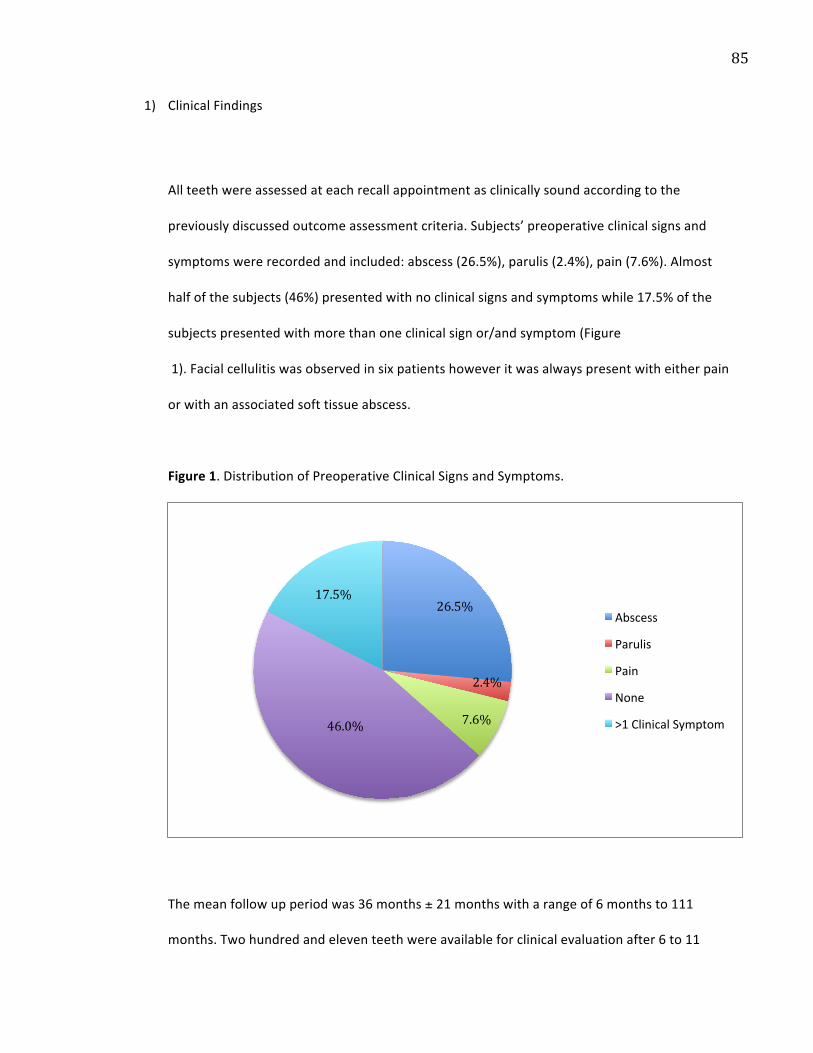
85
1) Clinical Findings
All teeth were assessed at each recall appointment as clinically sound according to the
previously discussed outcome assessment criteria. Subjects’ preoperative clinical signs and
symptoms were recorded and included: abscess (26.5%), parulis (2.4%), pain (7.6%). Almost
half of the subjects (46%) presented with no clinical signs and symptoms while 17.5% of the
subjects presented with more than one clinical sign or/and symptom (Figure
1). Facial cellulitis was observed in six patients however it was always present with either pain
or with an associated soft tissue abscess.
Figure 1. Distribution of Preoperative Clinical Signs and Symptoms.
The mean follow up period was 36 months ± 21 months with a range of 6 months to 111
months. Two hundred and eleven teeth were available for clinical evaluation after 6 to 11
26.5%
2.4%
7.6% 46.0%
17.5%
Abscess
Parulis
Pain
None
>1 Clinical Symptom
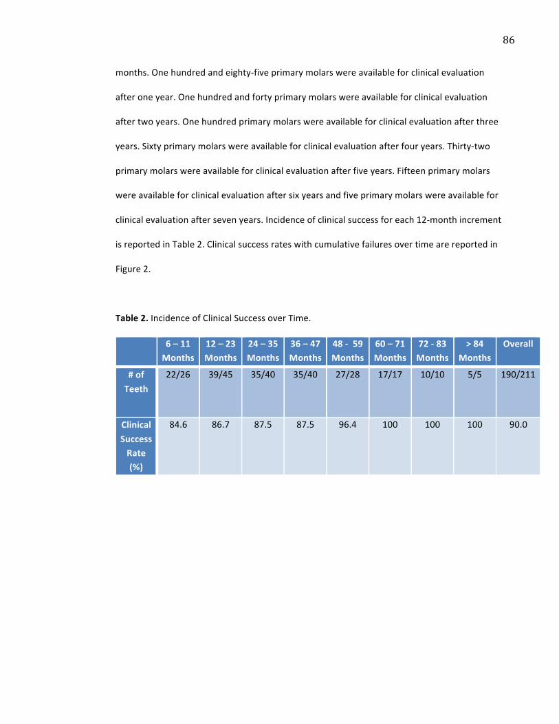
86
months. One hundred and eighty-‐five primary molars were available for clinical evaluation
after one year. One hundred and forty primary molars were available for clinical evaluation
after two years. One hundred primary molars were available for clinical evaluation after three
years. Sixty primary molars were available for clinical evaluation after four years. Thirty-‐two
primary molars were available for clinical evaluation after five years. Fifteen primary molars
were available for clinical evaluation after six years and five primary molars were available for
clinical evaluation after seven years. Incidence of clinical success for each 12-‐month increment
is reported in Table 2. Clinical success rates with cumulative failures over time are reported in
Figure 2.
Table 2. Incidence of Clinical Success over Time.
6 – 11 Months
12 – 23 Months
24 – 35 Months
36 – 47 Months
48 -‐ 59 Months
60 – 71 Months
72 -‐ 83 Months
> 84 Months
Overall
# of Teeth
22/26 39/45 35/40 35/40 27/28
17/17 10/10 5/5 190/211
Clinical Success Rate (%)
84.6 86.7 87.5 87.5 96.4 100 100 100 90.0
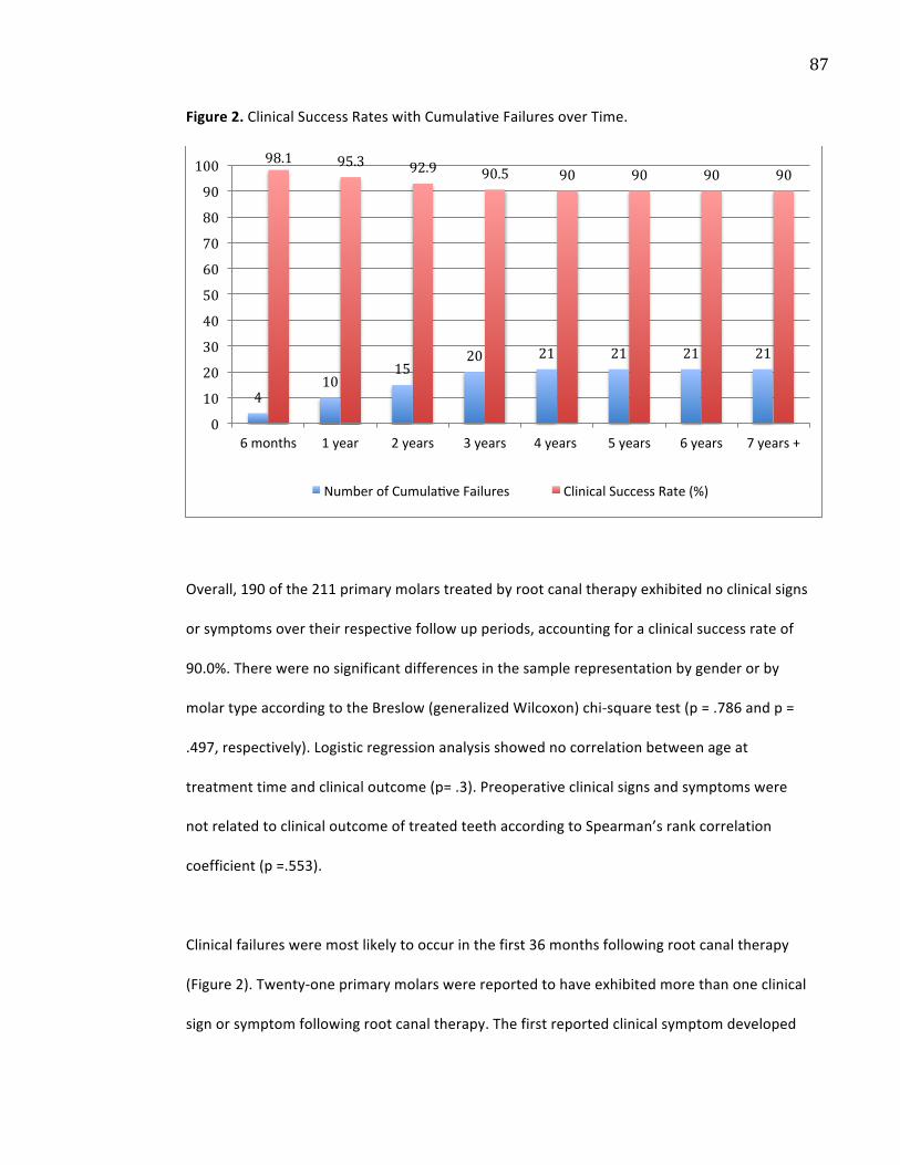
87
Figure 2. Clinical Success Rates with Cumulative Failures over Time.
Overall, 190 of the 211 primary molars treated by root canal therapy exhibited no clinical signs
or symptoms over their respective follow up periods, accounting for a clinical success rate of
90.0%. There were no significant differences in the sample representation by gender or by
molar type according to the Breslow (generalized Wilcoxon) chi-‐square test (p = .786 and p =
.497, respectively). Logistic regression analysis showed no correlation between age at
treatment time and clinical outcome (p= .3). Preoperative clinical signs and symptoms were
not related to clinical outcome of treated teeth according to Spearman’s rank correlation
coefficient (p =.553).
Clinical failures were most likely to occur in the first 36 months following root canal therapy
(Figure 2). Twenty-‐one primary molars were reported to have exhibited more than one clinical
sign or symptom following root canal therapy. The first reported clinical symptom developed
4 10
15 20 21 21 21 21
98.1 95.3 92.9 90.5 90 90 90 90
0
10
20
30
40
50
60
70
80
90
100
6 months 1 year 2 years 3 years 4 years 5 years 6 years 7 years +
Number of Cumulazve Failures Clinical Success Rate (%)

88
at one month postoperatively and the last reported clinical symptom developed at 49 months
postoperatively. The type of clinical signs and symptoms with associated time periods are
represented in Table 3. The clinical success data from all of the treated molars was used to
perform a Kaplan-‐Meier survival analysis (Table 4).
Table 3. Distribution of Types of Clinical Signs and Symptoms Over Time*.
6 to 11 Months
12 to 23 Months
24 to 35 Months
36 to 47 Months
48 to 59 Months
Total
Abscess 1 4 3 1 -‐ 9
Parulis 1 -‐ 1 -‐ -‐ 2
Mobility 2 -‐ 3 1 1 7
Pain 2 1 -‐ 1 1 5
Cellulitis -‐ 1 -‐ 1 -‐ 2
Total 6 6 7 4 2 25
* More than one clinical sign(s) and/or symptom(s) may have been reported in individual teeth.

89
Table 4. Estimated Kaplan-‐Meier Survival for Clinical Failures.
Time (Months)
Probability of Survival
Standard Error Time (Months)
Probability of Survival
Standard Error
6 .995 .005 54 .950 .016
12 .995 .005 60 .950 .016
18 .995 .005 66 .944 .017
24 .979 .010 72 .937 .018
30 .974 .012 78 .923 .021
36 .968 .013 84 .923 .021
42 .968 .013 90 .923 .021
48 .956 .015 96 .907 .023
The cumulative probability that any one tooth would be clinically successful at one year was
0.995. According to the Kaplan-‐Meier survival test the mean clinical survival time was 97
months with a standard error of 2.686. The associated survival analysis is plotted in Figure 3.
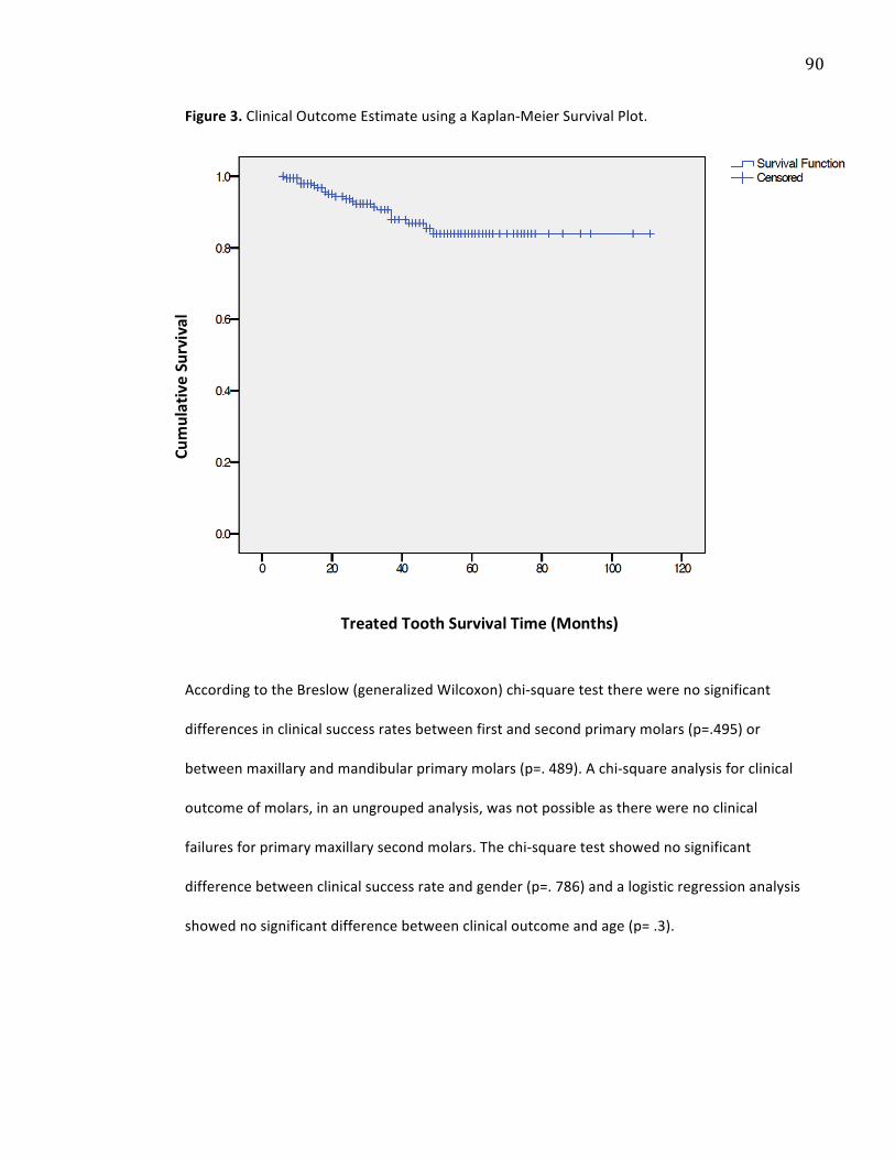
90
Figure 3. Clinical Outcome Estimate using a Kaplan-‐Meier Survival Plot.
According to the Breslow (generalized Wilcoxon) chi-‐square test there were no significant
differences in clinical success rates between first and second primary molars (p=.495) or
between maxillary and mandibular primary molars (p=. 489). A chi-‐square analysis for clinical
outcome of molars, in an ungrouped analysis, was not possible as there were no clinical
failures for primary maxillary second molars. The chi-‐square test showed no significant
difference between clinical success rate and gender (p=. 786) and a logistic regression analysis
showed no significant difference between clinical outcome and age (p= .3).
Cumulative Survival
Treated Tooth Survival Time (Months)

91
2) Radiographic Findings
All treated teeth were assessed radiographically both preoperatively and postoperatively
when films were available using the previously discussed outcome assessment criteria. Highly
significant inter-‐rater reliability was observed with a score of 0.81 (p<0.001). Likewise, intra-‐
rater reliability scored 0.84 (p<0.001). Two hundred and six teeth had available preoperative
radiographs and 176 treated teeth had appropriate postoperative radiographs.
As previously mentioned, the mean follow up period was 36 months ± 21 months with a range
of 6 months to 111 months. One hundred and seventy six teeth were available for
radiographic evaluation after 6 to 11 months. One hundred and fifty nine teeth were available
for radiographic evaluation after one year. One hundred and twenty eight primary molars
were available for radiographic evaluation after two years. Ninety-‐five primary molars were
available for radiographic evaluation after three years. Fifty-‐six primary molars were available
for radiographic evaluation after four years. Twenty-‐nine primary molars were available for
radiographic evaluation after five years. Fifteen primary molars were available for
radiographic evaluation after six years and five primary molars were available for radiographic
evaluation after seven years. In the event that the outcome of a treated tooth changed, its
most recent outcome was reported. All teeth that were considered clinical failures were also
found to be radiographic failures (Figure 4). Incidence of radiographic success for each 12-‐
month increment is reported in Table 5. Radiographic success rates with cumulative failures
over time are reported in Figure 5.
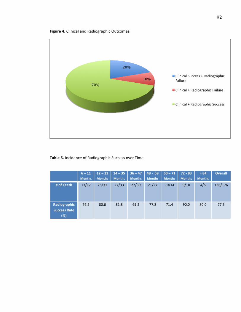
92
Figure 4. Clinical and Radiographic Outcomes.
Table 5. Incidence of Radiographic Success over Time.
20%
10% 70%
Clinical Success + Radiographic Failure
Clinical + Radiographic Failure
Clinical + Radiographic Success
6 – 11 Months
12 – 23 Months
24 – 35 Months
36 – 47 Months
48 -‐ 59 Months
60 – 71 Months
72 -‐ 83 Months
> 84 Months
Overall
# of Teeth 13/17 25/31 27/33 27/39 21/27
10/14
9/10 4/5 136/176
Radiographic Success Rate
(%)
76.5 80.6 81.8 69.2 77.8 71.4 90.0 80.0 77.3
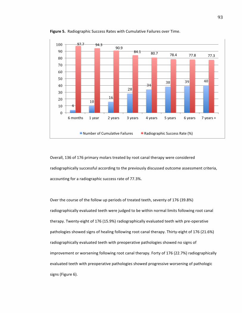
93
Figure 5. Radiographic Success Rates with Cumulative Failures over Time.
Overall, 136 of 176 primary molars treated by root canal therapy were considered
radiographically successful according to the previously discussed outcome assessment criteria,
accounting for a radiographic success rate of 77.3%.
Over the course of the follow up periods of treated teeth, seventy of 176 (39.8%)
radiographically evaluated teeth were judged to be within normal limits following root canal
therapy. Twenty-‐eight of 176 (15.9%) radiographically evaluated teeth with pre-‐operative
pathologies showed signs of healing following root canal therapy. Thirty-‐eight of 176 (21.6%)
radiographically evaluated teeth with preoperative pathologies showed no signs of
improvement or worsening following root canal therapy. Forty of 176 (22.7%) radiographically
evaluated teeth with preoperative pathologies showed progressive worsening of pathologic
signs (Figure 6).
4 10
16
28 34
38 39 40
97.7 94.3 90.9
84.1 80.7 78.4 77.8 77.3
0
10
20
30
40
50
60
70
80
90
100
6 months 1 year 2 years 3 years 4 years 5 years 6 years 7 years +
Number of Cumulazve Failures Radiographic Success Rate (%)
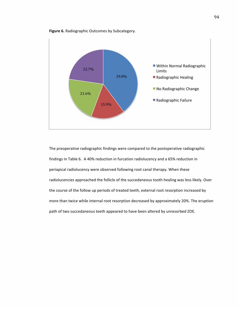
94
Figure 6. Radiographic Outcomes by Subcategory.
The preoperative radiographic findings were compared to the postoperative radiographic
findings in Table 6. A 40% reduction in furcation radiolucency and a 65% reduction in
periapical radiolucency were observed following root canal therapy. When these
radiolucencies approached the follicle of the succedaneous tooth healing was less likely. Over
the course of the follow up periods of treated teeth, external root resorption increased by
more than twice while internal root resorption decreased by approximately 20%. The eruption
path of two succedaneous teeth appeared to have been altered by unresorbed ZOE.
39.8%
15.9%
21.6%
22.7% Within Normal Radiographic Limits Radiographic Healing
No Radiographic Change
Radiographic Failure

95
Table 6. Preoperative and Postoperative Radiographic Healing*.
Preoperative (%) Postoperative (%)
Furcation Radiolucency 74.8 44.9
Periapical Radiolucency 38.8 13.6
Radiolucency near Follicle of Succedaneous Tooth
16.5 14.8
Widened PDL or Loss of Lamina Dura 11.2 6.8
External Root Resorption 4.4 9.7
Internal Root Resorption 5.8 4.5
Within Normal Limits 14 40
Altered Eruption Succedaneous Tooth n/a 1.1
* More than one type of radiographic finding may have been observed in individual teeth.
Of the 211 treated teeth in this study, 108 teeth were followed to the end of their survival
time, which was until a tooth was either extracted or exfoliated. The survival time of these
teeth was distinguished from those that were lost to follow up and recorded in Figure 7. Five
teeth or less required an extraction in each of the first three years of follow up. None of
treated teeth required an extraction after the fourth year of follow up. The third year of follow
up was the year with the highest rate of exfoliating treated teeth.

96
Figure 7. Survival of Treated Teeth Followed until Exfoliation or Extraction.
According to the Breslow (generalized Wilcoxon) chi-‐square test there were no significant
differences in radiographic success rates of the four primary molar types treated (p=.088). This
was also true when molars were grouped as maxillary versus mandibular primary molars
(p=.249). When molars were grouped as first versus second primary molars, a significant
difference was recorded (p=. 037). The chi-‐square test showed no significant difference
between radiographic success rate and gender (p=. 645) and a logistic regression analysis
showed no significant difference between radiographic outcome and age (p=. 764).
The radiographic outcome data from all of the treated molars was used to perform a Kaplan-‐
Meier survival analysis (Table 7). The cumulative probability that any one tooth would be
radiographically successful at one year was 0.826. The probability declined at a fairly steady
0
12 11
22
17
13
8
5 4 5 5 5
1 0 0 0 0
5
10
15
20
25
6 -‐ 11 months
1+ year 2+ years 3+ years 4+ years 5+ years 6+ years 7+ years
# Exfoliated Treated Teeth # Extracted Treated Teeth

97
rate with each subsequent year. According to the Kaplan-‐Meier survival test the mean
radiographic survival time was 82 months with a standard error of 6.413. The survival analysis
is plotted in Figure 8.
Table 7. Estimated Kaplan-‐Meier Survival for Radiographic Failures.
Time (Months)
Probability of Survival
Standard Error Time (Months)
Probability of Survival
Standard Error
6 .893 .045 54 .950 .016
12 .826 .056 60 .950 .016
18 .802 .059 66 .944 .017
24 .692 .072 72 .937 .018
30 .655 .077 78 .923 .021
36 .655 .077 84 .923 .021
42 .968 .013 90 .923 .021
48 .956 .015 96 .907 .023
Antibiotic use had a negative correlation with radiographic success according to Spearman’s
rank correlation coefficient (p < .05), which suggests that patients who presented with clinical
symptoms requiring antibiotic use were less likely to have a successful outcome by
radiographic standards. This correlation was not significant for clinical outcome.

98
Figure 8. Radiographic Outcome Estimate using a Kaplan-‐Meier Survival Plot.
3) Radiographic Assessment of Obturation Material
All treated teeth with radiographic follow up were assessed for degree of obturation in each
canal using the previously described assessment criteria. Twenty-‐eight of the treated teeth
were categorized as underfilled. One hundred and twenty treated teeth were categorized as
being adequately filled. Of these, two of the adequately filled teeth had intracanal ZOE that
resisted physiologic resorption. Forty-‐eight treated teeth were categorized as being overfilled.
Of these, ZOE resorbed in 30 treated teeth; while in 18 teeth, the ZOE did not resorb by the
Cumulative Survival
Treated Tooth Survival Time (Months)
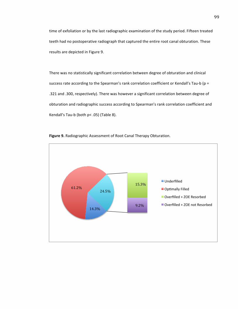
99
time of exfoliation or by the last radiographic examination of the study period. Fifteen treated
teeth had no postoperative radiograph that captured the entire root canal obturation. These
results are depicted in Figure 9.
There was no statistically significant correlation between degree of obturation and clinical
success rate according to the Spearman’s rank correlation coefficient or Kendall’s Tau-‐b (p =
.321 and .300, respectively). There was however a significant correlation between degree of
obturation and radiographic success according to Spearman’s rank correlation coefficient and
Kendall’s Tau-‐b (both p< .05) (Table 8).
Figure 9. Radiographic Assessment of Root Canal Therapy Obturation.
14.3%
61.2% 15.3%
9.2%
24.5%
Underfilled
Opzmally Filled
Overfilled + ZOE Resorbed
Overfilled + ZOE not Resorbed

100
Table 8. Clinical and Radiographic Success Rates Relative to Root Canal Therapy Obturation.
Overfill Adequate Fill Underfill
Clinical Success Rate (%) 85.4 91.2 91.8
Radiographic Success Rate (%) 61.7* 73.5* 79.4*
* p < .05
4) Condition of Succedaneous Teeth.
Enamel defects were observed on seven of the first premolars that succeeded treated primary
molars. No enamel defects were observed on second premolars. The defects ranged from
hypocalcified or hypoplastic enamel lesions to frank carious lesions (Table 9).
Table 9. Clinical Charting Descriptions of Affected Succedaneous Teeth.
Tooth # Notes from Clinical Charts Report of Caries or Restoration
19A “Hypoplastic” Restoration placed 6 months after eruption
90B “Enamel chipping off... hypoplastic” None 119 “Hypocalcified” None 122 “Pre-‐eruptive caries” Restoration placed at time of
eruption/first clinical exam 136A “Pre-‐eruptive caries” Restoration placed at time of
eruption/first clinical exam 140B “Hypoplasia” None 156 “Hypoplasia” Restoration placed 12 months after
eruption
Of the 211 teeth treated, only 108 teeth were observed until exfoliation or extraction and
their succedaneous teeth were only followed clinically in 103 cases. Therefore the calculated
enamel defect rate was 7/103 or 6.8%.

101
When age at time of treatment was considered for these 103 subjects, those who developed
enamel defects in their succedaneous teeth had a mean age of 54.1 months, while those that
did not develop enamel defects had a mean age of 77.2 months. According to the
independent student’s T-‐test, this difference was significant (p ≤ .001, equal variances
assumed) and suggests that enamel defects were more likely to occur in younger subjects
requiring root canal therapy (Table 10).
Table 10. Enamel Defects in Succedaneous Teeth versus Age at Time of Root Canal Therapy. Number of Teeth Affected Mean Age at Treatment
(months) Permanent Teeth with Enamel Defects
7 54.1
Permanent Teeth without Enamel Defects
96 77.2*
• = p ≤ .001
A chi-‐square analysis revealed that there was no systematic relationship between radiographic
success rate and the presence of enamel defects on succedaneous teeth (Cramer's V = .096, p
= .206). Degree of ZOE obturation (including ZOE extrusion) was also unrelated to enamel
defects on succedaneous teeth (Cramer’s V = .118, p = .120).
5) Exfoliation of Treated Teeth.
In order to study the effect that root canal therapy had on the timing of exfoliation, treated
teeth were compared to unrestored, contralateral teeth. According to the paired Student’s T-‐
test, this revealed no significant difference between the survival times of treated teeth
compared to unrestored teeth (p = .228). The sample size however, was only 16 teeth. In
order to increase the sample size, treated teeth were then compared to contralateral teeth

102
that were either unrestored or restored (without pulp therapy). According to the paired
Student’s T-‐test, this analysis showed a significantly longer survival time for contralateral teeth
relative to root canal therapy treated teeth (p< .001). The results of these two analyses are
shown in Table 11.
Table 11. Survival Time of Root Canal Therapy Treated Teeth vs. Contralateral Teeth.
N = 16 Mean Survival (months) N = 79 Mean Survival (months)
Treated Teeth
29.8 Treated Teeth 38.6
Unrestored Contralateral Teeth
36.9
Unrestored or Restored Contralateral Teeth
46.2*
* p < .001
When this same sample of 79 teeth was divided according to clinical outcome and assessed for
differences in survival time it was found that only teeth deemed as clinical failures had a
significantly shorter survival time (p = .049, paired T-‐test, equal variances assumed). Treated
teeth that were considered clinically successful had a mean survival time of 40.5 months that
was similar to the 44.4-‐month survival time of the contralateral tooth group (p= .777, equal
variances assumed). This comparison differed markedly for treated teeth that were
considered clinical failures since their mean survival time was 23.4 months compared to the
contralateral tooth group with a mean survival time of 60.2 months (Table 12). The same
analysis was performed for radiographic outcome but no significant differences in survival
time were found (Table 13).

103
Table 12. Survival Time vs. Clinical Outcome in Treated vs. Contralateral Teeth.
Sample Size
Survival Time (months)
Significance
Treated Tooth – Success 70 40.5 p = 0.777
Contralateral Unrestored or Restored – Success
70 44.4
Treated Tooth – Failure 9 23.4 p = 0.049 Contralateral Unrestored or Restored -‐ Failure
9 60.2
Table 13. Survival Time vs. Radiographic Outcome in Treated vs. Contralateral Teeth.
Sample Size
Survival Time (months)
Significance
Treated Tooth – Success 49 44.2 p = 0.460
Contralateral Unrestored or Restored – Success
49 48.6
Treated Tooth – Failure 17 36.2 p = 0.570 Contralateral Unrestored or Restored -‐ Failure
17 56.4

104
G. DISCUSSION
Preservation of an intact primary dentition until the eruption of the permanent successors is
very important in maintaining the arch form. In the event of a pulpal insult to a primary tooth,
resolution of the pathologic process through pulp therapy can help to preserve arch space and
restore function. In addition to preserving arch form, utilization of pulp therapy to maintain
the integrity of the primary dentition may also prevent aberrant tongue habits, prevent
speech problems, maintain normal masticatory function and preserve esthetics (Goerig &
Camp 1983, Barr et al. 1991).
Despite the inarguable benefits of preserving an intact primary dentition, root canal therapy in
primary teeth was reportedly one of the approaches to pulp therapy with the least amount of
consensus amongst educators in a 1997 survey by Primosch et al. (1997). Comparing the
results of this 1997 survey to a similar 2005 survey, primary tooth root canal therapy was
taught and used less by 2005 directors of predoctoral pediatric dental school programs and
diplomates of the American Academy of Pediatric Dentistry (AAPD). This difference was not
however significantly different from the 1997 results (94% versus 85%) (Primosch et al. 1997,
Dunston & Coll 2008).
Significantly more respondents in the 2005 survey reported using other non-‐ZOE filler pastes
(i.e. calcium hydroxide and iodoform) than did the 1997 directors and diplomates (6% versus
33%). However ZOE, with or without the use of formocresol, remained the material of choice
for obturation of root canals in primary teeth in 2005 by directors of pediatric dentistry

105
programs and diplomates of the American Academy of Pediatric Dentistry (66% and 68%,
respectively) (Primosch et al. 1997, Dunston & Coll 2008).
In this study a retrospective chart review was used to evaluate the clinical and radiographic
outcomes of a formocresol and ZOE root canal therapy technique in 211 non-‐vital primary
molars. In addition this study evaluated the clinical impact of variable obturation lengths for
endodontically-‐treated primary molars, the rate of enamel defects of the succedaneous teeth
following root canal therapy and the effect of root canal therapy on the timing of exfoliation
of treated teeth. All aims and objectives of this study were met.
The root canal therapy technique in this investigation was carried out on all teeth by a single
operator (PA). A study design with a single operator offers the advantage of consistent and
reproducible technique. However a potential disadvantage of this study design would be that
the outcome is due in part to a superior operator rather than a superior technique.
Other potential limitations of this study are related to its retrospective nature. All data relied
upon the accuracy of the clinical records and the radiographs that were available in charts. A
control group could not be added to the study sample and follow up periods were pre-‐
determined. Patients did not always have routine follow up. Ideally patients would have had
routine clinical and radiographic follow up of their treated teeth and their contralateral until
exfoliation, as well as a consistent clinical assessment of their succedaneous teeth. As for
other comparable studies, a minimum follow up period of six months was required to be
included in the study. However any treated tooth that required an extraction prior to six
months follow up would not have been identified (Coll et al. 1985, Coll & Sadrian 1996, Fuks et
106
al. 2002). Although it would not have been appropriate to perform statistical analysis on teeth
that had less than 6 months follow up, it would have been interesting to note how many
treated teeth failed within the first 6 months.
Furthermore, chart and radiographic analysis relied solely upon the principal investigator (KS).
In order to standardize the principal investigator’s radiographic analysis, a standardization
exercise was completed. Highly significant inter-‐rater reliability was observed with a score of
0.81 (p< .001). Likewise, intra-‐rater reliability scored 0.84 (p< .001). Despite these high kappa
scores, the standard in this field of research is to have more than one investigator evaluating
clinical and radiographic data in order to reduce potential for bias (Barr et al. 1991, Yacobi et
al. 1991, Coll & Sadian 1996, Bawazir & Salama 2006).
The present retrospective study design has the advantage of allowing a larger sample size and
thus detecting smaller differences in outcome. It was previously determined that a sample size
of 91 teeth was required to achieve 80% power and a 5% statistically significant level. The final
sample size of this study was 211 primary molars from 161 healthy subjects (Appendix II).
1) Clinical Outcomes
In the present investigation treated teeth were considered clinically successful according to
the following criteria:
i) Absence of reported pain (spontaneous or stimulated)
ii) Absence of soft tissue abscess

107
iii) Absence of parulis
iv) Absence of facial cellulitis
v) Absence of pathologic tooth mobility
vi) Absence of extraction of treated tooth
Preoperative signs and symptoms were reported in 54% of patients according to clinical
records. Postoperatively, only 10% of patients exhibited signs and symptoms. Therefore the
overall clinical success rate was 90.0% based on the 21 clinical failures of 211 treated teeth
followed over a mean period of 36 months (range 6 to 111 months). Clinical failures occurred
within the first four years following root canal therapy (Figure 2 and Table 2). In the event of a
clinical failure, the signs and symptoms most commonly reported included abscess, pathologic
tooth mobility and pain (Table 3). This clinical success outcome is among the higher outcomes
for root canal therapy studies, which range from 65% to 95.5% (Gould 1972, Yacobi et al.
1991, Holan & Fuks 1993, Payne et al. 1993, Coll & Sadrian 1996, Nadkarni & Damie 2000, Fuks
et al. 2002, Mortazavi & Mesbahi 2004, Bawazir & Salama 2006).
To compare the present clinical success rate of 90% directly to other studies is difficult, and
even inappropriate, given the variability in study designs (sample size, inclusion criteria, follow
up period, definitions of successful outcome, choice of restoration following root canal
therapy) and in the variability of the reported findings.
Clinical outcomes for specifically formocresol and ZOE root canal therapy studies range from
74.5% to 95.5% (Table 14) (Coll & Sadrian 1996, Mortazavi & Mesbahi 2004, Bawazir & Salama
2006). The formocresol and ZOE root canal therapy study by Barr et al. (1991) did not report

108
specific clinical outcomes. Coll and Sadrian (1996), who presented their results for ZOE and
formocresol root canal therapy without differentiating between clinical and radiographic
failures, reported an overall success rate of 74.5% (38/51) over a mean follow-‐up period of 90
months.
Bawazir and Salama (2006) reported a 93.6% overall clinical success rate of formocresol and
ZOE root canal therapy after a 6 month follow up period of 47 treated primary molars. More
specifically a clinical success rate of 95.5% (21/22) was observed in teeth that were obturated
with a slow-‐speed handpiece-‐mounted lentulo spiral while the clinical success rate with a
hand-‐held lentulo spiral was 92.0% (23/25). These results should be interpreted cautiously due
to the very short 6 month follow up period observed in the study.
The clinical inclusion criteria were usually broader and the definitions of clinical success were
equivalent in the present investigation to published studies for root canal therapy (Gould
1972, Yacobi et al. 1991, Holan & Fuks 1993, Payne et al. 1993, Coll & Sadrian 1996, Nadkarni
& Damie 2000, Fuks et al. 2002, Mortazavi & Mesbahi 2004, Bawazir & Salama 2006). As a
result, the clinical outcome of 90% for formocresol and ZOE root canal therapy is a highly
acceptable outcome.
2) Radiographic Outcomes
In the present investigation treated teeth were considered radiographically successful
according to the following criteria:

109
i) Absence of new furcation or periapical radiolucency
ii) Absence of pathologic external root resorption (either new or worsening of
existing)
iii) Absence of pathologic internal root resorption (either new or worsening of
existing)
iv) Improvement or stasis of a pathologic radiolucency when present
preoperatively
v) Succedaneous tooth and follicle were not affected by the treated primary
molar
vi) Root filling did not alter the path of eruption of the succedaneous tooth
An overall radiographic success rate of 77.3% was calculated based on the 40 radiographic
failures of 176 treated teeth over a mean follow up period of 36 months (range 6 to 111
months). Radiographic failures occurred most often in the third and fifth years after treatment
(Figure 5 and Table 5). Just under 40% of treated teeth had normal radiographic findings
postoperatively. Another 15.9% of teeth showed healing or improvement in their radiographic
pathologies relative to their preoperative radiographic conditions while 21.6% of treated teeth
had static radiographic pathologic findings. Finally 22.7% of treated teeth showed worsening
of preoperative radiographic findings or development of new radiographic pathologies over
their respective follow up periods (Figure 6). There was no significant difference in follow up
time in any of the above-‐listed groups therefore bias in follow up times was not a factor.

110
The radiographic success rate of this investigation is near the average for comparable
formocresol and ZOE root canal therapy studies, which range from 72% to 90.9% (Barr et al.
1991, Coll & Sadrian 1996, Mortazavi & Mesbahi 2004, Bawazir & Salama 2006). Table 14
summarizes the main differences between the relevant formocresol and ZOE root canal
therapy studies. Of particular note is the fact all of the listed studies restored endodontically-‐
treated primary molars with stainless steel crowns, with the exception of Mortazavi and
Mesbahi (2004), who restored all endodontically-‐treated primary molars with amalgam
restorations. Coll and Sadrian (1996) did not clearly state how their treated teeth were
restored. The figures in their studies implied that treated molars were restored with stainless
steel crowns, however this information was disclosed in their 1996 publication or their
previous publications of the same technique (Coll et al. 1985, Sadrian & Coll 1993).
In terms of sample size, the current investigation has the largest reported for formocresol and
ZOE root canal therapy – three to seven times the sample size of previous reports.
Follow up time is a critical assessment factor in the determination of a successful root canal
therapy treatment. Figures 2 and 5 clearly demonstrate the gradual decline in success rate
observed in the current study.
Coll and Sadrian (1996) reported the longest follow up time for formocresol and ZOE root
canal therapy with an overall 7.5 year (90 months) follow up period. The mean reported
patient age at treatment time for this study was 52 months (4 years), with a range of 19 to 111
months. Technically a 7.5 year follow up period would be possible since most primary molars
exfoliate between age 10 and 12. However the incongruity lies in the fact that the study also

111
included 30 primary incisors. With 36% (30/84) of the overall sample exfoliating between age
6 and 8, it would seem implausible that treated teeth could be followed up for an average of
7.5 years.
Table 14. Summary of Formocresol (FC) and ZOE Root Canal Therapy Studies.
Sample Size Follow Up Time (months)
Technique
Clinical Success Rate
Radiographic Success Rate
Barr et al. 1991
62 primary molars
Range = 12 to 74 Mean = 40
FC mixed with ZOE
Not reported
82.3%
Coll & Sadrian 1996
54 primary molars
Range = 20 to 177 Mean = 90
FC paper points then
ZOE
Overall 74.5%
Overall 74.5%
Mortazavi & Mesbahi 2004
29 primary molars
Range = 10 to 16 No mean given
FC pledget sealed in chamber for 1-‐2
weeks then ZOE
78.9% 75.0%
Bawazir & Salama 2006
47 primary molars
6 FC paper points then
ZOE
Overall 93.6% -‐ handpiece + lentulo = 95.5% -‐ handheld lentulo = 92%
Overall 80.9% -‐ handpiece + lentulo = 90.9% -‐ handheld lentulo = 72%
Stallaert et al. 2011
211 primary molars (clinical) 176 primary molars (radiographic)
Range = 6 to 111 Mean = 36
FC mixed with ZOE
90.0% 77.3%
112
Bawazir and Salama (2006) reported an overall 80.9% radiographic success rate of formocresol
and ZOE root canal therapy after a 6 month follow up period. More specifically a radiographic
success rate of 90.9% was observed in teeth that were obturated with a slow-‐speed
handpiece-‐mounted lentulo spiral while the radiographic success rate with a hand-‐held lentulo
spiral was only 72.0%. Had the present investigation only considered radiographic failures that
occurred in its initial 6 to 11 month follow up period the reported radiographic success rate
would have been 97.7% (172/176) (Figure 5). This was far from the study’s final reported
radiographic success rate with over 7 years of follow up, demonstrating how inaccurate a
short follow up period can be.
In the present investigation, preoperative and postoperative radiographic findings were
compared for two reasons. The first reason for this analysis was to emphasize the wide
inclusion criteria of this study. The high rate of preoperative radiographic findings in the
present investigation was indicative of the degree of pathoses present in both the pulpal
tissues of treated teeth and their surrounding supporting structures. Variable inclusion criteria
in primary tooth root canal therapy studies are a significant outcome bias, which are often
overlooked in the interpretation of reported outcomes.
The second reason to analyse preoperative and postoperative radiographic findings was to
determine the rate of healing in the surrounding structures of treated teeth following root
canal therapy treatment. It is well established that radiographic healing in permanent teeth
with apical periodontitis is a slow process (Ørstavik 1996, Dugas et al. 2002, Molven et al.
2002, Fristad et al. 2004, Metzger et al. 2009). In fact, less than half of permanent teeth with
apical periodontitis are healed one year after root canal therapy; approximately 90% are
113
healed after four years and approximately 95% are healed after 6 years. The remaining
approximate 5% require an even longer period to heal completely (Ørstavik 1996, Dugas et al.
2002, Molven et al. 2002, Fristad et al. 2004, Metzger et al. 2009).
There are no studies that extensively assess the rate of healing following root canal therapy in
primary teeth. However one may postulate that endodontic healing should occur over time in
primary teeth as it does in permanent teeth. It is for this reason that in the present
investigation, stasis of preoperative radiographic pathologies were considered to be successful
treatments. Other studies, also with wide inclusion criteria, defined improvement or stasis of
preoperative radiographic findings as a successful outcome (Barr et al. 1991, Fuks et al. 2002,
Prabhakar et al. 2008, Moskovitz et al. 2010, Nakornchai et al. 2010).
In the present investigation, 74.8% of the teeth had preoperative furcation radiolucencies and
38.8% had preoperative periapical radiolucencies. Postoperatively furcation radiolucencies
were observed in 44.9% of treated teeth and periapical radiolucencies were observed in only
13.6% of treated teeth. This was a 29.9% reduction in furcation radiolucencies and a 25.2%
reduction in periapical radiolucencies (Table 6).
Preoperative furcation and/or periapical radiolucencies were observed to extend near the
follicle of the succedaneous tooth in 16.5% of treated teeth however only 1.7% of these
lesions improved or resolved. When these radiolucencies approached the follicle of the
succedaneous tooth healing was much less likely. External root resorption increased by 5.3%
following root canal therapy, however this maybe due to the fact that it was difficult to
differentiate from physiologic root resorption.
114
The greatest number of radiographic failures tended to occur within the first year and then
again between three and six years following treatment. Patients who were prescribed
antibiotics were more likely to have negative radiographic outcomes (p< .05). As a result, in
the event that a child presented with a fever and a non-‐draining abscess and/or cellulitis,
careful radiographic diagnosis should be used in determining if the associated tooth is suitable
for root canal therapy treatment. Long term clinical and radiographic follow up of these teeth
is advisable if root canal therapy treatment is performed.
3) Effects of Obturation on Root Canal Therapy Outcome.
Degree of canal obturation was statistically unrelated to clinical outcome in this investigation.
However there was a trend towards higher clinical success rates for treated teeth that were
underfilled or adequately filled (91.5%) relative to teeth, which had been overfilled (85.4%).
Degree of canal obturation was statistically correlated to radiographic outcome (Table 8).
These results are consistent with both Coll and Sadrian (1996) and Fuks et al. (2002), who
reported a success rate of 57 to 58% for teeth with overfilled canals and an 83 to 87% success
rate for teeth with adequately filled or underfilled canals. It is possible that the degree of
pathosis may have contributed to overfilling of canals and the decreased success rates.
In the present investigation, 9% of treated teeth exhibited obturation material that was
expressed beyond the apex of at least one canal and did not resorb by the time of exfoliation
or by the last radiographic examination of the study period. The only observed consequence
115
of this was the deflection of the eruption path of two succedaneous teeth. Ectopic eruption of
succedaneous premolars following root canal therapy of primary molars was observed in
21.6% of cases in a Coll and Sadrian (1996) study (n= 51). The authors themselves reported
that this rate was unusually high and postulated that retained ZOE was the reason for the high
incidence of ectopic eruptions. Barr et al. (1991) reported that they had never observed
retained ZOE after the exfoliation of a primary molar treated by formocresol and ZOE root
canal therapy, not only in their study but also in 15 years of practice.
Retention of obturation material particles was not statistically related to clinical or
radiographic success of root canal therapy (p = .361 and .130, respectively, which coincided
with the Coll and Sadrian (1996) findings (p = .11). It is possible that the degree of pathosis of
a treated tooth may have contributed to the overfill of obturation material and the decreased
success rate.
4) Effect on Enamel of Succedaneous Teeth.
One hundred and three succedaneous teeth of endodontically-‐treated primary molars were
assessed clinically. Of these, seven premolars had enamel defects ranging from mild to severe
enamel hypocalcification or enamel hypoplasia. As a result some of these newly erupted teeth
were more susceptible to caries. Age at time of root canal therapy treatment proved to be a
significant factor in determining which patients might develop enamel defects in their
succedaneous teeth (p ≤ .001). A survey of the literature indicates there are no previous
studies that evaluate the effect of age at the time of irreversible pulpitis and root canal

116
therapy treatment in primary molars and the incidence of enamel defects in succedaneous
teeth.
The overall mean age at time of treatment for primary molar root canal therapy in this
investigation was 72 months. However age at time of treatment for the 103 aforementioned
subjects was 72.9 months (6 years of age) for those that did not develop enamel defects in
their succedaneous teeth and 54.1 months (4 years of age) for those that did develop enamel
defects in their succedaneous teeth (p ≤ .001). All enamel defects were observed in first
premolars while no enamel defects were observed in second premolars.
A possible explanation for these observations may be related to the age at which the enamel
of premolars calcifies. First premolars calcify between 18 months and 6 years of age while
second premolars calcify between 24 months and 7 years of age (AAPD 2011). Therefore a
younger child would be more vulnerable to errors in enamel calcification. Likewise a younger
child would be more likely to have an error in enamel calcification in a first premolar than a
second premolar since enamel calcification is completed earlier in first premolars.
Other contributing factors could be that first primary molars erupt, on average one year
earlier than second primary molars (AAPD 2011) and that 69% of primary molars included in
this investigation were first primary molars. Yet another possible reason that only first and not
second premolars were found to have enamel hypoplasia may be due to the anatomical
differences between first and second primary molars (Zurcher 1925):
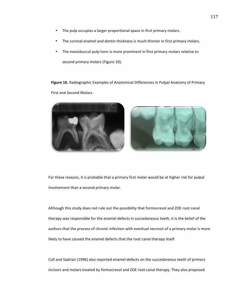
117
• The pulp occupies a larger proportional space in first primary molars.
• The coronal enamel and dentin thickness is much thinner in first primary molars.
• The mesiobuccal pulp horn is more prominent in first primary molars relative to
second primary molars (Figure 10).
Figure 10. Radiographic Examples of Anatomical Differences in Pulpal Anatomy of Primary
First and Second Molars.
For these reasons, it is probable that a primary first molar would be at higher risk for pulpal
involvement than a second primary molar.
Although this study does not rule out the possibility that formocresol and ZOE root canal
therapy was responsible for the enamel defects in succedaneous teeth, it is the belief of the
authors that the process of chronic infection with eventual necrosis of a primary molar is more
likely to have caused the enamel defects that the root canal therapy itself.
Coll and Sadrian (1996) also reported enamel defects on the succedaneous teeth of primary
incisors and molars treated by formocresol and ZOE root canal therapy. They also proposed
118
that enamel defects were the result of infection prior to root canal therapy rather than the
endodontic procedure itself. They also proposed that excessive (>1mm) preoperative root
resorption could be indicative of pre-‐existing infection and that primary teeth with greater
than 1mm of root resorption were consequently at increased risk of enamel defects on the
succedaneous teeth. The authors did not describe how they assessed minute degrees of root
resorption nor did they describe how they differentiated between pathologic root resorption
and physiologic root resorption.
Degree of ZOE obturation was unrelated to the occurrence of enamel defects in this
investigation, which coincided with the results from Coll and Sadrian (1996).
Given the evidence presented thus far, one may propose that a patient’s age should be
considered in planning the treatment of a hyperemic or necrotic primary molar. Since the
enamel of first premolars develops until the age of five years and the enamel of second
premolars develops until the age of six years, these along with clinical signs and symptoms and
radiographic findings could be important in predicting the outcome of root canal therapy
treatment.
5) Effect of Root Canal Therapy on Exfoliation Times.
The exfoliation times of teeth treated by root canal therapy were compared to the exfoliation
times of contralateral teeth that were unrestored or restored without pulp therapy. On
average, treated teeth exfoliated 7.6 months earlier than their contralateral teeth, which was
statistically significant (p < .001) but not clinically significant. However, when this same group

119
of paired 79 teeth was compared for clinical outcome, it was found that treated teeth
considered clinical failures were significantly more likely to exfoliate earlier (p < .05), on
average 36.8 months earlier (n= 9). If treated teeth were rated as clinical successes then no
significant difference was found in the timing of their exfoliation (p = .777).
Coll et al. (1985) also found that teeth treated by formocresol and ZOE root canal therapy
were significantly more likely to exfoliate earlier than their contralateral teeth (p= .01).
Likewise treated teeth that were rated failures were also more likely to exfoliate sooner than
their respective contralateral teeth, but similar if rated successes. One difference in the
comparison by Coll et al. (1985) was the inclusion of all contralateral teeth, including teeth
that were extracted, pulpotomized or untreated by pulp therapy. As a result their analysis of
contralateral teeth may have been biased by the presence of inflammatory mediators
produced by both carious processes and the consequent dental therapy.
Barr et al. (1991) reported that 56% of teeth treated by ZOE and formocresol root canal
therapy exhibited advanced exfoliation relative to their contralateral teeth. The authors did
not describe the condition of the contralateral teeth, which again, is a potential bias and
reduced the validity of their statement. In addition they did not characterize the range of
reported advanced exfoliation times or mention if it was statistically relevant.
120
SUMMARY
In summary, the results from the present investigation suggest that the described formocresol
and ZOE root canal therapy technique described herein is a viable alternative to extraction and
space maintenance for primary molars.
Preoperative risk factors for radiographic outcome include: radiolucencies that approach the
follicle of the succedaneous teeth, particularly if the enamel of the succedaneous tooth is not
completely developed, and patients who would require antibiotic therapy due to the presence
of a fever and a non-‐draining abscess and/or facial cellulitis. Postoperatively, periodic
radiographic evaluation following root canal therapy is critical to ascertain that inflammatory
processes are healing and not posing risk to the follicles of the succedaneous teeth.
CONCLUSIONS
1. Root canal therapy in primary molars using one drop of full strength Buckley’s
formocresol, one drop of eugenol and pure zinc oxide powder as the root filler
demonstrated a high clinical success rate (90.0%) and a moderate radiographic
success rate (77.3%).
2. Length of ZOE obturation and presence of ZOE expressed beyond the apex of a canal
had no effect on clinical success rate. However a significant correlation between
degree of obturation and radiographic outcome was observed with higher
radiographic success rates observed for teeth that were underfilled or adequately
filled (p < .05).

121
3. Enamel defects in first premolars were rarely observed (6.8%) following root canal
therapy in antecedent primary first molars but when present, they were more likely to
occur in a younger child.
4. Root canal therapy treatment may shorten the lifespan of primary tooth relative to a
contralateral tooth, particularly if the tooth has a negative clinical outcome.

122
APPENDIX I: INFORMED CONSENT

123

124

125
126
APPENDIX II: LETTER TO PARENTS

127
APPENDIX III: SAMPLE SIZE CALCULATION
1. The following is a sample size calculation for a study with one proportion known (chi-‐
square test).
2. The research question is “Does the described ZOE and formocresol root canal therapy
technique perform better than the existing one?”
3. The most recent and often cited root canal therapy study using formocresol and ZOE
reports a success rate of 74.5% (Coll et al. 1996, Nurko & Garcia-‐Godoy 1999, Nadkarni et
al. 2000, Primosch et al. 2005, Prabhakar et al. 2008, Sari & Okte 2008, Nakornchai et al.
2010, AAPD 2010).
4. It was estimated that the proposed technique from this study would generate an 85%
success rate.
5. What sample size would be required in order to detect the above-‐stated differences with
80% power and a 5% level of statistical significance?
N =(Z1 ! known (1!! known ) + Z2 ! 2 (1!! 2 ))
2
" 2, where:
Z1 is a Z critical value, based upon α = .05, one-‐tailed.
πknown refers to the proportion of success using the traditional method (74.5%)
Z2 is a Z critical value, based upon 1 – β (or, power), which is set at .80, one-‐tailed.
π2 refers to the expected proportion of successes, using the new method (80%)
δ refers to the difference between the two proportions (.745 -‐ .80)

128
N =(Z.95 .745(1!.745) + Z.80 .85(1!.85))2
(.745!.85)2
=(1.64 .189975 +.815 .1275)2
(0.011025)
=[1.64(0.436)+.815(.357)]2
0.011025= (.715+.291)2 / 0.011025=1.006 / 0.011025= 91.25
The pre-‐determined sample size calculation to obtain an 80% power at a 5% level of statistical
significance in 91 teeth.
Adapted from: Sample Size Tables for Clinical Studies (2nd Edition, 1997) by D. Machin, M. Campbell, P. Fayers & A. Pinol – published by Blackwell Science Ltd.

129
APPENDIX IV: INTER-‐ AND INTRA-‐RATER RELIABILITY
Kappa Test KS 1 KS 2 KS 1 -‐-‐-‐ 0.84 KS 2 0.84 -‐-‐-‐ PA/HN 0.84 0.79
130
LITERATURE CITED
Allen KR. Endodontic treatment of primary teeth. Aust Dent J 24(5):347-‐351. American Academy of Pediatric Dentistry (AAPD): The Handbook of Pediatric Dentistry, 3rd edition, 25, 2007. American Academy of Pediatric Dentistry (AAPD): Guideline on pulp therapy for primary and young permanent teeth – Adopted 1991 Revised 1998, 2001, 2004, 2009, 2010 Pediatric Dentistry http://www.aapd.org/media/Policies_Guidelines/G_Pulp.pdf, accessed December 12, 2010. American Academy of Pediatric Dentistry (AAPD): Dental Growth and Development. http://www.aapd.org/members/referencemanual/default.asp, accessed April 1, 2011. Acquilino SA and Caplan DJ. Relationship between crown placement and the survival of endodontically treated teeth. J Prosthet Dent 87(3):256-‐263, 2002. Aylard S and Johnson R. Assessment of filling techniques for primary teeth. Pediatr Dent 9:195-‐198, 1987. Barker BC et al. The demonstration of root canal anatomy. Aust Dent J 14(1):37-‐41, 1969. Barker BC and Lockett BC. Endodontic experiments with resorbable paste. Aust Dent J 16(6):364-‐72, 1971. Barker BC et al. Anatomy of root canals. IV. Deciduous Teeth. Aust Dent J 20(2):101-‐106, 1975. Barr ES et al. A retrospective radiographic evaluation of primary molar pulpectomies. Pediatr Dent 13(1):4-‐9, 1991. Barr ES et al. The authors respond. Pediatr Dent 13:249, 1991. Bawazir OA and Salama S. Clinical evaluation of root canal obturation methods in primary teeth. Pediatr Dent 28(1) 39-‐47, 2006. Bauer WH. Apical periodontitis of deciduous teeth. Ztscr. F. Stomatol. 30:721, 1932. (Quoted in Bauer WH. Effect of periapical processes of deciduous teeth on the buds of permanent teeth. Am J Orthod 32:232-‐241, 1946.) Bauer WH. Effect of periapical processes of deciduous teeth on the buds of permanent teeth. Am J Orthod 32:232-‐241, 1946. Bevelander G and Benzer S. Morphology and incidence of secondary dentin in human teeth. JADA 30:1079, 1081-‐1082, 1943. Bhatt HS et al. Effect of glutathione depletion on aminopyrine and formaldehyde metabolism. Biochem Pharmacol 37:1581-‐1589, 1988.
131
Binder RE. Correction of posterior crossbites: diagnosis and treatment. Pediatr Dent 26:266-‐272, 2004. Brilliant JD et al. Further studies on the quantitation of root canal medicaments. J Br Endod Soc 7(1)29-‐36, 1974. Buckley JP. A rational treatment for putrescent pulps. Dent Rev 18:1193-‐1197, 1904. Buckley JP. The rational treatment of putrescent pulps and their sequelae. Dent Cosmos 48:537-‐543, 1905.
Campbell AD et al. Cell-‐mediated response to endodontic cements: Research report. J Endod 4:147-‐150, 1978.
Casanova-‐Schmitz M et al. Differentiation between metabolic incorporation and covalent binding in the labeling of macromolecules in the rat nasal mucosa and bone marrow for inhaled 14C-‐ and 3H-‐formaldehyde. Toxicol Appl Pharmacol 76:26-‐44, 1984. Casas MJ et al. Do we still need formocresol in pediatric dentistry? JCDA 71(10): 749-‐750, 2005. Castagnola L and Orlay H. Treatment of gangrene of the pulp by the Walkhoff method. Br Dent J 93:93-‐102, 1952. Chopra I. The increasing use of silver-‐based products as antimicrobial agents: a useful development or a cause for concern? J Antimicrob Chemother 59(4):587-‐590, 2007. Cohen MM et al. Bacteriologic Study of Infected Deciduous Molars. Oral Surg Oral Med Oral Path 13:1382-‐1386, 1960. Coll JA et al. Evaluation of a one-‐appointment formocresol root canal therapy technique for primary molars. Pediatr Dent 7(2)123-‐129, 1985. Coll JA et al. An evaluation of pulpal therapy in primary incisors. Pediatr Dent 10(3):178-‐184, 1988. Coll JA and Sadrian R. Predicting pulpectomy success and its relationship to exfoliation and succedaneous dentition. Pediatr Dent 18(1)57-‐63, 1996. Cox ST Jr. et al. The bactericidal potential of various endodontic materials for primary teeth. Oral Surg Oral Path Oral Med 45(6):947-‐54, 1978. Dandashi et al. An in vitro comparison of three endodontic techniques for primary incisors. Pediatr Dent 15:254-‐257, 1993. daSilva Andrade A et al. Posterior Crossbite and Functional Changes – a systematic review. Angle Orthod 70(2):380-‐386, 2009. Dominguez Reyes A and Solano R. Root canal treatment in necrotic primary molars. J Pedod 14(1):36-‐39, 1989.

132
Dugas NN et al. Quality of life and satisfaction outcomes of endodontic treatment. J Endod 28(12):819-‐827, 2002. Dunston B and Coll JA. A survey of primary tooth pulp therapy as taught in US dental schools and practiced by diplomats of the American Board of Pediatric Dentistry. Pediatr Dent 30(1):42-‐48, 2008. Engstrom B. The significance of enterococci in root canal treatment. Odont Revy 15:87, 1964. Erausquin J and Muruzabel M. Root canal fillings in primary teeth: report of two cases. J Dent Child 24:547-‐548, 1967. Fanning E. Effect of extraction of deciduous molars on the formation and eruption of their successors. Angle Orthod 32(1):44-‐54, 1962. Fayle SA et al. British Society of Paediatric Dentistry: a policy document on management of caries in the primary dentition. Int J Paed Dent 11:153-‐157, 2001. Feng QL et al. A mechanistic study of the antibacterial effect of silver ions on Escherichia coli and Staphylococcus aureus. J Biomed Mater Res 52(4):662-‐668, 2000. Foreman PC and Barnes I. A review of calcium hydroxide. Int Endod J 23:283-‐297, 1990. Fristad I et al. Nonsurgically retreated rootfilled teeth – radiographic findings after 20-‐27 years. Int Endod J. 37(1):12-‐18, 2004. Fuakami K et al. Detection of buccal perimandibular neurovascularisation associated with accessory foramina using limited cone-‐beam computed tomography and gross anatomy. Surg Radiol Anat Aug 21. [Epub ahead of print] 2010. Fuks AB and Bimstein E. Clinical evaluation of diluted formocresol pulpotomies in primary teeth of school children. Pediatr Dent 3:321-‐324, 1981. Fuks AB and Eidelman E. Pulp therapy in the primary dentition. Curr Opin in Dent 1:556-‐563, 1991. Fuks AB. Pulp therapy for the primary and permanent dentitions. Dent Clin North Amer 44:571-‐596, 2000. Fuks AB et al. Root fillings with Endoflas in primary teeth: A retrospective study. J Clin Pediatr Dent 27(1): 41-‐46, 2002. Full CA. Pulpotomy treatment of fistulated primary molars. Quintessence Int 10:73-‐78, Oct, 1979. Garcia-‐Godoy F. Evaluation of an iodoform paste in root canal therapy for infected primary teeth. J Dent Child 54:30-‐34, 1987. Gerlach E. Root canal therapeutics in deciduous teeth. Dent Surv 8:68-‐74, 1932.

133
Goerig AC and Camp JH. RCT in primary teeth: a review. Pediatr Dent 5: 33-‐37, 1983. Goldman M and Pearson AH. A preliminary investigation of the “Hollow Tube” theory in endodontics. J Oral Therap Pharmacol 6:618-‐626, 1965. Gould JM. Root canal therapy for infected primary molar teeth-‐ preliminary report. J Dent Child 39:269-‐273, 1972.
Guelmann M et al. Pulpectomies in primary incisors using three delivery systems: an in vitro study. J Clin Pediatr Dent 28:323-‐326, 2004.
Guelmann M et al. Permanent versus temporary restorations after emergency pulpotomies in primary molars. Pediatr Dent 27(6):478-‐481, 2005.
Harrison JW. Irrigation of the root canal system. Dent Clin North Am 28:797-‐808, 1984.
Health Canada -‐ http://www.hc-‐sc.gc.ca/ewh-‐semt/air/in/poll/construction/formaldehyde-‐eng.php#a1, and http://www.hc-‐sc.gc.ca/ewh-‐semt/pubs/water-‐eau/formaldehyde/health_effects-‐effets_sante-‐eng.php, accessed February 23, 2011. Hermann BW. Dentin obliteration der wurzelkanale nach behandlung mit calcium. Zohnartzl Rundscau 39:888, 1930, cited in Martin DM and Crabb HSM. Calcium hydroxide in root canal therapy. A review. Brit Dent J 142(9):277-‐283, 1977. Hibbard, ED and Ireland RL: Morphology of the root canals of the primary molar teeth. J Dent Child, 24:250-‐257, 4th quar 1957. Hileman B. Formaldehyde: assessing the risk. Environ Sci Technol 18:216a-‐221a, 1984. Hobson P. Pulp treatment of deciduous teeth. Br Dent J 128:232-‐238 and 275-‐282, 1970. Holan G and Fuks AB. A comparison of root canal therapies using ZOE and KRI paste in primary molars: A retrospective study. Pediatr Dent 15(6)403-‐407, 1993. Holan G et al. Success rate of formocresol pulpotomy in primary molars restored with stainless steel crown versus amalgam. Pediatr Dent 24(3):212-‐216, 2002
Holland R et al. Root canal treatment with calcium hydroxide. 1-‐ Effect of overfilling and refilling. Oral Surg Oral Med Oral Pathol 47(1):87-‐92, 1979.
Hoppe U. The Lanolin Book. Published by Beiersdorf AG, Hamburg 1999.
Hoshino E et al. Bactericidal efficacy of metronidazole against bacteria of human carious dentin in vitro. Caries Res 22:280-‐282, 1988. Hoshino E et al. Sterilization of carious lesions by antibacterial drugs. New attempt to conserve pulp. The basic approach. Dent Outlook 75:1379-‐1386, 1990.

134
Hoshino et al. In vitro antibacterial susceptibility of bacteria taken from infected root dentine to a mixture of ciprofloxacin, metronidazole and minocycline. Int Endod J 29:125-‐130, 1996. Huang TH et al. Biocompatibility of various root canal filling materials in primary teeth. J Biomed Mater Res B Appl Biomater 80(2):486-‐90, 2007. Ingham HR et al. The antibacterial activity of metronidazole. J Antimicrob Chemother 1:355-‐361, 1975. Innes NPT et al. Preformed metal crowns for decayed primary molar teeth. Cochrane Database Syst Rev 24:(1)CD005512, 2007. International Agency for Research on Cancer. IARC Monographs on the Evaluation of Carcinogenic Risks to Humans Volume 88 (2006): Formaldehyde, 2-‐Butoxyethanol and 1-‐tert-‐Butoxypropan-‐2-‐ol. http://monographs.iarc.fr/ENG/Monographs/vol88/index.php. Accessed October 14, 2010. Ireland RL. Secondary dentin formation in the deciduous teeth. JADA 28:1626-‐1632, 1941.
Jerrell RG and Ronk SL. Developmental arrest of a succedaneous tooth following pulpectomy in a primary tooth. J Pedod 6:337-‐342, 1982.
Jorgensen KD. The deciduous dentition. Acta Odont Scand 14, Suppl 20, 1956. Judd PL et al. Formocresol concerns – A review. JCDA 53:401-‐404, 1987. Kahl J et al. Formocresol blood levels in children receiving dental treatment under general anesthesia. Pediatr Dent 30(5):393-‐399, 2008. Kawakami T et al. Effects of the penetration of a root canal filling material into the mandibular canal. 1. Tissue reaction to the material. Endod Dent Traumatol 7:36-‐41, 1991. Kilpatrick NM. Durability of restorations in primary molars. J Dent 21(2):67-‐73, 1993. Kirkevang LL et al. Periapical status and quality of root fillings and coronal restorations in a Danish population. Int Endod J 33:509-‐515, 2000. Kronfeld R. Histopathology of the teeth and their surrounding structures. 3rd edition, Philadelphia and Febiger, 271, 1949.
Kubota et al. Root canal filling materials for primary teeth: a review of the literature. J Dent Child 59(3):225-‐227, 1992.
Kutin G and Hawes RR. Posterior cross-‐bites in the deciduous and mixed dentitions. Amer J Orthod 56(5):491-‐504, 1969. Kuttler Y. Classification of dentine into primary, secondary and tertiary. Oral Surg Oral Med Oral Path 12(8):996-‐1001, 1959.

135
Landis JR and Koch GG. The measurement of observer agreement for categorical data. Biometrics 33:159–174, 1977. Magnusson TE. An epidemiologic study of dental space anomalies in Icelandic schoolchildren. Community Dent Oral Epidemiol 6:292-‐300, 1977. Markowitz K et al. Biologic properties of eugenol and zinc oxide eugenol. Oral Surg Oral Med Oral Path 73:729-‐737, 1992. Marsh SJ and Largent MD. A bacteriologic study of pulp canals in infected primary molars. J Dent Child, 34:460-‐470, 1967. Marshall FJ, Massler M. The sealing of pulpless teeth evaluated with radioisotopes. J Dent Med 16:172-‐184, 1961. Martin DM and Crabb HSM. Calcium hydroxide in root canal therapy. A review. Brit Dent J 142(9):277-‐283, 1977. Mass E and Zilberman LU. Endodontic treatment of infected primary teeth using Maisto's paste. J Dent Child 56:117-‐120, 1989. Matsumiya S. Experimental pathological study on the effect of treatment of infected root canals in the deciduous tooth on growth of the permanent tooth germ. Int J Endod 18(3):546-‐559, 1968. McDonald RE et al. Dentistry for the child and adolescent, 8th edition, p117, 177 (Mosby) 2004. McMartin KE et al. Lack of role for formaldehyde in methanol poisoning in the monkey. Biochem Pharmacol 28:645-‐649, 1979. Meeker HG and Linke HA. The antibacterial action of eugenol, thyme oil and related essential oils used in dentistry. Compend Contin Educ Dent 9(1):32-‐41, 1988. Mehdipour O et al. Anatomy of sodium hypochlorite accidents. Compend Contin Educ Dent 28(10):548-‐550, 2007. Mejare I and Mejare B. An in vitro study with various vehicles of diffusion of formocresol and its components. Scand J Dent Res 86:259-‐266, 1978. Metzger Z et al. Healing of periapical lesions enhanced by the apexum procedure: a clinical trial. J Endod 35(2):153-‐159, 2009. Meyer FW and Sayegh FS. Wound healing following curettement of bifurcation abscesses of human primary molars. Oral Surg Oral Med Oral Pathol 47(3):267-‐74, 1979. Michetti J et al. Validation of cone beam computed tomography as a tool to explore root canal anatomy. J Endod 36(7):1187-‐90, 2010. Milnes AR. Persuasive evidence that formocresol use in pediatric dentistry is safe. JCDA 72(3):247-‐248, 2006.
136
Molven O et al. Periapical changes following root canal treatment observed 20-‐27 years postoperatively. Int Endod J. 35(9):784-‐790, 2002. Morawa AP et al. Clinical evaluation of pulpotomies using dilute formocresol. J Dent Child 42:360-‐363, 1975. Mortazavi M and Mesbahi M. Comparison of ZOE and Vitapex® for root canal treatment of necrotic primary teeth. Int J Ped Dent 14:417-‐424, 2004. Moskovitz M et al. Success of root canal treatment in primary molars. J Dent 33:41-‐47, 2005. Moskovitz M et al. Long term follow up of root canal treated primary molars. Int J Paed Dent 20:207-‐213, 2010. Murata SS et al. Histological analysis of the periapical tissues of dog deciduous teeth after root canal filling with different materials. J Appl Oral Sci 13(3):318-‐324, 2005. Muruzabal M et al. A study of overfilling in root canal treatment in the molar of rat. Arch Oral Biol 11:373-‐383, 1966. Myers DR et al. The acute toxicity of high doses of systemically administered formocresol in dogs. Pediatr Dent 3(1):37-‐41, 1981. Myers DR et al. Tissue changes induced by the absorption of formocresol from pulpotomy sites in dogs. Pediatr Dent 5:6-‐8, 1983. Nadkarni U and Damie SG. Comparative evaluation of calcium hydroxide and zinc oxide eugenol as root canal filling materials for primary molars: A clinical and radiographic study. J Indian Soc Pedod Prev Dent 18:1-‐10, 2000. Nagasiri R et al. Long-‐term survival of endodontically treated molars without crown coverage: A retrospective cohort study. J Prosthet Dent 93(2):164-‐170, 2005. Nakornchai S et al. Clinical evaluation of 3Mix and Vitapex® as treatment options for pulpally involved primary molars. Int J Pediatr Dent 20:214-‐221, 2010. Nowak AJ and Casamassimo PS. The Handbook of Pediatric Dentistry, AAPD, 3rd Edition, 25, 2007. Nurko C and Garcia-‐Godoy F. Evaluation of a calcium hydroxide/iodoform paste (Vitapex®) in root canal therapy for primary teeth. J Clin Ped Dent 23(4):289-‐294, 1999. Nurko C et al. Resorption of a calcium hydroxide/iodoform paste (Vitapex®) in root canal therapy for primary teeth: a case report. Pediatr Dent 22:517-‐520, 2000. Nunn JH et al. Prevalence of developmental defects of enamel in areas with differing water fluoride levels and socio-‐economic groups in Sri Lanka and England. Int Dent J 44: 165-‐173, 1994.

137
Obersztyn A. Experimental investigation of factors causing resorption of deciduous teeth. J Dent Res 42:660-‐674, 1963. O’Riordan MW and Coll J. Pulpectomy procedure for deciduous teeth with severe pulpal necrosis. JADA 99:480-‐482, 1979. Orstavik D. Time-‐coure and risk analyses of the development and healing of chronic apical periodontitis in man. Int Endod J 29(3):150-‐155, 1996. Ozalp N et al. Evaluation of various root canal filling materials in primary molars pulpectomies: an in vivo study. Am J Dent 18:347-‐350, 2005. Owen BA et al. Formaldehyde in drinking water: comparative hazard evaluation and an approach to regulation. Regul Toxicol Pharmacol 11:220-‐236, 1990. Ozalp N et al. Evaluation of various root canal filling materials in primary molar pulpectomies: an in vivo study. Am J Dent 18:347-‐350, 2005. Pashley EL et al. Systemic distribution of 14C-‐formaldehyde from formocresol-‐treated pulpotomy sites. J Dent Res 59:602-‐607, 1980. Pashley EL et al. Cytotoxic effects of NaOCL on vital tissues. J Endod 11(12):525-‐529, 1985. Payne RG et al. Two-‐year outcome study of ZOE RCT for vital primary teeth JCDA 59:528-‐536, 1993. Pear JR. Bacterial effects of some drugs used in pulp canal therapy. JADA 29:244, 1942. Perrera F and Petito C. Formaldehyde: A question of cancer policy Science 216:1285-‐1291, 1982. Prabhakar AR et al. Endodontic treatment of primary teeth using combination of antibacterial drugs: an in vivo study. J Indian Soc Pedod Prevent Dent 26(5): 5-‐10, 2008. Primosch RE et al. Primary tooth pulp therapy as taught in predoctoral pediatric dental programs in the United States. Pediatr Dent 19(2):118-‐122, 1997. Primosch RE et al. A retrospective assessment of zinc-‐oxide eugenol pulpectomies in vital maxillary primary incisors successfully restored with composite resin crowns. Pediatr Dent 27(6):470-‐477, 2007. Prove SA et al. Physiologic root resorption of primary molars. J Clin Ped Dent 16(3)202-‐206, 1992. Pruhs RJ et al. Relationship between formocresol pulpotomies on primary teeth and enamel defects on their permanent successors. JADA 94:698-‐700, 1977. Rabinowitch BZ. Pulp management in primary teeth. Oral Surg Oral Med Oral Path 6:542-‐50, 1953.
Rabinowitch BZ. Pulp management in primary teeth. Oral Surg Oral Med Oral Pathol 6(5):671-‐6, 1953.
138
Rajab et al. Clinical performance and survival of space maintainer: evaluation over 5 years. J Dent Child 69:156-‐160, 2002. Ranly DM et al. The Loss of 3H-‐Formaldehyde from Zinc Oxide Eugenol Cement-‐ an In Vitro Study. J Dent Child 42:128-‐132, 1975. Ranly DM. Formocresol toxicity: Current knowledge. Acta Odont Pediat 5:93-‐98, 1984. Ranly DM and Horn D. Assessment of the systemic distribution and toxicity of formaldehyde following pulpotomy treatment; part two. J Dent Child 54:40-‐44, 1987. Ray HA and Trope M. Periapical status of endodontically treated teeth in relation to the technical quality of the root filling and the coronal restoration. Int Endod J 28:12-‐18, 1995. Reddy VVS and Fernandes. Clinical and radiological evaluation of zinc oxide and Maisto's paste as obturating materials in infected primary molars -‐ nine month study. J Indian Soc Pedo Prev Dent 14(2):39-‐44, 1996. Reddy S and Ramakrishna Y: Evaluation of antimicrobial efficacy of various root canal filling materials used in primary teeth: A microbiological study. J Clin Ped Dent. 31(3):193-‐198, 2007. Ribeiro DA et al. Lack of genotoxicity of formocresol, paramonochlorophenol and calcium hydroxide on mammalian cells by comet assay. J Endod 30:593-‐596, 2004. Ribeiro DA et al. Genotoxicity of antimicrobial endodontic compounds by single cell gel (comet) assay in Chinese hamster ovary (CHO) cells. Oral Surg Oral Med Oral Path Oral Radiol Endod 99:637-‐640, 2005. Rifkin A. A simple, effective, safe technique for the root canal treatment of abscessed primary teeth. J Dent Child 47:435-‐41, Nov-‐Dec 1980. Rifkin A. The root canal treatment of abscessed primary teeth -‐ a three to four year follow-‐up. J Dent Child 48:428-‐431, 1982. Rimondini L and Baroni C. Morphologic criteria for root canal treatment of primary molars undergoing resorption. Endod Dent Traumatol 11:136-‐141, 1995. Roberts JF and Sherriff M. The fate and survival of amalgam and preformed crown molar restorations placed in a specialist pediatric dental practice. Br Dent J 169(8):237-‐44, 1990. Robertson D et al. A clearing technique for study of root canal systems. J Endod 6(1)421-‐425, 1980. Rolling I et al. Allergy test against formaldehyde, cresol and eugenol in children with formocresol pulpotomized primary teeth. Scand J Dent Res 84:345-‐347, 1976. Rosendahl R and Weinart-‐Grodd A. Root canal treatment of primary molars with infected pulps using calcium hydroxide as a root canal filling. J Clin Pediatr Dent 19(4):255-‐258, 1995.
139
‘s-‐Gravenmade EJ. Some biochemical considerations of fixation in endodontics. J Endod 1(7):233-‐237, 1975. Sadrian R and Coll JA. A long term follow-‐up on the retention rate of ZOE filler after primary tooth pulpectomy. Pediatr Dent 15:249-‐253, 1993. Salama FS et al. Anatomy of primary incisor and molar root canals. Pediatr Dent 14(2):117-‐118, 1992. Sari S and Okte Z. Success rate of Sealapex in root canal treatment for primary teeth: a 3 year follow-‐up. Oral Surg Oral Med Oral Path Oral Radiol Endod 105:e93-‐96, 2008. Sato T et al. Bactericidal efficacy of a mixture of ciprofloxacin, metronidazole, minocycline and rifampicin against bacteria of carious and endodontic lesions of human deciduous teeth in vitro. Microb Ecol Health Dis 5(4):171-‐177, 1992. Sato T et al. In vitro antimicrobial susceptibility to combinations of drugs on bacteria from carious and endodontic lesions of human deciduous teeth. Oral Microbiol Immunol 8:172-‐176, 1993. Sato I et al. Sterilization of infected root canal dentine by topical application of a mixture of ciprofloxacin, metronidazole and minocycline. Int Endod J 29:118-‐124, 1996. Sigurdsson A et al. Intracanal placement of Ca(OH)2: a comparison of techniques. J Endod 18:367-‐370, 1992. Spedding RH. Incomplete resorption of resorbable zinc oxide root canal fillings in primary teeth: report of two cases. ASDC J Dent Child 52:214-‐16, 1985.
Stuart WW et al. Humoral response to endodontic cements. J Endod 5(7):214-‐217, 1979.
Sundquvist G. Bacteriological studies of necrotic dental pulps. Umea Sweden, Umea Universisty Odontologisk Dissertations, 1976. Sweet CA. Procedure for treatment of exposed and pulpless deciduous teeth. JADA 17:1150-‐1153, 1930. Tagger E and Sarnat H. Root canal therapy of infected primary teeth. Acta Odont Pediatr 5(2):63-‐66, 1984. Takushige T et al. Endodontic treatment of primary teeth using a combination of antibacterial drugs. Int Endod J 37:132-‐138, 2004. Tchaou WS et al. In vitro inhibition of bacteria from root canals of primary teeth by various dental materials. Pediatr Dent 17:351-‐355, 1995. Tchaou WS et al. Inhibition of pure cultures of oral bacteria by root canal filling materials. Pediatr Dent 18:444-‐449, 1996.

140
Threlfall AG et al. General dental practitioners’ views on the use of stainless steel crowns to restore primary molars. Br Dent J 199(7):453-‐455, 2005. Trairatkoravul C and Chunlasikaiwan S. Success of pulpectomy with zinc oxide-‐eugenol vs calcium hydroxide/iodoform paste in primary molars: a clinical study. Pediatr Dent 30(4): 303-‐308, 2008. Tronstad L et al. Influence of coronal restroations on the periapical health of endodontically treated teeth. Endod Dent Traumatol 16(5):218-‐221, 2000. Turner JG. Two cases of hypoplasia of enamel. Br J Dent Sci 55:227-‐228, 1912. United States Environmental Protection Agency, Office of Air and Radiation. Report to Congress on Indoor Air Quality, Volume II: Assessment and Control of Indoor Air Pollution, 1989. Velling RJ. A study of the treatment of infected and necrotic primary teeth. J Dent Child 28:213-‐217, 1961. Verco JW. Microbiological effectiveness of a reduced concentration of Buckley’s formocresol. Pediatr Dent 7(2): 130-‐133. Via WF. Evaluation of deciduous molars treated by pulpotomy and calcium hydroxide. JADA 5:34-‐43, 1955. Walkhoff O. Mein system der medicin Behandlung schwere erkrankungen der zahupulpen und des periodontum. Berlin: Meusser, 1928. Wesley DJ et al. The quantitation of formocresol as a root canal medicament. Oral Surg 29(4):603-‐607, 1970. Winter GB and Kramer IRH. Changes in periodontal membrane, bone and permanent teeth following experimental pulpal injury in deciduous molar teeth of monkeys (Macaca Irus). Arch Oral Biol 17:1771-‐1779, 1972. Wolfsohn NL. Effectiveness of a group of endodontic therapeutic agents. Oral Surg Oral Med Oral Path 11:1394, 1958. World Health Organization. Formaldehyde: environmental health criteria 89, International Programme on Chemical Safety Geneva, 1989. Available at: http://www.inchem.org/documents/ehc/ehc/ehc89.htm. Accessed October 13, 2010. Wright FAC and Widmer RP. Pulpal therapy in primary molar teeth: a retrospective study. J Pedod 3:195-‐206, 1979. Wright K et al. CAPD’s position on formocresol. JCDA 72(11):13-‐14, 2006. Yacobi R et al. Evolving primary pulp therapy technique. JADA 122(2):83-‐85, 1991. Zarzar PA et al. Formocresol mutagenicity following primary tooth pulp therapy: an in vivo study. J Dent 31:479-‐485, 2003.

141
Zehnder M. Root canal irrigants. J of Endod 32(5):389-‐398, 2007. Zurcher E. The anatomy of the root canals of the teeth of the deciduous dentition and of the first permanent molars. New York, William Wood 1925, pgs 164-‐183 and photos.