Embed Size (px)
Citation preview
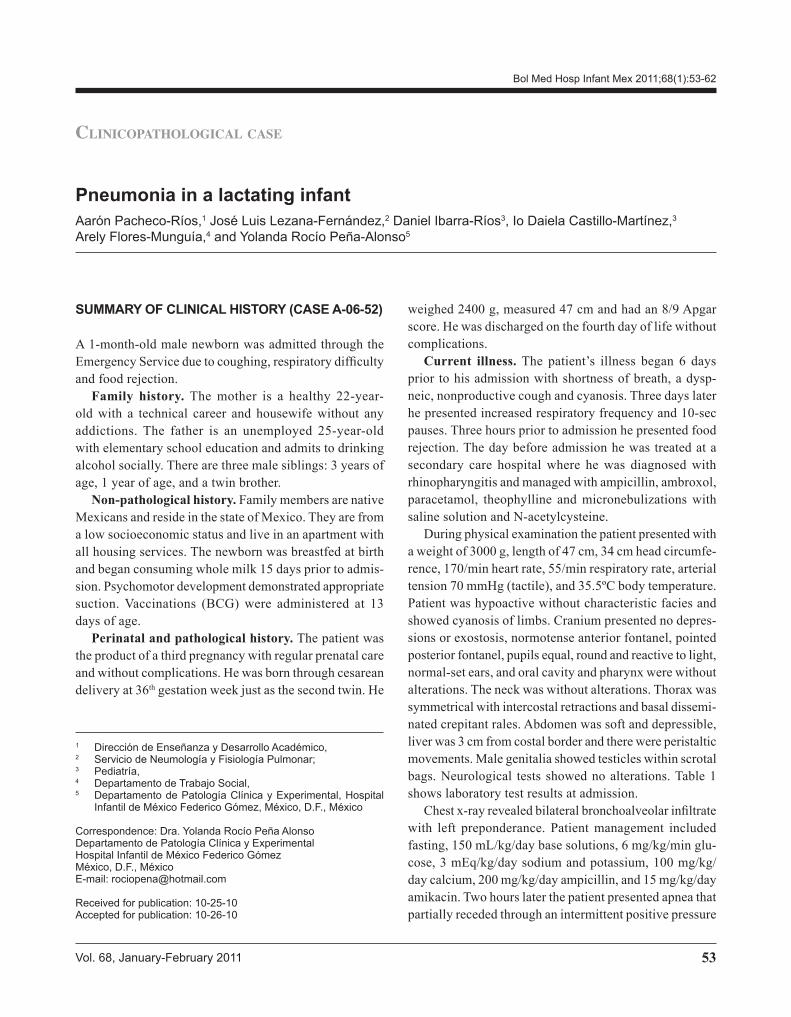
53Vol. 68, January-February 2011
Pneumonia in a lactating infant
clinicopathological case
Bol Med Hosp Infant Mex 2011;68(1):53-62
Aarón Pacheco-Ríos,1 José Luis Lezana-Fernández,2 Daniel Ibarra-Ríos3, Io Daiela Castillo-Martínez,3 Arely Flores-Munguía,4 and Yolanda Rocío Peña-Alonso5
1 Dirección de Enseñanza y Desarrollo Académico, 2 Servicio de Neumología y Fisiología Pulmonar;3 Pediatría, 4 Departamento de Trabajo Social, 5 Departamento de Patología Clínica y Experimental, Hospital
Infantil de México Federico Gómez, México, D.F., México
Correspondence: Dra. Yolanda Rocío Peña AlonsoDepartamento de Patología Clínica y ExperimentalHospital Infantil de México Federico Gómez México, D.F., MéxicoE-mail: [email protected]
Received for publication: 10-25-10Accepted for publication: 10-26-10
SummARy OF CLINICAL HISTORy (CASE A-06-52)
A 1-month-old male newborn was admitted through the Emergency Service due to coughing, respiratory difficulty and food rejection.
Family history. The mother is a healthy 22-year-old with a technical career and housewife without any addictions. The father is an unemployed 25-year-old with elementary school education and admits to drinking alcohol socially. There are three male siblings: 3 years of age, 1 year of age, and a twin brother.
Non-pathological history. Family members are native Mexicans and reside in the state of Mexico. They are from a low socioeconomic status and live in an apartment with all housing services. The newborn was breastfed at birth and began consuming whole milk 15 days prior to admis-sion. Psychomotor development demonstrated appropriate suction. Vaccinations (BCG) were administered at 13 days of age.
Perinatal and pathological history. The patient was the product of a third pregnancy with regular prenatal care and without complications. He was born through cesarean delivery at 36th gestation week just as the second twin. He
weighed 2400 g, measured 47 cm and had an 8/9 Apgar score. He was discharged on the fourth day of life without complications.
Current illness. The patient’s illness began 6 days prior to his admission with shortness of breath, a dysp-neic, nonproductive cough and cyanosis. Three days later he presented increased respiratory frequency and 10-sec pauses. Three hours prior to admission he presented food rejection. The day before admission he was treated at a secondary care hospital where he was diagnosed with rhinopharyngitis and managed with ampicillin, ambroxol, paracetamol, theophylline and micronebulizations with saline solution and N-acetylcysteine.
During physical examination the patient presented with a weight of 3000 g, length of 47 cm, 34 cm head circumfe-rence, 170/min heart rate, 55/min respiratory rate, arterial tension 70 mmHg (tactile), and 35.5ºC body temperature. Patient was hypoactive without characteristic facies and showed cyanosis of limbs. Cranium presented no depres-sions or exostosis, normotense anterior fontanel, pointed posterior fontanel, pupils equal, round and reactive to light, normal-set ears, and oral cavity and pharynx were without alterations. The neck was without alterations. Thorax was symmetrical with intercostal retractions and basal dissemi-nated crepitant rales. Abdomen was soft and depressible, liver was 3 cm from costal border and there were peristaltic movements. Male genitalia showed testicles within scrotal bags. Neurological tests showed no alterations. Table 1 shows laboratory test results at admission.
Chest x-ray revealed bilateral bronchoalveolar infiltrate with left preponderance. Patient management included fasting, 150 mL/kg/day base solutions, 6 mg/kg/min glu-cose, 3 mEq/kg/day sodium and potassium, 100 mg/kg/day calcium, 200 mg/kg/day ampicillin, and 15 mg/kg/day amikacin. Two hours later the patient presented apnea that partially receded through an intermittent positive pressure

54 Bol med Hosp Infant mex
Aarón Pacheco-Ríos, José Luis Lezana-Fernández, Daniel Ibarra-Ríos, Io Daiela Castillo-Martínez, Arely Flores-Munguía and Yolanda Rocío Peña-Alonso
(IPP) cycle. Patient was intubated with a #3 cannula and managed with controlled mechanical ventilation (CMV). Lumbar puncture was performed and revealed cerebros-pinal fluid without alterations (Table 1).
Three hours after admission, arterial tension was 70 mmHg, therfore three dosages of saline solution were administered at 0.9%, 20 mL/kg. Three hours later a Hart-man solution (20 mL/kg) was administered due to high lactate level (6.1); 0.4 µg/kg/min midazolam was started with 3 mEq/kg/dose sodium carbonate for 8 h.
The patient was assessed by the Infectology Department and managed with 150 mg/kg/day cefotaxime and 100 mg/kg/day dicloxacillin to treat multifocal pneumonia. Capi-llary puncture showed 461 mg/dL glycemia and the patient was managed with rapid insulin and then received sodium bicarbonate. Table 2 shows gasometry results.
On the second day after admission, the patient was transferred to the intensive care unit where he presented 3-sec capillary refill, 176/min heart rate, and 54/min respiratory rate. Mean arterial tension was 28 mmHg. Central venous catheter and arterial line were placed, and the patient was managed with fasting, base solutions, dobutamine, milrinone, norepinephrine and epinephrine. Central venous pressure (CVP) was 6 cm/H2O. The pa-tient received 15 mL/kg/dose red cell concentrate. Patient presented 12 h anuria and was managed with furosemide
infusion and two doses of bumetanide. Urinary output was 0.5 mL/kg/h. Due to persistent hypotension, the patient received vasopressin as infusion with the addition of cla-rithromycin and 3 mEq/kg/doses of sodium bicarbonate. Hours later the patient presented persistent hypoxemia, high ventilation parameters (assisted/controlled), 100% FiO2, 24 MIP, 4 PEEP, 0.5 sec IT, 35/min heart rate, and 1:2.4 I:E ratio. High frequency ventilation was begun and the patient received nitric oxide.
On the third day after admission, the newborn presented 3-sec capillary refill, 178/min heart rate, 37 mmHg mean arterial tension and 10 cm/H2O CVP. Due to supraven-tricular tachycardia, the patient received lidocaine and presented cardiac arrest managed through advanced resus-citation techniques for 5 min and epinephrine. The patient presented seizures that were managed with 0.15 mg/kg/dose midazolam. Transfontanel ultrasonography revealed severe brain edema impacting the ventricular system. The newborn deteriorated progressively and presented cardiac arrest without responding to resuscitation maneuvers on the third day of admission (Table 3). Postmortem study was authorized by patient’s parents.
Clinical Case Description Aaron Pacheco Rios. I would like to clarify some aspects of this patient’s clinical history. The newborn arrived at
Table 1. Laboratory tests at admission
Hb Hct Leukocytes Bands Seg Lymph Mon Plat BUN Creat Capillary glucose
11.9 g/dL 35.9% 17,900/mm3
17% 16% 55% 12% 405,000 16 mg/dL 0.7 mg/dL 164 mg/dL
Na K Cl Ca P124 mEq/L 5.8 mEq/L 90 mEq/L 8.9 mg/dL 5.8 mg/dL
CSF Aspect Color Prot Gluc Leuk Gram
Transparent Colorless 98 mg/dL 95 mg/dL 2/c No bacteria observed
CSF, cerebrospinal fluid.
Table 2. Gasometry summary
pH PaO2 PaCO2 HCO3 CO2T AG SaO2 Lact
Admission 7.14 87.2 36.7 12.1 11.7 -15.2 97.4% 9.73:00:00 PM 7.18 65.5 34.7 ---- 13.5 -14.7 94.9% 6.16:00:00 PM 7.23 29.8 34.6 13.9 33.6 -12.3 69.0% 5.021:00 arterial 7.20 25.5 28 11.1 ---- -15.5 66% 10.6venous 7.24 42.0 25.5 10.6 ---- -15.5 95% 10.1
55Vol. 68, January-February 2011
Pneumonia in a lactating infant
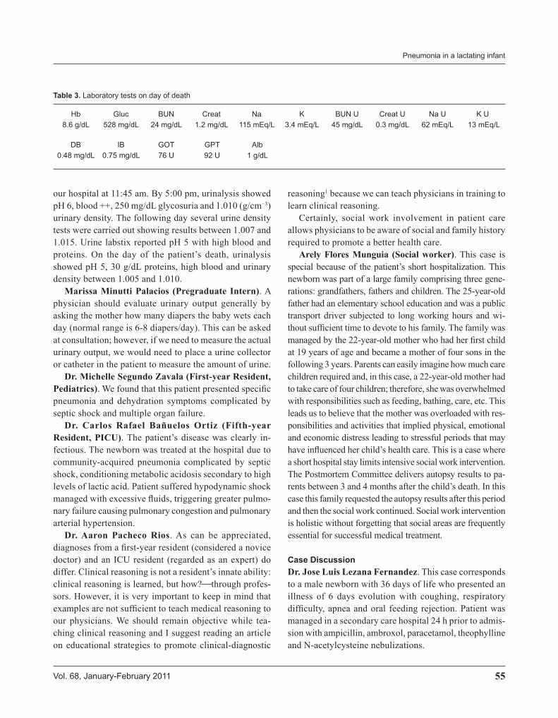
our hospital at 11:45 am. By 5:00 pm, urinalysis showed pH 6, blood ++, 250 mg/dL glycosuria and 1.010 (g/cm−3) urinary density. The following day several urine density tests were carried out showing results between 1.007 and 1.015. Urine labstix reported pH 5 with high blood and proteins. On the day of the patient’s death, urinalysis showed pH 5, 30 g/dL proteins, high blood and urinary density between 1.005 and 1.010.
Marissa Minutti Palacios (Pregraduate Intern). A physician should evaluate urinary output generally by asking the mother how many diapers the baby wets each day (normal range is 6-8 diapers/day). This can be asked at consultation; however, if we need to measure the actual urinary output, we would need to place a urine collector or catheter in the patient to measure the amount of urine.
Dr. Michelle Segundo Zavala (First-year Resident, Pediatrics). We found that this patient presented specific pneumonia and dehydration symptoms complicated by septic shock and multiple organ failure.
Dr. Carlos Rafael Bañuelos Ortiz (Fifth-year Resident, PICU). The patient’s disease was clearly in-fectious. The newborn was treated at the hospital due to community-acquired pneumonia complicated by septic shock, conditioning metabolic acidosis secondary to high levels of lactic acid. Patient suffered hypodynamic shock managed with excessive fluids, triggering greater pulmo-nary failure causing pulmonary congestion and pulmonary arterial hypertension.
Dr. Aaron Pacheco Rios. As can be appreciated, diagnoses from a first-year resident (considered a novice doctor) and an ICU resident (regarded as an expert) do differ. Clinical reasoning is not a resident’s innate ability: clinical reasoning is learned, but how?through profes-sors. However, it is very important to keep in mind that examples are not sufficient to teach medical reasoning to our physicians. We should remain objective while tea-ching clinical reasoning and I suggest reading an article on educational strategies to promote clinical-diagnostic
reasoning1 because we can teach physicians in training to learn clinical reasoning.
Certainly, social work involvement in patient care allows physicians to be aware of social and family history required to promote a better health care.
Arely Flores Munguia (Social worker). This case is special because of the patient’s short hospitalization. This newborn was part of a large family comprising three gene-rations: grandfathers, fathers and children. The 25-year-old father had an elementary school education and was a public transport driver subjected to long working hours and wi-thout sufficient time to devote to his family. The family was managed by the 22-year-old mother who had her first child at 19 years of age and became a mother of four sons in the following 3 years. Parents can easily imagine how much care children required and, in this case, a 22-year-old mother had to take care of four children; therefore, she was overwhelmed with responsibilities such as feeding, bathing, care, etc. This leads us to believe that the mother was overloaded with res-ponsibilities and activities that implied physical, emotional and economic distress leading to stressful periods that may have influenced her child’s health care. This is a case where a short hospital stay limits intensive social work intervention. The Postmortem Committee delivers autopsy results to pa-rents between 3 and 4 months after the child’s death. In this case this family requested the autopsy results after this period and then the social work continued. Social work intervention is holistic without forgetting that social areas are frequently essential for successful medical treatment.
Case DiscussionDr. Jose Luis Lezana Fernandez. This case corresponds to a male newborn with 36 days of life who presented an illness of 6 days evolution with coughing, respiratory difficulty, apnea and oral feeding rejection. Patient was managed in a secondary care hospital 24 h prior to admis-sion with ampicillin, ambroxol, paracetamol, theophylline and N-acetylcysteine nebulizations.
Table 3. Laboratory tests on day of death
Hb Gluc BUN Creat Na K BUN U Creat U Na U K U8.6 g/dL 528 mg/dL 24 mg/dL 1.2 mg/dL 115 mEq/L 3.4 mEq/L 45 mg/dL 0.3 mg/dL 62 mEq/L 13 mEq/L
DB IB GOT GPT Alb0.48 mg/dL 0.75 mg/dL 76 U 92 U 1 g/dL
56 Bol med Hosp Infant mex
Aarón Pacheco-Ríos, José Luis Lezana-Fernández, Daniel Ibarra-Ríos, Io Daiela Castillo-Martínez, Arely Flores-Munguía and Yolanda Rocío Peña-Alonso
Because the patient presented no improvement, he was referred to Hospital Infantil de Mexico Federico Gomez (HIMFG). Upon admission the patient weighed 3000 g (3150 g expected) with a 9% weight deficit placing him at the 15th percentile. Patient measured 47 cm (third per-centile) and head circumference was less than the third percentile although Millard’s index was 1.38 (1.36-1.54) being the minimum expected for his age.2,3 Sequential approach is based on the following problems.
Community-acquired pneumonia This was based on clinical data of coughing, rejection of feeding, tachypnea 55/min (31 ± 2), and respiratory insufficiency with cyanosis. From physical examination and x-rays, the most frequent bacterial agents that affect this age group are Chlamydia trachomatis, group B Strep-tococcus, Staphylococcus aureus, type-B Haemophilus influenzae and, less frequently, Streptococcus pneumoniae. Apnea does not rule out the possibility of viral agents (respiratory syncytial virus-RSV).4
Severe sepsisAccording to 2008 guidelines, it is required that children diagnosed with sepsis present other symptoms such as cardiovascular dysfunction, acute respiratory distress syndrome or failure of two or more organs.5 This patient presented systemic symptoms associated with sepsis from the time of admission: hypothermia, respiratory difficul-ty, apnea, cyanosis, oral rejection, evident respiratory infection and metabolic distress (hyperglycemia). Hema-tological criteria for sepsis include the following: total leukocytes <5000 or >18,000, absolute neutrophils <1800, total band cells ≥1,000, thrombocytopenia <100,000 platelets, band cells/total neutrophil ratio >0.2 for age, microerythrocyte sedimentation rate >15 mm during the first hour and C-reactive protein (CRP) >1 g/dL. Presence of two of these abnormal parameters is associated with sepsis with a 93% sensitivity and 83% specificity, 27% positive predictive value (PPV) and 100% negative pre-dictive value (NPV).6 Upon admission, the patient had total band cells >3000 and a band cell/total neutrophil ratio = 1.0. Leukocyte count was 17,900.
According to international guidelines, blood culture is the gold standard. It has recently been suggested, howe-ver, that CRP may be a more specific marker for sepsis diagnosis with 97% sensitivity.7
Initial metabolic response to sepsis is closely regulated by specific endocrine changes that deactivate anabolic pathways and increase anterior pituitary activity. Septic patients show resistance to insulin and regulatory hor-mones such as cortisol, glucagon, growth hormone and catecholamines. Stress-induced insulin resistance prevents glycogen synthesis and increases synthesis of pyruvate, lactate, free fatty acids and triglycerides. In the studies by Van den Berghe et al., these authors establish a direct rela-tionship between hyperglycemia and increased mortality rate with higher organic dysfunction.8-9 Patients with sepsis who present glucose >178 mg/dL increase their mortality rate by 2.6 times (OR).
A review by Andersen et al. on the role of insulin and hyperglycemia in sepsis pathogenesis emphasizes metabolic alterations of sepsis and their connection with hyperglycemia on the release of cortisol, growth hormone, glycagon, amino acids and lipid metabolism, catecholami-nes, altered functional interaction between leukocytes and capillary endothelium on the over-regulation of adhesion molecules, chemotaxis ability of polymorphonuclear cells and cytokine production.10
Tissue hypoperfusion, renal insufficiency, lactic acidosis and septic shock dataAt admission, patient presented arterial hypotension with increased creatinine, hyperkalemia and 21.4 Schwartz clearance, which leads to hypoperfusion and renal insuffi-ciency. Gasometry at admission does not include use of FiO2, so it was not possible to determine ventilatory and respiratory indexes. However, the patient presented mixed acidosis11 that did not recede during evolution. Anionic gap was high and possibly associated with a higher lactate production secondary to hypoxia from a possible decreased production from renal failure.
Hypoperfusion with slow capillary refill can be obser-ved in patients with shock, and it is one of the final events to take place in critical patients. Breathing difficulty is a final pathway for many different pathologies because, on one hand, energy requirements are increased and oxygen is demanded from all tissues and, on the other hand, a higher metabolism leads to a higher carbon dioxide pro-duction that increases respiratory demands. During tissue hypoperfusion, anaerobic metabolism with acid production is predominant and pulmonary compensation implies an increased CO2 clearance rate. In cases of serious infection,
57Vol. 68, January-February 2011
Pneumonia in a lactating infant
chiefly from gram-negative bacteria, there is an increased capillary permeability with pulmonary leakage, which reduces hematosis and increases ventilatory demand.7
ManagementClinical and laboratory data suggest the patient presen-ted severe sepsis and shock symptoms from the time of admission. The patient was administered crystaloid loads after 5 g, whereas international guidelines recommend that therapy is initiated within the first hour.7 Antimicrobial management was insufficient.
Shock management should be aimed towards normali-zation of arterial tension and tissue perfusion. Diagnosis of fluid-refractory shock should be established within 15 min and immediate administration of vasopressors and inotropic drugs should be initiated, using dopamine as the first choice in patients with refractory hypotension, even if hypovolemia has not been solved. Dopamine refractory shock requires the use of epinephrine or norepinephrine to keep ScvO2 ≥70%.7,12 Because of nonresponsiveness with low systemic vascular resistances, vasopressin has been used in isolated cases without having confirming evidence on its pediatric application. When shock is re-sistant to catecholamines (60 min), recommended therapy includes hydrocortisone because of renal insufficiency and consideration of extra-body membrane oxygenation.7,13-15
Evolution Serum osmolarity was <280 mOsm/L with persistent hyponatremia and sodium in urine reached >60 mEq/L, possibly because of tubular dysfunction and poor respon-se to aldosterone. Other causes of hyponatremia can be vasopressin administration or hyperglycemia because of intracellular displacement of water.
Independent of management, the patient continued with low-output data represented by a difference in arterial and venous contents of O2 >16, refractory hypoxemia expres-sed by breathing index >5 and gas exchange index of 0.08, >23% intrapulmonary short circuits and an increased V/Q relationship in spite of 1.0 FiO2, and 11.1 mL/dL O2 in blood.15 Finally, the patient developed tubular necrosis, adrenal gland insufficiency, organ failure and death.
Final diagnoses are as follows:
1. Male newborn with low birthweight and small size 2. Community-acquired pneumonia
3. Sepsis4. Metabolic acidosis and renal insufficiency5. Refractory septic shock6. Tubular necrosis and adrenal gland insufficiency7. Pulmonary edema8. Brain edema9. Multiple organ failure
Dr. Ricardo Munoz Arizpe. Patient arrived with renal insufficiency (RI), hydroelectrolytic imbalance and hypo-natremia, which was aggravated because of management. I disagree with the fluid and electrolyte management of this case. This newborn presented a sudden onset of severe RI possibly associated with sepsis and dehydration. First we should deal with the septic shock and then carry out dialysis as soon as possible. If we focus on renal failure, it may be associated with renal tubular necrosis although it is more likely that it was associated with renal infarction or renal thrombosis as revealed by urinary findings, RI data, anemia and the rapid evolution.
Dr. Aaron Pacheco Rios. It is important to analyze how we teach clinical reasoning to physicians and I ask Dr. Daniel Ibarra Rios to identify key elements in the clinical history of this case that lead diagnostic reasoning.
Dr. Daniel Ibarra Rios (Second-year resident, Pedia-trics). In this session Dr. Pacheco conducted an interesting exercise. First, he asked a first-year resident to synthesize the patient’s problem. Then he repeated the same question to a fifth-year resident in Intensive Care, identifying a hypodynamic septic shock. This contrasts recognition of the key elements that lead to diagnosis.
Evidence-based medicine triggered interest in how phy-sicians learn. We are used to learning from our superiors but frequently we do not realize that there are others who are learning from us. First-year residents have interns who observe them to interrogate, explore, diagnose and make decisions. Therefore, we all act as clinical teachers and our objective should be that physicians in training learn how to extract information and reason in the same way we do. In this way, they will be able to become independent so that when facing a similar problem they are able to establish diagnosis and make appropriate decisions.
When a case is summarized, we should consider the main elements of the clinical history that are combined through a database that will lead us to a hypothesis and, finally, to a diagnosis. In order to join all these concepts
58 Bol med Hosp Infant mex
Aarón Pacheco-Ríos, José Luis Lezana-Fernández, Daniel Ibarra-Ríos, Io Daiela Castillo-Martínez, Arely Flores-Munguía and Yolanda Rocío Peña-Alonso
we use links known as semantic qualifiers such as “acu-te”, “chronic”, “relapsing”, “severe”, etc., which help us differentiate between one pathology and another and we use anchoring points between different concepts. A simple example can be a child with acute petechiae whose com-plete blood count shows thrombocytopenia. This leads me to think of possibly idiopathic thrombocytopenic purpura. On the other hand, if the child presents acute petechiae and complete blood count shows anemia, thrombocyto-penia and hepatosplenomegaly, I consider a completely different diagnostic pathway. Finally, with all the training we receive in basic sciences and the contact with patients, this helps us create the so-called ìdisease scriptsî, which are signs, symptoms and laboratory findings we use to diagnose a particular disease.
We always start from clinical history and physical exa-mination to create a database. Experience helps us interpret these data appropriately to have a clear presentation of the problem. Therefore, a hypothesis is created that allows us to search for and select an appropriate algorithm to identify the disease. We usually consider the patient’s predisposing factors, make suppositions on pathophysiological results and clinical consequences, which are collected by clinical history.
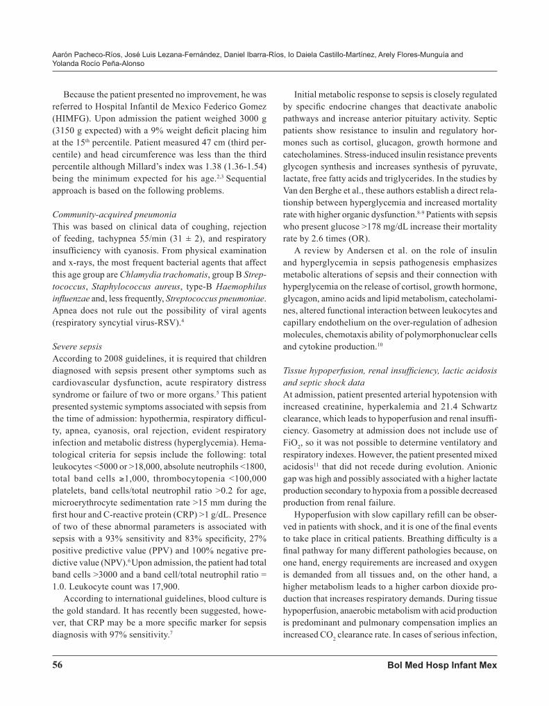
Focusing on this case, we dealt with a premature newborn with coughing, respiratory difficulty and apnea; the latter is regarded as an “apparent life-threatening event” (ALTE). We should have kept in mind primary or secondary causes that condition apnea. When conducting a study on secondary causes of apnea, we should have considered sepsis. It is possible that by having a more appropriate management we would have obtained a better outcome according to the patient’s history. By using this approach we would have discarded anemia, hypothermia, hydroelectrolytic imbalance, gastroesophageal reflux disease, metabolic errors, cardiopathies or neurological anomalies. The message is that we make diagnoses, take decisions or follow algorithms but seldom worry about making ourselves clear to the person who is observing us (Figure 1).
Dr. Aaron Pacheco Rios. Dr. Ibarra has revealed the importance of teaching clinical reasoning and not always by physicians or professors. Resident physicians can act as professors and guides so that new residents develop an appropriate clinical diagnostic reasoning.
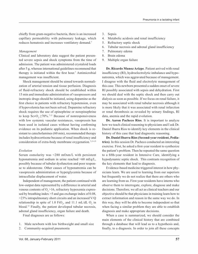
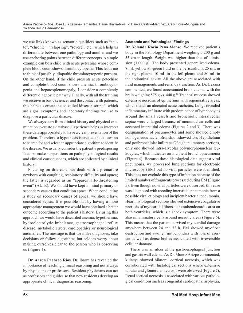
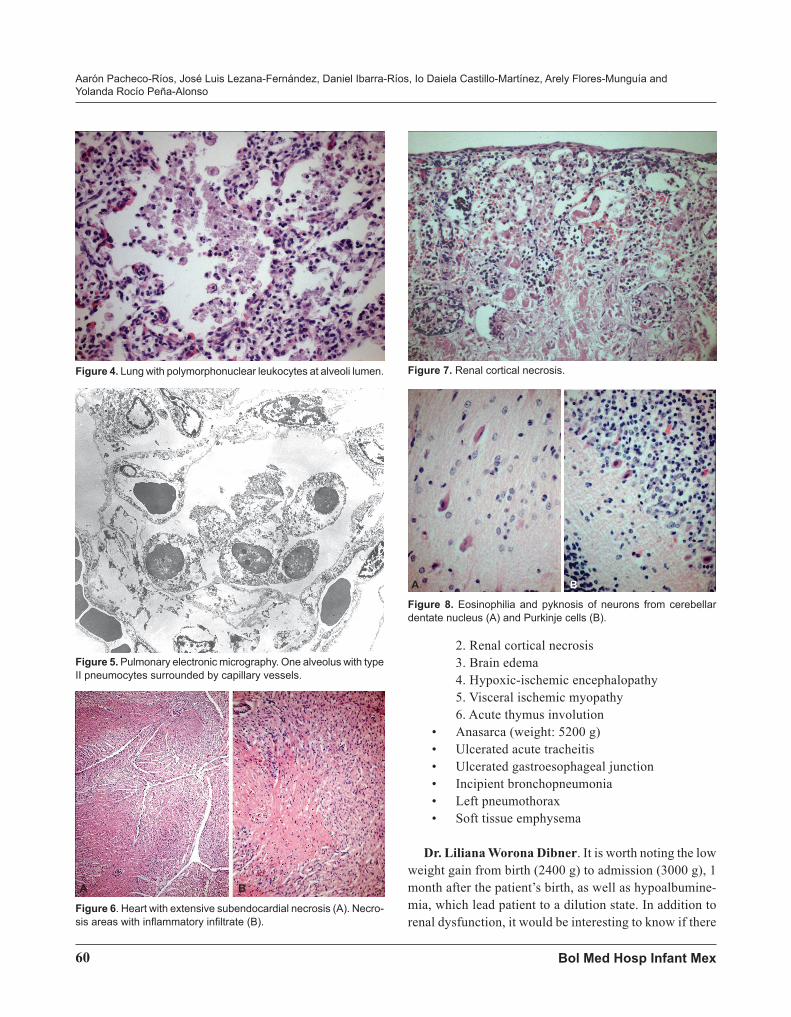
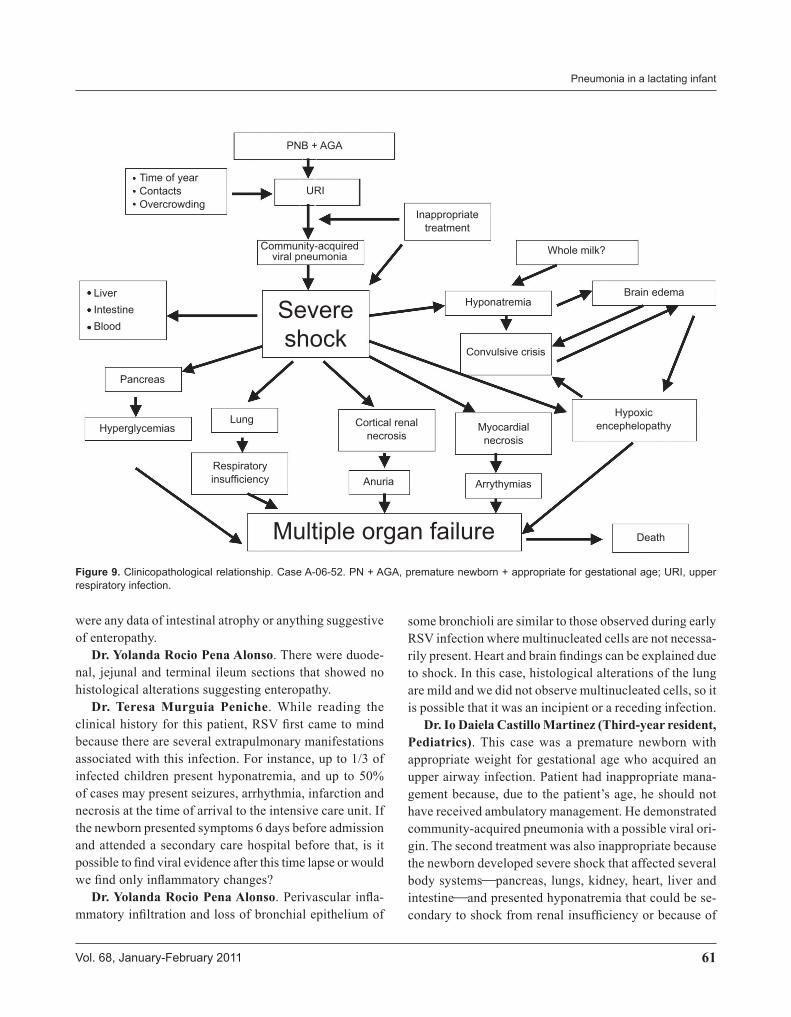
Anatomic and Pathological Findings Dr. Yolanda Rocio Pena Alonso. We received patient’s body in the Pathology Department weighing 5,200 g and 53 cm in length. Weight was higher than that of admis-sion (3,000 g). The body presented generalized edema, 8 mL yellowish-green fluid in the pericardium, 25 mL in the right pleura, 10 mL in the left pleura and 80 mL in the abdominal cavity. All the above are associated with fluid managements and renal dysfunction. As Dr. Lezana commented, we found accentuated brain edema, with the brain weighing 575 g vs. 440 g.16 Tracheal mucosa showed extensive necrosis of epithelium with regenerative areas, which match an ulcerated acute tracheitis. Lungs revealed inflammatory infiltrate with predominance of lymphocytes around the small vessels and bronchioli; interalveolar septae were enlarged because of mononuclear cells and accented interstitial edema (Figures 2 and 3). There was desquamation of pneumocytes and some showed empty homogeneous nuclei. Bronchioli showed loss of epithelium and peribronchiolar infiltrate. Of eight pulmonary sections, only one showed intra-alveolar polymorphonuclear leu-kocytes, which indicates an incipient bronchopneumonia (Figure 4). Because these histological data suggest viral pneumonia, we processed lung sections for electronic microscopy (EM) but no viral particles were identified. This does not exclude this type of infection because of the limited number of fragments processed during EM (Figure 5). Even though no viral particles were observed, this case was diagnosed with receding interstitial pneumonia from a possible viral etiology and incipient bacterial pneumonia. Heart histological sections showed extensive coagulative necrosis of myocardial fibers at the subendocardic area on both ventricles, which is a shock symptom. There were also inflammatory cells around necrotic areas (Figure 6). This means that the patient survived myocardial damage anywhere between 24 and 32 h. EM showed myofiber destruction and swollen mitochondria with loss of cres-tae as well as dense bodies associated with irreversible cellular damage.
There was an ulcer at the gastroesophageal junction and gastric wall edema. As Dr. Munoz Arizpe commented, kidneys showed bilateral cortical necrosis, which was corroborated with histological sections where extensive tubular and glomerular necrosis were observed (Figure 7). Renal cortical necrosis is associated with various patholo-gical conditions such as congenital cardiopathy, asphyxia,
59Vol. 68, January-February 2011
Pneumonia in a lactating infant
Figure 1. Algorithm to establish diagnosis.
Knowledge
Context
Experience
Knowledge
Context
Experience
Patient’s history
Data acquistion
Generation of a hypothesis
Search and select an archetype or algorithm of disease
Diagnosis
ALTE:APNEA
Premature newborn with cough, respiratory
difficulty and ALTE
1st collection• Perinatal history• Time of evolution• Epidemiological history• Initial disease
2nd collectionLaboratory, blood panel and deliberate physi-cal exploration
• Sepsis, meningitis or pneumonia• Anemia• Hypothermia• Hydroelectrolytic imbalance• Gastroesophageal reflux
disease• Inborn errors of metabolism• Cardiopathy• Convulsions or neurologic anomaly
APNEA 1st or 2nd
DIAGNOSIS
sepsis, hypovolemia and shock. All share alterations such as vasospasm, hypoxemia, acidosis and alteration in renal flow that contribute to lesion pathogenesis.17 Severe tissue damage occurs during shock associated with ischemia and also with reperfusion. We observed other shock data such as hypoxic visceral myopathy, intestinal ischemia and acute thymus involution.
This patient presented hyponatremia, which is a risk factor for the development of brain edema. Brain weighed 585 g, >95th percentile according to Coppoleta and Wol-
Figure 2. Lung with perivascular and interstitital mononuclear infiltrate.
A B
Figure 3. Lung with interstitial edema (A) and loss of bronchiolar epithelium and mononuclear inflammatory infiltrate (B).
bach charts of body length and organ weights of infants.16 Brain was softened and presented neuronal loss of the third layer of brain cortex, eosinophilia and retraction of hippo-campal neurons, cerebellar dentate nucleus and Purkinje cells, all signs of acute hypoxia (Figure 8).
Final Diagnoses• Interstitial pneumonia, possibly viral• Prolonged shock data:
1. Subendocardial necrosis
60 Bol med Hosp Infant mex
Aarón Pacheco-Ríos, José Luis Lezana-Fernández, Daniel Ibarra-Ríos, Io Daiela Castillo-Martínez, Arely Flores-Munguía and Yolanda Rocío Peña-Alonso
A B
2. Renal cortical necrosis3. Brain edema 4. Hypoxic-ischemic encephalopathy5. Visceral ischemic myopathy 6. Acute thymus involution
• Anasarca (weight: 5200 g)• Ulcerated acute tracheitis• Ulcerated gastroesophageal junction • Incipient bronchopneumonia• Left pneumothorax • Soft tissue emphysema
Dr. Liliana Worona Dibner. It is worth noting the low weight gain from birth (2400 g) to admission (3000 g), 1 month after the patient’s birth, as well as hypoalbumine-mia, which lead patient to a dilution state. In addition to renal dysfunction, it would be interesting to know if there
A B
Figure 4. Lung with polymorphonuclear leukocytes at alveoli lumen.
Figure 5. Pulmonary electronic micrography. One alveolus with type II pneumocytes surrounded by capillary vessels.
Figure 6. Heart with extensive subendocardial necrosis (A). Necro-sis areas with inflammatory infiltrate (B).
Figure 7. Renal cortical necrosis.
Figure 8. Eosinophilia and pyknosis of neurons from cerebellar dentate nucleus (A) and Purkinje cells (B).
61Vol. 68, January-February 2011
Pneumonia in a lactating infant
were any data of intestinal atrophy or anything suggestive of enteropathy.
Dr. Yolanda Rocio Pena Alonso. There were duode-nal, jejunal and terminal ileum sections that showed no histological alterations suggesting enteropathy.
Dr. Teresa Murguia Peniche. While reading the clinical history for this patient, RSV first came to mind because there are several extrapulmonary manifestations associated with this infection. For instance, up to 1/3 of infected children present hyponatremia, and up to 50% of cases may present seizures, arrhythmia, infarction and necrosis at the time of arrival to the intensive care unit. If the newborn presented symptoms 6 days before admission and attended a secondary care hospital before that, is it possible to find viral evidence after this time lapse or would we find only inflammatory changes?
Dr. Yolanda Rocio Pena Alonso. Perivascular infla-mmatory infiltration and loss of bronchial epithelium of
some bronchioli are similar to those observed during early RSV infection where multinucleated cells are not necessa-rily present. Heart and brain findings can be explained due to shock. In this case, histological alterations of the lung are mild and we did not observe multinucleated cells, so it is possible that it was an incipient or a receding infection.
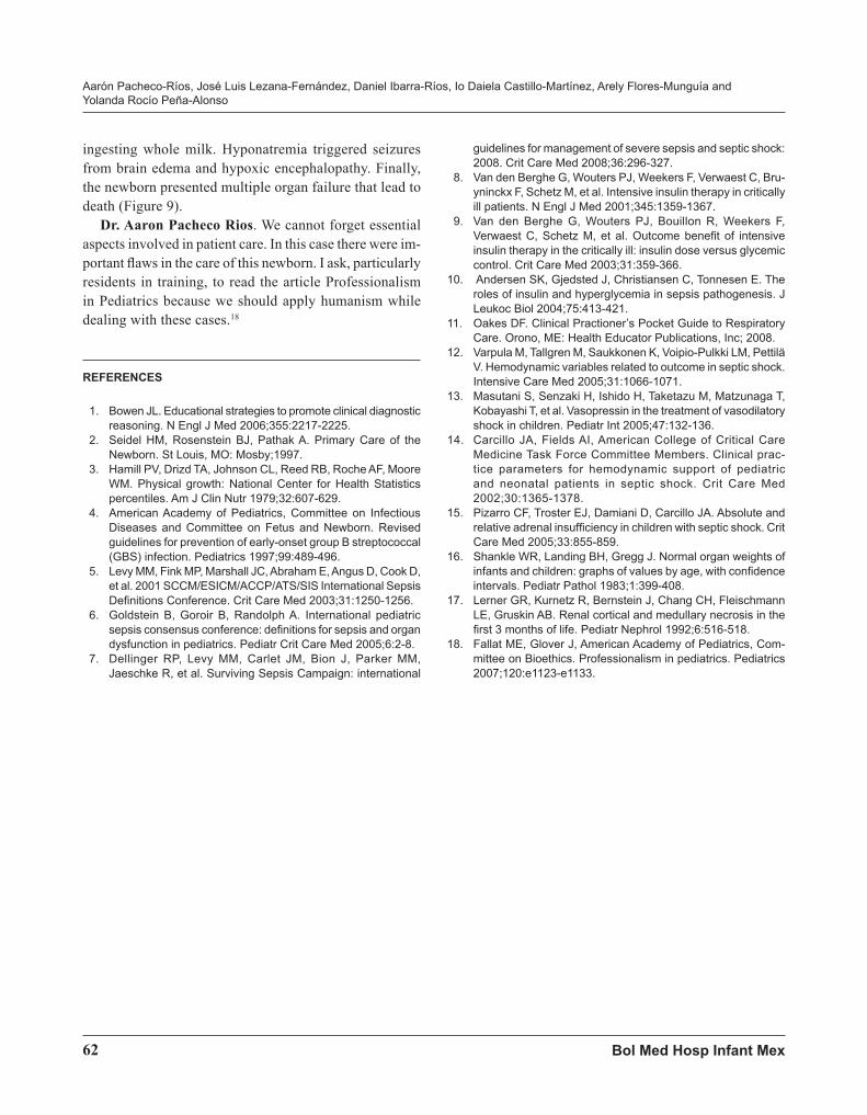
Dr. Io Daiela Castillo Martinez (Third-year resident, Pediatrics). This case was a premature newborn with appropriate weight for gestational age who acquired an upper airway infection. Patient had inappropriate mana-gement because, due to the patient’s age, he should not have received ambulatory management. He demonstrated community-acquired pneumonia with a possible viral ori-gin. The second treatment was also inappropriate because the newborn developed severe shock that affected several body systemspancreas, lungs, kidney, heart, liver and intestineand presented hyponatremia that could be se-condary to shock from renal insufficiency or because of
Figure 9. Clinicopathological relationship. Case A-06-52. PN + AGA, premature newborn + appropriate for gestational age; URI, upper respiratory infection.
Time of yearContactsOvercrowding
LiverIntestineBlood
PNB + AGA
URI
Community-acquired viral pneumonia
Inappropriate treatment
Severe shock
Pancreas
HyperglycemiasLung
Anuria
Whole milk?
Hyponatremia
Arrythymias
Death
Brain edema
Convulsive crisis
Hypoxic encephelopathyMyocardial
necrosis
Respiratory insufficiency
Cortical renal necrosis
Multiple organ failure
62 Bol med Hosp Infant mex
Aarón Pacheco-Ríos, José Luis Lezana-Fernández, Daniel Ibarra-Ríos, Io Daiela Castillo-Martínez, Arely Flores-Munguía and Yolanda Rocío Peña-Alonso
ingesting whole milk. Hyponatremia triggered seizures from brain edema and hypoxic encephalopathy. Finally, the newborn presented multiple organ failure that lead to death (Figure 9).
Dr. Aaron Pacheco Rios. We cannot forget essential aspects involved in patient care. In this case there were im-portant flaws in the care of this newborn. I ask, particularly residents in training, to read the article Professionalism in Pediatrics because we should apply humanism while dealing with these cases.18
REFERENCES
1. Bowen JL. Educational strategies to promote clinical diagnostic reasoning. N Engl J Med 2006;355:2217-2225.
2. Seidel HM, Rosenstein BJ, Pathak A. Primary Care of the Newborn. St Louis, MO: Mosby;1997.
3. Hamill PV, Drizd TA, Johnson CL, Reed RB, Roche AF, Moore WM. Physical growth: National Center for Health Statistics percentiles. Am J Clin Nutr 1979;32:607-629.
4. American Academy of Pediatrics, Committee on Infectious Diseases and Committee on Fetus and Newborn. Revised guidelines for prevention of early-onset group B streptococcal (GBS) infection. Pediatrics 1997;99:489-496.
5. Levy MM, Fink MP, Marshall JC, Abraham E, Angus D, Cook D, et al. 2001 SCCM/ESICM/ACCP/ATS/SIS International Sepsis Definitions Conference. Crit Care Med 2003;31:1250-1256.
6. Goldstein B, Goroir B, Randolph A. International pediatric sepsis consensus conference: definitions for sepsis and organ dysfunction in pediatrics. Pediatr Crit Care Med 2005;6:2-8.
7. Dellinger RP, Levy MM, Carlet JM, Bion J, Parker MM, Jaeschke R, et al. Surviving Sepsis Campaign: international
guidelines for management of severe sepsis and septic shock: 2008. Crit Care Med 2008;36:296-327.
8. Van den Berghe G, Wouters PJ, Weekers F, Verwaest C, Bru-yninckx F, Schetz M, et al. Intensive insulin therapy in critically ill patients. N Engl J Med 2001;345:1359-1367.
9. Van den Berghe G, Wouters PJ, Bouillon R, Weekers F, Verwaest C, Schetz M, et al. Outcome benefit of intensive insulin therapy in the critically ill: insulin dose versus glycemic control. Crit Care Med 2003;31:359-366.
10. Andersen SK, Gjedsted J, Christiansen C, Tonnesen E. The roles of insulin and hyperglycemia in sepsis pathogenesis. J Leukoc Biol 2004;75:413-421.
11. Oakes DF. Clinical Practioner’s Pocket Guide to Respiratory Care. Orono, ME: Health Educator Publications, Inc; 2008.
12. Varpula M, Tallgren M, Saukkonen K, Voipio-Pulkki LM, Pettilä V. Hemodynamic variables related to outcome in septic shock. Intensive Care Med 2005;31:1066-1071.
13. Masutani S, Senzaki H, Ishido H, Taketazu M, Matzunaga T, Kobayashi T, et al. Vasopressin in the treatment of vasodilatory shock in children. Pediatr Int 2005;47:132-136.
14. Carcillo JA, Fields AI, American College of Critical Care Medicine Task Force Committee Members. Clinical prac-tice parameters for hemodynamic support of pediatric and neonatal patients in septic shock. Crit Care Med 2002;30:1365-1378.
15. Pizarro CF, Troster EJ, Damiani D, Carcillo JA. Absolute and relative adrenal insufficiency in children with septic shock. Crit Care Med 2005;33:855-859.
16. Shankle WR, Landing BH, Gregg J. Normal organ weights of infants and children: graphs of values by age, with confidence intervals. Pediatr Pathol 1983;1:399-408.
17. Lerner GR, Kurnetz R, Bernstein J, Chang CH, Fleischmann LE, Gruskin AB. Renal cortical and medullary necrosis in the first 3 months of life. Pediatr Nephrol 1992;6:516-518.
18. Fallat ME, Glover J, American Academy of Pediatrics, Com-mittee on Bioethics. Professionalism in pediatrics. Pediatrics 2007;120:e1123-e1133.