Embed Size (px)
Citation preview
MICROBIOLOGY AND MOLECULAR BIOLOGY REVIEWS, Sept. 2003, p. 400–428 Vol. 67, No. 31092-2172/03/$08.00�0 DOI: 10.1128/MMBR.67.3.400–428.2003Copyright © 2003, American Society for Microbiology. All Rights Reserved.
Candida albicans Secreted Aspartyl Proteinases in Virulenceand Pathogenesis
Julian R. Naglik,1* Stephen J. Challacombe,1 and Bernhard Hube2
Department of Oral Medicine, Pathology & Immunology, GKT Dental Institute, Kings College London,London, United Kingdom,1 and Robert Koch Institut, D-13353 Berlin, Germany2
INTRODUCTION .......................................................................................................................................................401PATHOGENESIS AND VIRULENCE OF CANDIDA INFECTIONS .................................................................401HYDROLYTIC ENZYMES........................................................................................................................................402
Extracellular Proteinases of Pathogenic Fungi ..................................................................................................402SECRETED ASPARTYL PROTEINASES OF CANDIDA.....................................................................................402
Molecular and Biochemical Properties of the Candida SAP Family ...............................................................403Basic structure of the C. albicans SAP family.................................................................................................403Processing, activation, and regulation of the C. albicans proteinases.........................................................403Biochemical properties of the C. albicans proteinases...................................................................................404
SECRETED ASPARTYL PROTEINASES AND C. ALBICANS PATHOGENESIS...........................................404Correlation between Sap Production In Vitro and Candida Virulence ...........................................................405
Main focus points ...............................................................................................................................................405Sap production by clinical C. albicans isolates from humans ......................................................................405C. albicans strain selection in HIV infection...................................................................................................406
Degradation of Human Proteins and Structural Analysis in Determining Sap Substrate Specificity.......406Main focus points ...............................................................................................................................................406Broad substrate specificity of Sap2..................................................................................................................407Deducing proteinase specificity via three-dimensional structure and molecular modeling.....................408Discussion ............................................................................................................................................................408
Association of Sap Production with Other Virulence Processes of C. albicans..............................................408Main focus points ...............................................................................................................................................408Sap production and C. albicans adherence......................................................................................................409
(i) How do Sap proteins contribute to adherence?....................................................................................409Sap production and yeast-to-hypha transition ...............................................................................................409
(i) Coordinate regulation of hypha-formation and SAP4 to SAP6 expression.......................................409Sap production and phenotypic switching ......................................................................................................410
(i) Which SAP genes are regulated by phenotypic switching? .................................................................410Discussion ............................................................................................................................................................410
Sap Protein Production and Sap Immune Responses in Animal and Human Infections ...........................410Main focus points ...............................................................................................................................................410Sap protein production during C. albicans infections....................................................................................411Sap localization to the cell wall ........................................................................................................................411Antibodies against Sap induced by C. albicans infections.............................................................................411
(i) Antibodies produced during systemic infection ....................................................................................411(ii) Antibodies produced during mucosal infection ...................................................................................411
Functional anti-Sap antibodies.........................................................................................................................411(i) Systemic infections ....................................................................................................................................412(ii) Mucosal infections ...................................................................................................................................412(iii) Inhibitory antibodies ..............................................................................................................................412(iv) Sap B-cell epitopes ..................................................................................................................................412
SAP Gene Expression during Candida Infections ..............................................................................................412Main focus points ...............................................................................................................................................412Human infections................................................................................................................................................412In vitro artificial experimental infections based on RHE.............................................................................414Animal experimental models.............................................................................................................................414
(i) Mucosal models .........................................................................................................................................414(ii) Systemic models .......................................................................................................................................415
Universal expression of the SAP4 to SAP6 subfamily at mucosal surfaces................................................415
* Corresponding author. Mailing address: Department of Oral Med-icine, Pathology & Immunology, GKT Dental Institute, Kings CollegeLondon (Guy’s Campus), Floor 28, Guy’s Tower, London SE1 9RT,United Kingdom. Phone: 44 20 7955 5000, ext. 3797. Fax: 44 20 79554455. E-mail: [email protected].
400
on April 26, 2019 by guest
http://mm
br.asm.org/
Dow
nloaded from
Discussion and future directions for SAP expression studies ......................................................................415Modulation of C. albicans Virulence by Aspartyl Proteinase Inhibitors .........................................................416
Main focus points ...............................................................................................................................................416Pepstatin ..............................................................................................................................................................416
(i) Use of pepstatin in vitro...........................................................................................................................416(ii) Use of pepstatin in vivo...........................................................................................................................416
HIV PIs.................................................................................................................................................................417(i) Inhibition of Candida proteinase activity by HIV PIs in vitro............................................................417(ii) Effectiveness of HIV PIs against Candida infection in RHE and animal models ...........................418(iii) Effectiveness of HIV PIs against Candida infection in humans .......................................................418
Computer-assisted structure-based designed inhibitors ...............................................................................418Other inhibitors of Candida Sap proteins .......................................................................................................419Discussion and future directions for PI studies.............................................................................................419
Use of SAP-Disrupted Mutants To Analyze C. albicans Virulence...................................................................419Main focus points ...............................................................................................................................................419Contribution of Sap proteins to mucosal infections using SAP-deficient strains .....................................420Contribution of Sap proteins to systemic infections using SAP-deficient strains .....................................421Contribution of Sap proteins to evasion of host immune responses using SAP-deficient strains ..........421Conclusions from SAP-deficient mutant studies ............................................................................................422
FUNCTIONAL GENOMICS AND CANDIDA ........................................................................................................422Candida DNA Microarrays ....................................................................................................................................422
FUTURE DIRECTIONS IN SAP RESEARCH.......................................................................................................423ACKNOWLEDGMENTS ...........................................................................................................................................423REFERENCES ............................................................................................................................................................423
INTRODUCTION
Medical mycology is a relatively new field within the area ofmedical microbiology. Fungal diseases became recognized asbeing of clinical importance in the second half of the lastcentury, mainly due to advances in medical technologies. How-ever, within the last 20 years, the advent of the AIDS epidemichas opened up the clinical mycology field. The discovery thatreduction of the CD4� lymphocyte population of the cell-mediated immune system could predispose patients to a mul-titude of opportunistic fungal infections uncovered a wholenew area of host susceptibility and disease. As a result, anotable increase in basic research on pathogenic fungi, pre-dominantly Candida species, Cryptococcus neoformans, and As-pergillus fumigatus, has taken place (162). The outcome of thisresearch has led to the unraveling of many fundamental bio-logical processes that take place in the main fungal pathogens,particularly Candida albicans.
Candida infections are a problem of growing clinical impor-tance. The incidence of infections has increased dramaticallyover the past two to three decades, and this trend will inevita-bly continue into the 21st century. C. albicans is the mostcommon fungal pathogen of humans and has become thefourth leading cause of nosocomial infections (59, 167). At themost serious level, mortality rates from systemic candidiasisare high. However, the majority of patients, notably immuno-suppressed individuals with human immunodeficiency virus(HIV) infection, experience some form of superficial mucosalcandidiasis, most commonly thrush, and many suffer from re-current infections. In addition, nearly three-quarters of allhealthy women experience at least one vaginal yeast infectionand about 5% endure recurrent bouts of disease (211, 212).
Candida species usually reside as commensal organisms aspart of an individual’s normal microflora and can be detectedin approximately 50% of the population in this form. However,if the balance of the normal flora is disrupted or the immunedefenses are compromised, Candida species often become
pathogenic. Determining exactly how this transformation fromcommensal to pathogen takes place and how it can be pre-vented is a continuing challenge for the medical mycology field.Given the limited number of suitable and effective antifungaldrugs, the continuing increase in the incidence of Candidainfections, together with increasing drug resistance, highlightsthe need to discover new and better agents that target funda-mental biological processes and/or pathogenic determinants ofC. albicans.
PATHOGENESIS AND VIRULENCE OFCANDIDA INFECTIONS
The physiological status of the host is the primary factorgoverning the etiology of candidiasis. However, the observa-tion that only slight alterations in the host can turn normallyharmless commensal yeasts into agents able to inflict severelydebilitating illness points to the pathogenic potential of Can-dida species. Indeed, it appears that the transition from harm-less commensal to unrelenting pathogen is a fine line and onethat is attributable to an extensive repertoire of virulence de-terminants selectively expressed under suitable predisposingconditions (232).
All pathogenic microorganisms have developed mechanismsthat allow successful colonization or infection of the host (69).As a result, most pathogens, including Candida species, havedeveloped an effective battery of putative virulence factors andspecific strategies to assist in their ability to colonize hosttissues, cause disease, and overcome host defenses. The viru-lence factors expressed or required by Candida species, and inparticular C. albicans, to cause infections may well vary de-pending on the type of infection (i.e., mucosal or systemic), thesite and stage of infection, and the nature of the host response.It seems apparent that a panel of virulence attributes are in-volved in the infective process, but no single factor accounts forCandida virulence and not all expressed virulence attributes
VOL. 67, 2003 SECRETED ASPARTYL PROTEINASES AND C. ALBICANS VIRULENCE 401
on April 26, 2019 by guest
http://mm
br.asm.org/
Dow
nloaded from
may be necessary for a particular stage of infection (40, 161).Although many factors have been suggested to be virulenceattributes for C. albicans, hyphal formation, surface recogni-tion molecules, phenotypic switching, and extracellular hydro-lytic enzyme production have been the most widely studied inrecent years (24). The reader is guided to several excellentreviews on the topics of hyphal formation, surface recognitionmolecules, and phenotypic switching listed in Table 1.
The significance of these different putative virulence factorsto C. albicans pathogenicity can possibly be ascertained bydetermining whether similar homologous attributes exist inother nonpathogenic or less pathogenic yeasts such as Saccha-romyces cerevisiae (145). Sequencing of the Candida genomewith 10.4 coverage has recently been completed (http://www-sequence.stanford.edu/group/candida), and a comparativegenomic analysis between C. albicans and S. cerevisiae has beenperformed (239). Preliminary information on the Candida ge-nome suggested that although approximately 90% of all S.cerevisiae genes have a counterpart in C. albicans, 6 to 7% of C.albicans genes are not found in S. cerevisiae (135). Interest-ingly, the genes that appear to have no equivalent in S. cerevi-siae tend to be grouped in protein families, such as the agglu-tinin-like sequence (ALS) and secreted aspartyl proteinase(SAP) families, and are implicated in C. albicans virulence.
HYDROLYTIC ENZYMES
One factor that contributes to the process of virulence ishydrolytic enzyme production, which is known to play a centralrole in the pathogenicity of bacteria (69), protozoa (141), andpathogenic yeasts (163). Although many microorganisms pos-sess a variety of hydrolytic enzymes, proteinases are by far themost commonly associated with virulence.
All proteinases catalyze the hydrolysis of peptide bonds(CO—NH) in proteins but can differ markedly in specificityand mechanism of action (7). Proteinases are classified on thebasis of their catalytic mechanism and not according to theiranatomical origin, substrate specificity, or physiological func-tion. In 1978, Enzyme Nomenclature distinguished four classesof proteinases: serine, cysteine, and aspartyl proteinases andmetalloproteinases. Examples of serine proteinases are thedivergent trypsin, chymotrypsin, and subtilisin subfamilies; cys-teine proteinases include streptococcal proteinase and papain;and metalloproteinases include collagenases and microvillusproteinases (6). Aspartyl proteinases are ubiquitous in natureand are involved in a myriad of biochemical processes (41).Well-known aspartyl proteinases include the HIV aspartyl pro-teinase, and pepsin and renin in humans.
Extracellular Proteinases of Pathogenic Fungi
Extracellular proteinases of saprophytic fungi such as As-pergillus niger or Neurospora crassa are secreted primarily toprovide nutrients for the cells; however, pathogenic fungi ap-pear to have adapted this biochemical property to fulfill anumber of specialized functions during the infective process inaddition to the simple role of digesting molecules for nutrientacquisition. These more direct virulence functions may includedigesting or distorting host cell membranes to facilitate adhe-sion and tissue invasion, which has been demonstrated inplants (35, 155) and insects (209), or damaging cells and mol-ecules of the host immune system to avoid or resist antimicro-bial attack by the host (191).
Most studies investigating the role of extracellular hydrolyticenzymes in fungal pathogenicity have focused on human-pathogenic fungi, including the filamentous fungus Aspergillusfumigatus (104, 176, 208), the dermatophytes Trichophytonrubrum (5) and Trichophyton mentagrophytes (236), and thedimorphic yeasts Cryptococcus neoformans (21), Coccidioidesimmitis (253), and C. albicans (50, 75, 94, 97, 145). While littleis known about the extracellular proteinases of most dimorphichuman pathogenic fungi, the proteolytic system of C. albicansis well described.
SECRETED ASPARTYL PROTEINASES OF CANDIDA
The three most significant extracellular hydrolytic enzymesproduced by C. albicans are the secreted aspartyl proteinases(Sap), phospholipase B enzymes, and lipases. Of these, the Sapproteins, encoded by a family of 10 SAP genes (66, 146, 147),have been the most comprehensively studied as key virulencedeterminants of C. albicans and are the subject of this review.For more information on phospholipases and lipases, thereader is guided to references 75 and 98.
C. albicans is not the only Candida species known to produceextracellular proteinases. Many of the pathogenic Candidaspecies have been shown to posses SAP genes, including C.dubliniensis (76), C. tropicalis (147, 234, 254), and C. parapsi-losis (53, 147), all of which produce active extracellular pro-teinases in vitro (76, 185). C. tropicalis is thought to possessfour SAP genes (254), whereas C. parapsilosis possesses at leasttwo SAP genes (53). Little published information is availablewith regard to the importance of Sap proteins in the virulenceof C. dubliniensis. However, since C. dubliniensis probably pos-sesses at least nine SAP genes (76) (J. R. Naglik, unpublisheddata), it is highly likely that proteinase production contributesto the virulence of this fungus. Less pathogenic or nonpatho-
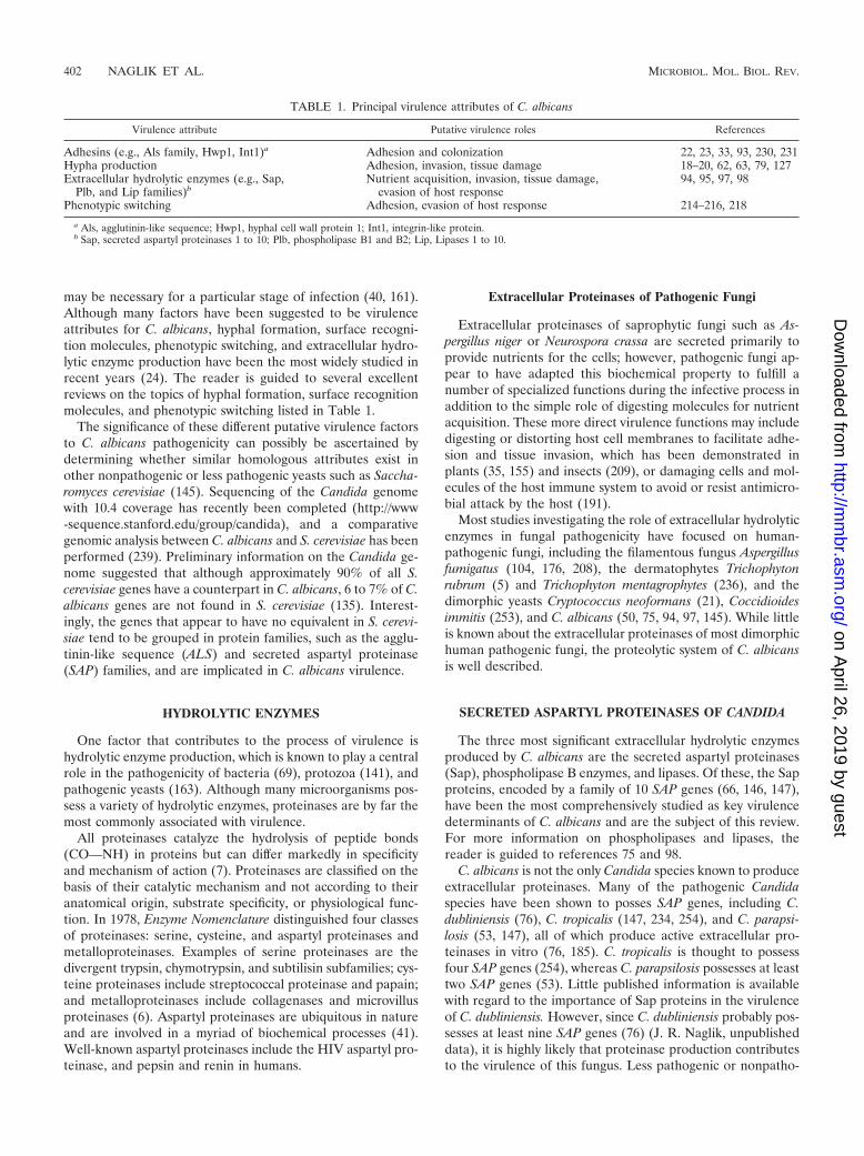
TABLE 1. Principal virulence attributes of C. albicans
Virulence attribute Putative virulence roles References
Adhesins (e.g., Als family, Hwp1, Int1)a Adhesion and colonization 22, 23, 33, 93, 230, 231Hypha production Adhesion, invasion, tissue damage 18–20, 62, 63, 79, 127Extracellular hydrolytic enzymes (e.g., Sap,
Plb, and Lip families)bNutrient acquisition, invasion, tissue damage,
evasion of host response94, 95, 97, 98
Phenotypic switching Adhesion, evasion of host response 214–216, 218
a Als, agglutinin-like sequence; Hwp1, hyphal cell wall protein 1; Int1, integrin-like protein.b Sap, secreted aspartyl proteinases 1 to 10; Plb, phospholipase B1 and B2; Lip, Lipases 1 to 10.
402 NAGLIK ET AL. MICROBIOL. MOL. BIOL. REV.
on April 26, 2019 by guest
http://mm
br.asm.org/
Dow
nloaded from
genic Candida species do not appear to produce significantamounts of proteinase, even though they may possess aspartylproteinase genes (see “Correlation between Sap production invitro and Candida virulence” below). Finally, one should notethat all secreted Candida secreted proteinases belong to thesame class of enzyme: the aspartyl proteinases. Neither extra-cellular serine nor cysteine proteinases nor metalloproteinaseshave been identified in pathogenic Candida species.
Molecular and Biochemical Properties of theCandida SAP Family
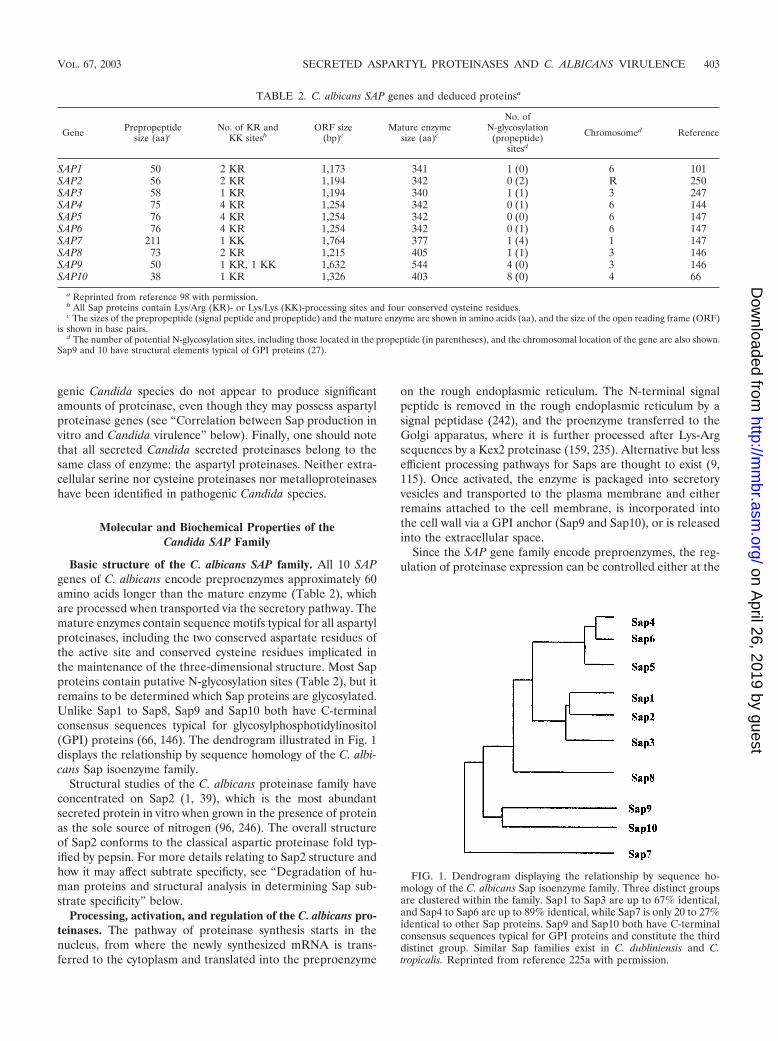
Basic structure of the C. albicans SAP family. All 10 SAPgenes of C. albicans encode preproenzymes approximately 60amino acids longer than the mature enzyme (Table 2), whichare processed when transported via the secretory pathway. Themature enzymes contain sequence motifs typical for all aspartylproteinases, including the two conserved aspartate residues ofthe active site and conserved cysteine residues implicated inthe maintenance of the three-dimensional structure. Most Sapproteins contain putative N-glycosylation sites (Table 2), but itremains to be determined which Sap proteins are glycosylated.Unlike Sap1 to Sap8, Sap9 and Sap10 both have C-terminalconsensus sequences typical for glycosylphosphotidylinositol(GPI) proteins (66, 146). The dendrogram illustrated in Fig. 1displays the relationship by sequence homology of the C. albi-cans Sap isoenzyme family.
Structural studies of the C. albicans proteinase family haveconcentrated on Sap2 (1, 39), which is the most abundantsecreted protein in vitro when grown in the presence of proteinas the sole source of nitrogen (96, 246). The overall structureof Sap2 conforms to the classical aspartic proteinase fold typ-ified by pepsin. For more details relating to Sap2 structure andhow it may affect subtrate specificty, see “Degradation of hu-man proteins and structural analysis in determining Sap sub-strate specificity” below.
Processing, activation, and regulation of the C. albicans pro-teinases. The pathway of proteinase synthesis starts in thenucleus, from where the newly synthesized mRNA is trans-ferred to the cytoplasm and translated into the preproenzyme
on the rough endoplasmic reticulum. The N-terminal signalpeptide is removed in the rough endoplasmic reticulum by asignal peptidase (242), and the proenzyme transferred to theGolgi apparatus, where it is further processed after Lys-Argsequences by a Kex2 proteinase (159, 235). Alternative but lessefficient processing pathways for Saps are thought to exist (9,115). Once activated, the enzyme is packaged into secretoryvesicles and transported to the plasma membrane and eitherremains attached to the cell membrane, is incorporated intothe cell wall via a GPI anchor (Sap9 and Sap10), or is releasedinto the extracellular space.
Since the SAP gene family encode preproenzymes, the reg-ulation of proteinase expression can be controlled either at the
FIG. 1. Dendrogram displaying the relationship by sequence ho-mology of the C. albicans Sap isoenzyme family. Three distinct groupsare clustered within the family. Sap1 to Sap3 are up to 67% identical,and Sap4 to Sap6 are up to 89% identical, while Sap7 is only 20 to 27%identical to other Sap proteins. Sap9 and Sap10 both have C-terminalconsensus sequences typical for GPI proteins and constitute the thirddistinct group. Similar Sap families exist in C. dubliniensis and C.tropicalis. Reprinted from reference 225a with permission.
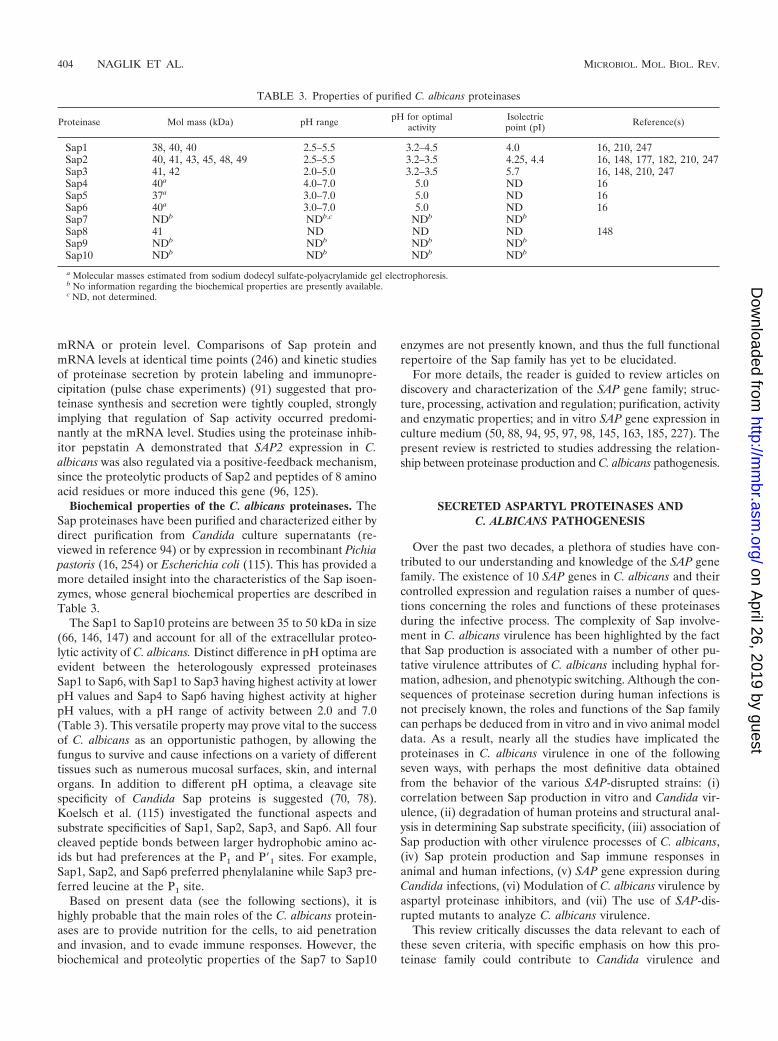
TABLE 2. C. albicans SAP genes and deduced proteinsa
Gene Prepropeptidesize (aa)c
No. of KR andKK sitesb
ORF size(bp)c
Mature enzymesize (aa)c
No. ofN-glycosylation
(propeptide)sitesd
Chromosomed Reference
SAP1 50 2 KR 1,173 341 1 (0) 6 101SAP2 56 2 KR 1,194 342 0 (2) R 250SAP3 58 1 KR 1,194 340 1 (1) 3 247SAP4 75 4 KR 1,254 342 0 (1) 6 144SAP5 76 4 KR 1,254 342 0 (0) 6 147SAP6 76 4 KR 1,254 342 0 (1) 6 147SAP7 211 1 KK 1,764 377 1 (4) 1 147SAP8 73 2 KR 1,215 405 1 (1) 3 146SAP9 50 1 KR, 1 KK 1,632 544 4 (0) 3 146SAP10 38 1 KR 1,326 403 8 (0) 4 66
a Reprinted from reference 98 with permission.b All Sap proteins contain Lys/Arg (KR)- or Lys/Lys (KK)-processing sites and four conserved cysteine residues.c The sizes of the prepropeptide (signal peptide and propeptide) and the mature enzyme are shown in amino acids (aa), and the size of the open reading frame (ORF)
is shown in base pairs.d The number of potential N-glycosylation sites, including those located in the propeptide (in parentheses), and the chromosomal location of the gene are also shown.
Sap9 and 10 have structural elements typical of GPI proteins (27).
VOL. 67, 2003 SECRETED ASPARTYL PROTEINASES AND C. ALBICANS VIRULENCE 403
on April 26, 2019 by guest
http://mm
br.asm.org/
Dow
nloaded from
mRNA or protein level. Comparisons of Sap protein andmRNA levels at identical time points (246) and kinetic studiesof proteinase secretion by protein labeling and immunopre-cipitation (pulse chase experiments) (91) suggested that pro-teinase synthesis and secretion were tightly coupled, stronglyimplying that regulation of Sap activity occurred predomi-nantly at the mRNA level. Studies using the proteinase inhib-itor pepstatin A demonstrated that SAP2 expression in C.albicans was also regulated via a positive-feedback mechanism,since the proteolytic products of Sap2 and peptides of 8 aminoacid residues or more induced this gene (96, 125).
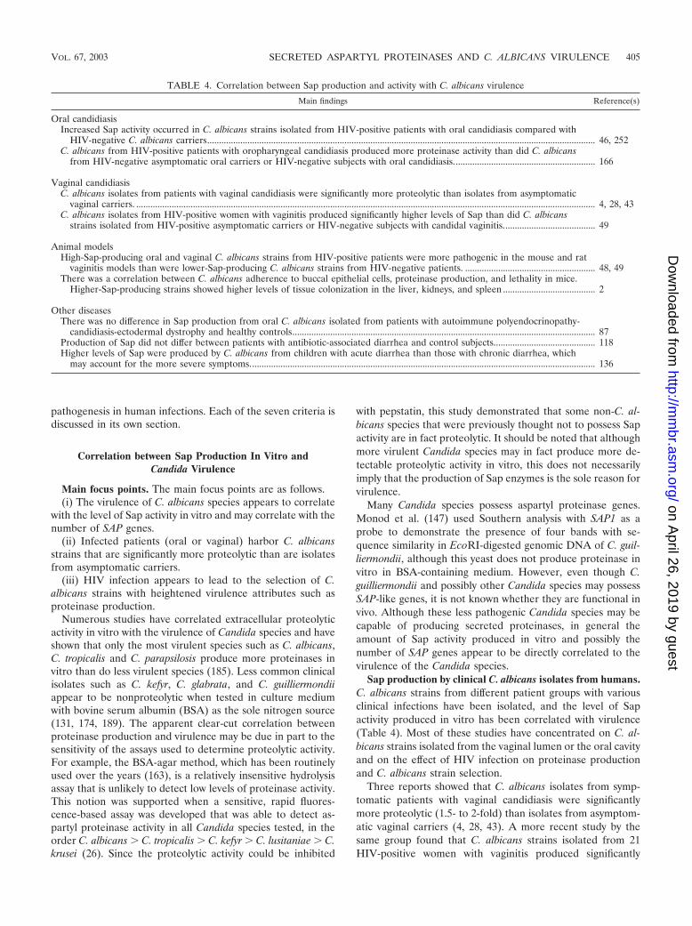
Biochemical properties of the C. albicans proteinases. TheSap proteinases have been purified and characterized either bydirect purification from Candida culture supernatants (re-viewed in reference 94) or by expression in recombinant Pichiapastoris (16, 254) or Escherichia coli (115). This has provided amore detailed insight into the characteristics of the Sap isoen-zymes, whose general biochemical properties are described inTable 3.
The Sap1 to Sap10 proteins are between 35 to 50 kDa in size(66, 146, 147) and account for all of the extracellular proteo-lytic activity of C. albicans. Distinct difference in pH optima areevident between the heterologously expressed proteinasesSap1 to Sap6, with Sap1 to Sap3 having highest activity at lowerpH values and Sap4 to Sap6 having highest activity at higherpH values, with a pH range of activity between 2.0 and 7.0(Table 3). This versatile property may prove vital to the successof C. albicans as an opportunistic pathogen, by allowing thefungus to survive and cause infections on a variety of differenttissues such as numerous mucosal surfaces, skin, and internalorgans. In addition to different pH optima, a cleavage sitespecificity of Candida Sap proteins is suggested (70, 78).Koelsch et al. (115) investigated the functional aspects andsubstrate specificities of Sap1, Sap2, Sap3, and Sap6. All fourcleaved peptide bonds between larger hydrophobic amino ac-ids but had preferences at the P1 and P�1 sites. For example,Sap1, Sap2, and Sap6 preferred phenylalanine while Sap3 pre-ferred leucine at the P1 site.
Based on present data (see the following sections), it ishighly probable that the main roles of the C. albicans protein-ases are to provide nutrition for the cells, to aid penetrationand invasion, and to evade immune responses. However, thebiochemical and proteolytic properties of the Sap7 to Sap10
enzymes are not presently known, and thus the full functionalrepertoire of the Sap family has yet to be elucidated.
For more details, the reader is guided to review articles ondiscovery and characterization of the SAP gene family; struc-ture, processing, activation and regulation; purification, activityand enzymatic properties; and in vitro SAP gene expression inculture medium (50, 88, 94, 95, 97, 98, 145, 163, 185, 227). Thepresent review is restricted to studies addressing the relation-ship between proteinase production and C. albicans pathogenesis.
SECRETED ASPARTYL PROTEINASES ANDC. ALBICANS PATHOGENESIS
Over the past two decades, a plethora of studies have con-tributed to our understanding and knowledge of the SAP genefamily. The existence of 10 SAP genes in C. albicans and theircontrolled expression and regulation raises a number of ques-tions concerning the roles and functions of these proteinasesduring the infective process. The complexity of Sap involve-ment in C. albicans virulence has been highlighted by the factthat Sap production is associated with a number of other pu-tative virulence attributes of C. albicans including hyphal for-mation, adhesion, and phenotypic switching. Although the con-sequences of proteinase secretion during human infections isnot precisely known, the roles and functions of the Sap familycan perhaps be deduced from in vitro and in vivo animal modeldata. As a result, nearly all the studies have implicated theproteinases in C. albicans virulence in one of the followingseven ways, with perhaps the most definitive data obtainedfrom the behavior of the various SAP-disrupted strains: (i)correlation between Sap production in vitro and Candida vir-ulence, (ii) degradation of human proteins and structural anal-ysis in determining Sap substrate specificity, (iii) association ofSap production with other virulence processes of C. albicans,(iv) Sap protein production and Sap immune responses inanimal and human infections, (v) SAP gene expression duringCandida infections, (vi) Modulation of C. albicans virulence byaspartyl proteinase inhibitors, and (vii) The use of SAP-dis-rupted mutants to analyze C. albicans virulence.
This review critically discusses the data relevant to each ofthese seven criteria, with specific emphasis on how this pro-teinase family could contribute to Candida virulence and
TABLE 3. Properties of purified C. albicans proteinases
Proteinase Mol mass (kDa) pH range pH for optimalactivity
Isolectricpoint (pI) Reference(s)
Sap1 38, 40, 40 2.5–5.5 3.2–4.5 4.0 16, 210, 247Sap2 40, 41, 43, 45, 48, 49 2.5–5.5 3.2–3.5 4.25, 4.4 16, 148, 177, 182, 210, 247Sap3 41, 42 2.0–5.0 3.2–3.5 5.7 16, 148, 210, 247Sap4 40a 4.0–7.0 5.0 ND 16Sap5 37a 3.0–7.0 5.0 ND 16Sap6 40a 3.0–7.0 5.0 ND 16Sap7 NDb NDb,c NDb NDb
Sap8 41 ND ND ND 148Sap9 NDb NDb NDb NDb
Sap10 NDb NDb NDb NDb
a Molecular masses estimated from sodium dodecyl sulfate-polyacrylamide gel electrophoresis.b No information regarding the biochemical properties are presently available.c ND, not determined.
404 NAGLIK ET AL. MICROBIOL. MOL. BIOL. REV.
on April 26, 2019 by guest
http://mm
br.asm.org/
Dow
nloaded from
pathogenesis in human infections. Each of the seven criteria isdiscussed in its own section.
Correlation between Sap Production In Vitro andCandida Virulence
Main focus points. The main focus points are as follows.(i) The virulence of C. albicans species appears to correlate
with the level of Sap activity in vitro and may correlate with thenumber of SAP genes.
(ii) Infected patients (oral or vaginal) harbor C. albicansstrains that are significantly more proteolytic than are isolatesfrom asymptomatic carriers.
(iii) HIV infection appears to lead to the selection of C.albicans strains with heightened virulence attributes such asproteinase production.
Numerous studies have correlated extracellular proteolyticactivity in vitro with the virulence of Candida species and haveshown that only the most virulent species such as C. albicans,C. tropicalis and C. parapsilosis produce more proteinases invitro than do less virulent species (185). Less common clinicalisolates such as C. kefyr, C. glabrata, and C. guilliermondiiappear to be nonproteolytic when tested in culture mediumwith bovine serum albumin (BSA) as the sole nitrogen source(131, 174, 189). The apparent clear-cut correlation betweenproteinase production and virulence may be due in part to thesensitivity of the assays used to determine proteolytic activity.For example, the BSA-agar method, which has been routinelyused over the years (163), is a relatively insensitive hydrolysisassay that is unlikely to detect low levels of proteinase activity.This notion was supported when a sensitive, rapid fluores-cence-based assay was developed that was able to detect as-partyl proteinase activity in all Candida species tested, in theorder C. albicans � C. tropicalis � C. kefyr � C. lusitaniae � C.krusei (26). Since the proteolytic activity could be inhibited
with pepstatin, this study demonstrated that some non-C. al-bicans species that were previously thought not to possess Sapactivity are in fact proteolytic. It should be noted that althoughmore virulent Candida species may in fact produce more de-tectable proteolytic activity in vitro, this does not necessarilyimply that the production of Sap enzymes is the sole reason forvirulence.
Many Candida species possess aspartyl proteinase genes.Monod et al. (147) used Southern analysis with SAP1 as aprobe to demonstrate the presence of four bands with se-quence similarity in EcoRI-digested genomic DNA of C. guil-liermondii, although this yeast does not produce proteinase invitro in BSA-containing medium. However, even though C.guilliermondii and possibly other Candida species may possessSAP-like genes, it is not known whether they are functional invivo. Although these less pathogenic Candida species may becapable of producing secreted proteinases, in general theamount of Sap activity produced in vitro and possibly thenumber of SAP genes appear to be directly correlated to thevirulence of the Candida species.
Sap production by clinical C. albicans isolates from humans.C. albicans strains from different patient groups with variousclinical infections have been isolated, and the level of Sapactivity produced in vitro has been correlated with virulence(Table 4). Most of these studies have concentrated on C. al-bicans strains isolated from the vaginal lumen or the oral cavityand on the effect of HIV infection on proteinase productionand C. albicans strain selection.
Three reports showed that C. albicans isolates from symp-tomatic patients with vaginal candidiasis were significantlymore proteolytic (1.5- to 2-fold) than isolates from asymptom-atic vaginal carriers (4, 28, 43). A more recent study by thesame group found that C. albicans strains isolated from 21HIV-positive women with vaginitis produced significantly
TABLE 4. Correlation between Sap production and activity with C. albicans virulence
Main findings Reference(s)
Oral candidiasisIncreased Sap activity occurred in C. albicans strains isolated from HIV-positive patients with oral candidiasis compared with
HIV-negative C. albicans carriers.................................................................................................................................................................... 46, 252C. albicans from HIV-positive patients with oropharyngeal candidiasis produced more proteinase activity than did C. albicans
from HIV-negative asymptomatic oral carriers or HIV-negative subjects with oral candidiasis............................................................ 166
Vaginal candidiasisC. albicans isolates from patients with vaginal candidiasis were significantly more proteolytic than isolates from asymptomatic
vaginal carriers. .................................................................................................................................................................................................. 4, 28, 43C. albicans isolates from HIV-positive women with vaginitis produced significantly higher levels of Sap than did C. albicans
strains isolated from HIV-positive asymptomatic carriers or HIV-negative subjects with candidal vaginitis....................................... 49
Animal modelsHigh-Sap-producing oral and vaginal C. albicans strains from HIV-positive patients were more pathogenic in the mouse and rat
vaginitis models than were lower-Sap-producing C. albicans strains from HIV-negative patients. ....................................................... 48, 49There was a correlation between C. albicans adherence to buccal epithelial cells, proteinase production, and lethality in mice.
Higher-Sap-producing strains showed higher levels of tissue colonization in the liver, kidneys, and spleen ....................................... 2
Other diseasesThere was no difference in Sap production from oral C. albicans isolated from patients with autoimmune polyendocrinopathy-
candidiasis-ectodermal dystrophy and healthy controls................................................................................................................................ 87Production of Sap did not differ between patients with antibiotic-associated diarrhea and control subjects........................................... 118Higher levels of Sap were produced by C. albicans from children with acute diarrhea than those with chronic diarrhea, which
may account for the more severe symptoms.................................................................................................................................................. 136
VOL. 67, 2003 SECRETED ASPARTYL PROTEINASES AND C. ALBICANS VIRULENCE 405
on April 26, 2019 by guest
http://mm
br.asm.org/
Dow
nloaded from
higher levels of Sap (fourfold) than did C. albicans strainsisolated from either 7 HIV-positive asymptomatic carriers or31 HIV-negative subjects with candidal vaginitis (49) (Table 4).
A similar approach has been applied to oral isolates of C.albicans, mainly from HIV-positive individuals. C. albicans iso-lates from 100 HIV-positive patients with oropharyngeal can-didiasis produced significantly more proteinase activity thandid isolates from 122 patients without HIV infection (50 withoral candidiasis and 72 asymptomatic Candida carriers) (166).The higher level of proteinase activity correlated with the in-creased level of cell surface-associated and secreted Sap, asrevealed by cytofluorometry and Western blotting, respec-tively. A similar study, but using fewer patients, reported acomparable increase in Sap activity in C. albicans strains iso-lated from the oral cavities of 44 HIV-positive patients (ad-vanced disease) with oral candidiasis compared with that in 30HIV-negative C. albicans carriers (46). However, since a con-trol group of HIV-negative subjects with oral candidiasis wasnot included, it is unclear whether the observed increase in Sapproduction resulted from the advanced HIV status of the in-dividuals or from the Candida infection. Likewise, Wu et al.(252) found that oral C. albicans isolates from HIV-positivesubjects (n � 18) produced significantly more proteinase thandid isolates from HIV-negative individuals (n � 18) when theywere investigated in a BSA agar plate assay. Finally, highSap-producing oral (48) and vaginal (49) C. albicans strainsisolated from HIV-positive patients were more pathogenic inthe mouse and rat vaginitis models, respectively, than werelesser Sap-producing C. albicans strains from HIV-negativepatients (Table 4).
In summary, these studies using oral and vaginal clinicalisolates showed a positive correlation between the level of Sapproduction in vitro and the virulence of C. albicans. Whetherthese observations reflect an elevated “fitness” or a specificadapted response of C. albicans strains during infection is notclear, but the data tentatively support a role for the proteinasesduring the infective process in vivo.
C. albicans strain selection in HIV infection. There is mount-ing evidence that Candida species colonizing the oral cavitiesof HIV-infected individuals are subject to selective pressuresthat may lead to the emergence of strains with altered geno-typic and phenotypic characteristics and enhanced expressionof known and putative virulence determinants. Studies haveshown that the genotype of the infecting Candida cells in HIVinfection is stable, and, as a result, HIV-infected patients tendto be colonized by a single endogenous strain of Candida thatpersists throughout recurrent bouts of oral candidiasis, evenafter antifungal therapy (34, 130, 143, 169, 175, 204, 245, 248).However, these and other studies also suggest that in the ma-jority of AIDS patients the original commensal strains arereplaced and that this replacement of genotypes occurs onlyonce, early in the course of HIV infection, producing a genet-ically conserved population (140, 204).
Both Ollert et al. (166) and De Bernardis et al. (46) showedthat the increase in Sap activity was observed only in patientswith advanced HIV infection and not in those with earlierstages of HIV infection or HIV-negative subjects. This indi-cated that more virulent biotypes of C. albicans with height-ened proteinase production might be selected in HIV-infectedpatients. However, it should be pointed out that this selection
appeared to occur before patients developed AIDS and wasindependent of CD4� counts.
One intriguing possibility for the observed differences in Sapproduction between HIV-positive and HIV-negative patientsmay be due to the direct binding of HIV proteins to Candidacells. Treatment of C. albicans with gp160, but not with gp120,led to an elevation of free and cell-bound aspartyl proteinase(82). In addition, culture supernatants obtained from C. albi-cans treated with gp160 or gp41, but not with gp120, showed astrong increase in proteinase activity. Why or how HIV gp160or gp41, but not gp120, influences proteinase production andwhether they modulate Sap secretion directly or indirectlythrough another mechanism remain to be elucidated. HIVinfection might also promote C. albicans virulence in anotherway, since the HIV transactivating protein Tat binds RGDsequences present on the surface of C. albicans to inducehyphal production (81), a process known to be linked withvirulence and the expression of the SAP4 to SAP6 subfamily(96, 246).
The pathobiological effects of HIV infection, including pos-sible epithelial cell surface changes (170), reduced salivary flowrate (203), and alterations in the oral microflora (171), mightalso influence the candidal microenvironment. Together withimpaired humoral or cell-mediated mucosal immunity and/orimpaired nonspecific host defenses, these selective pressures inHIV infection are likely to contribute to the selection of Can-dida strains, some of which may possess altered or heightenedvirulence attributes such as proteinase production (232). How-ever, the mechanism by which these selective pressures con-tribute to strain selection in HIV infection remains to be elu-cidated and may prove particularly challenging to resolve.
Degradation of Human Proteins and Structural Analysis inDetermining Sap Substrate Specificity
Main focus points. The main focus points are as follows.(i) Sap2 has very broad substrate specificity and can degrade
many human proteins.(ii) The crystal structure of Sap2 indicates that the C. albicans
proteinase family is unique among the aspartyl proteinases.(iii) Computer modeling suggests that the electrostatic
charge of the different Sap proteins may contribute to differentsubstrate specificities and tissue targeting.
The first observation of proteolytic activity in C. albicans wasdemonstrated by Staib (222) when yeast cells were grown inmedia containing BSA as the sole source of protein. Threeyears later, Remold et al. (177) attributed this activity to theproduction of an extracellular proteinase. Since then and up tothe early 1990s, a plethora of studies reported on the purifi-cation and biochemical properties of an extracellular protein-ase from C. albicans and the effect of environmental factorssuch as pH and temperature on proteolytic activity (Table 3).The culture conditions used to induce proteinase activity inthese early reports have subsequently been shown to favorSAP2 expression (96, 246). Therefore, any attempts to deter-mine the substrate specificities and potential targets of the Sapfamily in vivo were based on the activity of Sap2 in vitro. Atpresent, it is not clear whether the digestion of substrates bySap2 in vivo is similar to that shown in vitro or whether thesubstrates for Sap2 are similar or different from those of the
406 NAGLIK ET AL. MICROBIOL. MOL. BIOL. REV.
on April 26, 2019 by guest
http://mm
br.asm.org/
Dow
nloaded from
other proteinases in the Sap family. The full range of substratespecificities for all the secreted proteinases has not been ade-quately studied, but the in vitro proteolytic properties of Sap2have been described in some detail.
Broad substrate specificity of Sap2. One of the most notice-able properties of Sap2 is the variety of proteins it can cleave.The contribution of this broad activity to Candida pathogene-sis, along with other virulence attributes of C. albicans, is il-lustrated in Fig. 2. Sap2 is known to degrade many humanproteins including molecules that protect mucosal surfacessuch as mucin (36, 52) and secretory immunoglobulin A (IgA)(78, 184). Not only could this provide essential nitrogen forgrowth, but also it could enhance attachment, colonization,and penetration of host tissue by the removal of host barriers.Digestion of secretory IgA is particularly noteworthy because itis considerably more resistant to proteolysis than are mono-meric or serum immunoglobulins, is able to neutralize manytoxins and enzymes (109), and can inhibit C. albicans attach-ment to buccal epithelial cells (243). Supporting these data,Wu and Samaranayake (251) noted that reduction of totalsalivary protein concentration correlated with the degree ofSap expression, suggesting that Candida Sap proteins degradesalivary proteins in the oral cavity. Sap2 can also degrade
molecules of the extracellular matrix such as keratin, collagenand vimentin (94, 163, 174). Morschhauser et al. (152) showedthat induction of C. albicans proteinase caused digestion ofsoluble and immobilized extracellular matrix proteins pro-duced by a human endothelial cell line, suggesting that Sapproteins may facilitate the dissemination of C. albicans via thecirculatory system.
C. albicans proteinases may also evade host defenses bydirectly degrading molecules such as salivary lactoferrin, lac-toperoxidase, cathepsin D (an intracellular lysosomal enzymeof leukocytes), and complement (72, 94, 106). In addition,Sap2 can degrade �2-macroglobulin, a natural proteinase in-hibitor in human plasma (187), and cystatin A, a cysteineproteinase inhibitor found in human epidermal tissues andfluids (238). Furthermore, the proinflammatory cytokine inter-leukin-1� can be activated from its precursor by Sap2, suggest-ing a role for proteinases in the activation and maintenance ofthe inflammatory response at epithelial surfaces in vivo (8).Under certain conditions, Sap2 can also activate Hagemanfactor, a serine proteinase of the kallikrein-kinin system, whichmay cause increased vascular permeability in vivo (107). Sim-ilarly, Sap2 may also act on the blood clotting system by acti-vating coagulation cofactor X (183), clotting factor XII, or
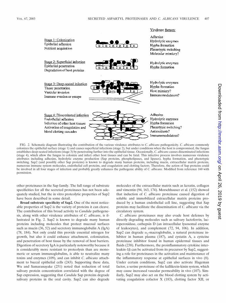
FIG. 2. Schematic diagram illustrating the contribution of the various virulence attributes to C. albicans pathogenicity. C. albicans commonlycolonizes the epithelial surface (stage 1) and causes superficial infections (stage 2), but under conditions when the host is compromised, the fungusestablishes deep-seated infections (stage 3) by penetrating further into the epithelial tissue. Occasionally, C. albicans causes disseminated infections(stage 4), which allow the fungus to colonize and infect other host tissues and can be fatal. This infective process involves numerous virulenceattributes including adhesins, hydrolytic enzyme production (Sap proteins, phospholipases, and lipases), hypha formation, and phenotypicswitching. Sap2 (and possibly other Sap proteins) is known to degrade many human proteins, including mucin, extracellular matrix proteins,numerous immune system molecules, endothelial cell proteins, and coagulation and clotting factors. Therefore, the action of Sap proteins couldbe involved in all four stages of infection and probably greatly enhances the pathogenic ability of C. albicans. Modified from reference 160 withpermission.
VOL. 67, 2003 SECRETED ASPARTYL PROTEINASES AND C. ALBICANS VIRULENCE 407
on April 26, 2019 by guest
http://mm
br.asm.org/
Dow
nloaded from
prothrombin, which may in turn result in the generation ofthrombin and hence blood clotting (105). The activation ofsuch host proteolytic cascades may not appear to be advanta-geous to C. albicans; however, the resulting deleterious effectsto the host may have some “downstream” beneficial affects thatmay assist or promote C. albicans infections.
Deducing proteinase specificity via three-dimensional struc-ture and molecular modeling. The substrate specificity of Sap2is noticeably very broad, and some researchers thought thatthis broad specificity could be deduced from its three-dimen-sional structure. Accordingly, two reports on the crystal struc-ture of Sap2 complexed with a potent inhibitor (A-70450 [see“Modulation of C. albicans virulence by aspartyl proteinaseinhibitors” below]) were published (1, 39), which indicated thatthe Sap2 structure conforms to the classical aspartyl proteinasefold typified by pepsin. However, comparisons of Sap2 withpepsin have revealed a number of major differences that maycontribute to the broad substrate specificity of Sap2 and whichmake the C. albicans proteinase family unique among the as-partyl proteinases (1, 39). Specifically, Sap2 has an enlargedand well-defined cavity for binding the third residue N-termi-nal to the cleaved bond in the substrate and two “flaps” over-lying this cavity, the latter observation being a hallmark of theSap family.
At present, the structure of Sap2 alone has been determined,and although other members of the Sap family are known tocontribute to C. albicans virulence, very few data are availableregarding their structures or substrate specificities. To addressthis, Stewart et al. (227) undertook a comparative structuralstudy with the sequences of SAP1 SAP6 by molecular model-ing. Although the structures of Sap1 to Sap6 and the electro-static charge of their active sites were generally similar, suffi-cient differences existed to allow for different substratespecificities, with the difference between Sap1 to Sap3 andSap4 to Sap6 being clearly evident. Furthermore, a potentiallysignificant trend in the total electrostatic charge of the Sap1 toSap6 enzymes was observed; the six enzymes had an overall netcharge of �8, �21, �22, �5, �2, and �2, respectively. It issomewhat puzzling that the charge of the non-active-site re-gions of Sap1 to Sap6 varies so much, but it might contributein part to our understanding of the different pH optima andrange of pH activities of the Sap enzymes (ranging from pH 2.0to 7.0). One more interesting observation resulting from themolecular modeling of Sap1 to Sap6 was the clear difference inthe carboxy end-terminal extension between SAP1 to SAP3and SAP4 to SAP6 at amino acid positions 323 to 324 and 335(SAP1 to SAP3, NE and A, SAP4 to SAP6 � RK and Q, E)(227). While speculative, this carboxy-terminal extension ap-pears to resemble an “attachment” anchor (C. Abad-Zapatero,personal communication). If true, this may support the hypoth-esis that the C. albicans proteinases may target specific cellproteins or tissue compartments during the infective process.
Discussion. Although proteinases other than Sap2 (specifi-cally Sap1 and Sap3 to Sap6) have recently been purified (16,210, 246), it remains to be determined whether they have thesame broad substrate specificities as Sap2. Furthermore, puri-fied proteins of Sap7, Sap8, Sap9, and Sap10 have not beenisolated or biochemically characterized, and thus the proteo-lytic properties of these proteinases remain totally unknown.On the one hand, it might seem unlikely that the different
members of the Sap family have the same broad substratespecificities as Sap2, since it would seem unnecessary for C.albicans to possess a family of 10 proteins which are differen-tially expressed under a variety of environmental conditionsand in different tissues (see “SAP gene expression during Can-dida infections” below) simply to digest the same substrates.On the other hand, C. albicans may require a family of extra-cellular proteolytic enzymes, each optimized to certain envi-ronmental conditions or different local pH values and/or par-ticular tissues, to help the fungus colonize and infect multiplesites of the body. However, there are clearly differences in thesubstrate specificities of the Sap proteins; for example, se-quence similarities of C. albicans Sap9 and Sap10 to the S.cerevisiae yapsins, including potential C-terminal consensus se-quences for GPI anchors, suggest that Sap9 and Sap10 mayhave different specificities and functions from the other C.albicans Sap proteins (A. Albrecht, I. Pichova, M. Monod, andB. Hube, unpublished data).
In all likelihood, there is probably considerable overlap inthe substrate specificities of many of the Sap members. Sincethe pH activity of the individual hydrolytic enzymes rangebetween pH 2.0 and 7.0 (75, 182, 210, 247), this would allow forthe concomitant expression of a number of similar SAP genesat environments with different pH values. In addition, there aredifferences in the promoter sequences of the different SAPgenes, which indicates that their expression might be con-trolled by different SAP-specific transcriptional regulators andpossibly suggests that the SAP genes might have evolved topossess distinct functions. Moreover, the coordinated regula-tion of the SAP genes with other virulence factors, includinghyphal formation and phenotypic switching, would permit sev-eral proteinases to act in unison to carry out a series of tasks tonot only digest a complex mixture of target proteins but also toprovide C. albicans with a biological advantage to specificallyenhance the pathogenic ability of the fungus (97). With theseconsiderations in mind, it is entirely plausible that C. albicanshas adapted to certain niche sites by expressing a combinationof SAP genes (and other virulence genes), which are calledupon as and when required.
Association of Sap Production with Other VirulenceProcesses of C. albicans
Main focus points. The main focus points are as follows.(i) Sap proteins facilitate C. albicans adherence to many host
tissues and cell types.(ii) Hypha formation and SAP4 to SAP6 expression are
coordinately regulated, but the signaling pathways remain tobe elucidated.
(iii) SAP1 appears to be regulated by phenotypic switching,but the contribution of switching to C. albicans virulence invivo is not yet clear.
Many of the early proteinase studies focused on the influ-ence of culture conditions on Sap expression and proteolyticactivity in vitro (reviewed in reference 94). However, after thediscovery of a SAP gene family, it became apparent that thisenzyme family had a more significant and complex contribu-tion to C. albicans pathogenicity. C. albicans is a polymorphicpathogen, which can exist in a yeast or a hyphal state and canundergo phenotypic switching (214). Therefore, it seemed log-
408 NAGLIK ET AL. MICROBIOL. MOL. BIOL. REV.
on April 26, 2019 by guest
http://mm
br.asm.org/
Dow
nloaded from
ical to assume that due to the large number of proteinasespresent in C. albicans, the SAP gene family may be differen-tially expressed in the different morphological forms. As aresult, the relationship between proteinase production and hy-phal production, phenotypic switching, and other putative vir-ulence attributes of C. albicans including adherence wasinvestigated.
Sap production and C. albicans adherence. Adhesion ofCandida to host tissues allows the fungus to attain a footholdand to colonize a specific niche environment. Under suitablepredisposing conditions when the host is compromised, thiscolonized site provides the base for candidal proliferation,invasion, and, in some instances, dissemination. Adherence ofC. albicans to host cells is a complex, multifactorial processinvolving several types of candidal adhesins on a morphogeni-cally changing cell surface (reviewed in the references in Table1), and one mechanism through which Candida adherencemight be promoted is via the production of proteinases.
One of the first early studies to link proteinase production toadherence in C. albicans showed that strongly proteolyticstrains of C. albicans adhered significantly more strongly tohuman buccal epithelial cells in vitro than did strains produc-ing less proteinase (74). A more recent report correlated pro-teinase production with increased adherence to buccal epithe-lial cells and death of mice; the higher-Sap-producing strainsshowed greater levels of tissue colonization in the liver, kid-neys, and spleen (2). However, the majority of studies linkingSap production with C. albicans adherence have been per-formed using the proteinase inhibitor pepstatin, which inhibitsSap2 (and probably Sap1 and Sap3) very efficiently (168). Borgand Ruchel (12) demonstrated a marked reduction in C. albi-cans adhesion and invasion of human mucosa by pepstatin, anda similar reduction of C. albicans adherence using pepstatinwas also shown with human epidermal cells (60, 165). Pepstatincould also inhibit the development of cavitations after yeastcells adhered to epidermal corneocytes (173). Some years ear-lier, Klotz et al. (114) observed that yeast cells formed cavita-tions and burrowed rapidly into vascular endothelium in vitroby a mechanism independent of germ tube formation. How-ever, at that time the burrowing was not associated with pro-teinase production, but the results of the work by Ray andPayne (173) clearly implicated proteinases in the process.
The actual Sap proteins involved in adherence and cavita-tion (and possibly subsequent penetration) of host tissues werenot studied, but a recent report by Kvaal et al. (121) indicatedthat Sap1 might be involved. Using a gene misexpression strat-egy in the switching strain WO-1, in which white-phase cellsmisexpressed the opaque-specific gene SAP1, the authors dem-onstrated in a cutaneous mouse model that SAP1 conferredtwo opaque-specific characteristics upon white cells: increasedadhesion and the capacity to cavitate skin (237). Interestingly,the addition of pepstatin inhibited cavitation but not the en-hanced adhesion (which confirmed the data of Ray and Payne[173]), suggesting that cavitation was the consequence of se-creted Sap1 enzyme, while increased adhesion was the result ofother cell-associated factors. Other, more recent studies usingHIV aspartyl proteinase inhibitors have also implicated Sap1to Sap3 in C. albicans adherence; however, these studies areexplained in full in “Modulation of C. albicans virulence byaspartyl proteinase inhibitors” (below).
(i) How do Sap proteins contribute to adherence? Thesepepstatin studies demonstrating the inhibition of C. albicansadherence clearly indicate that the Sap family plays some kindof role in C. albicans adherence. Although the precise mech-anisms by which Sap proteins contribute to the adherenceprocess are not clear, two hypotheses are currently favored. Inthe first, C. albicans proteinases could act as ligands to surfacemoieties on host cells, which does not necessarily require ac-tivity of the enzymes. In the second, C. albicans utilizes Sapproteins as active enzymes to modify target proteins or ligandson the fungal surface or on host cells (i.e., epithelial cells),which may alter surface hydrophobicity or lead to conforma-tional changes, thus allowing better adhesion of the fungus(145). If the Sap proteins can indeed function directly as C.albicans adhesins, this will add to the growing number of vir-ulence properties already possessed by the Sap family (i.e.,tissue damage, invasion, and evasion of host defenses) andestablish the proteinases as one of the most versatile and mul-tifunctional virulence gene families possessed by C. albicans.
Sap production and yeast-to-hypha transition. Research ef-forts by many investigators in different laboratories have con-centrated on the study of C. albicans morphogenesis, as well asthe identification and characterization of cell wall componentsthat are growth phase (yeast and hypha) specific and associatedwith virulence. The foundation of these studies is based on twofactors: (i) the common acceptance that the hyphal form isrelated to the invasive properties of C. albicans and (ii) theimportance of morphogenesis as a biological phenomenon.
The ability of C. albicans to transform into hyphae may beconsidered a pathogenic determinant in the initial processes ofsuperficial tissue invasion, whereby hyphae may promote theadherence and penetration of C. albicans to host tissues. Inculture medium, the main proteinases associated with hyphalformation are the SAP4 to SAP6 subfamily (96, 246), and pHand hypha induction alone are sufficient for the induction ofSAP4 to SAP6 (it should be noted that SAP4 transcripts werenot detected in several experiments).
(i) Coordinate regulation of hypha-formation and SAP4 toSAP6 expression. Although hypha formation and SAP4 toSAP6 expression were linked, more direct proof was requiredto determine whether the two phenomena were coordinatelyregulated. Sequence analysis of the promoter regions of SAP4to SAP6 revealed the presence of consensus sequences [CATTC(A/C)] for the TEA/ATTS transcription factor Tec1 (206).C. albicans mutant strains lacking TEC1 failed to producehyphal cells in vitro and were not able to express SAP4 toSAP6, suggesting the existence of joint or coordinated regula-tory pathways for hypha production and proteinase expression.The concept of coordinated pathways was supported by theobservation that SAP4 to SAP6 expression increased in a hy-perfilamentous strain lacking CPP1 (a mitogen-activated pro-tein kinase phosphatase) (205) and had a modified expressionpattern in a strain lacking EFG1 (a key transcriptional regula-tor of dimorphism), which has a strongly reduced ability toform hyphae (228). Moreover, in a murine systemic intraperi-toneal model, the EFG1-deficient mutant had a strongly re-duced ability to produce hyphae, which was associated withreduced expression of SAP4 to SAP6 and an inability to invadeor damage parenchymal organs including the liver and pan-creas (65). Interestingly, a triple null C. albicans mutant lacking
VOL. 67, 2003 SECRETED ASPARTYL PROTEINASES AND C. ALBICANS VIRULENCE 409
on April 26, 2019 by guest
http://mm
br.asm.org/
Dow
nloaded from
SAP4 to SAP6 showed strongly reduced invasiveness but stillproduced hyphal cells. Finally, SAP5 activation during in vivoinfection was shown not to depend on growth of C. albicans inthe hyphal form; however, the two major hyphal signalingpathways in C. albicans (defined by Cph1 and Efg1) wererequired for SAP5 expression (224). Together, these studiesindicate that not only are hypha formation and proteinaseproduction coordinately regulated but also C. albicans hyphalcells require the support of hydrolytic enzymes (specificallySAP4 to SAP6) in order to be fully invasive in vivo.
The observation that certain transcriptional factors whichregulate the yeast-to-hypha transition also regulate proteinaseexpression has recently been addressed using C. albicans DNAmicroarrays. Transcriptional profiling of C. albicans mutantslacking factors that regulate the dimorphic transition hashelped to elucidate signaling pathways and to clarify the coor-dinated regulation between morphology and proteinase pro-duction. The reader is guided to “Functional genomics andCandida” (below) for more details.
Sap production and phenotypic switching. The selection ofphenotypically altered strains may be enhanced in C. albicansby a phenomenon known as high-frequency phenotypic switch-ing, whereby Candida cells randomly switch their phenotype,especially in response to stress (217, 218). While many pro-karyotic and eukaryotic microorganisms can switch betweenalternate phenotypes under different environmental conditions(207), C. albicans appears to have an enhanced ability forchromosomal rearrangement and genetic reorganization. Un-like switching in other microbial pathogens, switching in C.albicans is pleiotropic, affecting several morphological andphysiological parameters and a number of virulence traits(214), all of which may allow the fungus to adapt to differenthost environments during the course of an infection. Recentwork suggests that phenotypic switching is based on heritablechanges in chromatin structure and supports the notion thatacetylation of histones plays a selective role in regulating theswitching process (113, 220).
(i) Which SAP genes are regulated by phenotypic switching?In a C. albicans strain named WO-1, the discovery and char-acterization of the white (W)-to-opaque (O) transition indi-cated that switching could affect a variety of cellular charac-teristics, including proteinase production (214). As a result, acorrelation between Sap secretion and switching was subse-quently described in C. albicans strains WO-1 (151) and 3153A(149). In both strains, transcripts of SAP1 were abundant inspecific switching-regulated forms and Sap1 was primarily re-sponsible for the higher extracellular proteolytic activity ob-served in these switching states. As a result, SAP1 was the firstcloned switching-regulated gene detected in C. albicans (151),a year after the gene was first isolated by Hube et al. (101).
Sequence analysis of the 5�-untranslated regions of SAP1from different C. albicans strains indicated that during switch-ing, SAP1 expression was regulated by activation or deactiva-tion of phase-specific trans-acting factors, which in turn wereregulated by a “master switch” event (215). Since SAP1 wasshown to be switching regulated, it was not surprising that theexpression of this gene was not dependent on the presence ofexogenous protein (96, 151, 246). This is in contrast to SAP2(not switching regulated), which is expressed in both the white
and opaque phenotypes of C. albicans strain WO-1 but only inthe presence of exogenous protein (96, 246).
SAP3 expression may also be regulated by phenotypicswitching (150, 247), but its regulation is different from that ofSAP1 and SAP2 in that SAP3 is detected in C. albicans strainswhen SAP2 is expressed (96, 210, 246). Another SAP gene thatis differentially expressed during switching is SAP8, since tran-scripts were detected in the opaque but not the white pheno-type (99). However, since SAP8 was shown to be up-regulatedat 25°C compared with 37°C (146) and since the opaque phe-notype is stable only at 25°C (149, 151, 219), this suggested thatSAP8 expression in opaque cells may be temperature regulatedrather than switching regulated.
At present, in C. albicans strain WO-1, SAP1 is the onlyproteinase that is strictly regulated by phenotypic switching.However, phenotypic switching is a very complicated process,which is by no means fully understood. In fact, very little isknown about this phenomenon outside of C. albicans strainWO-1. Other clinical or laboratory strains may have switchingprocesses divergent from or even unrelated to that of WO-1,each affecting SAP gene expression and other virulence genesin distinctive ways. Therefore, it cannot yet be concluded whichproteinases are regulated by switching in vivo or what contri-bution this phenomenon makes, in terms of SAP gene expres-sion, to the virulence of C. albicans.
Discussion. In summary, laboratory studies have indicatedthat the C. albicans SAP gene family is differentially expressedin the yeast, hyphal, and phenotypically switched states andmay contribute to C. albicans adherence. At the most basiclevel, one could conclude that yeast cells predominantly ex-press one set of SAP genes (SAP1 to SAP3), hyphae predom-inantly express another (SAP4 to SAP6), and phenotypicallyswitched cells predominantly express yet another (SAP1 andSAP3). Although this might be attractive, it is almost certainlytoo simplistic, since these conclusions have usually been drawnfrom the use of one strain of C. albicans grown under labora-tory-controlled conditions. In vivo, the environmental milieuand immune selective pressures may affect SAP gene expres-sion and phenotypic switching in individual yeast and hyphalcells in a unique fashion, which cannot be tested or controlledfor in the laboratory. Therefore, it is quite possible that theSAP genes expressed by C. albicans cells in the laboratory maynot equate to the SAP genes expressed in vivo. Determinationof exactly which SAP genes are expressed by the two morpho-logical forms and during phenotypic switching at the single-celllevel in vivo may provide a significant step forward in elucidat-ing the complex interaction between the host environment andSAP gene regulation.
Sap Protein Production and Sap Immune Responses inAnimal and Human Infections
Main focus points. The main focus points are as follows.(i) Sap proteins are produced in vivo during mucosal and
systemic infections.(ii) Proteinases are localized to the cell wall during C. albi-
cans infections.(iii) C. albicans proteinases are immunogenic and elicit mu-
cosal and systemic antibody responses.
410 NAGLIK ET AL. MICROBIOL. MOL. BIOL. REV.
on April 26, 2019 by guest
http://mm
br.asm.org/
Dow
nloaded from

(iv) The inhibitory and protective effects of Sap antibodiesagainst Candida infections remain unclear and the protectiveB- and T-cell epitopes of the Sap family are unknown.
Sap protein production during C. albicans infections. Sev-eral studies have provided strong evidence demonstrating theproduction of Sap protein in vivo. Early work using murinemodels of disseminated candidiasis revealed the presence ofSap proteins on the surface of C. albicans cells in murinekidneys (116, 132). Using indirect-immunofluroescence mi-croscopy, the presence of Sap proteins was also detected withinthe cell wall of yeast and hyphal cells in all organs of immu-nocompromised patients who had succumbed to systemic C.albicans infections, including the mucosa, central nervous sys-tem, lungs, heart, liver, pancreas, and kidneys (190). Withregard to mucosal infections, elevated levels of Sap proteinswere observed in vaginal fluids of candidiasis patients com-pared with Candida carrier subjects as determined by enzyme-linked immunosorbent assay and immunoblotting using Sap2polyclonal antibodies (43), indicating a link between Sap pro-duction in vivo and infection.
The presence of Sap protein has also been demonstratedduring phagocytosis of C. albicans by leukocytes. Macdonaldand Odds (134) were the first to show that the resistance of C.albicans to phagocytosis was associated with Sap expression.Some years later, it was observed that C. albicans and C.tropicalis yeast cells that resisted phagocytic killing germinatedintracellularly and expressed Sap on their surface (13). Re-cently, the Sap4 to Sap6 family have been implicated in theevasion of phagocytosis by C. albicans, since the expression ofSap4 to Sap6 but not Sap1 to Sap3 was upregulated on yeastand germ tubes after phagocytosis by murine peritoneal mac-rophages (16). In the same study, C. albicans mutants lackingSAP4 to SAP6 were significantly more susceptible to phagocy-tosis than were wild-type cells. These results strongly indicatethat by preventing macrophage killing, Sap4 to Sap6 play atleast one significant role in evading host immune defenses.
Sap localization to the cell wall. More recent studies usingimmunogold-labeling techniques demonstrated that Sap pro-teins are localized to the cell wall during C. albicans infections.In a rat vaginitis model, Sap1 to Sap3 were present in the yeastcell wall during early stages of infection, a pattern that corre-lated with the in vitro localization of Sap (229). Three studiesusing polyclonal antibodies raised against Sap1 to Sap6 dem-onstrated the presence of Sap1 to Sap3 on the surface of bothyeast and hyphal cells, while Sap4 to Sap6 antigens were foundpredominantly on hyphal cells (65, 119, 202). Also, in biopsyspecimens of oral epithelial lesions collected from three HIV-infected patients with oropharyngeal candidiasis, most Sap wassecreted at the locations where C. albicans directly adhered toepithelial cells or at sites in close contact between C. albicansand epithelial cells (199). However, it is important to recognizethat the Sap1 to Sap3 and Sap4 to Sap6 antibodies used werenot able to differentiate between the individual Sap proteinswithin each of the two subfamilies investigated. Therefore, theidentities of the individual proteinases that localize to the cellwall or are detected in vivo during experimental infectionscould not be determined. Antibodies specific for individual Sapproteins do not yet exist, but the development of such antibod-ies would be a valuable addition to existing molecular tech-niques in determining the localization patterns of the individ-
ual proteinases as well as the expression patterns of theproteinases during different stages and types of C. albicansinfections.
Antibodies against Sap induced by C. albicans infections. (i)Antibodies produced during systemic infection. Numerousstudies have described the presence of Sap protein during C.albicans infections; however, few studies have described anti-body responses to the C. albicans proteinases in human pa-tients. Macdonald and Odds (133) were the first to detectproteinase-specific IgG antibodies in sera of patients with dis-seminated candidiasis at significantly higher levels than thosefound in healthy individuals. These findings were later con-firmed by Ray and Payne (172) and Ruchel et al. (186, 187),further demonstrating the production of proteinases duringsystemic candidiasis. However, in the latter study, sera of a fifthof the patients suffering from candidiasis did not produce hightiters of antibodies against purified Sap2, probably reflectingthe inability of many high-risk patients to mount a normalimmune response (185).
(ii) Antibodies produced during mucosal infection. Theabove studies investigated anti-Sap IgG responses during sys-temic infections, but few studies have investigated the IgAresponse, and in particular secretory IgA, to Sap proteins dur-ing mucosal Candida infections, such as those in the oral cavityand vaginal lumen. This would clearly be more relevant thanIgG responses, since IgA is the predominant antibody presentat mucosal surfaces and is known to prevent the attachment ofC. albicans to the mucosal epithelium (243).
Two reports have recently addressed this issue. Using atime-resolved immunofluorometric assay, total levels of IgAagainst Sap1, Sap2, and Sap6 were found to be higher in salivafrom HIV-positive patients with oral and oropharyngeal can-didiasis than from HIV-positive patients without oral candidi-asis or HIV-negative healthy controls (57, 142). The authorsconcluded that during oral infection, HIV-positive patientshave an increased mucosal antibody response specifically di-rected against C. albicans virulence antigens, in this case theproteinases (57). However, this study did not include an HIV-negative patient group with oral candidiasis, so the observedincrease in the level of salivary IgA against the proteinases maybe related to the HIV status of the individual as well as tocandidiasis. Interestingly, over the 1-year period, variations inCandida colonization levels in the oral cavity and episodes oforopharyngeal candidiasis correlated with variations in salivaryanti-Sap6 IgA antibody levels (142). This may indicate a directrelationship whereby as C. albicans numbers proliferate duringmucosal infections, more Sap6 is produced, resulting in theinduction of a corresponding mucosal IgA antibody response.However, since it is highly likely that the polyclonal antibodiesinduced by certain Sap proteins during human mucosal infec-tions cross-react with other Sap proteins (especially withinhomologous subfamilies), more work is clearly needed beforethis hypothesis can be substantiated.
Functional anti-Sap antibodies. Antibodies have many func-tions in many diseases, but the role of antibody immunity inprotection against mucosal and systemic candidiasis is unclear.Indeed, the majority of patients with mucosal Candida infec-tions have normal or even elevated levels of both serum andmucosal anti-Candida antibodies (37, 112, 137, 233). This in-dicates that although patients are able to produce high levels of
VOL. 67, 2003 SECRETED ASPARTYL PROTEINASES AND C. ALBICANS VIRULENCE 411
on April 26, 2019 by guest
http://mm
br.asm.org/
Dow
nloaded from
antibodies in response to Candida infection, these high anti-body titers are not able to clear candidal infection. However,secretory IgA antibodies are able to bind to Candida andreduce the adherence of Candida to epithelial cells, theoreti-cally preventing or maintaining low levels of Candida coloni-zation (126, 243).
(i) Systemic infections. In patients with systemic candidiasis,there is some evidence for a role of anti-C. albicans antibodiesin recovery from infection. Development of antibodies againstC. albicans heat shock protein 90 (HSP90) appear to be pro-tective against human and animal Candida infections; highertiters of antibodies against a 47-kDa breakdown product of C.albicans HSP90 were present in patients recovering from can-didemia than in the early stages of infection (108, 139), andantibodies against this 47-kDa product were protective in amurine model of systemic candidiasis (138). In addition, Cutlerand coworkers identified an IgM (84, 85) and IgG3 (86) anti-body-recognizing phosphomannoprotein that is protectiveagainst systemic (84, 86) and vaginal (85, 86) candidiasis. Thesestudies, at the very least, indicate that antibodies can be pro-tective against Candida infections. However, very little infor-mation is available concerning a protective role of Sap anti-bodies against systemic Candida infections.
(ii) Mucosal infections. More data are available with regardto the protective affects of mucosal Sap antibodies against C.albicans infections. De Bernardis et al. (45) published somepromising data obtained with the rat vaginitis model, showingthat immunization with Sap2 antigen, or administration of ananti-Sap2 monoclonal antibody or anti-Sap2 antibody-contain-ing vaginal fluids, partially protected rats against candidal vag-initis. Protection by antibody was T-cell dependent and wasconferred by the Sap2 antibodies, since preabsorption of thefluids with Sap2 antigen reduced the level of protection. How-ever, it is likely that the protective anti-Sap2 antibodies cross-reacted with other proteinases such as Sap1 and Sap3, sincethese are thought to have similar epitopes (210, 246, 247).Although the mechanism of protection was not elucidated, thiswas the first demonstration that anti-Sap antibodies could af-ford protection against C. albicans infections in vivo and indi-cated that not only might Sap2 be the main proteinase con-tributing to rat vaginal infections but also that Sap2 could bethe main target of the host immune response affording protec-tion at mucosal sites.
(iii) Inhibitory antibodies. While C. albicans proteinases areknown to induce antibody responses in humans (57, 133, 142,186), the inhibitory and protective effects of Sap antibodiesproduced by infected hosts remain unclear. While one study byBorg et al. (14) demonstrated that three monoclonal IgM an-tibodies raised against Sap2 did not inhibit enzyme activity, itis not generally known whether antibody responses to Sapproteins include inhibitory antibodies. The demonstration ofdirect inhibition of Sap activity by antibodies resulting in aprotective effect in vivo would open up a new avenue of re-search and is an important area for further study.
(iv) Sap B-cell epitopes. Only one study has endeavored todetermine the B-cell epitopes of Candida Sap proteins fornondiagnostic purposes. Using sera from patients with oral andsystemic candidiasis, Ghadjari et al. (73) delineated six Sap2IgG and IgM epitopes. Human recombinant antibodies againsttwo of these epitopes, though, were not protective in a mouse
model of lethal systemic candidiasis. Nevertheless, this studyprovided some information regarding the Sap2 epitopes rec-ognized by serum IgG and IgM antibodies. As yet, the mappingof epitopes relevant to mucosal immune protection by usingsaliva or vaginal secretions has not been performed for animalsor humans. This would allow the identification of IgA epitopes,which would be particularly relevant to C. albicans infection atmucosal sites and would be more relevant for mucosal infec-tions than would identifying serum IgG and IgM epitopes.Furthermore, B-cell and T-cell mapping of other members ofthe Sap family has not yet been undertaken, even though thereis now solid evidence to indicate that they probably play sig-nificant but distinct roles during different C. albicans infec-tions. Identification of such epitopes does not necessarily implythat they will be relevant to infection or host protection, but,given the crucial relationship between C. albicans pathogenesisand the immune status of the host, further studies in this areaare clearly required.
SAP Gene Expression during Candida Infections
Main focus points. The main focus points are as follows.(i) SAP expression has been detected in all types of C.
albicans infections by using various gene expression detectiontechniques.
(ii) In vitro reconstituted human epithelium (RHE) modelsappear to be good surrogates of human infections.
(iii) C. albicans expresses the SAP4 to SAP6 subfamily in allmucosal and systemic infections examined.
(iv) Future molecular studies will provide a more accuraterepresentation of SAP gene expression during various types ofC. albicans infections, particularly in humans.
The discovery that C. albicans possessed a multitude of pro-teinase genes that were differentially expressed under a varietyof environmental conditions in vitro (96, 246) led to the at-tractive proposition that different members of the Sap familymight also be differentially expressed in vivo and might con-tribute to different C. albicans infections. This concept, to-gether with the knowledge that C. albicans inhabits a diversenumber of host niches, was the driving force behind subse-quent studies that investigated SAP gene expression in severalmodels to ascertain which proteinases were expressed in whichinfections. This research involved human samples, animalmodels, and in vitro experimental human infections, and thefindings are summarized in Table 5.
Human infections. The precise roles and functions of the C.albicans proteinases during human infections are currently notclear (although information can be extrapolated from in vitroand in vivo animal model data [see below]). However, beforethese roles and functions can be investigated, the expression ofthe SAP gene family during different human infections needsto be assessed in order to determine which proteinases areproduced in vivo. Naglik et al. (156) published the only de-tailed study of humans, in which SAP1 to SAP7 expression inthe oral cavities of both patients with oral candidiasis (n � 10)and asymptomatic Candida carriers (n � 8) was analyzed (Ta-ble 5). SAP2 and SAP4 to SAP6 (SAP4 to SAP6 were detectedtogether as a subfamily and not individually) were the predom-inant proteinase genes expressed in the oral cavities of infectedpatients and Candida carriers, while SAP1 and SAP3 tran-
412 NAGLIK ET AL. MICROBIOL. MOL. BIOL. REV.
on April 26, 2019 by guest
http://mm
br.asm.org/
Dow
nloaded from
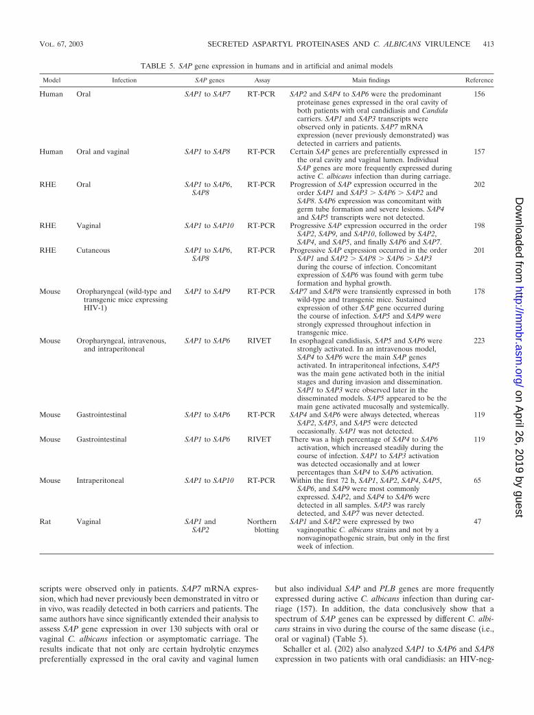
scripts were observed only in patients. SAP7 mRNA expres-sion, which had never previously been demonstrated in vitro orin vivo, was readily detected in both carriers and patients. Thesame authors have since significantly extended their analysis toassess SAP gene expression in over 130 subjects with oral orvaginal C. albicans infection or asymptomatic carriage. Theresults indicate that not only are certain hydrolytic enzymespreferentially expressed in the oral cavity and vaginal lumen
but also individual SAP and PLB genes are more frequentlyexpressed during active C. albicans infection than during car-riage (157). In addition, the data conclusively show that aspectrum of SAP genes can be expressed by different C. albi-cans strains in vivo during the course of the same disease (i.e.,oral or vaginal) (Table 5).
Schaller et al. (202) also analyzed SAP1 to SAP6 and SAP8expression in two patients with oral candidiasis: an HIV-neg-
TABLE 5. SAP gene expression in humans and in artificial and animal models
Model Infection SAP genes Assay Main findings Reference
Human Oral SAP1 to SAP7 RT-PCR SAP2 and SAP4 to SAP6 were the predominantproteinase genes expressed in the oral cavity ofboth patients with oral candidiasis and Candidacarriers. SAP1 and SAP3 transcripts wereobserved only in patients. SAP7 mRNAexpression (never previously demonstrated) wasdetected in carriers and patients.
156
Human Oral and vaginal SAP1 to SAP8 RT-PCR Certain SAP genes are preferentially expressed inthe oral cavity and vaginal lumen. IndividualSAP genes are more frequently expressed duringactive C. albicans infection than during carriage.
157
RHE Oral SAP1 to SAP6,SAP8
RT-PCR Progression of SAP expression occurred in theorder SAP1 and SAP3 � SAP6 � SAP2 andSAP8. SAP6 expression was concomitant withgerm tube formation and severe lesions. SAP4and SAP5 transcripts were not detected.
202
RHE Vaginal SAP1 to SAP10 RT-PCR Progressive SAP expression occurred in the orderSAP2, SAP9, and SAP10, followed by SAP2,SAP4, and SAP5, and finally SAP6 and SAP7.
198
RHE Cutaneous SAP1 to SAP6,SAP8
RT-PCR Progressive SAP expression occurred in the orderSAP1 and SAP2 � SAP8 � SAP6 � SAP3during the course of infection. Concomitantexpression of SAP6 was found with germ tubeformation and hyphal growth.
201
Mouse Oropharyngeal (wild-type andtransgenic mice expressingHIV-1)
SAP1 to SAP9 RT-PCR SAP7 and SAP8 were transiently expressed in bothwild-type and transgenic mice. Sustainedexpression of other SAP gene occurred duringthe course of infection. SAP5 and SAP9 werestrongly expressed throughout infection intransgenic mice.
178
Mouse Oropharyngeal, intravenous,and intraperitoneal
SAP1 to SAP6 RIVET In esophageal candidiasis, SAP5 and SAP6 werestrongly activated. In an intravenous model,SAP4 to SAP6 were the main SAP genesactivated. In intraperitoneal infections, SAP5was the main gene activated both in the initialstages and during invasion and dissemination.SAP1 to SAP3 were observed later in thedisseminated models. SAP5 appeared to be themain gene activated mucosally and systemically.
223
Mouse Gastrointestinal SAP1 to SAP6 RT-PCR SAP4 and SAP6 were always detected, whereasSAP2, SAP3, and SAP5 were detectedoccasionally. SAP1 was not detected.
119
Mouse Gastrointestinal SAP1 to SAP6 RIVET There was a high percentage of SAP4 to SAP6activation, which increased steadily during thecourse of infection. SAP1 to SAP3 activationwas detected occasionally and at lowerpercentages than SAP4 to SAP6 activation.
119
Mouse Intraperitoneal SAP1 to SAP10 RT-PCR Within the first 72 h, SAP1, SAP2, SAP4, SAP5,SAP6, and SAP9 were most commonlyexpressed. SAP2, and SAP4 to SAP6 weredetected in all samples. SAP3 was rarelydetected, and SAP7 was never detected.
65
Rat Vaginal SAP1 andSAP2
Northernblotting
SAP1 and SAP2 were expressed by twovaginopathic C. albicans strains and not by anonvaginopathogenic strain, but only in the firstweek of infection.
47
VOL. 67, 2003 SECRETED ASPARTYL PROTEINASES AND C. ALBICANS VIRULENCE 413
on April 26, 2019 by guest
http://mm
br.asm.org/
Dow
nloaded from
ative female and an HIV-positive male, both suffering frompseudomembranous candidiasis. This analysis was undertakento confirm the SAP expression data obtained using an in vitromodel of oral candidiasis based on RHE and to determinewhether the RHE models could be used as surrogates forhuman infections (see below). However, since only two patientsamples were analyzed, no conclusions could be drawn regard-ing which SAP genes were associated with human oralcandidiasis.
These are the only studies to have analyzed the expression ofa C. albicans virulence gene family in any human infection,which highlights the distinct lack of human gene expressiondata available. Future experiments using patient samplesshould bring us one step closer to identifying the genes andproteins (SAP or otherwise) that are directly involved in Can-dida pathogenesis in humans.
In vitro artificial experimental infections based on RHE.Most studies that analyze the virulence of Candida species useanimal models (see below) rather than human subjects. How-ever, recently a number of researchers have taken advantage ofartificial RHE models and used them as an alternative tosamples isolated directly from human patients. The RHE mod-els (oral, vaginal, skin, and other cell types) can be obtainedcommercially and are prepared on an inert supporting mem-brane (190a). The models are highly reproducible, closely rep-resent the human epithelium in vivo, and provide clear answersregarding gene expression that are not complicated by non-epithelial-cell factors. This, therefore, allows the direct analysisof the interaction between C. albicans and epithelial cells at themucosal surface. Furthermore, it allows postulates to beproved experimentally.
Using an RHE model of oral candidiasis and the applicationof reverse transcription-PCR (RT-PCR), Schaller et al. (202)showed a distinct order of SAP gene expression (SAP1 andSAP3 � SAP6 � SAP2 and SAP8) during the course of infec-tion. In this model, Sap1 to Sap3, but not Sap4 to Sap6, wereassociated with tissue damage (200). Interestingly, a differentSAP expression pattern was observed using an RHE model ofvaginal candidiasis: SAP2, SAP9, and SAP10 transcripts wereobserved initially, followed by SAP1, SAP4, and SAP5 (con-comitant with lesions), with SAP6 and SAP7 being expressedduring the later stages of infection (198). However, althoughthe gene expression pattern was different from that in the oralRHE model, studies with mutants indicated that Sap1 andSap2 were again the proteinases associated with tissue damage(198). Lastly, in an RHE model of cutaneous candidiasis, ex-pression of SAP genes in the order SAP1 and SAP2 � SAP8 �SAP6 � SAP3 was demonstrated when Candida cells pene-trated the corneal layer of the artificial skin (201). These stud-ies with RHE models clearly suggest that Sap1 to Sap3 are themain C. albicans proteinases contributing to the early stages ofmucocutaneous infections (Table 5).
Although the in vitro RHE models closely represent thehuman epithelium in vivo, it would be prudent to interpret thegene expression data with caution since the RHE models fallshort of directly representing the in vivo situation in humanpatients. Parameters such as pH changes, nutrient limitation,competition with oral flora, and humoral, cell-mediated, andcytokine responses are likely not only to have a profound effecton the ability of C. albicans to grow unchallenged on the
mucosal surface (as in the models) but also to provide stronglocal pressures that may influence the differential expression ofthe SAP gene family. However, despite this cautionary note,these RHE models have proved very effective for the directobservation of SAP gene expression in the initial stages ofsuperficial infections and, at present, are the best in vitrosurrogates for in vivo human Candida infections. The use ofthese artificial RHE models in the future should certainly leadto the discovery of new Candida genes that are expressed inhuman mucosal infections and should enlighten our under-standing of host-Candida interactions.
Animal experimental models. The development of animalmodels for specific diseases that precisely mimic their humanequivalent is the “gold standard” of animal research. However,given the dissimilar nature of humans and since C. albicans isan opportunistic pathogen that adapts continuously to its en-vironment, it is improbable that animal models can be devel-oped that are fully representative of human Candida infec-tions. Nevertheless, in truth, the use of animals is the best invivo alternative to study mucosal and systemic candidiasis, withthe mouse and rat models being the most commonly used. SAPgene expression in both mucosal and systemic C. albicans in-fections are described in Table 5.
(i) Mucosal models. De Bernardis et al. (47) were the first todemonstrate SAP gene expression in vivo in an estrogen-de-pendent rat vaginitis model. Northern analysis indicated thatSAP1 and SAP2 were expressed by two vaginopathic C. albi-cans strains but not by a nonvaginopathogenic strain, whichsupported a link between SAP expression and the virulencepotential of certain C. albicans strains. However, now that 10members of the SAP family have been identified, it would beinteresting to examine the SAP1 to SAP10 expression patternin this well-established rat model and compare the profileswith the RT-PCR and in vivo expression technology (IVET)mouse data described below. A comparison between humans,mice, and rats would provide an opportunity to determine howrepresentative the rat or mouse models are to human Candidainfections. The reader is guided to a detailed review recentlypublished by Samaranayake and Samaranayake (192), whichdiscusses the pros and cons of different animal models thathave been used to study Candida infections.
Several groups have reported detailed SAP gene expressionstudies in murine mucosal models. In a mouse model of gas-trointestinal infection, SAP4 and SAP6 were constitutively ex-pressed whereas SAP2, SAP3, and SAP5 mRNA were onlyoccasionally detected (119). Interestingly, SAP1 mRNA wasnot detected in this model, which is worthy of note becauseSAP1 expression has been observed in clinical C. albicans sam-ples from infected human patients and carriers (156), a mouseoropharyngeal model (178), a rat vaginitis model (47), and oral(202) and vaginal RHE models (198) (Table 5), all of which aremucosal infections.
In a mouse model of oropharyngeal candidiasis, a sequentialanalysis of the temporal expression of SAP1 to SAP9 in normaland transgenic mice expressing HIV-1 showed that SAP1 toSAP6 and SAP9 transcripts were detected continuouslythroughout the course of infection, with SAP5 and SAP9 beingmost strongly expressed in transgenic mice (178). SAP7 andSAP8 were the only genes to be expressed transiently, support-
414 NAGLIK ET AL. MICROBIOL. MOL. BIOL. REV.
on April 26, 2019 by guest
http://mm
br.asm.org/
Dow
nloaded from
ing the current evidence that members of the SAP family aredifferentially expressed during C. albicans infections (Table 5).
The SAP expression data described above were obtainedusing RT-PCR, but IVET, a relatively new technique, hasrecently been developed to analyze gene activation during in-fective processes (241). One of these technologies, the recom-bination-based IVET technique (RIVET), uses site-specificrecombination as a reporter of gene activation and is especiallysuited to detect the in vivo induction of genes that are onlytransiently expressed at a certain stage of infection (25),RIVET using FLP-recombinase has recently been adapted foruse with C. albicans by Staib et al. (223) to analyze the expres-sion of SAP1 to SAP6 in various mouse infection models. In amouse model of esophageal candidiasis, SAP5 and SAP6 werestrongly activated, indicating that these two SAP genes mightbe directly involved in the colonization and perhaps penetra-tion of mucosal surfaces (Table 5).
(ii) Systemic models. All SAP expression studies with animalsystemic models have been performed with mice. In an intra-peritoneal-infection model, SAP1, SAP2, SAP4, SAP5, SAP6,and SAP9 were discovered to be the most commonly expressedproteinase genes within the first 72 h (65). SAP2 and SAP4 toSAP6 transcripts were identified in all infected tissues at 4, 8,24, and 72 h, SAP3 and SAP10 were less frequently expressed(in less than 50% of the organs), and SAP7 was never detected.The lack of SAP7 expression is noteworthy since this gene isexpressed in human oral and vaginal infection and carriage(156, 157), in a mouse model of oropharyngeal candidiasis(178), and in the RHE vaginal model (198) (Table 5). It may bepremature to conclude that this implicates SAP7 specifically inmucosal rather than systemic infections, but it tentatively sup-ports some kind of role for this proteinase during humanmucosal infections.
Using RIVET, SAP4 to SAP6 were the main SAP genesactivated in an intravenous model of disseminated candidiasisand SAP5 was the main gene activated in disseminated intra-peritoneal infections, both at the initial time of inoculation andduring invasion and subsequent dissemination to the kidneys(223). However, SAP1 to SAP3 were also observed later in theinfective process (223, 225). This pattern of SAP gene activa-tion during systemic infections suggested a role for SAP4 toSAP6 during the initial phases of organ invasion and SAP1 toSAP3 in later phases, when C. albicans had already establishedinfection.
RIVET was also used to assess SAP1 to SAP6 gene activa-tion in a murine gastrointestinal model (119). SAP4 to SAP6activation was detected in a high percentage of C. albicanscells, which increased steadily during the course of infection. Incontrast, SAP1 to SAP3 activation was detected only occasion-ally and at lower percentages than SAP4 to SAP6. These resultscorrespond well to the RT-PCR expression analysis in thesame model, which demonstrated constitutive expression ofSAP4 and SAP6 and only the occasional detection of SAP2 andSAP3, with SAP1 transcripts never being observed (119). TheRT-PCR and RIVET studies thus appear to support some kindof role for SAP4 to SAP6 in both mucosal and systemic C.albicans infections.
Universal expression of the SAP4 to SAP6 subfamily at mu-cosal surfaces. A common feature of all the studies describedabove is the ubiquitous expression of the SAP4 to SAP6 sub-
family, regardless of the infection model (human or mouse,systemic or mucosal) or the assay technique used to detect SAPgene expression (RT-PCR or RIVET) (Table 5). Although adirect role for SAP4 to SAP6 in systemic Candida infectionshas been indicated from studies using SAP4- to SAP6-deficientstrains (see “Use of SAP-disrupted mutants to analyze C. al-bicans virulence” below), the prevalent expression of SAP4 toSAP6 in mucosal infections but the lack of attenuation ofvirulence using SAP4 to SAP6-deficient strains poses numerousquestions. (i) Are certain members of the SAP4 to SAP6 sub-family required by C. albicans for a housekeeping role or forsome kind of essential or constant interaction with mucosalsurfaces? (ii) Are SAP4 to SAP6 required simply for nutritionalpurposes in vivo, hence the necessity for their expression, orare they in fact essential for Candida virulence (i.e., invasionand tissue damage)? (iii) Alternatively, do SAP4 to SAP6 havenonenzymatic functions at mucosal surfaces? It is entirely fea-sible that although SAP4 to SAP6 are expressed by C. albicansin mucosal infections, they may not be required for virulence asthey appear to be in systemic infections. At present, the rolesand functions of the SAP4 to SAP6 subfamily during mucosalinfections, and specifically during the infective process in hu-mans, are not clear, but future functional studies might shedlight on why SAP4 to SAP6 are so prevalent at mucosalsurfaces.
Discussion and future directions for SAP expression stud-ies. While these SAP expression studies have been vital indetermining which SAP genes are associated with which typesof C. albicans infections, the true depth of their involvement inC. albicans pathogenicity has yet to be determined. This isconfounded by the many differences in SAP gene expressionprofiles observed between the studies (Table 5). Although thismay simply allude to the intricate and complex relationshipbetween Sap production, Candida pathogenesis, and the host,a number of other explanations are possible. These include thetechnique used for SAP gene analysis (RT-PCR, IVET, orNorthern blot analysis), the model used (human, mouse, rat, orRHE), or the site of infection studied (oral, oropharyngeal,vaginal, gastrointestinal, cutaneous, or systemic). In fact, thereare no directly comparable studies by different authors usingthe same technique, model, or site of infection (Table 5).
In addition to these disparities, the environmental condi-tions alone at the different infection sites, including pH, sub-strate availability, and ionic content, can differ markedly andcould partly account for the observed differences in SAP ex-pression between the studies. With these considerations inmind, it is entirely probable that C. albicans has adapted tothese niche sites in different hosts by expressing a different setof SAP genes. Furthermore, even within a single infection site,one would expect different gene expression patterns withinsingle C. albicans cells, but all the studies to date have de-scribed the average SAP expression pattern of a population ofcells.
Therefore, we require studies that use a single technique(RT-PCR or IVET) to analyze SAP gene expression in numer-ous infection models (human, mouse, rat, and RHE) or thatuse various gene expression techniques in a single infectionmodel. Ideally, the former should reveal differences in SAPgene expression between infected sites and the latter shouldgenerate matching SAP expression data, since the infection is
VOL. 67, 2003 SECRETED ASPARTYL PROTEINASES AND C. ALBICANS VIRULENCE 415
on April 26, 2019 by guest
http://mm
br.asm.org/
Dow
nloaded from
the same. The availability of more of these “multimodel”and/or “multitechnique” studies will add to our understandingof SAP gene expression in the host and its relevance to Can-dida pathogenesis.
Finally, the reader should note that the quantitative expres-sion of the SAP genes or any other virulence gene family ofCandida has not yet been assessed, and this is an importantnext step (particularly at the single-cell level) in addressing theup- and down-regulation of these virulence genes during var-ious stages of different C. albicans infections. In addition, nowthat the C. albicans genome sequence is complete (http://www-sequence.stanford.edu/group/candida), the development ofDNA microarrays specifically for Candida research should sig-nificantly expand our knowledge of which genes may be con-tributing not only to C. albicans infections but also specificallyto different types of human infections.
Modulation of C. albicans Virulence by AspartylProteinase Inhibitors
Main focus points. The main focus points are as follows.(i) Aspartyl proteinase inhibitors can inhibit C. albicans ad-
herence and attenuate mucosal infections.(ii) Proteinase inhibitors appear ineffective at preventing
systemic C. albicans infections.(iii) More conclusive studies using all main classes of pro-
teinase inhibitors are required.Drug-resistant Candida infections are likely to pose a serious
therapeutic challenge over the next few decades. Conse-quently, discoveries of novel drug classes are crucial aims ofantifungal research. One of the opportunities for the develop-ment of new compounds active against Candida species in-cludes the development of drugs directed against the Candidaproteinases. This is particularly pertinent now that the three-dimensional structure of one member of the Sap family (Sap2)has been determined (1, 39). Over the last two decades, andparticularly within the last few years, two areas of interest—therole of Candida proteinases in pathogenesis and their potentialas antifungal targets—have driven the use of aspartyl protein-ase inhibitors (PIs) in Candida research. These studies usingthe classical aspartyl PI pepstatin, HIV PIs, and computer-assisted structure-based designed inhibitors have provided di-rect evidence demonstrating the contribution of Sap proteinsto C. albicans virulence and have indicated that the proteinasefamily may be a possible target for antifungal research.
Pepstatin. The contribution of proteinases to C. albicansadherence, invasion, and infection has long been advocated,but it was not until the use of the classical aspartyl PI pepstatinthat many of these associations were confirmed. The studiesthat used pepstatin to demonstrate a link between Sap proteinsand adherence are detailed in “Association of Sap productionwith other virulence processes of C. albicans” (above) and arenot readdressed here, but the investigations using pepstatin toassociate Sap proteins with Candida penetration and invasionof host tissues are described below.
(i) Use of pepstatin in vitro. Colina et al. (36) showed thatthe digestion of mucin could be inhibited by pepstatin, indi-cating that Candida Sap proteins may degrade mucosal barrierproteins. This may allow C. albicans to gain access to the oraland gastrointestinal mucosa and may consequently indicate a
role for Candida proteinases in dissemination from these col-onized sites. Likewise, the addition of pepstatin inhibited the invitro digestion of soluble and immobilized extracellular matrixproteins produced by a human endothelial cell line (152).Again, this suggests that Candida Sap proteins contribute tocell damage and invasion of the subendothelial extracellularmatrix, which in turn could facilitate dissemination via thecirculatory system. In an RHE model of oral candidiasis, pep-statin was used to demonstrate a role for Sap1 to Sap3 but notSap4 to Sap6 in early tissue damage of oral epithelium (200).Pepstatin noticeably reduced the tissue damage caused by Sap1to Sap3; however, even in the presence of pepstatin, epithelialtissue damage was observed in late stages of RHE infections.This suggested that certain members of the Sap family thatmight not be inhibited effectively by pepstatin or other hydro-lytic enzymes of C. albicans such as phospholipases and lipases(which are not inhibited by pepstatin) might also contribute tothe development of mucosal lesions.
(ii) Use of pepstatin in vivo. While in vitro assays evaluatingthe effect of pepstatin on Candida infections are necessary andprovide valuable information, the most compelling biologicaldata for antifungal research come from in vivo studies of Can-dida infection. The first report of experiments using pepstatinin vivo was published in 1988 and described the use of asystemic model of murine candidiasis (58); however, no realprotective effect was observed after intravenous challenge withC. albicans. A similar lack of protection was also reported bytwo subsequent studies, using an in vivo pepstatin concentra-tion of up to 9 � 10�4 M (188, 258). However, a recent studyof murine peritonitis demonstrated that the addition of pep-statin (1 mg/kg) significantly reduced the amount of liver andpancreatic tissue damage, as determined by decreased levels ofalanine aminotransferase and �-amylase enzyme activities, re-spectively (120). Tissue damage appeared to be caused by Sap4to Sap6, a conclusion obtained from using SAP-deficientstrains (120). These findings indicated that pepstatin couldattenuate C. albicans virulence in a systemic murine intraperi-toneal model, probably by inhibiting tissue damage and inva-sion by Sap4 to Sap6.
While the evidence supporting a protective role for pepsta-tin in systemic animal models remain unconvincing, particu-larly after intravenous challenge with Candida, its potential useat mucosal surfaces is more convincing. Protective effects ofpepstatin were demonstrated in a rat vaginitis model, wherepostinfective administration of pepstatin (1 mg/ml) greatly ac-celerated the clearance of C. albicans from the rat vagina (45,49). Fallon et al. (64) showed in a mouse model that inhibitionof Sap proteins by pepstatin (0.06 to 6 mg/kg) prevented theinitial penetration of C. albicans through mucosal surfaces butnot the dissemination of C. albicans once the cells had alreadyreached the blood vessels. Thus, one conclusion may be thatpepstatin can prevent disseminated infections by inhibitingCandida penetration through the mucosal route but cannotinhibit systemic infections when the Candida is administeredvia the intravenous route. In other words, the route of Candidachallenge rather than the route of pepstatin administrationmight be more relevant to observe protective effects bypepstatin.
These studies indicate that proteinase inhibition by pepsta-tin can reduce the ability of C. albicans to colonize and invade
416 NAGLIK ET AL. MICROBIOL. MOL. BIOL. REV.
on April 26, 2019 by guest
http://mm
br.asm.org/
Dow
nloaded from

host tissues and illustrate the potential benefits of pepstatin invivo, chiefly in mucosal models. Unfortunately, however, pep-statin is not selective and is slightly toxic in animals, probablydue to its inhibitory action on host aspartyl proteinases, includ-ing cathepsin D and renin (188). Moreover, pepstatin accumu-lates in the liver and not the kidneys, which is a major targetorgan of C. albicans in systemic infections, and is thus likely tobe rapidly cleared from the blood in vivo. Given that theconcentrations of pepstatin used in the above experiments aremanyfold higher than those required to effectively inhibit Sap2(inhibition constant [Ki] for Sap2 of 2.9 nM and 50% inhibitoryconcentrations [IC50s] of 27 nM [1, 226]), rapid clearance maypartly explain the ineffectiveness of pepstatin in systemic mod-els after intravenous administration of Candida. Therefore,while pepstatin is a potent inhibitor of the C. albicans aspartylproteinases in vitro, its suitability as an antifungal agent in vivois not compelling.
HIV PIs. The interest in discovering new compounds thatinhibit Sap activity was recently given a surprising and unex-pected boost as a result of the evolving HIV epidemic. Since1996, the treatment of HIV-positive patients with highly activeantiretroviral therapy (HAART), which includes a cocktail ofHIV reverse transcriptase and HIV PIs, has proved successfulin delaying the onset of AIDS. Administration of HAARTresults in a significant improvement in the immune status ofthe HIV-positive individual, reflected by an increase in theCD4� T-cell levels. These patients also tend to have a dramat-ically reduced incidence of oropharyngeal candidiasis (54, 92,240). Since the Candida proteinases and the HIV proteinaseare members of the same aspartyl proteinase family, thesefindings led to the hypothesis that HIV PIs may also act againstCandida aspartyl proteinases in vivo and consequently preventor reduce candidal infections directly (31, 83, 89). A spate ofstudies has followed to determine whether HIV PIs couldinhibit C. albicans Sap proteins and whether they had anypotential as therapeutic agents in the treatment of C. albicans
infections. The main conclusions from these studies are thatHIV PIs are able to inhibit Sap activity, C. albicans adherence,and tissue damage but have a limited effect on C. albicansviability (Table 6).
(i) Inhibition of Candida proteinase activity by HIV PIs invitro. A total of four HIV PIs, namely ritonavir, saquinavir,indinavir, and nelfinavir, have been investigated for their abil-ity to inhibit Candida proteinase activity (15, 29, 80, 83, 117,168). Ritonavir was consistency found to be the most potentinhibitor of C. albicans Sap2 activity (15, 80, 168), with Ki of0.34 M (168), while saquinavir, indinavir, and nelfinavir in-hibited Sap2 activity to differing extents. One study did notobserve inhibition of Sap2 activity with indinavir or nelfinavir,which equated well with the poor Ki values of these com-pounds against Sap2 (Ki � �1,000 M for both) (168). Of thefour HIV PIs, only ritonavir and saquinavir were able to inhibitthe activity of secreted proteinases purified from C. tropicalis,C. parapsilosis, and C. lusitaniae as well as those purified fromC. albicans (168) (Table 6).
With regard to the inhibition of different members of theSap family, Borg-von Zepelin et al. (15) discovered that all fourHIV PIs specifically inhibited the C. albicans Sap1 to Sap3 butnot Sap4 to Sap6 enzymes. Interestingly, ritonavir and saquina-vir, but not indinavir, inhibited the adherence of C. albicans toVero epithelial cell cultures in a dose-dependent manner, thussupporting a role for Sap1 to Sap3 in the adherence process(15). Similar results were also obtained by Bektic et al. (10)using HeLa S3 epithelial cell cultures; these investigatorsshowed that ritonavir and saquinavir, as well as indinavir onthis occasion, inhibited C. albicans adhesion, with ritonavirbeing the most potent. However, none of the HIV PIs modu-lated the phagocytosis of Candida by polymorphonuclear leu-kocytes (10). Since Sap4 to Sap6 are thought to assist C. albi-cans in evading phagocytosis by murine macrophages (16),these data support the notion that the HIV PIs might notinhibit Sap4 to Sap6 activity (15).
TABLE 6. Effect of HIV PIs on Sap activity and C. albicans virulence
HIV PI Main findings Reference(s)
In vitroRitonavir, indinavir, saquinavir All three inhibited Sap2 activity. Only saquinavir was fungicidal, but only at
high doses.80
Indinavir Indinavir weakly inhibited Sap2 and reduced the amount of cell-bound andsecreted proteinase. C. albicans viability and growth were markedly reduced.
83
Ritonavir, indinavir,saquinavir, nelfinavir
Ritonavir and saquinavir inhibited Sap2 at micromolar concentrations,whereas indinavir and nelfinavir had no effect. Ritonavir and saquinavir alsoinhibited Sap proteins from C. tropicalis, C. parapsilosis, and C. lusitaniae.
168
Ritonavir, indinavir,saquinavir, nelfinavir
All four specifically inhibited Sap1 to Sap3, but not Sap4 to Sap6. Ritonavirand saquinavir inhibited C. albicans adherence to epithelial cells, whereasindinavir had no effect. No effect on C. albicans viability.
15
Saquinavir, indinavir Both inhibited Sap2 activity. Saquinavir strongly attenuated tissue damage inan in vitro RHE model of oral candidiasis.
117
Indinavir, ritonavir Neither had an antifungal effect. 55, 56Ritonavir, indinavir, saquinavir All three inhibited C. albicans adhesion to human epithelial cells; ritonavir
was the most potent. None modulated phagocytosis of Candida.10
In vivo modelsIndinavir, ritonavir Both inhibited Sap2 activity. Both exerted a therapeutic effect in an estrogen-
dependent rat vaginitis model.29
Indinavir, nelfinavir, ritonavir The anti-Sap effect of PI-HAART was associated with clinical resolution oforal candidiasis in HIV-positive patients.
30
VOL. 67, 2003 SECRETED ASPARTYL PROTEINASES AND C. ALBICANS VIRULENCE 417
on April 26, 2019 by guest
http://mm
br.asm.org/
Dow
nloaded from
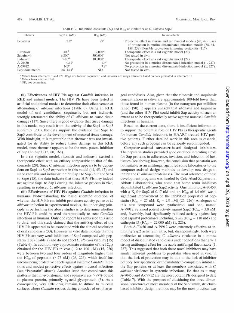
(ii) Effectiveness of HIV PIs against Candida infection inRHE and animal models. The HIV PIs have been tested inartificial and animal models to determine their effectiveness atattenuating C. albicans infections (Table 6). Using an RHEmodel of oral candidiasis, saquinavir, but not indinavir,strongly attenuated the ability of C. albicans to cause tissuedamage (117). Since there is good evidence that tissue damagein this model may result from the activity of the Sap1 to Sap3subfamily (200), the data support the evidence that Sap1 toSap3 contribute to the development of mucosal tissue damage.With hindsight, it is regrettable that ritonavir was not investi-gated for its ability to reduce tissue damage in this RHEmodel, since ritonavir appears to be the most potent inhibitorof Sap1 to Sap3 (15, 80, 168).
In a rat vaginitis model, ritonavir and indinavir exerted atherapeutic effect with an efficacy comparable to that of flu-conazole (29). Since C. albicans infection appears to be depen-dent on Sap1 to Sap3 expression in this model (44, 45, 47) andsince ritonavir and indinavir inhibit Sap1 to Sap3 but not Sap4to Sap6 (15), the data indicate that these HIV PIs may indeedact against Sap1 to Sap3 during the infective process in vivo,resulting in reduced C. albicans infection.
(iii) Effectiveness of HIV PIs against Candida infection inhumans. Notwithstanding the basic scientific question ofwhether the HIV PIs can inhibit proteinase activity per se or C.albicans infection in experimental models, the underlying prin-ciple in performing the above studies is to determine whetherthe HIV PIs could be used therapeutically to treat Candidainfections in humans. Only one report has addressed this issueto date, and this study claimed that the anti-Sap effect of theHIV PIs appeared to be associated with the clinical resolutionof oral candidiasis (30). However, in vitro data indicate that theHIV PIs are very weak inhibitors of Sap2 compared with pep-statin (168) (Table 7) and do not affect C. albicans viability (15)(Table 6). In addition, very approximate estimates of the IC50sobtained for the HIV PIs in vivo (2 to 100 M) (15, 226)were between two and four orders of magnitude higher thanthe IC50 of pepstatin (27 nM) (26, 226), which itself hasunconvincing protective effects against systemic Candida infec-tions and modest protective effects against mucosal infections(see “Pepstatin” above). Another issue that complicates thismatter is that in vivo ritonavir and saquinavir are �97% boundto plasma protein, primarily �1-acid glycoprotein (3). As aconsequence, very little drug remains to diffuse to mucosalsurfaces where Candida resides during episodes of oropharyn-
geal candidiasis. Also, given that the ritonavir and saquinavirconcentrations in saliva are approximately 100-fold lower thanthose found in human plasma (in the nanogram-per-milliliterrange) (90), it appears unlikely that ritonavir and saquinavir(and the other HIV PIs) could inhibit Sap activity to such anextent as to be therapeutically active against mucosal Candidainfections in humans.
Based on the current data, there is insufficient informationto support the potential role of HIV PIs as therapeutic agentsfor human Candida infections in HAART-treated HIV-posi-tive patients. Further detailed work in this area is essentialbefore any such proposal can be seriously recommended.
Computer-assisted structure-based designed inhibitors.The pepstatin studies provided good evidence indicating a rolefor Sap proteins in adherence, invasion, and infection of hosttissues (see above); however, the conclusion that pepstatin wasnot suitable as an antifungal agent led some laboratories to usecomputer-assisted design methods to develop new drugs toinhibit the C. albicans proteinases. The most advanced of thesedevelopment programs was headed by Cele Abad-Zapatero atAbbott Laboratories, who noted that some renin inhibitorsalso inhibited C. albicans Sap2 activity. One inhibitor, A-70450,with a Ki for Sap2 of 0.17 nM and an IC50 of 1.4 nM, was asignificant improvement on the inhibition properties of pep-statin (IC50 � 27 nM; Ki � 2.9 nM) (26, 226). Analogues ofthis new compound were synthesized, and one, namedA-79912, retained potent activity against Sap2 (IC50 � 3.8 nM)and, favorably, had significantly reduced activity against keyhost aspartyl proteinases including renin (IC50 � 110 nM) andcathepsin D (IC50 � 22,000 nM) (1, 226).
Both A-70450 and A-79912 were extremely effective at in-hibiting Sap2 activity in vitro, but, disappointingly, both wereineffective at attenuating C. albicans virulence in a mousemodel of disseminated candidiasis under conditions that give astrong antifungal effect for the azole antifungal fluconazole (1,227). This suggested that both these novel inhibitors may havesimilar inherent problems to pepstatin when used in vivo, inthat the lack of protection may be due to the lack of inhibitorpotency, low specificity, or the inability to completely inhibit allthe Sap proteins or at least the members associated with C.albicans virulence in systemic infections. Be that as it may,A-70450 and A-79912 are the most potent PIs designed to date(Table 7). With the prospect of elucidating the three-dimen-sional structures of more members of the Sap family, structure-based inhibitor design methods may be the most practical way
TABLE 7. Inhibition constants (Ki) and IC50 of inhibitors of C. albicans Sap2
Inhibitor Sap2 Ki (nM) IC50 (nM) In vivo effects
Pepstatin 2.9a 27a Protective effect in murine and rat mucosal models (45, 49). Lackof protection in murine disseminated-infection models (58, 64,188, 258). Possible protection in murine peritonitis (117).
Ritonavir 300b 2,000a Therapeutic effect in a rat vaginitis model (29).Saquinavir 6,800b 300,000a Not tested in vivo.Indinavir �106b 100,000a Therapeutic effect in a rat vaginitis model (29).A-70450 0.17a 1.4a No protection in a murine disseminated-infection model (1, 227).A-79912 NDc 3.8a No protection in a murine disseminated-infection model (1, 227).Peptidomimetics 0.6–14.4b ND Not tested in vivo.
a Values from references 1 and 226. IC50s of ritonavir, saquinavir, and indinavir are rough estimates based on data presented in reference 15.b Values from reference 168.c ND, not determined.
418 NAGLIK ET AL. MICROBIOL. MOL. BIOL. REV.
on April 26, 2019 by guest
http://mm
br.asm.org/
Dow
nloaded from
forward to generate inhibitors with the potency and selectivityneeded for the inhibition of the Sap family in vivo and poten-tially for the subsequent treatment of Candida infections (226).
Other inhibitors of Candida Sap proteins. The identificationof novel Candida Sap inhibitors, either through design by mod-ification of existing inhibitors or by isolation of the inhibitors intheir natural state from microbes, has been reported. Pichovaet al. (168) designed a series of peptidomimetic inhibitors,derived from the structure of pepstatin A, and showed thatmost of the inhibitors were essentially equally active againstfour purified secreted proteinases isolated from C. albicans(Sap2), C. tropicalis (Sapt1), C. parapsilosis (Sapp1), and C.lusitaniae (Sap1). Unfortunately, these peptidomimetic inhib-itors were not tested in animal models, but one might hypoth-esize that since they are based on pepstatin, similar ineffectiveprotective properties may be observed during systemic Can-dida infections to found previously (58, 64, 188, 258).
Very recently, the first potent inhibitor of S. cerevisiae yapsin1, termed Y1, with an apparent Ki of 64.5 nM, was reported(32). Y1 also inhibited Sap9 from C. albicans, which has se-quence homology to the S. cerevisiae yapsins including poten-tial GPI sites. Other groups have identified natural moleculesisolated from microorganisms with inhibitory activity againstthe C. albicans proteinases. These include compounds fromStreptomyces species (YF-0200R-A/B and YF-044P-D) (196,197), numerous xanthones from Tovomita krokovii (256), theserratene triterpenes from Lycopodium cernuum (255), andphenolic compounds from Miconia myriantha. The in vivo ef-ficacy of all these compounds was not investigated; however,the IC50s alone indicated that these compounds were not se-rious candidates as C. albicans Sap inhibitors.
Discussion and future directions for PI studies. Studies us-ing PIs have been instrumental in demonstrating the directcontribution of the Sap proteins to C. albicans virulence andhave confirmed their status as true virulence factors of C.albicans. In addition, the data specifically implicates the Sap1to Sap3 subfamily in causing mucosal tissue damage and dis-ease pathology. Despite this, the in vivo benefits of the majorityof these PIs has on the whole been rather disappointing. How-ever, there are numerous shortfalls in the literature regardingPIs, and so there is still plenty of scope in using current andpotentially new Sap inhibitors. For instance, the potency of Sapinhibition of the different classes of PIs has not been fullydetermined. Pepstatin inhibits Sap1 to Sap6 (16), but thestrength of inhibition has been determined only for Sap2. Sim-ilarly, the HIV PIs inhibit only Sap1 to Sap3 and not Sap4 toSap6 (15), and the potency of inhibition is weak compared withthat of pepstatin (168, 226). The designed drugs A-70450 andA-79912 strongly inhibit Sap2, and although one assumes thatthey also strongly inhibit Sap1 and Sap3 (within the samesubfamily as Sap2), their inhibitory activity against Sap4 toSap6 has not been tested. In addition, the inhibition potency ofall of these compounds against Sap7 to Sap10 are unknown.
This lack of information concerning the inhibition of activityof the different members of the C. albicans proteinase familyhas implications for the way in which data obtained from an-imal model experiments is interpreted. For example, sincepepstatin inhibits Sap1 to Sap6 (16) and with the knowledgethat Sap4 to Sap6 are readily expressed and contribute tosystemic infections (see “SAP gene expression during Candida
infections” above and “Use of SAP-disrupted mutants to ana-lyze C. albicans virulence” below), one might predict that pep-statin would attenuate systemic as well as mucosal infections,which does not appear to be the case. Also, why do the HIVPIs appear to be so effective at inhibiting C. albicans adherenceand attenuating experimental mucosal infections when theyare weak inhibitors of Sap1 to Sap3 compared with pepstatin?The reader should be aware that the HIV PIs have not beentested in a systemic model of Candida infection, but since theHIV PIs do not appear to inhibit Sap4 to Sap6 activity (15),one might predict little or no attenuation of C. albicans viru-lence. Similarly, A-70450 and A-79912 have not been examinedin a mucosal model of C. albicans infection, but only in sys-temic models, where, like pepstatin, they were shown to beineffective. Since A-70450 and A-79912 strongly inhibit Sap2(and in all probability Sap1 and Sap3) (26), it would be muchmore appropriate to test these two compounds in a mucosalmodel, where they might be predicted to be highly effective atinhibiting adhesion and tissue damage of the oral mucosa,similar to pepstatin (200).
More conclusive studies are required in which all threegroups of inhibitors (pepstatin, HIV PIs, and the designedinhibitors) are tested for their ability to inhibit the activity of allmembers of the C. albicans proteinase family and to attenuatemucosal and systemic C. albicans infections in numerous ex-perimental models. Only then will sufficient information beavailable to determine the efficacy of these agents at prevent-ing C. albicans infections. Furthermore, with the completion ofthe Candida genome sequence (http://www-sequence.stanford.edu/group/candida) and with the potential of elucidating thethree-dimensional structures of more members of the Sap fam-ily, the prospects of developing new and powerful inhibitorsspecific to the Candida proteinases seem excellent. Futurestudies will judge whether the Candida proteinases are genuinetargets for treatment therapies of mucosal or systemic infec-tions and will underline the therapeutic potential of drugs thatare targeted against virulence traits rather than essential pri-mary metabolic or biosynthetic processes.
Use of SAP-Disrupted Mutants To AnalyzeC. albicans Virulence
Main focus points. The main focus points are as follows.(i) Studies using C. albicans SAP-deficient strains have im-
plicated Sap1 to Sap3 in mucosal but not systemic infectionsand Sap4 to Sap6 in systemic but not mucosal infections.
(ii) Disruption of SAP genes may be compensated by the up-or down-regulation of other SAP genes.
(iii) Functional analysis of SAP7 to SAP10 using disruptedmutants has not yet been investigated.
Genetic manipulation of C. albicans has always been com-plicated by the diploid nature of the fungus and the fact that ithas an unproven naturally functional sexual stage. As a result,for a long time it was very difficult to create mutant strains forthe analysis of virulence properties. In addition, C. albicansexhibits unusual codon usage, inserting the amino acid serineinstead of leucine at the normally leucine-specific codon CTG(cytosine-thymine-guanine) by the use of a special tRNA (195).Another difficulty is that very few suitable dominant selectablemarkers are available for the selection of genetic transfor-
VOL. 67, 2003 SECRETED ASPARTYL PROTEINASES AND C. ALBICANS VIRULENCE 419
on April 26, 2019 by guest
http://mm
br.asm.org/
Dow
nloaded from
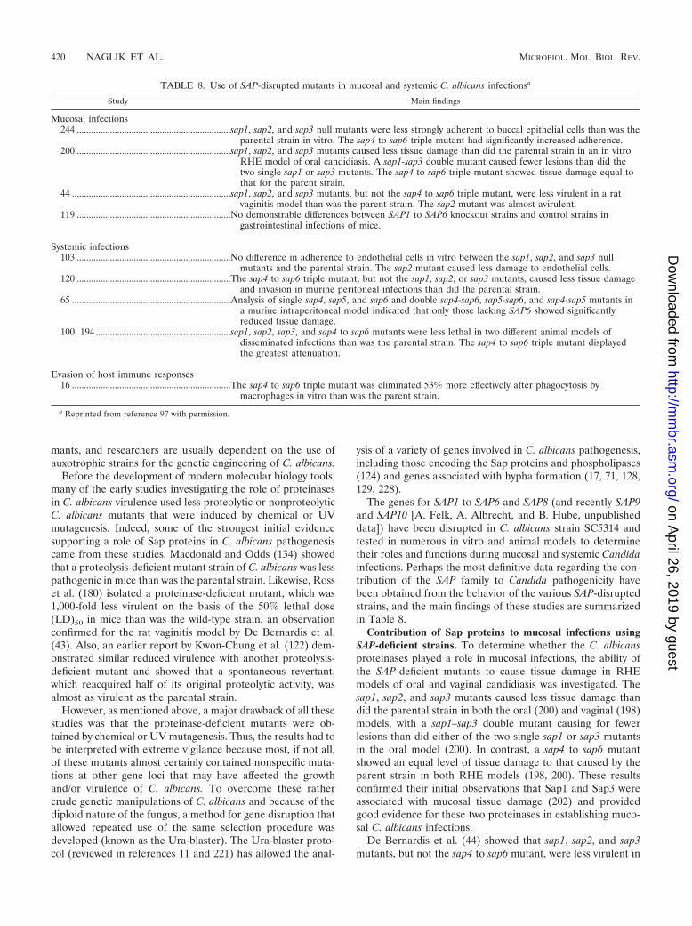
mants, and researchers are usually dependent on the use ofauxotrophic strains for the genetic engineering of C. albicans.
Before the development of modern molecular biology tools,many of the early studies investigating the role of proteinasesin C. albicans virulence used less proteolytic or nonproteolyticC. albicans mutants that were induced by chemical or UVmutagenesis. Indeed, some of the strongest initial evidencesupporting a role of Sap proteins in C. albicans pathogenesiscame from these studies. Macdonald and Odds (134) showedthat a proteolysis-deficient mutant strain of C. albicans was lesspathogenic in mice than was the parental strain. Likewise, Rosset al. (180) isolated a proteinase-deficient mutant, which was1,000-fold less virulent on the basis of the 50% lethal dose(LD)50 in mice than was the wild-type strain, an observationconfirmed for the rat vaginitis model by De Bernardis et al.(43). Also, an earlier report by Kwon-Chung et al. (122) dem-onstrated similar reduced virulence with another proteolysis-deficient mutant and showed that a spontaneous revertant,which reacquired half of its original proteolytic activity, wasalmost as virulent as the parental strain.
However, as mentioned above, a major drawback of all thesestudies was that the proteinase-deficient mutants were ob-tained by chemical or UV mutagenesis. Thus, the results had tobe interpreted with extreme vigilance because most, if not all,of these mutants almost certainly contained nonspecific muta-tions at other gene loci that may have affected the growthand/or virulence of C. albicans. To overcome these rathercrude genetic manipulations of C. albicans and because of thediploid nature of the fungus, a method for gene disruption thatallowed repeated use of the same selection procedure wasdeveloped (known as the Ura-blaster). The Ura-blaster proto-col (reviewed in references 11 and 221) has allowed the anal-
ysis of a variety of genes involved in C. albicans pathogenesis,including those encoding the Sap proteins and phospholipases(124) and genes associated with hypha formation (17, 71, 128,129, 228).
The genes for SAP1 to SAP6 and SAP8 (and recently SAP9and SAP10 [A. Felk, A. Albrecht, and B. Hube, unpublisheddata]) have been disrupted in C. albicans strain SC5314 andtested in numerous in vitro and animal models to determinetheir roles and functions during mucosal and systemic Candidainfections. Perhaps the most definitive data regarding the con-tribution of the SAP family to Candida pathogenicity havebeen obtained from the behavior of the various SAP-disruptedstrains, and the main findings of these studies are summarizedin Table 8.
Contribution of Sap proteins to mucosal infections usingSAP-deficient strains. To determine whether the C. albicansproteinases played a role in mucosal infections, the ability ofthe SAP-deficient mutants to cause tissue damage in RHEmodels of oral and vaginal candidiasis was investigated. Thesap1, sap2, and sap3 mutants caused less tissue damage thandid the parental strain in both the oral (200) and vaginal (198)models, with a sap1–sap3 double mutant causing for fewerlesions than did either of the two single sap1 or sap3 mutantsin the oral model (200). In contrast, a sap4 to sap6 mutantshowed an equal level of tissue damage to that caused by theparent strain in both RHE models (198, 200). These resultsconfirmed their initial observations that Sap1 and Sap3 wereassociated with mucosal tissue damage (202) and providedgood evidence for these two proteinases in establishing muco-sal C. albicans infections.
De Bernardis et al. (44) showed that sap1, sap2, and sap3mutants, but not the sap4 to sap6 mutant, were less virulent in
TABLE 8. Use of SAP-disrupted mutants in mucosal and systemic C. albicans infectionsa
Study Main findings
Mucosal infections244 .................................................................sap1, sap2, and sap3 null mutants were less strongly adherent to buccal epithelial cells than was the
parental strain in vitro. The sap4 to sap6 triple mutant had significantly increased adherence.200 .................................................................sap1, sap2, and sap3 mutants caused less tissue damage than did the parental strain in an in vitro
RHE model of oral candidiasis. A sap1-sap3 double mutant caused fewer lesions than did thetwo single sap1 or sap3 mutants. The sap4 to sap6 triple mutant showed tissue damage equal tothat for the parent strain.
44 ...................................................................sap1, sap2, and sap3 mutants, but not the sap4 to sap6 triple mutant, were less virulent in a ratvaginitis model than was the parent strain. The sap2 mutant was almost avirulent.
119 .................................................................No demonstrable differences between SAP1 to SAP6 knockout strains and control strains ingastrointestinal infections of mice.
Systemic infections103 .................................................................No difference in adherence to endothelial cells in vitro between the sap1, sap2, and sap3 null
mutants and the parental strain. The sap2 mutant caused less damage to endothelial cells.120 .................................................................The sap4 to sap6 triple mutant, but not the sap1, sap2, or sap3 mutants, caused less tissue damage
and invasion in murine peritoneal infections than did the parental strain.65 ...................................................................Analysis of single sap4, sap5, and sap6 and double sap4-sap6, sap5-sap6, and sap4-sap5 mutants in
a murine intraperitoneal model indicated that only those lacking SAP6 showed significantlyreduced tissue damage.
100, 194 .........................................................sap1, sap2, sap3, and sap4 to sap6 mutants were less lethal in two different animal models ofdisseminated infections than was the parental strain. The sap4 to sap6 triple mutant displayedthe greatest attenuation.
Evasion of host immune responses16 ...................................................................The sap4 to sap6 triple mutant was eliminated 53% more effectively after phagocytosis by
macrophages in vitro than was the parent strain.
a Reprinted from reference 97 with permission.
420 NAGLIK ET AL. MICROBIOL. MOL. BIOL. REV.
on April 26, 2019 by guest
http://mm
br.asm.org/
Dow
nloaded from

a rat vaginitis model than was the parent strain, with the sap2mutant being almost avirulent. Reintroduction of the SAP2gene recovered the vaginopathic potential of the sap2 mutant,showing that Sap2 may play a pathogenic role in rat vaginitis.This supports the data obtained with the RHE model and alsoimplicates the Sap1 to Sap3 subfamily in rat mucosal infections(Table 8).
The ability of the C. albicans SAP-deficient mutants to ad-here to epithelial cells was also investigated (244). Althoughthe sap1, sap2, and sap3 null mutants were moderately lessadherent to buccal epithelial cells than was the parent strain,the sap4 to sap6 triple mutant had significantly increased ad-herence. This was unexpected since one might have predictedthat the sap4 to sap6 mutant would have decreased adherence,given the association of SAP4 to SAP6 expression with thehyphal form (96, 246), which is known to be more adherentthan the yeast form (110, 111, 193). To explain the results, itwas suggested that one possible function of Sap4 to Sap6 mightbe to remove or degrade cell surface components on the yeastor epithelial cells, causing an inhibitory effect to the host-fungus recognition and adhesion mechanism. Although this isplausible, a more likely explanation would be that other pro-teinases or other unrelated adhesin genes might be up-regu-lated to compensate for the loss of Sap4 to Sap6 in the mutantstrain. Since Sap1 to Sap3 may contribute to C. albicans ad-herence (15, 121), up-regulation of one or more of these genesalone in the SAP4- to SAP6-deficient strain may account for atleast some of the increased adherence observed.
Studies using SAP-deficient strains have implicated the Sap1to Sap3 subfamily, either individually or collectively, in thevirulence of C. albicans infections at oral and vaginal sites(Table 8). This appears to be in contrast to murine gastroin-testinal infections (also mucosal), where no demonstrable dif-ferences between SAP1- to SAP6-deficient strains and the par-ent strain could be observed in the ability to invade thestomach or to disseminate to the brain or in the number offungi persisting in the feces (119). Although SAP gene andprotein expression were readily detectable in this model, therewas a notable absence of SAP1 expression. Since SAP1 con-tributes to oral (200) and vaginal (44) infections and might beinvolved in C. albicans adherence (15, 121), the absence ofSAP1 expression in the gastrointestinal tract may go some waytoward explaining why SAP1-deficient mutants were not atten-uated in virulence compared with the parent strain (119). It ispossible that the SAP genes used by C. albicans during gastro-intestinal infections differ in part from the SAP genes requiredfor oral or vaginal infections. This certainly appears to be thecase, at least for systemic candidiasis, where Sap4 to Sap6 andnot Sap1 to Sap3 appear to be more pertinent to infection (seebelow).
Contribution of Sap proteins to systemic infections usingSAP-deficient strains. The use of SAP-deficient strains has alsoimplicated the Sap family in systemic C. albicans infections(Table 8). When guinea pig and murine models of dissemi-nated candidiasis were infected with the sap1, sap2, sap3, andsap4 to sap6 mutants, all animals had increased survival ratescompared with those infected by the parent strain (100, 194).The sap4 to sap6 mutant displayed the greatest attenuation notonly in terms of lethality but also in terms of fungal burden inhost organs such as the kidneys, liver, and skin. However, the
authors concluded that although Sap4 to Sap6 appeared tocontribute more to systemic infections in both animal modelsthan did Sap1 to Sap3, none of the C. albicans proteinases wasa single dominant factor during disseminated infections.
In a model of murine peritonitis, the invasive properties ofsap1, sap2, or sap3 mutants were indistinguishable from thoseof wild-type cells, but the sap4 to sap6 triple mutant showedstrongly reduced invasiveness (65, 120). In addition, the sap4 tosap6 mutant induced significantly reduced levels of alaninetransferase and �-amylase activity (markers of liver and pan-creas damage, respectively) compared with the parent strain.Interestingly, when single sap4, sap5, sap6, sap4–sap5, sap5–sap6, and sap4–sap6 mutants were compared with wild-typecells, only mutants that lacked a functional SAP6 gene showedsignificantly reduced liver and pancreas tissue damage (65).The data indicated that the Sap4 to Sap6 subfamily, and prob-ably Sap6 specifically, contributes to tissue damage and inva-sion in peritoneal infections.
Ibrahim et al. (103) used an in vitro model of human um-bilical vein endothelial cell injury to show that although Sap1to Sap3 activity was not required for the adherence of C.albicans to endothelial cells, Sap2 activity was required for theinitial stages of cell injury. Extrapolated to the in vivo situation,the possible degradation of the endothelial barrier by Sap2may enable C. albicans to evade immune attack or to escapefrom blood vessels and cause disseminated infections. In con-trast to the individual sap1 to sap3 mutants, the sap4 to sap6mutant had increased adherence to endothelial cells but ex-hibited no differences in cell injury compared with the wildtype (A. S. Ibrahim, S. G. Filler, D. Sanglard, J. E. Edwards,Jr., and B. Hube, Program Abstr. 37th Intersci. Conf. Antimi-crob. Agents Chemother., abstr. B9, 1997). The increase inadherence is in line with the results of another study thatshowed enhanced adherence of the sap4 to sap6 mutant toepithelial cells (244). However, the lack of cell injury wasperhaps surprising, considering that contact (68) and candidalgermination (67, 102), which is associated with Sap4 to Sap6expression (96, 246), are required for the induction of endo-thelial cell injury. Furthermore, if other SAP genes (e.g., SAP1to SAP3) are up-regulated to compensate for the loss of Sap4to Sap6 in this mutant, this raises the question whether certainSAP genes play dual roles; e.g., Sap1 to Sap3 may contribute toC. albicans adhesion and tissue damage.
Contribution of Sap proteins to evasion of host immuneresponses using SAP-deficient strains. The sap mutants werealso instrumental in demonstrating a role for Sap4 to Sap6 inevading host immune responses (Table 8). In macrophage-killing assays, the sap4 to sap6 triple mutant was eliminated53% more effectively after phagocytosis by murine macro-phages than was the parent strain (16). Under normal circum-stances C. albicans is taken up for elimination into the phagoly-sosome, which has a pH of 4.7 to 4.8 (164). As a result ofingestion, C. albicans may germinate and secrete Sap4 to Sap6,which are optimally active at the same pH as that found in thephagolysosome (16). One should note that C. albicans germi-nation in vitro is normally observed at higher pH values (7.0)and is limited at lower pH values (5.0, the approximate pH ofthe phagolysosome). Nevertheless, in vivo, the production offunctional Sap4 to Sap6 proteinases within the phagolysosomemay result in the digestion of enzymes associated with candidal
VOL. 67, 2003 SECRETED ASPARTYL PROTEINASES AND C. ALBICANS VIRULENCE 421
on April 26, 2019 by guest
http://mm
br.asm.org/
Dow
nloaded from

killing and might render C. albicans more resistant to macro-phage attack. The macrophage proteins targeted by Sap4 toSap6 are not yet known, but Sap4 to Sap6 may act eitherdirectly on phagolysosomal enzymes involved in microbial kill-ing (cathepsin D and/or antifungal peptides) or on key en-zymes of macrophage metabolism that are essential for opti-mal microbial killing (145). The results of this study stronglyindicated that Sap4 to Sap6 might function in a way that en-ables C. albicans to evade host innate defenses by resistingmacrophage attack, a host evasion strategy also observed forother microorganisms, including the bacterium Listeria mono-cytogenes (51).
Conclusions from SAP-deficient mutant studies. These invitro and animal model studies using SAP-deficient C. albicansstrains have been instrumental in confirming a role for Sapproteins in oral, vaginal, and systemic Candida infections andhave begun to dissect some of the fundamental functions theproteinase family may possess in vivo. However, these resultsshould be interpreted with a little caution, since the disruptionof one SAP gene may be compensated for by the up-regulationor down-regulation of other SAP genes, which may account forsome of the observations reported. In addition, SAP gene dis-ruption may alter or modify the expression profiles of otherhydrolytic enzymes of C. albicans such as phospholipases andlipases or, indeed, other genes that may be associated withvirulence. These forced adaptations that C. albicans may needto undergo as a result of SAP gene disruption may mask ordisguise some of the roles and functions of individual Sapenzymes in the in vivo situation.
Furthermore, the contributions of Sap7, Sap8, Sap9, andSap10, nearly half the SAP gene family, during C. albicansinfections are presently unknown, and the role of these pro-teinases in pathogenesis have not yet been tested in animalmodels using the respective SAP-deficient strains. Data fromthese mutants should add significantly to our knowledge of Sapfunction in C. albicans pathogenesis, particularly with respectto Sap9 and Sap10, which both have C-terminal consensussequences typical for GPI-anchored proteins and may there-fore not be secreted from the cell. Preliminary data suggestthat Sap9 and Sap10 are regulatory proteinases that may playa role in the cell surface integrity of the cell, which is incontrast to the putative functions of the other Sap proteins (A.Albrecht, A. Felk, I. Pichova, M. Schaller, M. Monod, and B.Hube, unpublished data). It is clear that the use of SAP-defi-cient strains has significantly advanced our understanding ofthe possible roles and functions of the proteinases during Can-dida infections. Future experiments using SAP-deficient strainsshould lead to a better understanding of the complex interplaybetween C. albicans, the SAP gene family, and the host.
FUNCTIONAL GENOMICS AND CANDIDA
Our understanding of Candida pathogenesis in general hasadvanced considerably in the last 20 years, notably in the iden-tification of genes and proteins involved in cell signaling, mor-phogenesis, and virulence. However, we are presently witness-ing significant developments in molecular microbiology andproceeding toward a new era of fungal research, and this isprimarily due to one reason: the sequencing of a large numberof medically important fungal genomes. Complete genome
sequences of S. cerevisiae and Schizosaccharomyces pombe havealready been published (77, 249). The preliminary assembly forthe C. albicans genome sequence has been released (http://www-sequence.stanford.edu/group/candida), and sequencingprojects are currently under way for Candida glabrata, Crypto-coccus neoformans, Aspergillus sp. and several other fungalspecies (http://www.tigr.org/tdb/mdb/mdbinprogress.html). Therapidity of these sequencing initiatives is impressive, and with thedevelopment of new functional genomic technologies includingDNA microarrays, which has become an indispensable tool forhigh-throughput gene expression analysis, we will soon be able toanalyze the whole-genome responses of many fungal pathogens.As a result, our view and appreciation of fungal biology andpathogenesis will undergo something little short of a revolution inthe very near future.
Candida DNA Microarrays
The sequencing of the Candida genome has generated amountain of information, but the next critical step is to accu-rately decipher the biochemical and physiological meaning ofthe information (257). Gene function is the primary informa-tion that most researchers would like to extract from the Can-dida genome sequence; at present, DNA microarray analysis isone of the most valuable technologies available to achieve thison a genome-wide scale. Microarrays allow researchers tocompare the behavior of huge numbers of genes in differenttissues and under different environmental conditions to iden-tify virulence genes, gene pathways, and regulatory networks.
Due to the very recent date of the sequencing of the ge-nome, only a handful of Candida DNA microarray studies havebeen published; these include analysis of the genome-wideexpression profile of C. albicans when exposed to the azoleantifungal drugs (38, 42, 179), during the yeast-to- hypha tran-sition (123, 153, 154, 158, 213), during stress (61), and whenexposed to human blood (69a). The microarray studies de-scribed above have already been valuable in confirming thesuspected coordinated regulation between SAP gene expres-sion and hypha formation. However, because the current ar-rays are not highly effective at distinguishing between differentmembers of multigene families, one needs to be cautious ininterpreting the results.
In conclusion, transcript profiling is rapidly becoming a stan-dard tool in academic laboratories for the global analysis ofgene expression. Although microarray technology is a dynamicand developing process, its development ranks as one of thepioneering breakthrough technologies of the last decade. Inthe Candida field, most microarray data have been based onexperiments in vitro; the use of arrays to evaluate global generesponses during in vivo infections has not yet been reported.Given the complexity of Candida infections in vivo, includingthe influence of the local environment, the microflora, and thehost immune response on gene function, transcript profilingdata obtained from in vivo infections may well differ from thedata observed in vitro. In addition, mammalian gene arrayshave not yet been used to study host-pathogen interactionsfrom the host’s perspective, in order to determine host geneexpression profiles induced by the presence of Candida. Fur-thermore, the simultaneous analysis of both the host and Can-dida genome response using DNA microarrays during in vivo
422 NAGLIK ET AL. MICROBIOL. MOL. BIOL. REV.
on April 26, 2019 by guest
http://mm
br.asm.org/
Dow
nloaded from
infections is a goal well worth striving for. This should provideimportant information concerning which Candida and hostfactors interact during various stages of different infections andwill clearly lead to valuable insights into the delicate and com-plex relationship between Candida and the host.
FUTURE DIRECTIONS IN SAP RESEARCH
The presence of a SAP gene family in C. albicans clearlyprovides the fungus with an efficient and flexible proteolyticsystem that may prove vital to its success as an opportunisticpathogen. Sap production is probably a well-regulated processthat is activated at specific time points during colonization andinfection to obtain maximum benefits for C. albicans. Nearly allstudies implicating proteinases in C. albicans pathogenesishave done so by using one or more of the seven main criteriadescribed in this review, which demonstrate the multiple func-tions of the SAP genes and have established the proteinases asa very versatile and multifunctional virulence gene family of C.albicans. However, although it appears that we know a greatdeal about the C. albicans SAP family, our knowledge is actu-ally rather limited. In fact, a number of fundamental questionsneed to be addressed in future studies.
First, since SAP mRNA expression patterns in vivo appearto be different from those observed under in vitro conditions,what, exactly, are the environmental stimuli that influence SAPexpression in vivo? Also, do the same transcriptional factorscoordinately regulate the simultaneous expression of the SAPgenes with a combination of other Candida virulence geneswhen triggered by changes in the local environment?
Second, what are the signal transduction pathways that reg-ulate C. albicans proteinase expression? These pathways arecurrently unknown, and no receptor (if indeed a receptor isinvolved) has yet been implicated in the regulation of protein-ase secretion.
Third, what are the actual targets of the Sap family duringhuman infections? Although the possible targets for Sap2might be deduced from in vitro degradation studies, these needto be confirmed in the in vivo environment. Furthermore, thesubstrate specificities of Sap1, Sap3, and Sap4 to Sap10 are notyet known and may provide valuable insights into how theseenzymes might contribute enzymatically and nonenzymaticallyto different infections at various bodily sites.
Fourth, given the recognized importance of the host-patho-gen interaction in the development of candidiasis and the finebalance between commensialism and infection, our under-standing of the immune response to the Sap family is distinctlyunsatisfactory. The protective mechanisms of humoral andcell-mediated immune responses possibly elicited by differentmembers of the Sap family need to be thoroughly investigatedand may have the potential to open up a brand new avenue ofresearch and lead to the exciting possibility that Sap proteinscould be targeted for immunotherapeutic means.
Answers to these fundamental questions would significantlyadvance our understanding of the interplay between Candidaproteinases and the host and bring us a good deal closer todiscovering how important this protein family is to Candidapathogenicity. Future studies using DNA microarrays will al-low us to identify new and no doubt surprising correlationsbetween SAP gene expression and Candida biology and viru-
lence. In time, this may lead to the development of new pro-phylactic and therapeutic strategies targeting specific viru-lence-associated genes, as well as signaling pathways orregulatory networks, which would be a valuable addition to thelimited repertoire of antifungal agents currently available.
ACKNOWLEDGMENTS
We gratefully acknowledge Cele Abad Zapatero for helpful discus-sions regarding Sap2 structure.
Our own work was supported by the Dunhill Medical Trust, theEuropean commission (QLK2-2000-00795; “Galar Fungail consor-tium”), and the Deutsche Forschungsgemeinschaft (Hu 528/8).
REFERENCES
1. Abad-Zapatero, C., R. Goldman, S. W. Muchmore, C. Hutchins, K. Stew-art, J. Navaza, C. D. Payne, and T. L. Ray. 1996. Structure of a secretedaspartic protease from C. albicans complexed with a potent inhibitor: im-plications for the design of antifungal agents. Protein Sci. 5:640–652.
2. Abu-Elteen, K. H., A. Z. Elkarmi, and M. Hamad. 2001. Characterization ofphenotype-based pathogenic determinants of various Candida albicansstrains in Jordan. Jpn. J. Infect. Dis. 54:229–236.
3. Acosta, E. P., T. N. Kakuda, R. C. Brundage, P. L. Anderson, and C. V.Fletcher. 2000. Pharmacodynamics of human immunodeficiency virus type1 protease inhibitors. Clin. Infect. Dis. 30(Suppl. 2):S151–S159.
4. Agatensi, L., F. Franchi, F. Mondello, R. L. Bevilacqua, T. Ceddia, F. DeBernardis, and A. Cassone. 1991. Vaginopathic and proteolytic Candidaspecies in outpatients attending a gynaecology clinic. J. Clin. Pathol. 44:826–830.
5. Apodaca, G., and J. H. McKerrow. 1990. Expression of proteolytic activityby cultures of Trichophyton rubrum. J. Med. Vet. Mycol. 28:159–171.
6. Barrett, A. J. 1980. Introduction: the classification of proteinases, p. 1–13.In Symposium on protein degradation in health and disease. ExcerptaMedica, Amsterdam, The Netherlands.
7. Barrett, A. J., and N. D. Rawlings. 1991. Types and families of endopepti-dases. Biochem. Soc. Trans. 19:707–715.
8. Beausejour, A., D. Grenier, J. P. Goulet, and N. Deslauriers. 1998. Proteo-lytic activation of the interleukin-1 beta precursor by Candida albicans.Infect. Immun. 66:676–681.
9. Beggah, S., U. Lechenne, U. Reichard, S. Foundling, and M. Monod. 2000.Intra- and intermolecular events direct the propeptide-mediated matura-tion of the Candida albicans secreted aspartic proteinase Sap1p. Microbi-ology 146:2765–2773.
10. Bektic, J., C. P. Lell, A. Fuchs, H. Stoiber, C. Speth, C. Lass-Florl, M.Borg-von Zepelin, M. P. Dierich, and R. Wurzner. 2001. HIV proteaseinhibitors attenuate adherence of Candida albicans to epithelial cells invitro. FEMS Immunol. Med. Microbiol. 31:65–71.
11. Berman, J., and P. E. Sudbery. 2002. Candida albicans: a molecular revo-lution built on lessons from budding yeast. Nat. Rev. Genet. 3:918–930.
12. Borg, M., and R. Ruchel. 1988. Expression of extracellular acid proteinaseby proteolytic Candida spp. during experimental infection of oral mucosa.Infect. Immun. 56:626–631.
13. Borg, M., and R. Ruchel. 1990. Demonstration of fungal proteinase duringphagocytosis of Candida albicans and Candida tropicalis. J. Med. Vet. My-col. 28:3–14.
14. Borg, M., D. Watters, B. Reich, and R. Ruchel. 1988. Production andcharacterization of monoclonal antibodies against secretory proteinase ofCandida albicans CBS 2730. Zentbl. Bakteriol. Microbiol. Hyg. Ser. A268:62–73.
15. Borg-von Zepelin, M., I. Meyer, R. Thomssen, R. Wurzner, D. Sanglard, A.Telenti, and M. Monod. 1999. HIV-Protease inhibitors reduce cell adher-ence of Candida albicans strains by inhibition of yeast secreted asparticproteases. J. Investig. Dermatol. 113:747–751.
16. Borg-von Zepelin, M., S. Beggah, K. Boggian, D. Sanglard, and M. Monod.1998. The expression of the secreted aspartyl proteinases Sap4 to Sap6 fromCandida albicans in murine macrophages. Mol. Microbiol. 28:543–554.
17. Braun, B. R., and A. D. Johnson. 1997. Control of filament formation inCandida albicans by the transcriptional repressor TUP1. Science 277:105–109.
18. Brown, A. J., and N. A. Gow. 1999. Regulatory networks controlling Can-dida albicans morphogenesis. Trends Microbiol. 7:333–338.
19. Brown, A. J. P. 2002. Expression of growth-form specific factors duringmorphogenesis in Candida albicans, p. 87–93. In R. A. Calderone (ed.),Candida and candidiasis. ASM Press, Washington, D.C.
20. Brown, A. J. P. 2002. Morphogenetic signaling pathways in Candida albi-cans, p. 95–106. In R. A. Calderone (ed.), Candida and candidiasis. ASMPress, Washington, D.C.
21. Brueske, C. H. 1986. Proteolytic activity of a clinical isolate of Cryptococcusneoformans. J. Clin. Microbiol. 23:631–633.
VOL. 67, 2003 SECRETED ASPARTYL PROTEINASES AND C. ALBICANS VIRULENCE 423
on April 26, 2019 by guest
http://mm
br.asm.org/
Dow
nloaded from
22. Calderone, R., and N. A. R. Gow. 2002. Host recognition by Candidaspecies, p. 67–86. In R. A. Calderone (ed.), Candida and candidiasis. ASMPress, Washington, D.C.
23. Calderone, R. A., and P. C. Braun. 1991. Adherence and receptor relation-ships of Candida albicans. Microbiol. Rev. 55:1–20.
24. Calderone, R. A., and W. A. Fonzi. 2001. Virulence factors of Candidaalbicans. Trends Microbiol. 9:327–335.
25. Camilli, A., D. T. Beattie, and J. J. Mekalanos. 1994. Use of geneticrecombination as a reporter of gene expression. Proc. Natl. Acad. Sci. USA91:2634–2638.
26. Capobianco, J. O., C. G. Lerner, and R. C. Goldman. 1992. Application ofa fluorogenic substrate in the assay of proteolytic activity and in the dis-covery of a potent inhibitor of Candida albicans aspartic proteinase. Anal.Biochem. 204:96–102.
27. Caro, L. H., H. Tettelin, J. H. Vossen, A. F. Ram, H. van den Ende, andF. M. Klis. 1997. In silicio identification of glycosyl-phosphatidylinositol-anchored plasma-membrane and cell wall proteins of Saccharomyces cer-evisiae. Yeast 13:1477–1489.
28. Cassone, A., F. De Bernardis, F. Mondello, T. Ceddia, and L. Agatensi.1987. Evidence for a correlation between proteinase secretion and vulvo-vaginal candidosis. J. Infect. Dis. 156:777–783.
29. Cassone, A., F. De Bernardis, A. Torosantucci, E. Tacconelli, M. Tum-barello, and R. Cauda. 1999. In vitro and in vivo anticandidal activity ofhuman immunodeficiency virus protease inhibitors. J. Infect. Dis. 180:448–453.
30. Cassone, A., E. Tacconelli, F. De Bernardis, M. Tumbarello, A. Torosan-tucci, P. Chiani, and R. Cauda. 2002. Antiretroviral therapy with proteaseinhibitors has an early, immune reconstitution-independent beneficial effecton Candida virulence and oral candidiasis in human immunodeficiencyvirus-infected subjects. J. Infect. Dis. 185:188–195.
31. Cauda, R., E. Tacconelli, M. Tumbarello, G. Morace, De, F. Bernardis, A.Torosantucci, and A. Cassone. 1999. Role of protease inhibitors in prevent-ing recurrent oral candidosis in patients with HIV infection: a prospectivecase-control study. J. Acquir. Immun. Defic. Syndr. 21:20–25.
32. Cawley, N. X., M. Chino, A. Maldonado, Y. M. Rodriguez, Y. P. Loh, andJ. A. Ellman. 2003. Synthesis and characterization of the first potent inhib-itor of yapsin 1. Implications for the study of yapsin-like enzymes. J. Biol.Chem. 278:5523–5530.
33. Chaffin, W. L., J. L. Lopez-Ribot, M. Casanova, D. Gozalbo, and J. P.Martinez. 1998. Cell wall and secreted proteins of Candida albicans: iden-tification, function, and expression. Microbiol. Mol. Biol. Rev. 62:130–180.
34. Challacombe, S. J., J. Muir, S. A. Howell, and S. P. Sweet. 1995. Geneticvariability of Candida albicans in HIV infection. Microb. Ecol. Health Dis.8:63–70.
35. Choi, G. H., D. M. Pawlyk, B. Rae, R. Shapira, and D. L. Nuss. 1993.Molecular analysis and overexpression of the gene encoding endothiapep-sin, an aspartic protease from Cryphonectria parasitica. Gene 125:135–141.
36. Colina, A. R., F. Aumont, N. Deslauriers, P. Belhumeur, and L. de Repen-tigny. 1996. Evidence for degradation of gastrointestinal mucin by Candidaalbicans secretory aspartyl proteinase. Infect. Immun. 64:4514–4519.
37. Coogan, M. M., S. P. Sweet, and S. J. Challacombe. 1994. ImmunoglobulinA (IgA), IgA1, and IgA2 antibodies to Candida albicans in whole andparotid saliva in human immunodeficiency virus infection and AIDS. Infect.Immun. 62:892–896.
38. Cowen, L. E., A. Nantel, M. S. Whiteway, D. Y. Thomas, D. C. Tessier, L. M.Kohn, and J. B. Anderson. 2002. Population genomics of drug resistance inCandida albicans. Proc. Natl. Acad. Sci. USA 99:9284–9289.
39. Cutfield, S. M., E. J. Dodson, B. F. Anderson, P. C. Moody, C. J. Marshall,P. A. Sullivan, and J. F. Cutfield. 1995. The crystal structure of a majorsecreted aspartic proteinase from Candida albicans in complexes with twoinhibitors. Structure 3:1261–1271.
40. Cutler, J. E. 1991. Putative virulence factors of Candida albicans. Annu.Rev. Microbiol. 45:187–218.
41. Davies, D. R. 1990. The structure and function of aspartic proteinases.Annu. Rev. Biophys. Biophys. Chem. 19:189–215.
42. De Backer, M. D., T. Hyina, X. J. Ma, S. Vandoninck, W. H. Luyten, and H.Vanden Bossche. 2001. Genomic profiling of the response of Candidaalbicans to itraconazole treatment using a DNA microarray. Antimicrob.Agents Chemother. 45:1660–1670.
43. De Bernardis, F., L. Agatensi, I. K. Ross, G. W. Emerson, R. Lorenzini,P. A. Sullivan, and A. Cassone. 1990. Evidence for a role for secretedaspartate proteinase of Candida albicans in vulvovaginal candidiasis. J. In-fect. Dis. 161:1276–1283.
44. De Bernardis, F., S. Arancia, L. Morelli, B. Hube, Sanglard, W. Schafer,and A. Cassone. 1999. Evidence that members of the secretory aspartylproteinase gene family, in particular SAP2, are virulence factors for Can-dida vaginitis. J. Infect. Dis. 179:201–208.
45. De Bernardis, F., M. Boccanera, D. Adriani, E. Spreghini, G. Santoni, andA. Cassone. 1997. Protective role of antimannan and anti-aspartyl protein-ase antibodies in an experimental model of Candida albicans vaginitis inrats. Infect. Immun. 65:3399–3405.
46. De Bernardis, F., M. Boccanera, L. Rainaldi, C. E. Guerra, I. Quinti, and
A. Cassone. 1992. The secretion of aspartyl proteinase, a virulence enzyme,by isolates of Candida albicans from the oral cavity of HIV-infected sub-jects. Eur. J. Epidemiol. 8:362–367.
47. De Bernardis, F., A. Cassone, J. Sturtevant, and R. Calderone. 1995. Ex-pression of Candida albicans SAP1 and SAP2 in experimental vaginitis.Infect. Immun. 63:1887–1892.
48. De Bernardis, F., P. Chiani, M. Ciccozzi, G. Pellegrini, T. Ceddia, G.D’Offizzi, I. Quinti, P. A. Sullivan, and A. Cassone. 1996. Elevated asparticproteinase secretion and experimental pathogenicity of Candida albicansisolates from oral cavities of subjects infected with human immunodefi-ciency virus. Infect. Immun. 64:466–471.
49. De Bernardis, F., F. Mondello, G. Scaravelli, A. Pachi, A. Girolamo, L.Agatensi, and A. Cassone. 1999. High aspartyl proteinase production andvaginitis in human immunodeficiency virus-infected women. J. Clin. Micro-biol. 37:1376–1380.
50. De Bernardis, F., P. A. Sullivan, and A. Cassone. 2001. Aspartyl proteinasesof Candida albicans and their role in pathogenicity. Med. Mycol. 39:303–313.
51. de Chastellier, C., and P. Berche. 1994. Fate of Listeria monocytogenes inmurine macrophages: evidence for simultaneous killing and survival ofintracellular bacteria. Infect. Immun. 62:543–553.
52. de Repentigny, L., F. Aumont, K. Bernard, and P. Belhumeur. 2000. Char-acterization of binding of Candida albicans to small intestinal mucin and itsrole in adherence to mucosal epithelial cells. Infect. Immun. 68:3172–3179.
53. de Viragh, P. A., D. Sanglard, G. Togni, R. Falchetto, and M. Monod. 1993.Cloning and sequencing of two Candida parapsilosis genes encoding acidproteases. J. Gen. Microbiol. 139:335–342.
54. Diz Dios, P., A. Ocampo, C. Miralles, I. Otero, I. Iglesias, and N. Rayo.1999. Frequency of oropharyngeal candidiasis in HIV-infected patients onprotease inhibitor therapy. Oral Surg. Oral Med. Oral Pathol. Oral Radiol.Endodont. 87:437–441.
55. Diz Dios, P., I. Otero, I. Iglesias, A. Ocampo, and C. Martinez. 1999.Failure of indinavir to inhibit Candida albicans in vitro. Eur. J. Clin. Mi-crobiol. Infect. Dis. 18:755–756.
56. Diz Dios, P., A. Ocampo, I. Iglesias, and I. Otero. 1999. Lack of ritonavirantifungal effect in vitro. Antimicrob. Agents Chemother. 43:997.
57. Drobacheff, C., L. Millon, M. Monod, R. Piarroux, E. Robinet, R. Laurent,and D. Meillet. 2001. Increased serum and salivary immunoglobulinsagainst Candida albicans in HIV-infected patients with oral candidiasis.Clin. Chem. Lab Med. 39:519–526.
58. Edison, A. M., and M. Manning-Zweerink. 1988. Comparison of the extra-cellular proteinase activity produced by a low-virulence mutant of Candidaalbicans and its wild-type parent. Infect. Immun. 56:1388–1390.
59. Edmond, M. B., S. E. Wallace, D. K. McClish, M. A. Pfaller, R. N. Jones,and R. P. Wenzel. 1999. Nosocomial bloodstream infections in UnitedStates hospitals: a three-year analysis. Clin. Infect. Dis. 29:239–244.
60. el-Maghrabi, E. A., D. M. Dixon, and J. W. Burnett. 1990. Characterizationof Candida albicans epidermolytic proteases and their role in yeast-celladherence to keratinocytes. Clin. Exp. Dermatol. 15:183–191.
61. Enjalbert, B., A. Nantel, and M. Whiteway. 2003. Stress-induced geneexpression in Candida albicans: absence of a general stress response. Mol.Biol. Cell 14:1460–1467.
62. Ernst, J. F. 2000. Regulation of dimorphism in Candida albicans. Contrib.Microbiol. 5:98–111.
63. Ernst, J. F. 2000. Transcription factors in Candida albicans—environmentalcontrol of morphogenesis. Microbiology 146:1763–1774.
64. Fallon, K., K. Bausch, J. Noonan, E. Huguenel, and P. Tamburini. 1997.Role of aspartic proteases in disseminated Candida albicans infection inmice. Infect. Immun. 65:551–556.
65. Felk, A., M. Kretschmar, A. Albrecht, M. Schaller, S. Beinhauer, T. Nich-terlein, D. Sanglard, H. C. Korting, W. Schafer, and B. Hube. 2002. Can-dida albicans hyphal formation and the expression of the Efg1-regulatedproteinases Sap4 to Sap6 are required for the invasion of parenchymalorgans. Infect. Immun. 70:3689–3700.
66. Felk, A., W. Schafer, and B. Hube. 2000. Candida albicans secretory asparticproteinase (SAP10) gene. Accession number AF146440.
67. Filler, S. G., A. S. Pfunder, B. J. Spellberg, J. P. Spellberg, and J. E.Edwards, Jr. 1996. Candida albicans stimulates cytokine production andleukocyte adhesion molecule expression by endothelial cells. Infect. Im-mun. 64:2609–2617.
68. Filler, S. G., J. N. Swerdloff, C. Hobbs, and P. M. Luckett. 1995. Penetrationand damage of endothelial cells by Candida albicans. Infect. Immun. 63:976–983.
69. Finlay, B. B., and S. Falkow. 1989. Common themes in microbial pathoge-nicity. Microbiol. Rev. 53:210–230.
69a.Fradin, C., M. Kretschmar, T. Nichterlein, C. Gaillardin, C. d’Enfert, andB. Hube. 2003. Stage-specific gene expression of Candida albicans in humanblood. Mol. Microbiol. 47:1523–1543.
70. Fusek, M., E. A. Smith, M. Monod, B. M. Dunn, and S. I. Foundling. 1994.Extracellular aspartic proteinases from Candida albicans, Candida tropica-lis, and Candida parapsilosis yeasts differ substantially in their specificities.Biochemistry 33:9791–9799.
424 NAGLIK ET AL. MICROBIOL. MOL. BIOL. REV.
on April 26, 2019 by guest
http://mm
br.asm.org/
Dow
nloaded from
71. Gale, C. A., C. M. Bendel, M. McClellan, M. Hauser, J. M. Becker, J.Berman, and M. K. Hostetter. 1998. Linkage of adhesion, filamentousgrowth, and virulence in Candida albicans to a single gene, INT1. Science279:1355–1358.
72. Germaine, G. R., and L. M. Tellefson. 1981. Effect of pH and human salivaon protease production by Candida albicans. Infect. Immun. 31:323–326.
73. Ghadjari, A., R. C. Matthews, and J. P. Burnie. 1997. Epitope mappingCandida albicans proteinase (SAP 2). FEMS Immunol. Med. Microbiol.19:115–123.
74. Ghannoum, M., and K. Abu Elteen. 1986. Correlative relationship betweenproteinase production, adherence and pathogenicity of various strains ofCandida albicans. J. Med. Vet. Mycol. 24:407–413.
75. Ghannoum, M. A. 2000. Potential role of phospholipases in virulence andfungal pathogenesis. Clin. Microbiol. Rev. 13:122–143.
76. Gilfillan, G. D., D. J. Sullivan, K. Haynes, T. Parkinson, D. C. Coleman,and N. A. R. Gow. 1998. Candida dubliniensis: phylogeny and putativevirulence factors. Microbiology 144:829–838.
77. Goffeau, A., B. G. Barrell, H. Bussey, R. W. Davis, B. Dujon, H. Feldmann,F. Galibert, J. D. Hoheisel, C. Jacq, M. Johnston, E. J. Louis, H. W. Mewes,Y. Murakami, P. Philippsen, H. Tettelin, and S. G. Oliver. 1996. Life with6000 genes. Science. 274:546, 563–546, 567.
78. Goldman, R. C., D. J. Frost, J. O. Capobianco, S. Kadam, R. R. Rasmussen,and C. Abad-Zapatero. 1995. Antifungal drug targets: Candida secretedaspartyl protease and fungal wall beta-glucan synthesis. Infect. Agents Dis.4:228–247.
79. Gow, N. A. R., A. J. P. Brown, and F. C. Odds. 2002. Fungal morphogenesisand host invasion. Curr. Opin. Microbiol. 5:366–371.
80. Gruber, A., J. Berlit, C. Speth, C. Lass-Florl, G. Kofler, M. Nagl, M. Borgvon Zepelin, M. P. Dierich, and R. Wurzner. 1999. Dissimilar attenuation ofCandida albicans virulence properties by human immunodeficiency virustype 1 protease inhibitors. Immunobiology 201:133–144.
81. Gruber, A., C. P. Lell, C. Speth, H. Stoiber, C. Lass-Florl, A. Sonneborn,J. F. Ernst, M. P. Dierich, and R. Wurzner. 2001. Human immunodefi-ciency virus type 1 Tat binds to Candida albicans, inducing hyphae butaugmenting phagocytosis in vitro. Immunology 104:455–461.
82. Gruber, A., E. Lukasser-Vogl, M. Borg von Zepelin, M. P. Dierich, and R.Wurzner. 1998. Human immunodeficiency virus type 1 gp160 and gp41binding to Candida albicans selectively enhances candidal virulence in vitro.J. Infect. Dis. 177:1057–1063.
83. Gruber, A., C. Speth, E. Lukasser-Vogl, R. Zangerle, M. Borg von Zepelin,M. P. Dierich, and R. Wurzner. 1999. Human immunodeficiency virus type1 protease inhibitor attenuates Candida albicans virulence properties invitro. Immunopharmacology 41:227–234.
84. Han, Y., and J. E. Cutler. 1995. Antibody response that protects againstdisseminated candidiasis. Infect. Immun. 63:2714–2719.
85. Han, Y., R. P. Morrison, and J. E. Cutler. 1998. A vaccine and monoclonalantibodies that enhance mouse resistance to Candida albicans vaginal in-fection. Infect. Immun. 66:5771–5776.
86. Han, Y., M. H. Riesselman, and J. E. Cutler. 2000. Protection againstcandidiasis by an immunoglobulin G3 (IgG3) monoclonal antibody specificfor the same mannotriose as an IgM protective antibody. Infect. Immun.68:1649–1654.
87. Hannula, J., M. Saarela, B. Dogan, J. Paatsama, P. Koukila-Kahkola, S.Pirinen, H. L. Alakomi, J. Perheentupa, and S. Asikainen. 2000. Compar-ison of virulence factors of oral Candida dubliniensis and Candida albicansisolates in healthy people and patients with chronic candidosis. Oral Mi-crobiol. Immunol. 15:238–244.
88. Hoegl, L., M. Ollert, and H. C. Korting. 1996. The role of Candida albicanssecreted aspartic proteinase in the development of candidoses. J. Mol. Med.74:135–142.
89. Hoegl, L., E. Thoma-Greber, M. Rocken, and H. C. Korting. 1998. HIVprotease inhibitors influence the prevalence of oral candidosis in HIV-infected patients: a 2-year study. Mycoses 41:321–325.
90. Hoetelmans, R. M., M. van Essenberg, P. L. Meenhorst, J. W. Mulder, andJ. H. Beijnen. 1997. Determination of saquinavir in human plasma, saliva,and cerebrospinal fluid by ion-pair high-performance liquid chromatogra-phy with ultraviolet detection. J. Chromatogr. Ser. B 698:235–241.
91. Homma, M., H. Chibana, and K. Tanaka. 1993. Induction of extracellularproteinase in Candida albicans. J. Gen. Microbiol. 139:1187–1193.
92. Hood, S., A. Bonington, J. Evans, and D. Denning. 1998. Reduction inoropharyngeal candidiasis following introduction of protease inhibitors.AIDS 12:447–448.
93. Hostetter, M. K. 1994. Adhesins and ligands involved in the interaction ofCandida spp. with epithelial and endothelial surfaces. Clin. Microbiol. Rev.7:29–42.
94. Hube, B. 1996. Candida albicans secreted aspartyl proteinases. Curr. Top.Med. Mycolo. 7:55–69.
95. Hube, B. 2000. Extracellular proteinases of human pathogenic fungi, p.126–137. In J. F. Ernst and A. Schmidt (ed.), Contributions to microbiology.Dimorphism in human pathogenic and apathogenic yeasts. S. Karger AG,Basel, Switzerland.
96. Hube, B., M. Monod, D. A. Schofield, A. J. P. Brown, and N. A. R. Gow.
1994. Expression of seven members of the gene family encoding secretoryaspartyl proteinases in Candida albicans. Mol. Microbiol. 14:87–99.
97. Hube, B., and J. Naglik. 2001. Candida albicans proteinases: resolving themystery of a gene family. Microbiology 147:1997–2005.
98. Hube, B., and J. R. Naglik. 2002. Extracellular hydrolases, p. 107–122. InR. A. Calderone (ed.), Candida and candidiasis. ASM Press, Washington,D.C.
99. Hube, B., D. Sanglard, M. Monod, A. J. P. Brown, and N. A. R. Gow. 1997.Extracellular proteolytic activity of Candida species, p. 109–122. In H.Vanden Bossche, D. A. Stevens, and F. C. Odds (ed.), Host-fungus inter-play. Proceedings of the Fifth Symposium on Topics in Mycology, 1995.National Foundation for Infectious Diseases, Bethesda, Md.
100. Hube, B., D. Sanglard, F. C. Odds, D. Hess, M. Monod, W. Schafer, A. J.Brown, and N. A. Gow. 1997. Disruption of each of the secreted aspartylproteinase genes SAP1, SAP2, and SAP3 of Candida albicans attenuatesvirulence. Infect. Immun. 65:3529–3538.
101. Hube, B., C. J. Turver, F. C. Odds, H. Eiffert, G. J. Boulnois, H. Kochel, andR. Ruchel. 1991. Sequence of the Candida albicans gene encoding thesecretory aspartate proteinase. J. Med. Vet. Mycol. 29:129–132.
102. Ibrahim, A. S., S. G. Filler, M. A. Ghannoum, and J. E. Edwards, Jr. 1993.Interferon-� protects endothelial cells from damage by Candida albicans.J. Infect. Dis. 167:1467–1470.
103. Ibrahim, A. S., S. G. Filler, D. Sanglard, J. E. J. Edwards, and B. Hube.1998. Secreted aspartyl proteinases and interactions of Candida albicanswith human endothelial cells. Infect. Immun. 66:3003–3005.
104. Jaton-Ogay, K., S. Paris, M. Huerre, M. Quadroni, R. Falchetto, G. Togni,J. P. Latge, and M. Monod. 1994. Cloning and disruption of the geneencoding an extracellular metalloprotease of Aspergillus fumigatus. Mol.Microbiol. 14:917–928.
105. Kaminishi, H., H. Hamatake, T. Cho, T. Tamaki, N. Suenaga, T. Fujii, Y.Hagihara, H. Maeda, S. H. Miyasaki, T. C. White, and N. Agabian. 1994.Activation of blood clotting factors by microbial proteinases. FEMS Micro-biol. Lett. 176:1702–1710.
106. Kaminishi, H., H. Miyaguchi, T. Tamaki, N. Suenaga, M. Hisamatsu, I.Mihashi, H. Matsumoto, H. Maeda, and Y. Hagihara. 1995. Degradation ofhumoral host defense by Candida albicans proteinase. Infect. Immun. 63:984–988.
107. Kaminishi, H., M. Tanaka, T. Cho, H. Maeda, and Y. Hagihara. 1990.Activation of the plasma kallikrein-kinin system by Candida albicans pro-teinase. Infect. Immun. 58:2139–2143.
108. Kaufmann, S. H. 1990. Heat shock proteins and the immune response.Immunol. Today 11:129–136.
109. Kilian, M., J. Mestecky, and M. W. Russell. 1988. Defense mechanismsinvolving Fc- dependent functions of immunoglobulin A and their subver-sion by bacterial immunoglobulin A proteases. Microbiol. Rev. 52:296–303.
110. Kimura, L. H., and N. N. Pearsall. 1978. Adherence of Candida albicans tohuman buccal epithelial cells. Infect. Immun. 21:64–68.
111. Kimura, L. H., and N. N. Pearsall. 1980. Relationship between germinationof Candida albicans and increased adherence to human buccal epithelialcells. Infect. Immun. 28:464–468.
112. Kirkpatrick, C. H., R. R. Rich, and J. E. Bennett. 1971. Chronic mucocu-taneous candidiasis: model-building in cellular immunity. Ann. Intern.Med. 74:955–978.
113. Klar, A. J., T. Srikantha, and D. R. Soll. 2001. A histone deacetylationinhibitor and mutant promote colony-type switching of the human patho-gen Candida albicans. Genetics 158:919–924.
114. Klotz, S. A., D. J. Drutz, J. L. Harrison, and M. Huppert. 1983. Adherenceand penetration of vascular endothelium by Candida yeasts. Infect. Immun.42:374–384.
115. Koelsch, G., J. Tang, J. A. Loy, M. Monod, K. Jackson, S. I. Foundling, andX. Lin. 2000. Enzymic characteristics of secreted aspartic proteases ofCandida albicans. Biochim. Biophys. Acta 1480:117–131.
116. Kondoh, Y., K. Shimizu, and K. Tanaka. 1987. Proteinase production andpathogenicity of Candida albicans. II. Virulence for mice of C. albicansstrains of different proteinase activity. Microbiol. Immunol. 31:1061–1069.
117. Korting, H. C., M. Schaller, G. Eder, G. Hamm, U. Bohmer, and B. Hube.1999. Effects of the human immunodeficiency virus (HIV) proteinase in-hibitors saquinavir and indinavir on in vitro activities of secreted aspartylproteinases of Candida albicans isolates from HIV-infected patients. Anti-microb. Agents Chemother. 43:2038–2042.
118. Krause, R., E. Schwab, D. Bachhiesl, F. Daxbock, C. Wenisch, G. J. Krejs,and E. C. Reisinger. 2001. Role of Candida in antibiotic-associated diar-rhea. J. Infect. Dis. 184:1065–1069.
119. Kretschmar, M., A. Felk, P. Staib, M. Schaller, D. Hess, M. Callapina, J.Morschhauser, W. Schafer, H. C. Korting, H. Hof, B. Hube, and T. Nich-terlein. 2002. Individual acid aspartic proteinases (Saps) 1–6 of Candidaalbicans are not essential for invasion and colonization of the gastrointes-tinal tract in mice. Microb. Pathog. 32:61–70.
120. Kretschmar, M., B. Hube, T. Bertsch, D. Sanglard, R. Merker, M. Schro-der, H. Hof, and T. Nichterlein. 1999. Germ tubes and proteinase activitycontribute to virulence of Candida albicans in murine peritonitis. Infect.Immun. 67:6637–6642.
VOL. 67, 2003 SECRETED ASPARTYL PROTEINASES AND C. ALBICANS VIRULENCE 425
on April 26, 2019 by guest
http://mm
br.asm.org/
Dow
nloaded from
121. Kvaal, C., S. A. Lachke, T. Srikantha, K. Daniels, J. McCoy, and D. R. Soll.1999. Misexpression of the opaque-phase-specific gene PEP1 (SAP1) in thewhite phase of Candida albicans confers increased virulence in a mousemodel of cutaneous infection. Infect. Immun. 67:6652–6662.
122. Kwon-Chung, K. J., D. Lehman, C. Good, and P. T. Magee. 1985. Geneticevidence for role of extracellular proteinase in virulence of Candida albi-cans. Infect. Immun. 49:571–575.
123. Lane, S., C. Birse, S. Zhou, R. Matson, and H. Liu. 2001. DNA array studiesdemonstrate convergent regulation of virulence factors by Cph1, Cph2, andEfg1 in Candida albicans. J. Biol. Chem. 276:48988–48996.
124. Leidich, S. D., A. S. Ibrahim, Y. Fu, A. Koul, C. Jessup, Vitullo, W. Fonzi,F. Mirbod, S. Nakashima, Y. Nozawa, and M. A. Ghannoum. 1998. Cloningand disruption of caPLB1, a phospholipase B gene involved in the patho-genicity of Candida albicans. J. Biol. Chem. 273:26078–26086.
125. Lerner, C. G., and R. C. Goldman. 1993. Stimuli that induce production ofCandida albicans extracellular aspartyl proteinase. J. Gen. Microbiol. 139:1643–1651.
126. Liljemark, W. F., and R. J. Gibbons. 1973. Suppression of Candida albicansby human oral streptococci in gnotobiotic mice. Infect. Immun. 8:846–849.
127. Liu, H. 2001. Transcriptional control of dimorphism in Candida albicans.Curr. Opin. Microbiol. 4:728–735.
128. Liu, H., J. Kohler, and G. R. Fink. 1994. Suppression of hyphal formationin Candida albicans by mutation of a STE12 homolog. Science 266:1723–1726.
129. Lo, H. J., J. R. Kohler, B. DiDomenico, D. Loebenberg, A. Cacciapuoti, andG. R. Fink. 1997. Nonfilamentous C. albicans mutants are avirulent. Cell90:939–949.
130. Lupetti, A., G. Guzzi, A. Paladini, K. Swart, M. Campa, and S. Senesi. 1995.Molecular typing of Candida albicans in oral candidiasis: karyotype epide-miology with human immunodeficiency virus-seropositive patients in com-parison with that with healthy carriers. J. Clin. Microbiol. 33:1238–1242.
131. Macdonald, F. 1984. Secretion of inducible proteinase by pathogenic Can-dida species. Sabouraudia 22:79–82.
132. Macdonald, F., and F. C. Odds. 1980. Inducible proteinase of Candidaalbicans in diagnostic serology and in the pathogenesis of systemic candido-sis. J. Med. Microbiol. 13:423–435.
133. Macdonald, F., and F. C. Odds. 1980. Purified Candida albicans proteinasein the serological diagnosis of systemic candidosis. JAMA 243:2409–2411.
134. Macdonald, F., and F. C. Odds. 1983. Virulence for mice of a proteinase-secreting strain of Candida albicans and a proteinase-deficient mutant.J. Gen. Microbiol. 129:431–438.
135. Magee, P. T., and S. Scherer. 1998. Genome mapping and gene discoveryin Candida albicans. ASM News 64:505–511.
136. Mathaba, L. T., A. E. Paxman, P. B. Ward, D. A. Forbes, and J. R.Warmington. 2000. Genetically distinct strains of Candida albicans withelevated secretory proteinase production are associated with diarrhoea inhospitalized children. J. Gastroenterol. Hepatol. 15:53–60.
137. Mathur, S., G. V. Koistinen, E. O. Horger, T. A. Mahvi, and H. H. Fuden-berg. 1977. Humoral immunity in vaginal candidiasis. Infect. Immun. 15:287–294.
138. Matthews, R. C., J. P. Burnie, D. Howat, T. Rowland, and F. Walton. 1991.Autoantibody to heat-shock protein 90 can mediate protection againstsystemic candidosis. Immunology 74:20–24.
139. Matthews, R. C., J. P. Burnie, and S. Tabaqchali. 1984. Immunoblot anal-ysis of the serological response in systemic candidosis. Lancet ii:1415–1418.
140. McCullough, M. J., B. C. Ross, B. D. Dwyer, and P. C. Reade. 1994.Genotype and phenotype of oral Candida albicans from patients infectedwith the human immunodeficiency virus. Microbiology 140:1195–1202.
141. McKerrow, J. H., E. Sun, P. J. Rosenthal, and J. Bouvier. 1993. Theproteases and pathogenicity of parasitic protozoa. Annu. Rev. Microbiol.47:821–853.
142. Millon, L., C. Drobacheff, R. Piarroux, M. Monod, G. Reboux, R. Laurent,and D. Meillet. 2001. Longitudinal study of anti-Candida albicans mucosalimmunity against aspartic proteinases in HIV-infected patients. J. Acquir.Immune Defic. Syndr. 26:137–144.
143. Miyasaki, S. H., J. B. Hicks, D. Greenspan, I. Polacheck, L. A. MacPhail,T. C. White, N. Agabian, and J. S. Greenspan. 1992. The identification andtracking of Candida albicans isolates from oral lesions in HIV-seropositiveindividuals. J. Acquir. Immune Defic. Syndr. 5:1039–1046.
144. Miyasaki, S. H., T. C. White, and N. Agabian. 1994. A fourth secretedaspartyl proteinase gene (SAP4) and a CARE2 repetitive element arelocated upstream of the SAP1 gene in Candida albicans. J. Bacteriol. 176:1702–1710.
145. Monod, M., and M. Borg-von Zepelin. 2002. Secreted proteinases and othervirulence mechanisms of Candida albicans. Chem. Immunol. 81:114–128.
146. Monod, M., B. Hube, D. Hess, and D. Sanglard. 1998. Differential regula-tion of SAP8 and SAP9, which encode two new members of the secretedaspartic proteinase family in Candida albicans. Microbiology 144:2731–2737.
147. Monod, M., G. Togni, B. Hube, and D. Sanglard. 1994. Multiplicity of genesencoding secreted aspartic proteinases in Candida species. Mol. Microbiol.13:357–368.
148. Morrison, C. J., S. F. Hurst, S. L. Bragg, R. J. Kuykendall, H. Diaz, D. W.McLaughlin, and E. Reiss. 1993. Purification and characterization of theextracellular aspartyl proteinase of Candida albicans: removal of extraneousproteins and cell wall mannoprotein and evidence for lack of glycosylation.J. Gen. Microbiol. 139:1177–1186.
149. Morrow, B., H. Ramsey, and D. R. Soll. 1994. Regulation of phase-specificgenes in the more general switching system of Candida albicans strain3153A. J. Med. Vet. Mycol. 32:287–294.
150. Morrow, B., T. Srikantha, J. Anderson, and D. R. Soll. 1993. Coordinateregulation of two opaque-phase-specific genes during white-opaque switch-ing in Candida albicans. Infect. Immun. 61:1823–1828.
151. Morrow, B., T. Srikantha, and D. R. Soll. 1992. Transcription of the genefor a pepsinogen, PEP1, is regulated by white-opaque switching in Candidaalbicans. Mol. Cell. Biol. 12:2997–3005.
152. Morschhauser, J., R. Virkola, T. K. Korhonen, and J. Hacker. 1997. Deg-radation of human subendothelial extracellular matrix by proteinase-secret-ing Candida albicans. FEMS Microbiol. Lett. 153:349–355.
153. Murad, A. M., C. d’Enfert, C. Gaillardin, H. Tournu, F. Tekaia, D. Talibi,D. Marechal, V. Marchais, J. Cottin, and A. J. Brown. 2001. Transcriptprofiling in Candida albicans reveals new cellular functions for the tran-scriptional repressors CaTup1, CaMig1 and CaNrg1. Mol. Microbiol. 42:981–993.
154. Murad, A. M., P. Leng, M. Straffon, J. Wishart, S. Macaskill, D. MacCal-lum, N. Schnell, D. Talibi, D. Marechal, F. Tekaia, C. d’Enfert, C. Gail-lardin, F. C. Odds, and A. J. Brown. 2001. NRG1 represses yeast-hyphamorphogenesis and hypha-specific gene expression in Candida albicans.EMBO J. 20:4742–4752.
155. Murphy, J. M., and J. D. Walton. 1996. Three extracellular proteases fromCochliobolus carbonum: cloning and targeted disruption of ALP1. Mol.Plant-Microbe Interact 9:290–297.
156. Naglik, J. R., G. Newport, T. C. White, L. L. Fernandes-Naglik, J. S.Greenspan, D. Greenspan, S. P. Sweet, S. J. Challacombe, and N. Agabian.1999. In vivo analysis of secreted aspartyl proteinase expression in humanoral candidiasis. Infect. Immun. 67:2482–2490.
157. Naglik, J. R., C. A. Rodgers, P. J. Shirlaw, J. L. Dobbie, L. L. Fernandes-Naglik, D. Greenspan, N. Agabian, and S. J. Challacombe. 2003. Differen-tial expression of Candida albicans secreted aspartyl proteinase and phos-pholipase B genes in humans correlates with active oral and vaginalinfections. J. Infect. Dis. 188:465–475.
158. Nantel, A., D. Dignard, C. Bachewich, D. Harcus, A. Marcil, A. P. Bouin,C. W. Sensen, H. Hogues, H. M. Van Het, P. Gordon, T. Rigby, F. Benoit,D. C. Tessier, D. Y. Thomas, and M. Whiteway. 2002. Transcription pro-filing of Candida albicans cells undergoing the yeast-to-hyphal transition.Mol. Biol. Cell 13:3452–3465.
159. Newport, G., and N. Agabian. 1997. KEX2 influences Candida albicansproteinase secretion and hyphal formation. J. Biol. Chem. 272:28954–28961.
160. Odds, F. C. 1994. Candida species and virulence. ASM News 60:313–318.161. Odds, F. C. 1994. Pathogenesis of Candida infections. J. Am. Acad. Der-
matol. 31:S2–S5.162. Odds, F. C. 2000. Pathogenic fungi in the 21st century. Trends Microbiol.
8:200–201.163. Ogrydziak, D. M. 1993. Yeast extracellular proteases. Crit. Rev. Biotechnol.
13:1–55.164. Ohkuma, S., and B. Poole. 1978. Fluorescence probe measurement of the
intralysosomal pH in living cells and the perturbation of pH by variousagents. Proc. Natl. Acad. Sci. USA 75:3327–3331.
165. Ollert, M. W., R. Sohnchen, H. C. Korting, U. Ollert, S. Brautigam, and W.Brautigam. 1993. Mechanisms of adherence of Candida albicans to culturedhuman epidermal keratinocytes. Infect. Immun. 61:4560–4568.
166. Ollert, M. W., C. Wende, M. Gorlich, C. G. McMullan-Vogel, M. Borg-vonZepelin, C. W. Vogel, and H. C. Korting. 1995. Increased expression ofCandida albicans secretory proteinase, a putative virulence factor, in iso-lates from human immunodeficiency virus-positive patients. J. Clin. Micro-biol. 33:2543–2549.
167. Pfaller, M. A., R. N. Jones, S. A. Messer, M. B. Edmond, and R. P. Wenzel.1998. National surveillance of nosocomial blood stream infection due toCandida albicans: frequency of occurrence and antifungal susceptibility inthe SCOPE Program. Diagn. Microbiol. Infect. Dis. 31:327–332.
168. Pichova, I., L. Pavlickova, J. Dostal, E. Dolejsi, O. Hruskova-Heidingsfel-dova, J. Weber, T. Ruml, and M. Soucek. 2001. Secreted aspartic proteasesof Candida albicans, Candida tropicalis, Candida parapsilosis and Candidalusitaniae. Inhibition with peptidomimetic inhibitors. Eur. J. Biochem. 268:2669–2677.
169. Powderly, W. G. 1992. Mucosal candidiasis caused by non-albicans speciesof Candida in HIV-positive patients. AIDS 6:604–605.
170. Qureshi, M. N., C. E. Barr, T. Seshamma, J. Reidy, R. J. Pomerantz, andO. Bagasra. 1995. Infection of oral mucosal cells by human immunodefi-ciency virus type 1 in seropositive persons. J. Infect. Dis. 171:190–193.
171. Rams, T. E., M. J. Andriolo, D. Feik, S. N. Abel, T. M. McGivern, and J.Slots. 1991. Microbiological study of HIV-related periodontitis. J. Peri-odontol. 62:74–81.
426 NAGLIK ET AL. MICROBIOL. MOL. BIOL. REV.
on April 26, 2019 by guest
http://mm
br.asm.org/
Dow
nloaded from
172. Ray, T. L., and C. D. Payne. 1987. Detection of Candida acid proteinase(CAP) antibodies in systemic candidiasis by enzyme immunoassay. Clin.Res. 35:711.
173. Ray, T. L., and C. D. Payne. 1988. Scanning electron microscopy of epider-mal adherence and cavitation in murine candidiasis: a role for Candida acidproteinase. Infect. Immun. 56:1942–1949.
174. Ray, T. L., C. D. Payne, R. Ruchel, B. Ritter, and M. Schaffrinski. 1990.Comparative production and rapid purification of Candida acid proteinasefrom protein-supplemented cultures. Infect. Immun. 273:391–403.
175. Redding, S., J. Smith, G. Farinacci, M. Rinaldi, Fothergill, J. Rhine-Chal-berg, and M. Pfaller. 1994. Resistance of Candida albicans to fluconazoleduring treatment of oropharyngeal candidiasis in a patient with AIDS:documentation by in vitro susceptibility testing and DNA subtype analysis.Clin. Infect. Dis. 18:240–242.
176. Reichard, U., M. Monod, F. Odds, and R. Ruchel. 1997. Virulence of anaspergillopepsin-deficient mutant of Aspergillus fumigatus and evidence foranother aspartic proteinase linked to the fungal cell wall. J. Med. Vet.Mycol. 35:189–196.
177. Remold, H., H. Fasold, and F. Staib. 1968. Purification and characterizationof a proteolytic enzyme from Candida albicans. Biochim. Biophys. Acta167:399–406.
178. Ripeau, J. S., M. Fiorillo, F. Aumont, P. Belhumeur, and L. de Repentigny.2002. Evidence for differential expression of Candida albicans virulencegenes during oral infection in intact and human immunodeficiency virustype 1-transgenic mice. J. Infect. Dis. 185:1094–1102.
179. Rogers, P. D., and K. S. Barker. 2002. Evaluation of differential geneexpression in fluconazole-susceptible and -resistant isolates of Candidaalbicans by cDNA microarray analysis. Antimicrob. Agents Chemother.46:3412–3417.
180. Ross, I. K., F. De Bernardis, G. W. Emerson, A. Cassone, and P. A.Sullivan. 1990. The secreted aspartate proteinase of Candida albicans:physiology of secretion and virulence of a proteinase-deficient mutant.J. Gen. Microbiol. 136:687–694.
181. Reference deleted.182. Ruchel, R. 1981. Properties of a purified proteinase from the yeast Candida
albicans. Biochim. Biophys. Acta 659:99–113.183. Ruchel, R. 1983. On the renin-like activity of Candida proteinases and
activation of blood coagulation in vitro. Zentbl. Bakteriol. Mikrobiol. Hyg.Orig. A 255:368–379.
184. Ruchel, R. 1986. Cleavage of immunoglobulins by pathogenic yeasts of thegenus Candida. Microbiol. Sci. 3:316–319.
185. Ruchel, R. 1992. Proteinase, p. 17–31. In J. E. Bennett, R. J. Hay, and P. K.Peterson (ed.), New strategies in fungal disease. Churchill Livingstone,Edinburgh, United Kingdom.
186. Ruchel, R., B. Boning-Stutzer, and A. Mari. 1988. A synoptical approach tothe diagnosis of candidosis, relying on serological antigen and antibodytests, on culture, and on evaluation of clinical data. Mycoses 31:87–106.
187. Ruchel, R. and B. Boning. 1983. Detection of Candida proteinase by en-zyme immunoassay and interaction of the enzyme with alpha-2-macroglob-ulin. J. Immunol. Methods 61:107–116.
188. Ruchel, R., B. Ritter, and M. Schaffrinski. 1990. Modulation of experimen-tal systemic murine candidosis by intravenous pepstatin. Zentbl. Bakteriol.273:391–403.
189. Ruchel, R., K. Uhlemann, and B. Boning. 1983. Secretion of acid protein-ases by different species of the genus Candida. Zentbl. Bakteriol. Mikrobiol.Hyg. Orig. A 255:537–548.
190. Ruchel, R., F. Zimmermann, B. Boning-Stutzer, and U. Helmchen. 1991.Candidiasis visualised by proteinase-directed immunofluorescence. Vir-chows Arch. Ser. A 419:199–202.
190a.Rupniak, H. T., C. Rowlatt, E. B. Lane, J. G. Steele, Trejdosiewicz, LK, B.Laskiewicz, S. Povey, and B. T. Hill. 1985. Characteristics of four newhuman cell lines derived from squamous cell carcinomas of the head andneck. J. Natl. Cancer Inst. 75:621–635.
191. Salyers, A., and D. Witt. 1994. Virulence factors that promote colonization,p. 30–46. In A. Salyers and D. Witt (ed.), Bacterial pathogenesis: a molec-ular approach. ASM Press, Washington, D.C.
192. Samaranayake, Y. H., and L. P. Samaranayake. 2001. Experimental oralcandidiasis in animal models. Clin. Microbiol. Rev. 14:398–429.
193. Sandin, R. L., A. L. Rogers, R. J. Patterson, and E. S. Beneke. 1982.Evidence for mannose-mediated adherence of Candida albicans to humanbuccal cells in vitro. Infect. Immun. 35:79–85.
194. Sanglard, D., B. Hube, M. Monod, F. C. Odds, and N. A. Gow. 1997. Atriple deletion of the secreted aspartyl proteinase genes SAP4, SAP5, andSAP6 of Candida albicans causes attenuated virulence. Infect. Immun.65:3539–3546.
195. Santos, M. A., and M. F. Tuite. 1995. The CUG codon is decoded in vivo asserine and not leucine in Candida albicans. Nucleic Acids. Res. 23:1481–1486.
196. Sato, T., K. Nagai, M. Shibazaki, K. Abe, Y. Takebayashi, B. Lumanau, andR. M. Rantiatmodjo. 1994. Novel aspartyl protease inhibitors, YF-0200R-Aand B. J. Antibiot. (Tokyo) 47:566–570.
197. Sato, T., M. Shibazaki, H. Yamaguchi, K. Abe, H. Matsumoto, and M.
Shimizu. 1994. Novel Candida albicans aspartyl protease inhibitor. II. Anew pepstatin-ahpatinin group inhibitor, YF-044P-D. J. Antibiot. 47:588–590.
198. Schaller, M., M. Bein, H. C. Korting, S. Baur, G. Hamm, M. Monod, S.Beinhauer, and B. Hube. 2003. The secreted aspartyl proteinases Sap1 andSap2 cause tissue damage in an in vitro model of vaginal candidiasis basedon reconstituted human vaginal epithelium. Infect. Immun. 71:3227–3234.
199. Schaller, M., B. Hube, M. W. Ollert, W. Schafer, v. Borg, M. Zepelin, E.Thoma-Greber, and H. C. Korting. 1999. In vivo expression and localizationof Candida albicans secreted aspartyl proteinases during oral candidiasis inHIV-infected patients. J. Investig. Dermatol. 112:383–386.
200. Schaller, M., H. C. Korting, W. Schafer, J. Bastert, W. Chen, and B. Hube.1999. Secreted aspartic proteinase (Sap) activity contributes to tissue dam-age in a model of human oral candidosis. Mol. Microbiol. 34:169–180.
201. Schaller, M., C. Schackert, H. C. Korting, E. Januschke, and B. Hube. 2000.Invasion of Candida albicans correlates with expression of secreted asparticproteinases during experimental infection of human epidermis. J. Investig.Dermatol. 114:712–717.
202. Schaller, M., W. Schafer, H. C. Korting, and B. Hube. 1998. Differentialexpression of secreted aspartyl proteinases in a model of human oral can-didosis and in patient samples from the oral cavity. Mol. Microbiol. 29:605–615.
203. Schiodt, M. 1992. HIV-associated salivary gland disease: a review. OralSurg. Oral Med. Oral Pathol. 73:164–167.
204. Schmid, J., F. C. Odds, M. J. Wiselka, K. G. Nicholson, and D. R. Soll.1992. Genetic similarity and maintenance of Candida albicans strains froma group of AIDS patients, demonstrated by DNA fingerprinting. J. Clin.Microbiol. 30:935–941.
205. Schroppel, K., K. Sprosser, M. Whiteway, Y. Thomas, M. Rollinghoff, andC. Csank. 2000. Repression of hyphal proteinase expression by the mito-gen-activated protein (MAP) kinase phosphatase cpp1p of Candida albi-cans is independent of the MAP kinase cek1p. Infect. Immun. 68:7159–7161.
206. Schweizer, A., S. Rupp, B. N. Taylor, M. Rollinghoff, and K. Schroppel.2000. The TEA/ATTS transcription factor CaTec1p regulates hyphal de-velopment and virulence in Candida albicans. Mol. Microbiol. 38:435–445.
207. Shapiro, J. 1983. Mobile genetic elements. Academic Press, Inc., Orlando,Fla.
208. Smith, J. M., C. M. Tang, S. Van Noorden, and D. W. Holden. 1994.Virulence of Aspergillus fumigatus double mutants lacking restriction and analkaline protease in a low-dose model of invasive pulmonary aspergillosis.Infect. Immun. 62:5247–5254.
209. Smithson, S. L., I. C. Paterson, A. M. Bailey, S. E. Screen, B. A. Hunt, B. D.Cobb, R. M. Cooper, A. K. Charnley; and J. M. Clarkson. 1995. Cloningand characterisation of a gene encoding a cuticle-degrading protease fromthe insect pathogenic fungus Metarhizium anisopliae. Gene 166:161–165.
210. Smolenski, G., P. A. Sullivan, S. M. Cutfield, and J. F. Cutfield. 1997.Analysis of secreted aspartic proteinases from Candida albicans: purifica-tion and characterization of individual Sap1, Sap2 and Sap3 isoenzymes.Microbiology 143:349–356.
211. Sobel, J. D. 1988. Pathogenesis and epidemiology of vulvovaginal candidi-asis. Ann. N. Y. Acad. Sci. 544:547–557.
212. Sobel, J. D. 1992. Pathogenesis and treatment of recurrent vulvovaginalcandidiasis. Clin. Infect. Dis. 14(Suppl. 1):S148–S153.
213. Sohn, K., C. Urban, H. Brunner, and S. Rupp. 2003. EFG1 is a majorregulator of cell wall dynamics in Candida albicans as revealed by DNAmicroarrays. Mol. Microbiol. 47:89–102.
214. Soll, D. R. 1992. High-frequency switching in Candida albicans. Clin. Mi-crobiol. Rev. 5:183–203.
215. Soll, D. R. 1997. Gene regulation during high-frequency switching in Can-dida albicans. Microbiology 143:279–288.
216. Soll, D. R. 2002. Phenotypic switching, p. 123–142. In R. A. Calderone (ed.),Candida and candidiasis. ASM Press, Washington, D.C.
217. Soll, D. R., B. Morrow, and T. Srikantha. 1993. High-frequency phenotypicswitching in Candida albicans. Trends Genet. 9:61–65.
218. Soll, D. R., B. Morrow, T. Srikantha, K. Vargas, and P. Wertz. 1994.Developmental and molecular biology of switching in Candida albicans.Oral Surg. Oral Med. Oral Pathol. 78:194–201.
219. Srikantha, T., and D. R. Soll. 1993. A white-specific gene in the white-opaque switching system of Candida albicans. Gene 131:53–60.
220. Srikantha, T., L. Tsai, K. Daniels, A. J. Klar, and D. R. Soll. 2001. Thehistone deacetylase genes HDA1 and RPD3 play distinct roles in regulationof high-frequency phenotypic switching in Candida albicans. J. Bacteriol.183:4614–4625.
221. Staab, J. F., and P. Sundstrom. 2003. URA3 as a selectable marker fordisruption and virulence assessment of Candida albicans genes. TrendsMicrobiol. 11:69–73.
222. Staib, F. 1965. Serum-proteins as nitrogen source for yeastlike fungi. Sab-ouraudia 4:187–193.
223. Staib, P., M. Kretschmar, T. Nichterlein, H. Hof, and J. Morschhauser.2000. Differential activation of a Candida albicans virulence gene familyduring infection. Proc. Natl. Acad. Sci. USA 97:6102–6107.
VOL. 67, 2003 SECRETED ASPARTYL PROTEINASES AND C. ALBICANS VIRULENCE 427
on April 26, 2019 by guest
http://mm
br.asm.org/
Dow
nloaded from
224. Staib, P., M. Kretschmar, T. Nichterlein, H. Hof, and J. Morschhauser.2002. Transcriptional regulators Cph1p and Efg1p mediate activation of theCandida albicans virulence gene SAP5 during infection. Infect. Immun.70:921–927.
225. Staib, P., M. Kretschmar, T. Nichterlein, G. Kohler, S. Michel, H. Hof, J.Hacker, and J. Morschhauser. 1999. Host-induced, stage-specific virulencegene activation in Candida albicans during infection. Mol. Microbiol. 32:533–546.
225a.Stehr, F., A. Felk, M. Kretschmar, M. Schaller, W. Schafer, and B. Hube.2000. Extracellular hydrolytic enzymes and their relevance during Candidaalbicans infections. Mycoses 43(Suppl. 2):17–21.
226. Stewart, K., and C. Abad-Zapatero. 2001. Candida proteases and theirinhibition: prospects for antifungal therapy. Curr. Med. Chem. 8:941–948.
227. Stewart, K. W., R. C. Goldman, and C. Abad-Zapatero. 1999. The secretedproteinases from Candida: challenges for structure-aided drug design, p.117–138. In B. M. Dunn (ed.), Proteases of infectious agents. AcademicPress, Inc., San Diego, Calif.
228. Stoldt, V. R., A. Sonneborn, C. E. Leuker, and J. F. Ernst. 1997. Efg1p, anessential regulator of morphogenesis of the human pathogen Candida al-bicans, is a member of a conserved class of bHLH proteins regulatingmorphogenetic processes in fungi. EMBO J. 16:1982–1991.
229. Stringaro, A., P. Crateri, G. Pellegrini, G. Arancia, A. Cassone, and F. DeBernardis. 1997. Ultrastructural localization of the secretory aspartyl pro-teinase in Candida albicans cell wall in vitro and in experimentally infectedrat vagina. Mycopathologia 137:95–105.
230. Sundstrom, P. 1999. Adhesins in Candida albicans. Curr. Opin. Microbiol.2:353–357.
231. Sundstrom, P. 2002. Adhesion in Candida spp. Cell Microbiol. 4:461–469.232. Sweet, S. P. 1997. Selection and pathogenicity of Candida albicans in HIV
infection. Oral Dis. 3:S88–S95.233. Sweet, S. P., S. J. Challacombe, and J. R. Naglik. 1995. Whole and parotid
saliva IgA and IgA-subclass responses to Candida albicans in HIV infec-tion. Adv. Exp. Med. Biol. 371B:1031–1034.
234. Togni, G., D. Sanglard, R. Falchetto, and M. Monod. 1991. Isolation andnucleotide sequence of the extracellular acid protease gene (ACP) from theyeast Candida tropicalis. FEBS Lett. 286:181–185.
235. Togni, G., D. Sanglard, M. Quadroni, S. I. Foundling, and M. Monod. 1996.Acid proteinase secreted by Candida tropicalis: functional analysis of pre-proregion cleavages in C. tropicalis and Saccharomyces cerevisiae. Microbi-ology 142:493–503.
236. Tsuboi, R., I. Ko, K. Takamori, and H. Ogawa. 1989. Isolation of a kera-tinolytic proteinase from Trichophyton mentagrophytes with enzymatic ac-tivity at acidic pH. Infect. Immun. 57:3479–3483.
237. Tsuboi, R., H. Ogawa, K. Bramono, M. D. Richardson, G. S. Shankland,W. J. Crozier, Y. Sei, J. Ninomiya, A. Nakabayashi, and I. Takaiuchi. 1994.Pathogenesis of superficial mycoses. J. Med. Vet. Mycol. 32(Suppl. 1):91–104.
238. Tsushima, H., H. Mine, Y. Kawakami, F. Hyodoh, and A. Ueki. 1994.Candida albicans aspartic proteinase cleaves and inactivates human epider-mal cysteine proteinase inhibitor, cystatin A. Microbiology 140:167–171.
239. Tzung, K. W., R. M. Williams, S. Scherer, N. Federspiel, T. Jones, N.Hansen, V. Bivolarevic, L. Huizar, C. Komp, R. Surzycki, R. Tamse, R. W.Davis, and N. Agabian. 2001. Genomic evidence for a complete sexual cyclein Candida albicans. Proc. Natl. Acad. Sci. USA 98:3249–3253.
240. Valdez, H., B. M. Gripshover, R. A. Salata, and M. M. Lederman. 1998.Resolution of azole-resistant oropharyngeal candidiasis after initiation ofpotent combination antiretroviral therapy. AIDS 12:538.
241. Valdivia, R. H., and S. Falkow. 1997. Probing bacterial gene expressionwithin host cells. Trends Microbiol. 5:360–363.
242. von Heijne, G. 1985. Signal sequences. The limits of variation. J. Mol. Biol.184:99–105.
243. Vudhichamnong, K., D. M. Walker, and H. C. Ryley. 1982. The effect ofsecretory immunoglobulin A on the in-vitro adherence of the yeast Candidaalbicans to human oral epithelial cells. Arch. Oral Biol. 27:617–621.
244. Watts, H. J., F. S. Cheah, B. Hube, D. Sanglard, and N. A. Gow. 1998.
Altered adherence in strains of Candida albicans harbouring null mutationsin secreted aspartic proteinase genes. FEMS Microbiol. Lett. 159:129–135.
245. Whelan, W. L., D. R. Kirsch, K. J. Kwon-Chung, S. M. Wahl, and P. D.Smith. 1990. Candida albicans in patients with the acquired immunodefi-ciency syndrome: absence of a novel of hypervirulent strain. J. Infect. Dis.162:513–518.
246. White, T. C., and N. Agabian. 1995. Candida albicans secreted aspartylproteinases: isoenzyme pattern is determined by cell type, and levels aredetermined by environmental factors. J. Bacteriol. 177:5215–5221.
247. White, T. C., S. H. Miyasaki, and N. Agabian. 1993. Three distinct secretedaspartyl proteinases in Candida albicans. J. Bacteriol. 175:2997–3005.
248. White, T. C., M. A. Pfaller, M. G. Rinaldi, J. Smith, and S. W. Redding.1997. Stable azole drug resistance associated with a substrain of Candidaalbicans from an HIV-infected patient. Oral Dis. 3(Suppl 1):S102–S109.
249. Wood, V., R. Gwilliam, M. A. Rajandream, M. Lyne, R. Lyne, A. Stewart, J.Sgouros, N. Peat, J. Hayles, S. Baker, D. Basham, S. Bowman, K. Brooks,D. Brown, S. Brown, T. Chillingworth, C. Churcher, M. Collins, R. Connor,A. Cronin, P. Davis, T. Feltwell, A. Fraser, S. Gentles, A. Goble, N. Hamlin,D. Harris, J. Hidalgo, G. Hodgson, S. Holroyd, T. Hornsby, S. Howarth,E. J. Huckle, S. Hunt, K. Jagels, K. James, L. Jones, M. Jones, S. Leather,S. McDonald, J. McLean, P. Mooney, S. Moule, K. Mungall, L. Murphy, D.Niblett, C. Odell, K. Oliver, S. O’Neil, D. Pearson, M. A. Quail, E. Rabbi-nowitsch, K. Rutherford, S. Rutter, D. Saunders, K. Seeger, S. Sharp, J.Skelton, M. Simmonds, R. Squares, S. Squares, K. Stevens, K. Taylor, R. G.Taylor, A. Tivey, S. Walsh, T. Warren, S. Whitehead, J. Woodward, G.Volckaert, R. Aert, J. Robben, B. Grymonprez, I. Weltjens, E. Vanstreels,M. Rieger, M. Schafer, S. Muller-Auer, C. Gabel, M. Fuchs, C. Fritze, E.Holzer, D. Moestl, H. Hilbert, K. Borzym, I. Langer, A. Beck, H. Lehrach,R. Reinhardt, T. M. Pohl, P. Eger, W. Zimmermann, H. Wedler, R. Wam-buttt, B. Purnelle, A. Goffeau, E. Cadieu, S. Dreano, S. Gloux, V. Lelaure,S. Mottier, F. Galibert, S. J. Aves, Z. Xiang, C. Hunt, K. Moore, S. M.Hurst, M. Lucas, M. Rochet, C. Gaillardin, V. A. Tallada, A. Garzon, G.Thode, R. R. Daga, L. Cruzado, J. Jimenez, M. Sanchez, F. del Rey, J.Benito, A. Dominguez, J. L. Revuelta, S. Moreno, J. Armstrong, S. L.Forsburg, L. Cerrutti, T. Lowe, W. R. McCombie, I. Paulsen, J. Potashkin,G. V. Shpakovski, D. Ussery, B. G. Barrell, and P. Nurse. 2002. The genomesequence of Schizosaccharomyces pombe. Nature 415:871–880.
250. Wright, R. J., A. Carne, A. D. Hieber, I. L. Lamont, G. W. Emerson, andP. A. Sullivan. 1992. A second gene for a secreted aspartate proteinase inCandida albicans. J. Bacteriol. 174:7848–7853.
251. Wu, T., and L. P. Samaranayake. 1999. The expression of secreted aspartylproteinases of Candida species in human whole saliva. J. Med. Microbiol.48:711–720.
252. Wu, T., L. P. Samaranayake, B. Y. Cao, and J. Wang. 1996. In-vitro pro-teinase production by oral Candida albicans isolates from individuals withand without HIV infection and its attenuation by antimycotic agents.J. Med. Microbiol. 44:311–316.
253. Yuan, L., and G. T. Cole. 1987. Isolation and characterization of an extra-cellular proteinase of Coccidioides immitis. Infect. Immun. 55:1970–1978.
254. Zaugg, C., M. Borg-von Zepelin, D. Reichard, D. Sanglard, and M. Monod.2001. Secreted aspartic proteinase family of Candida tropicalis. Infect. Im-mun. 69:405–412.
255. Zhang, Z., H. N. El Sohly, M. R. Jacob, D. S. Pasco, L. A. Walker, and A. M.Clark. 2002. Natural products inhibiting Candida albicans secreted asparticproteases from Lycopodium cernuum. J. Nat. Prod. 65:979–985.
256. Zhang, Z., H. N. El Sohly, M. R. Jacob, D. S. Pasco, L. A. Walker, and A. M.Clark. 2002. Natural products inhibiting Candida albicans secreted asparticproteases from Tovomita krukovii. Planta Med. 68:49–54.
257. Zhou, J., and J. H. Miller. 2002. Microbial genomics—challenges andopportunities: the 9th International Conference on Microbial Genomes. J.Bacteriol. 184:4327–4333.
258. Zotter, C., U. F. Haustein, C. Schonborn, H. D. Grimmecke, and H. Wand.1990. Effect of pepstatin A on Candida albicans infection in the mouse.Dermatol. Monatsschrift 176:189–198.
428 NAGLIK ET AL. MICROBIOL. MOL. BIOL. REV.
on April 26, 2019 by guest
http://mm
br.asm.org/
Dow
nloaded from