Embed Size (px)
Citation preview
POSTMORTEM DEGRADATION OF MUSCLE PROTEINS
Role of the Neutral Proteinases in Postmortem Muscle Protein Degradation and Meat Tenderness
Mohammad Koohmaraie*
Introduction Consumers consider tenderness to be the single most
important component of meat quality. Recently, it has finally become clear that a lack of consistency in meat tenderness is one of the main problems facing the meat industry (Mor- gan et al., 1991b; Smith, 1992). A West coast supermarket chain (annual beef sales of $130 million) requests that its customers return any meat purchased, if they are not satis- fied. Over the last 3 years, customers have returned meat valued at $364,00O/year, of which $286,000 (Le., 78%) were related to inadequate or inconsistent beef tenderness, even after blade tenderization (Morgan, 1992). The magnitude of the tenderness problem is realized when one considers the following facts as stated by Wilks (1992), “Data reveal that: a) only one-tenth of one percent of tough, dry or bland steaks are returned for replacement or to get money back, b) for every one complaint that is vocalized, ten complaints are never heard, and c) most consumers who have such ex- periences don’t complain-they just don’t return.” Addition- ally, consumers are also demanding meat with a minimal quantity of visible fat. To satisfy this consumer demand, re- tailers have begun to trim excess fat. Clearly, this practice contributes to inefficient production and, therefore, meth- ods of producing lean carcasses is an intensive area of re- search. In many cases, lean carcasses are produced at the expense of tenderness (such as dietary administration of some O-adrenergic agonist) or inconsistent meat tenderness (intact male; Field, 1971; Seideman et al., 1982; Koohmaraie, 1988a). Therefore, i t is important to identify factors regulating meat tenderness so that methodology can be developed to manipulate the process advantageously. Such methodology may also be used to supplement the cur- rent U.S. quality grading system which is based primarily on the quantity of intramuscular fat. The inability of mar- bling to explain a large portion of total variation in tender- ness is well documented (for review, see Parrish, 1974).
Of all the observed variation in meat tenderness (obtained from animals of similar age), approximately 15% is explained by differences in marbling and connective tissue. I believe that differences in postmortem events that lead to improvement in meat tenderness probably explain the ma- jority, if not all, of the 85% of the variation in tenderness not
*M. Koohmaraie, USDA-ARS, Roman L. Hruska U. S. Meat Animal Research Center, P. 0. Box 7 66, Clay Center, NE 69833
Reciprocal Meat Conference Proceedings, Volume 45, 1992.
Figure 1
8 , , 12 I
I 1 1 4
7 14 1 7 14
Days Postmortem
The rate and extent of postmortem tenderization in beef (#1, from Koohmaraie et at., 1987); different species (#2, from Koohmaraie et ai., 1991a); bulls and steers (#3, from Morgan et al., 1992); 60s indicus and Bos taurus cattle (#4, from Whipple et ai., 1990); lambs fed B-adrenergic agonist, L644,969 (#5, from Koohmaraie et al., 1991 b); and control and lamb carcasses infused with zinc chloride (#6, from Koohmaraie, 1990).
accounted for (Figure 1). It is for this reason that we have studied and continue to study the mechanism of meat ten- derization during postmortem storage (for review, see Koohmaraie, 1988b, 1992a,b).
The objective of this manuscript is to discuss the role of neutral proteinases in postmortem protein degradation and meat tenderness.
Postmortem Changes in Skeletal Muscle Because postmortem changes in skeletal muscle were
the subject of a previous speaker (Bandman, 1992), only key postmortem changes relevant to this manuscript will be discussed. During postmortem storage of carcasses, nu-
63
64 American Meat Science Association
merous changes occur in skeletal muscle (for information on original source, see Goll et ai. 1983a; Koohmaraie, 1988b, 1992a), some of which result in the loss of tissue in- tegrity, which is translated into the improvement in meat tenderness. These changes include: 1) 2-disk weakening and/or degradation which leads to fragmentation of myofib- rils; 2) degradation of desmin which leads to fragmentation of myofibrils, probably through disruption of transverse crosslinking between myofibrils; 3) degradation of titin (speculatively, degradation of titin would improve meat ten- derness due to loss of tensile strength of myofibrils); 4) degradation of nebulin (because of the location of nebulin in the myofibrils [I-band], it is not clear how nebulin degra- dation will affect meat tenderness); 5) disappearance of troponin-T and simultaneous appearance of polypeptides with molecular weight of 28 to 32 (because of the location of troponin-T in myofibrils [i.e., I-band], it is doubtful that degradation of troponin-T by itself will have a direct effect on meat tenderness); however, these changes [i.e., the dis- appearance of troponin-T and appearance of 28 to 32 kDa polypeptides] seem to be good indicators of the extent of postmortem proteolysis); 6) appearance of a polypeptide with a molecular weight of 95 (neither the origin nor its sig- nificance to meat tenderness is known); and 7) perhaps the most important observation is that the major contractile pro- teins (myosin and actin) are not affected. One of the impor- tant changes that occurs in the tissue is the ease of frag- mentation of myofibrils under controlled homogenization, which does not occur in unaged tissue (Davey and Gilbert, 1969). The extent of myofibril fragmentation is now rou- tinely measured by a number of laboratories and is called myofibril fragmentation index (MFI). Speculatively, the weakening and/or degradation of Z-disks and degradation of desmin (and probably degradation of titin) are responsible for the increased fragility of myofibrils during postmortem storage.
Experimental Evidence Supporting Proteolysis Theory
Because all of the above changes are due to proteolytlc action, proteinases indigenous to skeletal muscle cells must have a major role in the regulation of these changes and ul- timate meat tenderness. There is substantial evidence indi- cating that the rate and extent of postmortem proteolysis are the principal causes of the observed variation in meat tenderness. Some of this evidence includes: 1 ) differences in the rate of muscle protein degradation are probably the reason for differences in the rate of postmortem tenderiza- tion in meat from pork, lamb and beef carcasses (Koohmaraie et al., 1991a); 2) infusion of carcasses with zinc chloride, which is a potent inhibitor of several classes of proteinases, prevents postmortem proteolysis and the tenderization process (Koohmaraie, 1990); 3) of all the pa- rameters that are thought to affect meat tenderness, re- duced rate and extent of postmortem proteolysis is the principal reason for the tenderness differences of meat from 60s faurus and Bos indicus breeds of cattle (Wheeler et al., 1990; Whipple et al., 1990; Shackelford et al., 1991); 4) nei- ther detectable postmortem proteolysis nor meat tender- ization occurs in muscle from carcasses of animals (lamb
and beef) fed some &adrenergic agonists (Fiems et al., 1990; Kretchmar et al., 1990; Koohmaraie and Shackelford, 1991; Koohmaraie et al., 1991b; Pringle et al., 1992; Wheeler and Koohmaraie, 1992); 5) incubation of muscle slices with calcium chloride induces 2-disk degradation, my- ofibril fragmentation (Busch et al., 1972; Koohmaraie et al., 1988a) and degradation of myofibrillar proteins (Koohmaraie, 1988a); 6) incubation of muscle slices with calcium chelators (EDTA or EGTA) prevents 2-disk degra- dation, myofibril fragmentation (Busch et al., 1972; Koohmaraie et al., 1988a) and degradation of myofibrillar proteins (Koohmaraie et al., 1988a); 7) infusion of lamb car- casses with calcium chloride accelerates postmortem pro- teolysis and tenderization (Koohmaraie et al., 1988b; Koohmaraie et al., 1989; Koohmaraie and Shackelford, 1991; St. Angelo et al., 1991); and 8) differences in tender- ness of meat obtained from intact and castrated males are probably due to differences in the rate and extent of post- mortem proteolysis (Morgan et al., 1992).
Proteinases Involved in Postmortem Proteolysis
Skeletal muscle contains numerous proteinases; how- ever, because of their ability to degrade myofibrillar pro- teins, two proteolytic systems have received considerable attention. These include the lysosomal cathepsins and the calpain proteolytic system. Recently, this list has been ex- panded to include the multicatalytic proteinase complex (MCP). Because the lysosomal proteinases were discussed by a previous speaker (Zeece, 1992), the focus of this man- uscript will be on the neutral proteinases which include both calpains and MCP. I shall first discuss the biochemical prop- erties of each of these proteolytic systems and then, using current experimental data, examine their possible roles in postmortem muscle protein degradation.
Biochemical Properties of the Calpain Proteolytic System
The first report documenting the existence of calpain is perhaps that of Guroff (1964) who reported the existence of a calcium- and sulfhydryl-dependent proteinase from rat brain. Meyer et al. (1964) reported the existence of a similar proteinase in skeletal muscle and it was later purified from porcine skeletal muscle by Dayton et al. (1976a,b). Since then, calpains have been shown to exist in a wide variety of cells, tissues and species. The following paragraphs will summarize some of the key characteristics of this prote- olytic system. The sources for this information are reviews written on calpain (Goll et al., 1983a,b, 1985, 1986, 1989, 1992; Mellgren, 1987; Murachi, 1983, 1984, 1989; Murachi et al., 1981a,b; Pontremoli and Melloni, 1986; Suzuki, 1987; Suzuki et al., 1987; CroalI and DeMartino, 1991). Original information sources will be given only when the subject has not been addressed in these review articles.
The calpain proteolytic system has been called by a va- riety of names including kinase activating factor (KAF), cal- cium-activated factor (CAF), calcium-activated neutral pro- tease (CANP), calcium-dependent sulfhydryl protease

45th Reciprocal Meat Conference 65
(CDSP) and calcium-dependent protease (CDP). It is now generally accepted to be called "calpain" by the Interna- tional Union of Biochemistry (EC 3.4.22.17). The calpain proteolytic system consists of p-calpain (the form of the proteinase active at micromolar concentration of calcium, thus p-calpain), m-calpain (the form of the proteinase ac- tive at millimolar concentration, thus called m-calpain), cal- pastatin (a protein that specifically inhibits both forms of calpain at their respective calcium concentration required for proteolytic activity) and an activator (a protein that greatly enhances calpain activity).
Skeletal muscle calpains (both y- and m-calpain) have a molecular weight of about 110 kDa under non-denaturing gel electrophoresis (PAGE) which is dissociated to two subunits with molecular weight of 80 kDa and 30 kDa, by SDS-PAGE. The large subunit which is different, though homologous, between p- and m-calpain is the catalytic subunit. The large subunit consists of four domains. Domain II has been as- signed as the catalytic subunit because of its amino acid sequence identity to other sulfhydryl proteinases such as papain. Within domain 11, cysteine-108 and histidine-265 are considered to be reactive amino acids. Domain IV is the cal- cium binding subunit because of its amino acid sequence identity to other calcium binding proteins such as calmod- ulin and troponin C. The function of domain I (the N termi- nus) and Ill (the C terminus) is not known at the present. cDNA for the large subunit of both p- and m-calpain has been cloned and sequenced. These results indicate that al- though there is similarity in nucleotide sequence, the large subunit of p- and m-calpain are clearly different gene prod- ucts. The small subunit, which is identical in both forms, is the regulatory subunit of the proteinase.
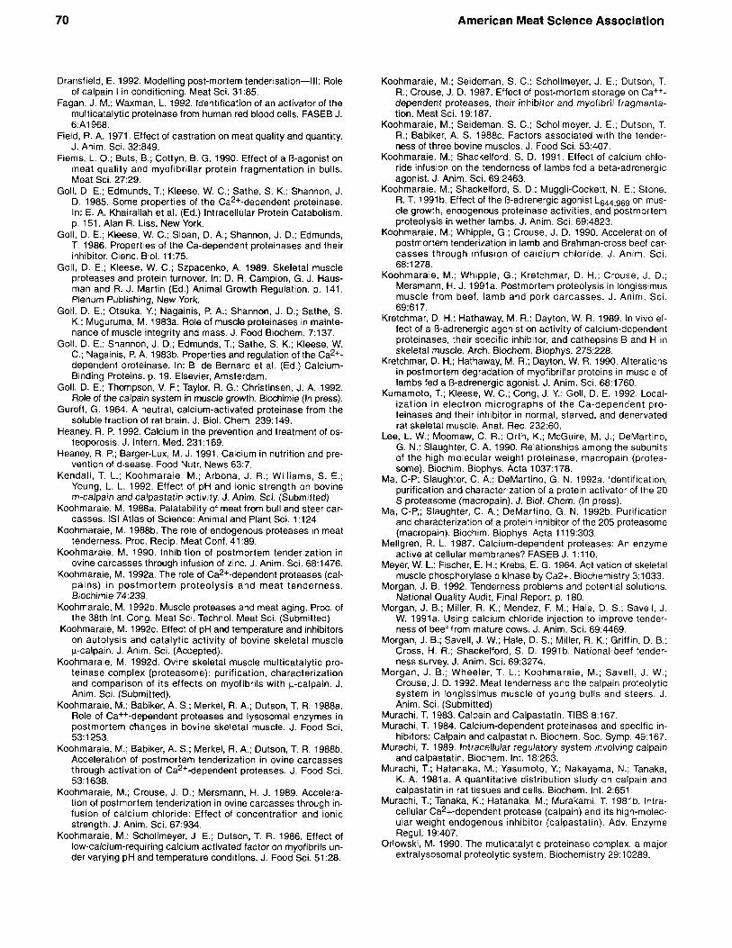
One of the important and well characterized properties of calpains is their susceptibility to calcium-induced auto- proteolysis. Prolonged exposure to sufficient calcium results in the loss of proteolytic activity and ultimate de- struction of the enzyme. As a result of autoproteolysis, both the 80 kDa and 30 kDa subunits are degraded to produce polypeptides with molecular mass ranging in size from 78 kDa to 18 kDa. The autolysis and subsequent loss of proteolytic activity of calpains is highly dependent on the presence of substrate, pH and temperature. The rate of autolysis and subsequent loss of proteolytic activity is decreased by the presence of substrate (Figure 2), de- creasing temperature and increasing pH (Koohmaraie, 1992~) . Autoproteolysis is one of the major factors that regulates the activity of the p calpain under postmortem conditions (Koohmaraie, 1992~). Though autolysis of p and m-calpain is similar for the most part, it differs in one significant way. Autolysis of p calpain is an intermolecular process (i.e., each p calpain will degrade its neighboring y-calpain molecules but not itself) and, therefore, autolysis will not go to completion. As a result, even after extensive autolysis either in situ or in vitro, some p-calpain activity will remain. On the other hand, because autolysis of m-cal- pain is both an inter- and intramolecular process (i.e., each m-calpain molecule will degrade itself and neighboring m- calpain molecules), autolysis of m-calpain proceeds to com- pletion. As a result, after extensive autolysis, no m-calpain activity can be detected (Figure 3).
Figure 2
std 0 5 10 15 30 60 120mIn
std 0 5 10 15 30 60 120mi.n
-t- r*) ----- -- -- Time course of bovine skeletal muscle p-calpain autolysis in the absence (top) and presence (bottom; 900 pg of ca- sein) of substrate at pH 7.0 and 25°C. Autolysis was done in 40 mM Tris-acetate, pH 7.0, 0.5 mM EDTA, 10 mM MCE with 1.58 mg/ml purified p-calpain. Tubes were preincubated at 25°C for 10 min and then the reaction was initiated with the addition of 3.8 mM CaCI,. The zero h samples were re- moved prior to the addition of CaCI,. At indicated times, af- ter vortexing an aliquot of the reaction was removed and immediately mixed with equal volume of protein denaturing buffer (to stop the reaction) protein denaturing buffer and heated in a boiling water bath for 5 min, cooled to room tem- perature and then electrophoresed on a 12.5% polyacry- lamide gel. Lane 1 is standard which consisted of Rabbit muscle phosphorylase b (97.4 kDa), Bovine serum albumin (66.2 kDa), Hen egg white ovalbumin (45.0 kDa), Bovine carbonic anhydrase (31 .kDa), Soybean trypsin inhibitor (21.5 kDa) and Hen egg white lysozyme (14.4 kDa). (Koohmaraie, unpublished data)

66
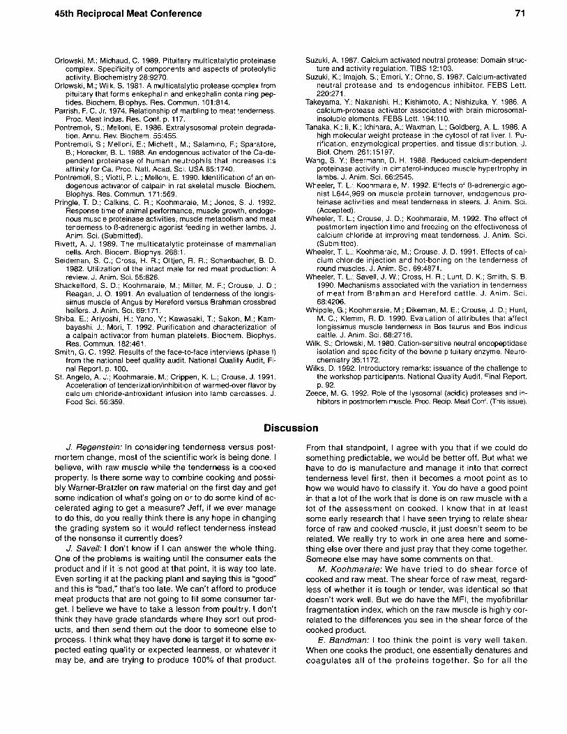
Figure 3
A
American Meat Science Association
Figure 4
C 0
Effect of autolysis at pH 7.0 and 5°C on the proteolytic activity of bovine skeletal muscle K-calpain (top) and m-calpain (bottom). For details see Figure 1. From Koohmaraie (1992~).
All cells that contain calpain also contain calpastatin, al- though the ratio of calpains to calpastatin varies from cell to cell. In skeletal muscle, this ratio is species dependent. The ratio of ca1pastatin:y-calpain + m-calpain is approximately 2.0 in bovine, 1.2 in ovine and 0.7 in porcine skeletal muscle (Koohmaraie et al., 1991 a). Calpastatin inhibits the activity of p- and m-calpain at the respective calcium concentration required for catalytic activity and has no inhibitory effect on any other known proteinase. There has been considerable confusion regarding the molecular weight of calpastatin. The molecular weight of calpastatin is reported to be be- tween 68 and 400 kDa. In meat animals (sheep, pigs, and cattle), we routinely isolate calpastatin with the molecular weight of 68 kDa (Kendall et al., 1992; Koohmaraie, 1992~). Calpastatin is an unusual protein, in that it is highly suscep- tible to proteolysis while in the tissue. It has been proposed that one of the major reasons for the large variation in mol- ecular weight of calpastatin is its degradation during ex- traction from the tissue. However, once isolated, it is very stable. For example, heating even at boiling temperature or SDS does not affect its activity.
In addition to calpastatin, there is some evidence indi- cating that calpains are also regulated by an activator pro- tein (DeMartino and Blumenthal, 1982; Takeyama et al., 1986; Pontremoli et al., 1988, 1990; Shiba et al., 1992). The first report of the existence of a calpain activator is that of DeMartino and Blumenthal (1982), who discovered it in ex- periments designed to examine the possible effects of calmodulin on the calpains. These so-called calpain activa- tors isolated from various sources have different properties and characteristics and, therefore, the field of calpain acti- vators (endogenous proteins) is not well understood.
Subcellular localization studies indicate that both p- and m-calpain and calpastatin are present throughout muscle cells, including nuclei and mitochondria, but they are local- ized at high concentrations at the Z-disk region (Kumamoto et al., 1992).
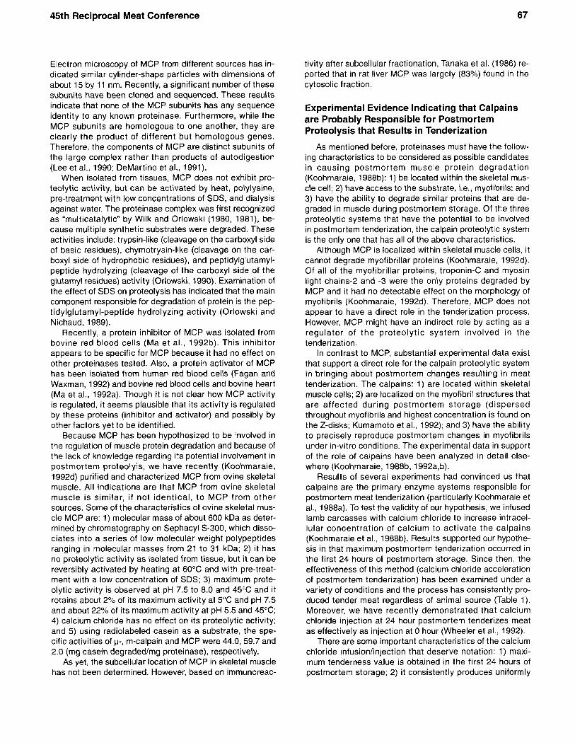
SDS-Polyacrylamide gel electrophoresis (PAGE) (C) and nondena- turing PAGE (D) analysis ovine skeletal muscle multicatalytic pro- teinase complex. Lane A is the molecular weight standards con- sisting of rabbit muscle phosphorylase b (97.4 kDa), bovine serum albumin (66.2 kDa), hen egg white ovalbumin (45.0 kDa), bovine carbonic anhydrase (31 kDa). soybean trypsin inhibitor (21.5 kDa) and hen egg white lysozyme (14.4 kDa). From Koohmaraie (1992d).
Biochemical Properties of the Multicatalyic Proteinase Complex
The multicatalytic proteinase complex (MCP) was first isolated from the bovine pituitary (Orlowski and Wilk, 1981) and, subsequently, purified from a number of mammalian tissues. All indications are that MCP is also a ubiquitous proteinase, found in every species and cell type examined from archaebacterium to man. The following paragraphs will summarize key characteristics of this proteolytic system. The sources for this information are recent reviews on MCP (Rivett, 1989; Orlowski, 1990). Original information sources will be given only when the subject has not been addressed in these review articles.
The proteinase complex has been referred to by a vari- ety of names, some of which include high-molecular-weight protease, high-molecular-weight cysteine proteinase, latent alkaline multifunctional proteinase, macropain, multicat- alytic proteinase, proteasome, 700-kDa multisubunit pro- teinase and 20s protease. However, there is now a general agreement to call it MCP. Though its function is not known, it is proposed to play a significant role in nonlysosomal pro- tein turnover. It has also been hypothesized that it could be involved in the degradation of muscle proteins (Goll et al., 1989).
MCP is a nonlysosomal proteinase with a native molec- ular weight of about 650 to 700 kDa. It is composed of a series of low molecular weight, nonidentical subunits.
45th Reciprocal Meat Conference 67
Electron microscopy of MCP from different sources has in- dicated similar cylinder-shape particles with dimensions of about 15 by 11 nm. Recently, a significant number of these subunits have been cloned and sequenced. These results indicate that none of the MCP subunits has any sequence identity to any known proteinase. Furthermore, while the MCP subunits are homologous to one another, they are clearly the product of different but homologous genes. Therefore, the components of MCP are distinct subunits of the large complex rather than products of autodigestion (Lee et al., 1990; DeMartino et al., 1991).
When isolated from tissues, MCP does not exhibit pro- teolytic activity, but can be activated by heat, polylysine, pre-treatment with low concentrations of SDS, and dialysis against water. The proteinase complex was first recognized as “multicatalytic” by Wilk and Orlowski (1980, 1981), be- cause multiple synthetic substrates were degraded. These activities include: trypsin-like (cleavage on the carboxyl side of basic residues), chymotrysin-like (cleavage on the car- boxyl side of hydrophobic residues), and peptidylglutamyl- peptide hydrolyzing (cleavage of the carboxyl side of the glutamyl residues) activity (Orlowski, 1990). Examination of the effect of SDS on proteolysis has indicated that the main component responsible for degradation of protein is the pep- tidylglutamyl-peptide hydrolyzing activity (Orlowski and Nichaud, 1989).
Recently, a protein inhibitor of MCP was isolated from bovine red blood cells (Ma et al., 199213). This inhibitor appears to be specific for MCP because it had no effect on other proteinases tested. Also, a protein activator of MCP has been isolated from human red blood cells (Fagan and Waxman, 1992) and bovine red blood cells and bovine heart (Ma et al., 1992a). Though it is not clear how MCP activity is regulated, it seems plausible that its activity is regulated by these proteins (inhibitor and activator) and possibly by other factors yet to be identified.
Because MCP has been hypothesized to be involved in the regulation of muscle protein degradation and because of the lack of knowledge regarding its potential involvement in postmortem proteolyis, we have recently (Koohmaraie, 1992d) purified and characterized MCP from ovine skeletal muscle. All indications are that MCP from ovine skeletal muscle is similar, i f not identical, to MCP from other sources. Some of the characteristics of ovine skeletal mus- cle MCP are: 1) molecular mass of about 600 kDa as deter- mined by chromatography on Sephacyl s-300, which disso- ciates into a series of low molecular weight polypeptides ranging in molecular masses from 21 to 31 kDa; 2) it has no proteolytic activity as isolated from tissue, but it can be reversibly activated by heating at 60°C and with pre-treat- ment with a low concentration of SDS; 3) maximum prote- olytic activity is observed at pH 7.5 to 8.0 and 45°C and it retains about 2% of its maximum activity at 5°C and pH 7.5 and about 22% of its maximum activity at pH 5.5 and 45°C; 4) calcium chloride has no effect on its proteolytic activity; and 5) using radiolabeled casein as a substrate, the spe- cific activities of p-, m-calpain and MCP were 44.0, 59.7 and 2.0 (mg casein degraded/mg proteinase), respectively.
As yet, the subcellular location of MCP in skeletal muscle has not been determined. However, based on immunoreac-
tivity after subcellular fractionation, Tanaka et al. (1986) re- ported that in rat liver MCP was largely (83%) found in the cytosolic fraction.
Experimental Evidence Indicating that Calpains are Probably Responsible for Postmortem Proteolysis that Results in Tenderization
As mentioned before, proteinases must have the follow- ing characteristics to be considered as possible candidates in causing postmortem muscle protein degradation (Koohmaraie, 1988b): 1) be located within the skeletal mus- cle cell; 2) have access to the substrate, Le., myofibrils; and 3) have the ability to degrade similar proteins that are de- graded in muscle during postmortem storage. Of the three proteolytic systems that have the potential to be involved in postmortem tenderization, the calpain proteolytic system is the only one that has all of the above characteristics.
Although MCP is localized within skeletal muscle cells, it cannot degrade myofibrillar proteins (Koohmaraie, 1992d). Of all of the myofibrillar proteins, troponin-C and myosin light chains-2 and -3 were the only proteins degraded by MCP and it had no detectable effect on the morphology of myofibrils (Koohmaraie, 1992d). Therefore, MCP does not appear to have a direct role in the tenderization process. However, MCP might have an indirect role by acting as a regulator of the proteolytic system involved in the tenderization.
In contrast to MCP, substantial experimental data exist that support a direct role for the calpain proteolytic system in bringing about postmortem changes resulting in meat tenderization. The calpains: 1) are located within skeletal muscle cells; 2) are localized on the myofibril structures that are affected during postmortem storage (dispersed throughout myofibrils and highest concentration is found on the Z-disks; Kumamoto et al., 1992); and 3) have the ability to precisely reproduce postmortem changes in myofibrils under in-vitro conditions. The experimental data in support of the role of calpains have been analyzed in detail else- where (Koohmaraie, 1988b, 1992a,b).
Results of several experiments had convinced us that calpains are the primary enzyme systems responsible for postmortem meat tenderization (particularly Koohmaraie et al., 1988a). To test the validity of our hypothesis, we infused lamb carcasses with calcium chloride to increase intracel- lular concentration of calcium to activate the calpains (Koohmaraie et al., 198813). Results supported our hypothe- sis in that maximum postmortem tenderization occurred in the first 24 hours of postmortem storage. Since then, the effectiveness of this method (calcium chloride acceleration of postmortem tenderization) has been examined under a variety of conditions and the process has consistently pro- duced tender meat regardless of animal source (Table 1). Moreover, we have recently demonstrated that calcium chloride injection at 24 hour postmortem tenderizes meat as effectively as injection at 0 hour (Wheeler et al., 1992).
There are some important characteristics of the calcium chloride infusion/injection that deserve notation: 1) maxi- mum tenderness value is obtained in the first 24 hours of postmortem storage; 2) it consistently produces uniformly
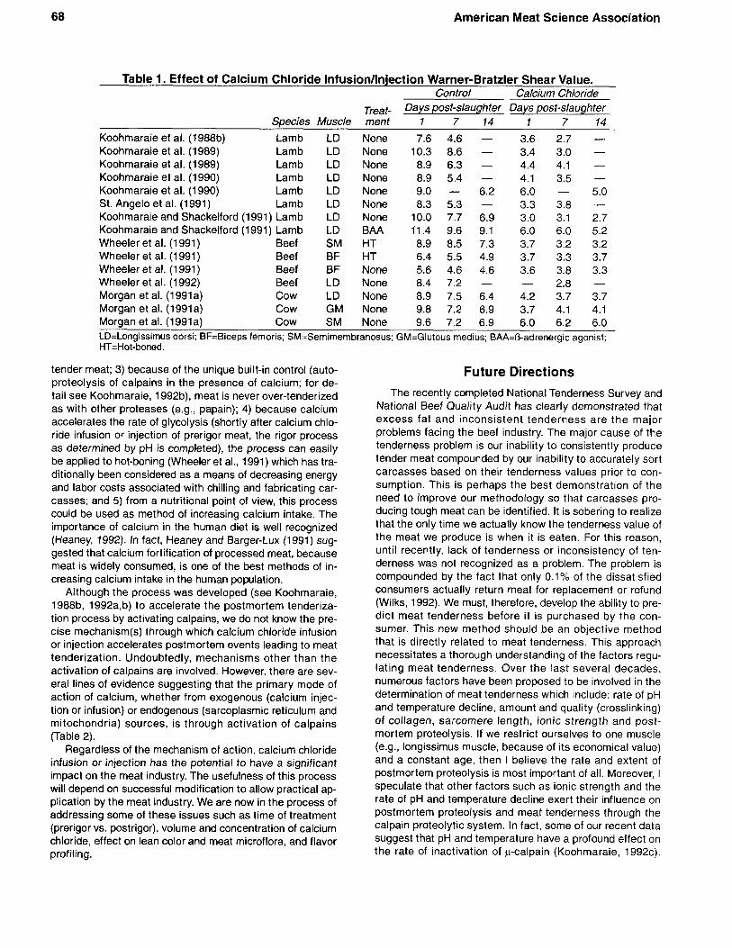
68 American Meat Science Association
Table 1. Effect of Calcium Chloride Infusion/lnjection Warner-Bratzler Shear Value. Control Calcium Chloride
Treat- Days post-slaughter Days post-slaughter Species Muscle ment 1 7 14 1 7 14
Koohmaraie et al. (1988b) Lamb LD None 7.6 4.6 - 3.6 2.7 - Koohmaraie et al. (1989) Lamb LD None 10.3 8.6 - 3.4 3.0 - Koohmaraie et al. (1989) Lamb LD None 8.9 6.3 - 4.4 4.1 - Koohmaraie et at. (1990) Lamb LD None 8.9 5.4 - 4.1 3.5 -
St. Angelo et at. (1991) Lamb LD None 8.3 5.3 - 3.3 3.8 - Koohmaraie et al. (1990) Lamb LD None 9.0 - 6.2 6.0 - 5.0
Koohmaraie and Shackelford (1991) Lamb LD None 10.0 7.7 6.9 3.0 3.1 2.7 Koohmaraie and Shackelford (1991) Lamb LD BAA 11.4 9.6 9.1 6.0 6.0 5.2 Wheeler et al. (1991) Beef SM HT 8.9 8.5 7.3 3.7 3.2 3.2 Wheeler et al. (1991) Beef BF HT 6.4 5.5 4.9 3.7 3.3 3.7 Wheeler et al. (1 991) Beef BF None 5.6 4.6 4.6 3.6 3.8 3.3
Morgan et al. (1991a) Cow LD None 8.9 7.5 6.4 4.2 3.7 3.7 Morgan et al. (1991a) Cow GM None 9.8 7.2 6.9 3.7 4.1 4.1 Morgan et al. (1991a) Cow SM None 9.6 7.2 6.9 6.0 6.2 6.0 LD=Longissirnus dorsi; BF=Biceps femoris: SM=Semimem branosus; GM=Gluteus medius; BAA=O-adrenergic agonist; HT=Hot-boned.
2.8 - Wheeler et al. (1 992) Beef LD None 8.4 7.2 - -
tender meat; 3) because of the unique built-in control (auto- proteolysis of calpains in the presence of calcium; for de- tail see Koohmaraie, 1992b), meat is never over-tenderized as with other proteases (e.g., papain); 4) because calcium accelerates the rate of glycolysis (shortly after calcium chlo- ride infusion or injection of prerigor meat, the rigor process as determined by pH is completed), the process can easily be applied to hot-boning (Wheeler et al., 1991) which has tra- ditionally been considered as a means of decreasing energy and labor costs associated with chilling and fabricating car- casses; and 5) from a nutritional point of view, this process could be used as method of increasing calcium intake. The importance of calcium in the human diet is well recognized (Heaney, 1992). In fact, Heaney and Barger-Lux (1991) sug- gested that calcium fortification of processed meat, because meat is widely consumed, is one of the best methods of in- creasing calcium intake in the human population.
Although the process was developed (see Koohmaraie, 1988b, 1992a,b) to accelerate the postmortem tenderiza- tion process by activating calpains, we do not know the pre- cise mechanism(s) through which calcium chloride infusion or injection accelerates postmortem events leading to meat tenderization. Undoubtedly, mechanisms other than the activation of calpains are involved. However, there are sev- eral lines of evidence suggesting that the primary mode of action of calcium, whether from exogenous (calcium injec- tion or infusion) or endogenous (sarcoplasmic reticulum and mitochondria) sources, is through activation of calpains (Table 2).
Regardless of the mechanism of action, calcium chloride infusion or injection has the potential to have a significant impact on the meat industry. The usefulness of this process will depend on successful modification to allow practical ap- plication by the meat industry. We are now in the process of addressing some of these issues such as time of treatment (prerigor vs. postrigor), volume and concentration of calcium chloride, effect on lean color and meat microflora, and flavor profiling.
Future Directions The recently completed National Tenderness Survey and
National Beef Quality Audit has clearly demonstrated that excess fat and inconsistent tenderness are the major problems facing the beef industry. The major cause of the tenderness problem is our inability to consistently produce tender meat compounded by our inability to accurately sort carcasses based on their tenderness values prior to con- sumption. This is perhaps the best demonstration of the need to improve our methodology so that carcasses pro- ducing tough meat can be identified. It is sobering to realize that the only time we actually know the tenderness value of the meat we produce is when it is eaten. For this reason, until recently, lack of tenderness or inconsistency of ten- derness was not recognized as a problem. The problem is compounded by the fact that only 0.1% of the dissatisfied consumers actually return meat for replacement or refund (Wilks, 1992). We must, therefore, develop the ability to pre- dict meat tenderness before i t is purchased by the con- sumer. This new method should be an objective method that is directly related to meat tenderness. This approach necessitates a thorough understanding of the factors regu- lating meat tenderness. Over the last several decades, numerous factors have been proposed to be involved in the determination of meat tenderness which include: rate of pH and temperature decline, amount and quality (crosslinking) of collagen, sarcomere length, ionic strength and post- mortem proteolysis. If we restrict ourselves to one muscle (e.g., longissimus muscle, because of its economical value) and a constant age, then I believe the rate and extent of postmortem proteolysis is most important of all. Moreover, I speculate that other factors such as ionic strength and the rate of pH and temperature decline exert their influence on postmortem proteolysis and meat tenderness through the calpain proteolytic system. In fact, some of our recent data suggest that pH and temperature have a profound effect on the rate of inactivation of p-calpain (Koohmaraie, 1992c).
45th Reciprocal Meat Conference 69
Table 2. Some of the Experimental Data Indicating that Calcium Acts through Activation of the Calpains.
1. Incubation of muscle slices with calcium chloride induces proteolysis of myofibrillar proteins, fragmen- tation and activates calpains (Koohmaraie et al., 1988a). However, incubation of muscle slices with cal- cium chelators (EDTA and EGTA) prevents proteolysis of myofibrillar proteins, myofibril fragmentation and calpains activation (Koohmaraie et al., 1988a). Davey and Gilbert (1969) demonstrated that incuba- tion of fiber pieces with EDTA prevented both weakening of lateral attachments and the disappearance of Z-disks. Busch et al. (1972) demonstrated that myofibrils fragmentation and disappearance of Z- disks were accelerated by incubating muscle slices with calcium chloride and both processes were in- hibited by using EDTA instead of calcium chloride.
2. When carcasses were infused with different concentrations of calcium chloride (75, 150 and 300 mM), acceleration of meat tenderness occurred only at the concentration which activated calpains (Koohmaraie et al., 1989).
3. Alarcon-Rojo and Dransfield (1989) reported that the calcium chloride acceleration of postmortem ten- derization was inhibited in the presence of inhibitors that are specific for calpains.
4. Infusion of carcasses with zinc chloride, which is a potent inhibitor of calpains, inhibited proteolysis of myofibrillar proteins, myofibril fragmentation and tenderization during 14-day postmortem storage (Koohmaraie, 1990).
5. Of the three proteolytic systems thought to be involved in postmortem proteolysis and tenderization, only calpains are activated by calcium. Calcium has no effect on the multicatalytic proteinase complex (Koohmaraie, 1992d) and has no stimulatory effect on the lysosomal proteinases. In fact at 10 mM, calcium chloride inhibits the activity of cathepsin B by 39% (Barrett, 1973).
6. Muscle from animals fed B-adrenergic agonist does not undergo (or undergoes minimal) postmortem proteolysis and produces tough meat. Lack of postmortem proteolysis and meat tenderization in BAA- fed animals has been attributed to a reduction in the activity of the calpain proteolytic system (Fiems et al., 1990; Wang and Beermann, 1988; Kretchmar et al., 1989, 1990, Koohmaraie and Shackelford, 1991 ; Koohmaraie et al., 1991 b; Pringle et al., 1992; Wheeler and Koohmaraie, 1992). However, cal- cium chloride infusion of carcasses from BAA-fed lambs induces activation of caiDains, deuradation of . -
myofibrillar proteins and meat tenderization (Koohmaraie and Shackelford, 1991).
Additional reasons for such speculations are discussed by Koohmaraie (1 992b).
As detailed in the previous section, current experimental data suggest that the calpain proteolytic system is probably the single most important proteolytic system directly involved in postmortem proteolysis and improvement in meat tenderness. Further support for our hypothesis (Koohmaraie, 1988b, 1990, 1992a,b; Koohmaraie et al., 1986, 1987, 1988a,b,c) has recently been reported by Dransfield (1992). He reported that 68% of the variation in meat toughness was accounted for by variation in the activ- ity of p-calpain. We have, therefore, decided that under- standing the regulation of calpain under postmortem conditions (entirely different than in living muscle,
Koohmaraie, 1992a) is perhaps one of the key items of knowledge required for the development of a method to pre- dict meat quality.
ACKNOWLEDGMENTS I am grateful to Drs. J. R. Arbona, T. L. Wheeler, and G.
Whipple for their constructive criticism and invaluable sug- gestions during preparation of this manuscript; to K. Theer for preparation of figures and slides; and to M. Bierman and C. Grummert for secretarial assistance. I am also grateful to Drs. E. Bandman, B. B. Marsh, R. A. Merkel, H. J. Mers- mann, G. C. Smith, S. 6. Smith, and J. D. Tatum for their review, constructive criticism, and invaluable suggestions prior to the submission of this manuscript.
References
Alarcon-Rojo, A,; Dransfield, E. 1989. Effect of calcium ions on tex- ture of beef during conditioning. Proc. Int. Cong. Meat Sci. Tech- nol. 35:1141.
Bandman, E. 1992. Changes in myofibrillar and cytoskeletal proteins in postmortem muscle. Proc. Recip. Meat Conf. (This issue).
Barrett, A. J. 1973. Human cathepsin B1: Purification and some of the properties of the enzyme. Biochem. J. 131:809.
Busch, W. A.; Stromer, M. H.; Goll, D. E.; Suzuki, A. 1972. Ca2+- specific removal of Z lines from rabbit skeletal muscle. J. Cell Biol. 52:367.
Croall, D. E.; DeMartino, G. N. 1991. Calcium-activated neutral pro- tease (calpain) system: Structure, function, and regulation. Physiol. Rev. 71:813.
Davey, C. L.; Gilbert, K. V. 1969. Studies in meat tenderness. 7. Changes in the fine structure of meat during aging. J. Food Sci. 3469.
Dayton, W. R.; Goll, D. E.; Zeece, M. G.; Robson, R. M.; Reville, W. J. 1976a. A Ca2+-activated protease involved in myofibrillar protein turnover. Purification from porcine muscle. Biochemistry 15:2150.
Dayton, W. R.; Reville, W. J.; Goll, D. E.; Stromer, M. H. 1976b. A Ca2+-activated protease possibly involved in myofibrillar protein turnover. Partial characterization of the purified enzyme. Bio- chemistry 152159.
DeMartino, G. N.; Blumenthal, D. K. 1982. Identification and partial purification of a factor that stimulates calcium-dependent pro- teases. Biochemistry 21 :4297.
DeMartino, G. N.; Orth, K.; McCullough, M. L.; Lee, L. W.; Munn, T. Z.; Moomaw, C. R.; Dawson, P. A,; Slaughter, C. A. 1991. The primary structures of four subunits of the human, high-molecu- lar-weight proteinase, macropain (proteasome), are distinct but homologous. Biochim. Biophys. Acta 1079:29.
70 American Meat Science Association
Dransfield, E. 1992. Modelling post-mortem tenderisation-Ill: Role of calpain I in conditioning. Meat Sci. 31 :85.
Fagan, J. M.; Waxman, L. 1992. Identification of an activator of the multicatalytic proteinase from human red blood cells. FASEB J. 6:A1968.
Field, R. A. 1971. Effect of castration on meat quality and quantity. J. Anim. Sci. 32:849.
Fiems, L. 0.; Buts, B.; Cottyn, B. G. 1990. Effect of a O-agonist on meat quality and myofibrillar protein fragmentation in bulls. Meat Sci. 27:29.
Goll, D. E.; Edmunds, T.; Kleese, W. C.; Sathe, S. K.; Shannon, J. D. 1985. Some properties of the Ca2+-dependent proteinase. In: E. A. Khairallah et al. (Ed.) Intracellular Protein Catabolism. p. 151. Alan R. Liss, New York.
Goll, D. E.; Kleese, W. C.; Sloan, D. A,; Shannon, J. D.; Edmunds, T. 1986. Properties of the Ca-dependent proteinases and their inhibitor. Cienc. Biol. 11:75.
Goll, D. E.; Kleese, W. C.; Szpacenko, A. 1989. Skeletal muscle proteases and protein turnover. In: D. R. Campion, G. J. Haus- man and R. J. Martin (Ed.) Animal Growth Regulation. p. 141. Plenum Publishing, New York.
Goll, D. E.; Otsuka, Y.; Nagainis, P. A.; Shannon, J. D.; Sathe, S. K.; Muguruma. M. 1983a. Role of muscle proteinases in mainte- nance of muscle integrity and mass. J. Food Biochem. 7:137.
Goll, D. E.; Shannon, J. D.; Edmunds, T.; Sathe, S. K.; Kleese, W. C.; Nagainis, P. A. 1983b. Properties and regulation of the Ca2+- dependent proteinase. In: B. de Bernard et al. (Ed.) Calcium- Binding Proteins. p. 19. Elsevier, Amsterdam.
Goll, D. E.; Thompson, V. F.; Taylor, R. G.; Christinsen, J. A. 1992. Role of the calpain system in muscle growth. Biochimie (In press).
Guroff, G. 1964. A neutral, calcium-activated proteinase from the soluble fraction of rat brain. J. Biol. Chem. 239:149.
Heaney, R. P. 1992. Calcium in the prevention and treatment of os- teoporosis. J. Intern. Med. 231:169.
Heaney, R. P.; Barger-Lux, M. J. 1991. Calcium in nutrition and pre- vention of disease. Food Nutr. News 63:7.
Kendall, T. L.; Koohmaraie, M.; Arbona, J. R.; Williams, S. E.; Young, L. L. 1992. Effect of pH and ionic strength on bovine m-calpain and calpastatin activity. J. Anim. Sci. (Submitted)
Koohmaraie, M. 1988a. Palatability of meat from bull and steer car- casses. IS1 Atlas of Science: Animal and Plant Sci. 1:124.
Koohmaraie, M. 1988b. The role of endogenous proteases in meat tenderness. Proc. Recip. Meat Conf. 41:89.
Koohmaraie, M. 1990. Inhibition of postmortem tenderization in ovine carcasses through infusion of zinc. J. Anim. Sci. 68:1476.
Koohmaraie, M. 1992a. The role of Ca2+-dependent proteases (cal- pains) in postmortem proteolysis and meat tenderness. Biochimie 74:239.
Koohmaraie, M. 1992b. Muscle proteases and meat aging. Proc. of the 38th Int. Cong. Meat Sci. Technol. Meat Sci. (Submitted)
Koohmaraie, M. 1992c. Effect of pH and temperature and inhibitors on autolysis and catalytic activity of bovine skeletal muscle p-calpain. J. Anim. Sci. (Accepted).
Koohmaraie, M. 1992d. Ovine skeletal muscle multicatalytic pro- teinase complex (proteasome): purification, characterization and comparison of its effects on myofibrils with k-calpain. J. Anim. Sci. (Submitted).
Koohmaraie, M.; Babiker, A. S.; Merkel, R. A,; Dutson, T. R. 1988a. Role of Ca++-dependent proteases and lysosomal enzymes in postmortem changes in bovine skeletal muscle. J. Food Sci. 53:1253.
Koohmaraie, M.; Babiker, A. S.; Merkel, R. A,; Dutson, T. R. 1988b. Acceleration of postmortem tenderization in ovine carcasses through activation of Ca2+-dependent proteases. J. Food Sci. 53:1638.
Koohmaraie, M.; Crouse, J. D.; Mersmann, H. J. 1989. Accelera- tion of postmortem tenderization in ovine carcasses through in- fusion of calcium chloride: Effect of concentration and ionic strength. J. Anim. Sci. 67:934.
Koohmaraie, M.; Schollmeyer, J. E.; Dutson, T. R. 1986. Effect of low-calcium-requiring calcium activated factor on myofibrils un- der varying pH and temperature conditions. J. Food Sci. 51:28.
Koohmaraie, M.; Seideman, S. C.; Schollmeyer, J. E.; Dutson, T. R.; Crouse, J. D. 1987. Effect of post-mortem storage on Ca++- dependent proteases, their inhibitor and myofibril fragmenta- tion. Meat Sci. 19:187.
Koohmaraie, M.; Seideman, S. C.; Schollmeyer, J. E.; Dutson, T. R.; Babiker, A. S. 1988c. Factors associated with the tender- ness of three bovine muscles. J. Food Sci. 53:407.
Koohmaraie, M.; Shackelford, S. D. 1991. Effect of calcium chlo- ride infusion on the tenderness of lambs fed a beta-adrenergic agonist. J. Anim. Sci. 69:2463.
Koohmaraie, M.; Shackelford, S. D.; Muggli-Cockett, N. E.; Stone, R. T. 1991 b. Effect of the &adrenergic agonist Le44 969 on mus- cle growth, endogenous proteinase activities, and postmortem proteolysis in wether lambs. J. Anim. Sci. 69:4823.
Koohmaraie, M.; Whipple, G.; Crouse, J. D. 1990. Acceleration of postmortem tenderization in lamb and Brahman-cross beef car- casses through infusion of calcium chloride. J. Anim. Sci. 68:1278.
Koohmaraie, M.; Whipple, G.; Kretchmar, D. H.; Crouse, J. D.; Mersmann, H. J. 1991a. Postmortem proteolysis in longissimus muscle from beef, lamb and pork carcasses. J. Anim. Sci. 69:617.
Kretchmar, D. H.; Hathaway, M. R.; Dayton, W. R. 1989. In vivo ef- fect of a B-adrenergic agonist on activity of calcium-dependent proteinases, their specific inhibitor, and cathepsins B and H in skeletal muscle. Arch. Biochem. Biophys. 275228.
Kretchmar, D. H.; Hathaway, M. R.; Dayton, W. R. 1990. Alterations in postmortem degradation of myofibrillar proteins in muscle of lambs fed a B-adrenergic agonist. J. Anim. Sci. 68:1760.
Kumamoto, T.; Kleese, W. C.; Cong, J. Y.; Goll, D. E. 1992. Local- ization in electron micrographs of the Ca-dependent pro- teinases and their inhibitor in normal, starved, and denervated rat skeletal muscle. Anat. Rec. 232:60.
Lee, L. W.; Moomaw, C. R.; Orth, K.; McGuire, M. J.; DeMartino, G. N.; Slaughter, C. A. 1990. Relationships among the subunits of the high molecular weight proteinase, macropain (protea- some). Biochim. Biophys. Acta 1037:178.
Ma, C-P; Slaughter, C. A,; DeMartino, G. N. 1992a. Identification, purification and characterization of a protein activator of the 20 S proteasome (macropain). J . Biol. Chern. (In press).
Ma, C-P,; Slaughter, C. A,; DeMartino, G. N. 1992b. Purification and characterization of a protein inhibitor of the 205 proteasome (macropain). Biochim. Biophys. Acta 11 19:303.
Mellgren, R. L. 1987. Calcium-dependent proteases: An enzyme active at cellular membranes? FASEB J. 1:llO.
Meyer, W. L.; Fischer, E. H.; Krebs, E. G. 1964. Activation of skeletal muscle phosphorylase b kinase by Ca2+. Biochemistry 3:1033.
Morgan, J. B. 1992. Tenderness problems and potential solutions. National Quality Audit, Final Report. p. 180.
Morgan, J. B.; Miller, R. K.; Mendez, F. M.; Hale, D. S.; Savell, J. W. 1991 a. Using calcium chloride injection to improve tender- ness of beef from mature cows. J. Anim. Sci. 69:4469.
Morgan, J. B.; Savell, J. W.; Hale, D. S.; Miller, R. K.; Griffin, D. B.; Cross, H. R.; Shackelford, S. D. 1991 b. National beef tender- ness survey. J. Anim. Sci. 69:3274.
Morgan, J. B.; Wheeler, T. L.; Koohmaraie, M.; Savell, J. W.; Crouse, J. D. 1992. Meat tenderness and the calpain proteolytic system in longissimus muscle of young bulls and steers. J. Anim. Sci. (Submitted)
Murachi, T. 1983. Calpain and Calpastatin. TlBS 8:167. Murachi, T. 1984. Calcium-dependent proteinases and specific in-
hibitors: Calpain and calpastatin. Biochem. SOC. Symp. 49:167. Murachi, T. 1989. Intracellular regulatory system involving calpain
and calpastatin. Biochem. Int. 18:263. Murachi, T.; Hatanaka, M.; Yasumoto, Y.; Nakayama, N.; Tanaka,
K. A. 1981a. A quantitative distribution study on calpain and calpastatin in rat tissues and cells. Biochem. Int. 2:651,
Murachi, T.; Tanaka, K.; Hatanaka, M.; Murakami, T. 1981b. Intra- cellular Ca2+-dependent protease (calpain) and its high-molec- ular weight endogenous inhibitor (calpastatin). Adv. Enzyme Regul. 19:407.
Orlowski, M. 1990. The muticatalytic proteinase complex, a major extralysosomal proteolytic system. Biochemistry 29: 10289.
45th Reciprocal Meat Conference 71
Orlowski, M.; Michaud, C. 1989. Pituitary multicatalytic proteinase complex. Specificity of components and aspects of proteolytic activity. Biochemistry 28:9270.
Orlowski, M.; Wilk, S. 1981. A multicatalytic protease complex from pituitary that forms enkephalin and enkephalin containing pep- tides. Biochem. Biophys. Res. Commun. 101:814.
Parrish, F. C. Jr. 1974. Relationship of marbling to meat tenderness. Proc. Meat Indus. Res. Conf. p. 117.
Pontremoli, S.; Melloni, E. 1986. Extralysosomal protein degrada- tion. Annu. Rev. Biochem. 55:455.
Pontremoli, S.; Melloni, E.; Michetti, M.; Salamino, F.; Sparatore, B.; Horecker, B. L. 1988. An endogenous activator of the Ca-de- pendent proteinase of human neutrophils that increases its affinity for Ca. Proc. Natl. Acad. Sci. USA 85:1740.
Pontremoli, S.; Viotti, P. L.; Melloni, E. 1990. Identification of an en- dogenous activator of calpain in rat skeletal muscle. Biochem. Biophys. Res. Commun. 171569.
Pringle, T. D.; Calkins, C. R.; Koohmaraie, M.; Jones, S. J. 1992. Response time of animal performance, muscle growth, endoge- nous muscle proteinase activities, muscle metabolism and meat tenderness to &adrenergic agonist feeding in wether lambs. J. Anim. Sci. (Submitted).
Rivett, A. J. 1989. The multicatalytic proteinase of mammalian cells. Arch. Biocem. Biophys. 268:l.
Seideman, S. C.; Cross, H. R.; Oltjen, R. R.; Schanbacher, B. D. 1982. Utilization of the intact male for red meat production: A review. J. Anim. Sci. 55:826.
Shackelford, S. D.; Koohmaraie, M.; Miller, M. F.; Crouse, J. D.; Reagan, J. 0. 1991. An evaluation of tenderness of the longis- simus muscle of Angus by Hereford versus Brahman crossbred heifers. J. Anim. Sci. 69:171.
Shiba, E.; Ariyoshi, H.; Yano, Y.; Kawasaki, T.; Sakon, M.; Kam- bayashi, J.; Mori, T. 1992. Purification and characterization of a calpain activator from human platelets. Biochem. Biophys. Res. Commun. 182:461.
Smith, G. C. 1992. Results of the face-to-face interviews (phase I) from the national beef quality audit. National Quality Audit, Fi- nal Report. p. 100.
St. Angelo, A. J.; Koohmaraie, M.; Crippen, K. L.; Crouse, J. 1991. Acceleration of tenderizationlinhibition of warmed-over flavor by calcium chloride-antioxidant infusion into lamb carcasses. J. Food Sci. 56:359.
Suzuki, A. 1987. Calcium activated neutral protease: Domain struc- ture and activity regulation. TlBS 12:103.
Suzuki, K.; Imajoh, S.; Emori, Y.; Ohno, S. 1987. Calcium-activated neutral protease and its endogenous inhibitor. FEBS Lett. 220:271.
Takeyama, Y.; Nakanishi, H.; Kishimoto, A.; Nishizuka, Y. 1986. A calcium-protease activator associated with brain microsomal- insoluble elements. FEBS Lett. 194:llO.
Tanaka, K.; li, K.; Ichihara, A,; Waxman, L.; Goldberg, A. L. 1986. A high molecular weight protease in the cytosol of rat liver. I. Pu- rification, enzymological properties, and tissue distribution. J. Biol. Chem. 261:15197.
Wang, S. Y.; Beermann, D. H. 1988. Reduced calcium-dependent proteinase activity in cimaterol-induced muscle hypertrophy in lambs. J. Anim. Sci. 66:2545.
Wheeler, T. L.; Koohmaraie, M. 1992. Effects of &adrenergic ago- nist L644,969 on muscle protein turnover, endogenous pro- teinase activities and meat tenderness in steers. J. Anim. Sci. (Accepted).
Wheeler, T. L.; Crouse, J. D.; Koohmaraie, M. 1992. The effect of postmortem injection time and freezing on the effectiveness of calcium chloride at improving meat tenderness. J. Anim. Sci. (Submitted).
Wheeler, T. L.; Koohmaraie, M.; Crouse, J. D. 1991. Effects of cal- cium chloride injection and hot-boning on the tenderness of round muscles. J. Anim. Sci. 69:4871.
Wheeler, T. L.; Savell, J. W.; Cross, H. R.; Lunt, D. K.; Smith, S. B. 1990. Mechanisms associated with the variation in tenderness of meat from Brahman and Hereford cattle. J. Anim. Sci. 68:4206.
Whipple, G.; Koohmaraie, M.; Dikeman, M. E.; Crouse, J. D.; Hunt, M. C.; Klemm, R. D. 1990. Evaluation of attributes that affect longissimus muscle tenderness in Bos taurus and Bos indicus cattle. J. Anim. Sci. 68:2716.
Wilk, S.; Orlowski, M. 1980. Cation-sensitive neutral endopeptidase isolation and specificity of the bovine pituitary enzyme. Neuro- chemistry 351172.
Wilks, D. 1992. Introductory remarks: issuance of the challenge to the workshop participants. National Quality Audit, Final Report. p. 92.
Zeece, M. G. 1992. Role of the lysosomal (acidic) proteases and in- hibitors in postmortem muscle. Proc. Recip. Meat Conf. (This issue).
Discussion
J . Regenstein: In considering tenderness versus post- mortem change, most of the scientific work is being done, I believe, with raw muscle while the tenderness is a cooked property. Is there some way to combine cooking and possi- bly Warner-Bratzler on raw material on the first day and get some indication of what’s going on or to do some kind of ac- celerated aging to get a measure? Jeff, i f we ever manage to do this, do you really think there is any hope in changing the grading system so it would reflect tenderness instead of the nonsense it currently does?
J . Savell: I don’t know if I can answer the whole thing. One of the problems is waiting until the consumer eats the product and if it is not good at that point, it is way too late. Even sorting it at the packing plant and saying this is “good” and this is “bad,” that’s too late. We can’t afford to produce meat products that are not going to fit some consumer tar- get. I believe we have to take a lesson from poultry. I don’t think they have grade standards where they sort out prod- ucts, and then send them out the door to someone else to process. I think what they have done is target it to some ex- pected eating quality or expected leanness, or whatever it may be, and are trying to produce 100°/o of that product.
From that standpoint, I agree with you that if we could do something predictable, we would be better off. But what we have to do is manufacture and manage it into that correct tenderness level first, then it becomes a moot point as to how we would have to classify it. You do have a good point in that a lot of the work that is done is on raw muscle with a lot of the assessment on cooked. I know that in at least some early research that I have seen trying to relate shear force of raw and cooked muscle, it just doesn’t seem to be related. We really try to work in one area here and some- thing else over there and just pray that they come together. Someone else may have some comments on that.
M. Koohmaraie: We have tried to do shear force of cooked and raw meat. The shear force of raw meat, regard- less of whether it is tough or tender, was identical so that doesn’t work well. But we do have the MFI, the myofibrillar fragmentation index, which on the raw muscle is highly cor- related to the differences you see in the shear force of the cooked product.
E. Bandman: I too think the point is very well taken. When one cooks the product, one essentially denatures and coagulates all of the proteins together. So for all the
72 American Meat Science Association
changes that occur in myofibrillar proteins, whether we would view them under a microscope or on gels, one really has to question the relationship of a Warner-Bratzler shear measure to what one is measuring biochemically. I think there may, however, be other approaches here, something along the lines that Mohammad suggested with myofibrillar fragmentation. It should be possible to determine the shear force of individual fibers using techniques other than Warner-Bratzler shear. There are methods which are used by physiologists to measure the tension generation. They are also used, in fact, by food scientists to measure the breaking force of the individual fibers. Perhaps something like that can be used for some of these samples which have undergone extensive degradation before they are cooked.
F.C. Parrish: Everett and Mohammad, I want to congrat- ulate you on confirming and extending many of the obser- vations that have been made by Iowa State University over the past 25 years. Since you didn’t mention us by name, I thought I might take just a minute or two to perhaps extend and clarify some of the things you have said. Everett, your slide of the French work in 1990, we presented in 1976 showing that troponin-T was degraded by calcium activated factor, the name used for this protease back in those days, to the 30,000 dalton component. Fourteen years before the data you presented there, Iowa State showed this informa- tion. Furthermore, it was good to note that you have con- firmed what we found also in the mid 197O’s, that myofibril- lar fragmentation index accounts for 50% of the variation in tenderness. This was on raw muscle related to cooking muscle using both a Warner-Bratzler shear and a sensory panel. Mohammad, using Crouse’s little circular diagram showing 10% due to connective tissue, 10% to fat and 80% unknown, we can fill this in very quickly using some data we published a number of years ago showing that 50% of the variation in rib and loin steaks can be accounted for by the myofibrillar fragmentation index. One last comment, we have also shown protein differences using what we classify on the basis of Warner-Bratzler shear and sensory data as tough versus tender muscle. In tender muscle, we have shown that there is a more rapid degradation of titin than in tough muscle. Similarly, we show that there is more rapid degradation and disappearance of nebulin from SDS gels of tender versus tough muscle. Obviously, we do not have the exact mechanism but we feel that the myofibrillar frag- mentation index is a result of calpain activity or calpastatin inhibition. I think, as you have indicated, it is a very fruitful area of research. Tenderness is very important and I appre- ciate the comments that you have made this morning.
P. Lewis: Rhodes in the Journal of Texture Studies indi- cated that he tried to break out shear into myofibrillar ten- derness and connective tissue tenderness. Have any of you tried to do this by looking at your shears? At least where I have looked at the shear curve, I can’t come up with what Rhodes did. He was working with cold-shortened meat only comparing cold shortening with non-shortened meat. I was wondering if it was possible with shear values to break out myofibrillar tenderness and total shear?
G. Trout: Over the last 20 years, Peter Harrison and others have worked in this area trying to break down the Warner-Bratzler shear into two components. One being at-
tributed to connective tissue and one to the myofibrillar pro- tein. They believe they have been very successful in doing that. One thing I must point out is the fact that it is quite a different type of Warner-Bratzler shear than what most of you would be familiar with and you have to refer to the pa- pers to see the quite drastic modifications to the technique. If you look at the graph that comes off of the Instron, there are two distinct peaks from the shear values. They use these two peaks to allocate forces due to connective tissue and not to the myofibrillar protein. We have, at the same lab, tried to reconfirm a lot of the data on different animals and different muscles and we can’t completely reconfirm all of that so while it is an indication of the contributions of those two components, I don’t think it’s completely accurate or absolute. Can I ask Mohammad a question? One of the things that is not fully explained in the aging process is the effect of pH on meat tenderness. If you look at some of the work done in Canada, New Zealand, and also our lab in Australia, it shows a curvilinear relationship where you get extremely tender meat with very high pH’s around 7, then the tenderness gradually decreases and the toughness in- creases up to pH 6 and then it declines or improves, whichever way you want to look at it, at 5.5 again. You have a maximum toughness at a pH of 6. That seems to be re- confirmed at several labs but it doesn’t seem to be consis- tent with what many people seem to say about the role of pH changes.
M. Koohmaraie: I think the pH that most of us are talking about is 3 hour pH as in Bruce Marsh’s data. But you are talking about ultimate pH. You seem to think that because you get tender meat at pH 5.7, then it is a dichotomy with the role of neutral protease and I will disagree with that. That’s what I meant by the importance of studying the cal- pains regulation in postmortem muscle. We think calpain is regulated in a major way by autoproteolysis that occurs dur- ing postmortem aging. Incidentally, calpain regulation is probably entirely different in living tissue. The two impor- tant changes that occur in tissue postmortem are the pH and the temperature. We have looked at the effect of pH and temperature on rate of autoproteolysis that results in inactivation of the p-calpain. The autoproteolysis is accel- erated as you decrease the pH from 7.5 to 5.8 but it also decreases when you decrease the temperature from 25” to 5°C. In fact, with autoproteolysis at 5°C and pH 5.8, it took almost 24 hours before all the calpains were lost. It is not a simple situation, you have to throw the temperature into that equation, there are a lot of interactions going on and I think just because it is more tender at 5.7 doesn’t exclude the neutral protease. It (p-calpain) certainly retains significant percentage of its activity, much more than is required to produce postmortem changes at acidic pH of 5.7 to 5.8.
J. Acton: Jeff, I want to take this opportunity to thank you for putting up a slide of poultry products there. That should be a new one for Texas A&M. I also want to caution you if you use that as a reference for a consumer standard for ten- derness, using a boneless breast type product. I wouldn’t dare say that we would come out with the same uniformity of product if we did everything the way they do. If you think about the way the poultry industry, in their slaughter processes, beat it to death, scald i t to death, and then chill
45th Reciprocal Meat Conference 73
it rapidly, so all of the factors that we talk about, heat ef- fects with stimulation type activity, particularly mechanical beating and the chill aspects, I really don’t see how that can be a standard. I realize this is a totally different muscular system in terms of fiber diameter, connective tissue in- volvement, and I realize there are a lot of spin-offs between red meats and poultry meats.1 don’t even want to get into Joe’s fish muscle type of a structure, but I think there is a lot to be gained by comparisons. Everett, you used information from Hayes’ work earlier with SDS-PAGE. There are a lot of things we can learn from each other, but I think I would rather go back and use my tenderloin as an example rather than the breast tissue because one of the things I hear a lot is that breast tissue from poultry meat is not as tender as i t was five years ago and I think you hear the same thing in the beef area.
J. Save//: Well, I finally figured out what the purpose of reciprocation is. It is really “hammering” speakers. Thank you, Jim, for those kind comments about Texas. I agree with you. I know in visiting with our poultry folks that when there is a problem, i t tends to be in fast processed products where they strip the breast off a bird before it is in full rigor. I guess from that standpoint, I do agree with you for that concern. I guess I really used the breast here more from a consumer standpoint. A consumer can care less how it was taken off, whether it was beaten up, heated, chilled, or whatever it was. The fact is that it is something that does have a more consistent eating quality. It may not be as good as five years ago, but I believe beef isn’t near as good as it was five years ago either, as far as what we need to be producing. If it’s not the ultimate standard, at least it’s something I wanted people to think about from a con- sumer’s standpoint. The playing field is changing and it may be different five years from now when there may be some of Joe’s orange ruffy or something that is the standard for ulti- mate eating quality and everything else has to be matched against that. That is more of the gist of what I think we need to look at. I don’t know i f we can go through and make everything as tender as a chicken breast. Maybe that should be our objective. But even with the tenderloin infor- mation I showed you today, there is quite a variation within that. What we have to do is try to reduce variation of all muscles and make them more consistent in tenderness and at least move it down the line and narrow it down. At least hopefully that is the part I want to share with you today and get people thinking about that direction.
J . Sebranek: I think we have time for one more question and really RMC discussions don’t seem complete without a question from Bob Kauffman.
R. Kauffman: I have two questions. I didn’t know our time was running out. Everett, thank you for your presentation. I wonder if you might clarify very briefly for us the differences between degradation and denaturation of proteins as far as their measurements postmortem are concerned. Secondly, for Koohmaraie or anyone else in the audience and that in- cludes Jeff, Graham and everyone else, I agree with Jeff that it would be nice to develop a product that was uni- formly tender from the time it was born until it actually was consumed. But until then, we need some application of what’s being said here. Is there anything we can apply to
hot boning, for instance, that would include the neutral pro- teases, Mohammad?
E. Bandman: The first question was what the difference is between degradation and denaturation. Degradation refers to a proteolytic break or cleavage of a peptide within an amino acid chain generating at least two fragments; one of which could be an amino acid if it was at the end of the chain or two larger polypeptide chains. Denaturation simply refers to the loss of the native structure of the polypeptide within the muscle. This is something that happens obviously when one cooks a piece of meat, coagulates the protein and ultimately winds up forming some type of matrix which in- trinsically would appear to be considerably more tough than the uncooked sample. There is another point which I will perhaps take the opportunity to make since I have the podium here. I want to pick up on something that Mike said that I would like to impress upon you. I think we are over- focusing perhaps on the myofibrillar proteins. Sure, they are correlated in their changes with the changes in postmortem muscle. But the connective tissue proteins, presumably col- lagen fibrils, will be considerably more tough than the my- ofibrils themselves. I don’t think we should eliminate looking at the contribution of the connective tissue components in degradation. Very little work, as Mike said, has been fo- cused on that particular aspect in tenderness. It may, in fact, play some kind of role in this variation that you see in shear values.
M. Koohmaraie: I see the quickest fix, right now for hot boning, in the application of calcium, calcium chloride ten- derization. Everett, for connective tissue, in fact, there are two laboratories that I know are spending a lot of time on this, one is in Australia. They are looking extensively at con- nective tissue. The reason we don’t put so much emphasis on this is because we see all these variations that Jeff talked about in animals that are within a year or two years of age. Connective tissue cannot, we think, play a major role in explaining these variations. But if you compare a one-year old animal to a seven-year old animal, sure. But if you want to try to explain the variation in meat tenderness and come up with a method to predict which animals produce tender meat or more than likely the tough one which you want to pull out, it has to be something that’s related to tenderness. When the differences between those animals are consid- ered, because all are fed alike, are the same age, and all that stuff, you can’t believe the connective tissue plays a major role.
L. Orme: Jeff, you showed variation in your figures there in tenderness. How much of that variation was due to the Warner-Bratzler shear variation and how much was due to the muscle itself?
J . Save//: Leon, that is a good point. You still have some variation that is experimental. Variation, from how you cook it, even though you try to go to a constant temperature and have shear force values there. No question, that is a com- ponent. I believe it is a very minor component compared to all of the other evaluations when you start evaluating that. Also, with sensory panel evaluation, there is still a pretty high correlation with shear force values so I believe that to be relatively small. One last thing while I have the podium. I think it is important to consider, as we look at this, that we

74 American Meat Science Association
tend to forget when we do things in the laboratory and stan- dardize cooking and all these things, how the products are prepared in the home. When we did the National Consumer Retail Beef Study six or seven years ago, we found that at- most all of the product was cooked to greater state of done- ness, a more well-advanced degree of doneness than what we had thought before. As we look at that, we know that dif-
ferent products and different muscles respond differently. You can find something that might be related to a 65°C end point, it may respond differently. We have to think about that too as we look at some of the basic things that are done in the home.
J. Sebranek: I would like to thank the speakers. You have each done a superb job on this session for us.