Embed Size (px)
Citation preview
CAPILLARY PERMEABILITY TO NARROW-RANGE
MACROMOLECULAR DEXTRANS AT NORMAL
AND HYPOBARIC PRESSURES
APPROVED:
Major Professor
Minor Professor
Comnwttee Mepvper
Director of the Department of Biology
Dean (of the Graduate School
Norris, John Anthony, Capillary Permeability to
Narrow-Range Macromolecular Dextrans at Normal and Hypobaric
Pressures. Master of Science (Biology), December, 1972,
130 pp., 3 tables, 15 illustrations, bibliography, 90 titles.
In view of its varied concepts and interpretations, and
because of the discrepancies produced by the previous
utilization of polydispersed dextrans, a study using
extremely narrow-range molecular weight dextran fractions
was initiated to reevaluate and consolidate some of the
aspects of capillary permeability. A portion of the study
was performed under decreased barometric pressure in order
to clarify further some of the mechanisms involved in
particulate transfer across the capillary endothelial
membranes. Gel filtration procedures augmented the study
as an assessment of the polydispersity effects of the
dextrans employed.
Mongrel dogs were chosen as the experimental animal.
Their capillary permeability to dextrans was facilitated
by monitoring dextran concentrations in plasma, thoracic
lymph, and leg lymph. Control and simulated altitude
groups of animals were examined in four molecular weight
fractions of dextran.
The concentrations of macromolecular, narrow-range
dextran fractions increase in the thoracic lymph during
exposure to a simulated altitude of 28,000 feet (247 mm Hg).
The increases are suggested to originate from a combination
of changes in the capillary endothelial membranes, increased
central blood volume, and changes in the gel reticulum and
pressure balances of the interstitial compartment.
The lymphatic network of the popliteal fossa is found
unsatisfactory for use in studying capillary permeability
at decreased barometric pressures. A possible alternate
method to monitor leg lymph would be the lymphatics of
the inguinal fossa.
Gel filtration procedures reveal substantial poly-
dispersity differences between the acquired, narrow-range
dextran fractions and commercially prepared dextran.
However, no significant differences are evident in their
capillary permeability.
It is suggested that further investigations should be
made to determine the pooling of dextran and the maximum
molecular weight limits of dextran for capillary transport.
An increase in both the control and treatment animals
would also enhance statistical evaluation.

CAPILLARY PERMEABILITY TO NARROW-RANGE
MACROMOLECULAR DEXTRANS AT NORMAL
AND HYPOBARIC PRESSURES
THESIS
Presented to the Graduate Council of the
North Texas State University in Partial
Fulfillment of the Requirements
For the Degree of
MASTER OF SCIENCE
By '
John Anthony Norris, B. A.
Denton, Texas
December, 1972

TABLE OF CONTENTS
Page
LIST OF TABLES iv
LIST OF ILLUSTRATIONS v
Chapter
I. INTRODUCTION . . . . 1 Capillary Permeability Altitude Studies Gel Filtration Statement of the Problem
II. METHODS AND MATERIALS . . . 37
Experimental Design Preparation of the Animals Decompression Chamber Sampling Procedure Dextran Analysis Molecular Weight Determination by
Gel Filtration
III. RESULTS . . . . . . . . . . . . 54
Capillary Permeability Physiological Parameters Molecular Weight Determination by
Gel Filtration
IV. DISCUSSION 93
Capillary Permeability Physiological Parameters Molecular Weight Determination by
Gel Filtration Summary
BIBLIOGRAPHY . . . . . . . . . . . . . . 123
111
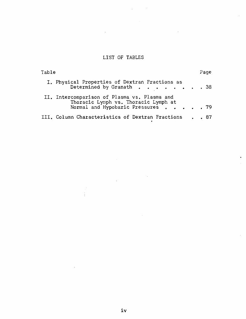
LIST OF TABLES
Table Page
I. Physical Properties of Dextran Fractions as Determined by Granath 38
II. Intercomparison of Plasma vs. Plasma and Thoracic Lymph vs. Thoracic Lymph at Normal and Hypobaric Pressures 79
III. Column Characteristics of Dextran Fractions . . 87
IV
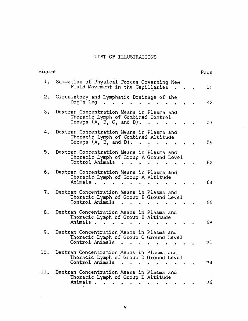
LIST OF ILLUSTRATIONS
Figure p a g e
1. Summation of Physical Forces Governing New Fluid Movement in the Capillaries . . . 10
2. Circulatory and Lymphatic Drainage of the Dog's Leg 42
3. Dextran Concentration Means in Plasma and Thoracic Lymph of Combined Control Groups (A, B, C, and D) 57
4. Dextran Concentration Means in Plasma and Thoracic Lymph of Combined Altitude Groups (A, B, and D) 59
5. Dextran Concentration Means in Plasma and Thoracic Lymph of Group A Ground Level Control Animals 62
6. Dextran Concentration Means in Plasma and Thoracic Lymph of Group A Altitude Animals 64
7. Dextran Concentration Means in Plasma and Thoracic Lymph of Group B Ground Level Control Animals 66
8. Dextran Concentration Means in Plasma and Thoracic Lymph of Group B Altitude Animals 68
9. Dextran Concentration Means in Plasma and Thoracic Lymph of Group C Ground Level Control Animals 71
10. Dextran Concentration Means in Plasma and Thoracic Lymph of Group D Ground Level Control Animals 74
11. Dextran Concentration Means in Plasma and Thoracic Lymph of Group D Altitude Animals 76
VI
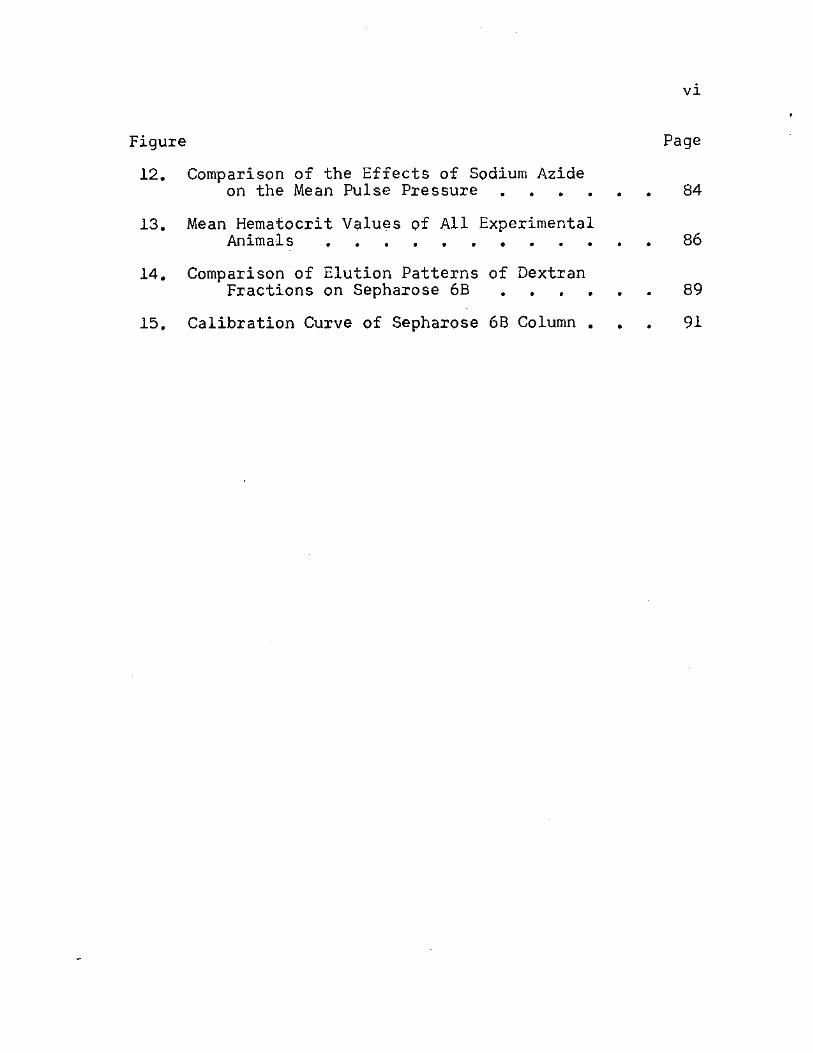
Figure Page
12. Comparison of the Effects of Sodium Azide on the Mean Pulse Pressure 84
13. Mean Hematocrit Values of All Experimental Animal s 86
14. Comparison of Elution Patterns of Dextran Fractions on Sepharose 6B 89
15. Calibration Curve of Sepharose 6B Column . . . 91
CHAPTER I
INTRODUCTION
The capillary wall forms the barrier between the
closed mammalian circulatory system and the tissues which
it serves. This wall has the distinct characteristic of
having a selectively restrictive permeability. With this
unique characteristic the wall functions to retain the
fluid volume of the blood, yet still allow the numerous
components of the circulating plasma to pass across in
amounts necessary to the physiological function of the
system. This special permeability of the capillary barrier
and the mechanisms by which materials are transported
across the barrier have long been the subject of physio-
logical investigation. The present work is submitted as
an extension of that investigation.
Capillary Permeability
Theory
Rudiments of the study of capillary permeability and
the related lymphatic system have been reported as early as
the seventeenth century. The most significant developments,
however, were brought forth in the middle of the nineteenth
century. With the theory of a closed lymphatic system

gaining impetus, related questions concerning the mechanism
of capillary permeability and lymph formation arose. Two
schools of thought developed to answer these questions (20).
The first group, those following the Heidenhain line of
thought, claimed that lymph was formed by active secretion
on the part of the lymphatic endothelial membrane. A
second group, led by Carl Ludwig, adhered to the idea that
lymph was formed by filtration. The latter line of
reasoning gained experimental prominence, and in 1873 one
of Ludwig's students, H. P. Bowditch (7), presented a paper
describing some of this experimental evidence. In this
paper the connections between the lymphatic vessels and what
is now called interstitial spaces in connective tissue were
established. It still remained, however, for Ernest Starling
to set forth the underlying principles that regulated the
dynamic process of capillary permeability.
Prior to Starling's basic pronouncement in 1896, experi-
mentation had been primarily involved with injecting foreign
substances into the previously described interstitial space
and observing their absorption through natural diffusion
into the blood vessels (52). With this in mind, Starling (53)
set out in a series of experiments to explain the absorption
effects of fluids having the same tonicity and same approx-
imate constitution as the circulating plasma. From the
results of these experiments he recognized the distinction
between total osmotic pressure and colloid osmotic pressure
as the force regulating fluid balance across the capillary
membrane. Thus he was able to establish the simplest
description of capillary membrane selectivity: free
permeability to crystalloids and water, and relative
impermeability to colloids. With a few minor additions,
the basic thrust of the Starling hypothesis has remained
valid through present investigations.
For several years after Starling submitted his
hypothesis to the scientific community, it remained
untested. It was not until 1926, when Landis (32) intro-
duced his micro-injection technique, that anyone had the
capability to test the theory. His method permitted
measurement of mean pressure flucuations in successive
periods without disturbance of blood flow in individual
capillaries. Through the use of this technique, he was able
to study and report several physiological parameters
involving the capillary dynamics in frog mesentery (33, 34).
In 1930 Landis (35) applied his technique to mammalian
mesenteric capillaries, and his observations indicated a
balance between the average capillary blood pressure and
the osmotic pressure of the plasma proteins. He reported
that the average arteriolar capillary blood pressure was
above the colloid osmotic pressure, and the average venous
capillary pressure was below this value. These findings
provided evidence in favor of Starling's hypothesis for
fluid interchange in mammals.
At the same time that Landis was setting forth the
results of his experiments, another group of investigators
was discovering some facts that did not seem to coincide
with some of Landis's findings. Churchill, Nakazawa, and
Drinker (10) measured colloid osmotic pressure of blood
serum and lymph from subcutaneous lymph spaces in frogs.
With this procedure they were able to show physical and
chemical forces acting to produce a normal movement of
blood plasma, including a fraction of its protein, from the
blood vessels into the lymphatic system. In a more
elaborate set of experiments, Conklin (11) demonstrated
the presence of serum proteins in lymph. She also
discovered that an almost total depletion of protein from
plasma and lymph occurred with injection of large amounts
of Ringer's solution into the frog's circulatory system.
In addition to this, foreign proteins, when injected into
the circulatory system, were found to pass through the
capillaries and to be recovered in the lymph. This latter
phase of experimentation was thought to be somewhat
dependent on molecular weight.
With this evidence of serum proteins in amphibian
lymph, Drinker and Field (13) published a paper citing
references of lymph content from various regions of
different mammals. In almost all instances, the lymph
contained serum proteins, usually in concentrations above
one per cent. This led Field and Drinker (14) to develop
a series of experiments involving the quantitative detection
of horse and dog serum in lymph. By ligating various
lymphatic branches and by injecting one of the above sera
into strategic locations, they were able to show a
generalized though graded permeability to protein on the
part of blood capillaries in different parts of the dog.
This and previous findings modified but did not invalidate
Starling's hypothesis. It was determined that the
difference between the osmotic pressures of the intra- and
extravascular fluids was the important factor. This meant
that the variations between the capillary pressure and this
"effective" osmotic pressure determined filtration and
absorption by the capillary endothelial wall.
This theory was further refined in 1948 by Pappenheimer
and Soto-Rivera (41). They developed a technique for
perfusion of dog and cat hindlegs so that the protein
osmotic pressure of the plasma could be independently
adjusted to desired constant values. Included in this
perfusion method was the delicate, individual control of
capillary and venous pressures. The resulting rate of
filtration of fluid from blood to tissues or the absorption

6
of fluid from tissues to blood was determined respectively
as a gain or loss in weight of the leg. By manipulating
each of the parameters individually or in pairs, these
investigators determined that with any given protein osmotic
pressure, there were an infinite number of pairs of values
of arterial and venous pressures at which no net transfer of
fluid occurs. Two or more pairs of such values were thus
defined as the mean hydrostatic pressure in the capillaries.
Using this method the two colleagues also established that
the principal osmotic factor regulating the fluid exchange
normally involves changes in the plasma protein concentration
rather than changes in the composition of tissue fluid. As
a result of these findings, a clarification and a delicate j
method of measurement were attained for capillary pressure
and effe tive osmotic pressure described by Field and
Drinker (14).
The study of the kinetic aspects of this effective
osmotic pressure was greatly enhanced by the use of radio-
isotopic tracers. In experiments using trace amounts of 131
albumin and globulin labeled with radioactive iodine (I ),
Wasserman and Mayerson (57) were able to follow the pathway «
of these serum proteins as they traveled from the plasma to
the lymph and back to the plasma. They discovered that
both proteins passed through the capillary wall, albumin
passing approximately 1.6 times faster than globulin.
After mixing with the pericapillary interstitial fluid,
about two-thirds of these "leaked" proteins were returned
to the circulatory system via the thoracic lymph duct.
Newly metabolized proteins were not detected in the thoracic
duct lymph.
In a later study using the same techniques, Wasserman,
Joseph, and Mayerson (60) established that the extravascular
albumin mass exists as a separate entity. Net movements
from this mass occurred when the equilibrium between the
vascular and extravascular masses was disturbed. With this
last information, the importance of "effective" osmotic
pressure, its role and its maintainance in capillary
permeability, was essentially determined.
This meant that the only aspect of the Starling hypo-
thesis left to be investigated was the effect of interstitial
pressure on the net transcapillary pressure. Early attempts
at actual measurement of this pressure centered around the
needle or micropuncture technique. It involved inserting a
very small needle into a tissue and then determining the
minimum pressure required to make fluid flow from the needle
into the tissue. A good example of this technique was given
by Swann and co-workers (56) in 1950. A major criticism of
the method, however, was the fact that at best the inserted
needle was still some 300 times as large as the widths of
the tissue spaces. As a result, distortion of these spaces
and false measurements were thought to occur.
8
In order to bypass this criticism, Guyton (22)
developed a technique using perforated capsules implanted
in the tissue spaces. Two to three weeks after implantation,
his histological examination of the capsules revealed that
they were filled with interstitial fluid, tissue and blood
vessels had begun to fill the capsule, and the intra- and
extracapsular fluids communicated freely. In essence, he
had created a comparatively large interstitial space into
which he could insert a small needle and measure the
pressure without distortion of the surrounding tissues.
Using this method the workers in Guyton's laboratory
set out a series of experiments in 1963 to investigate the
importance of interstitial fluid pressure on capillary
permeability. Guyton (22) initially established that this
pressure was negative and that it only became positive in
cases of tissue edema. He also demonstrated that the
pressure within the capsule was affected by the classic
permeability mediators of arterial pressure, venous pressure,
and effective osmotic pressure. A later study (23) depicted
these effects in the form of pressure volume curves.
In a series of experiments that climaxed their studies
in 1966, Guyton and several of his co-workers (24) used the
implanted capsules to compare the effects of changes in
interstitial fluid pressure with the effects of changes in
venous and arterial pressures. Used in this manner the
implanted capsules were described as internal plethysmo-
graphs, and the net movement of fluid through the capillary
membranes Wc(S measured. As a result of this modification,
they were able to demonstrate that a decrease in inter-
stitial fluid pressure of 1 mm Hg increased the filtration
out of the capillaries 1.20 times as much as did a 1 mm Hg
increase in venous pressure. In addition, a filtration
coefficient for fluid movement through the capillary wall
per unit change in interstitial fluid pressure was
calculated, and this value was compatible with the coeffic-
ient calculated by Pappenheimer and Soto-Rivera (41)
following changes that they induced in capillary and
osmotic pressures.
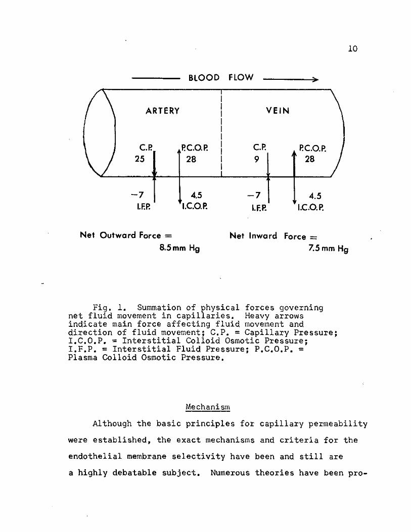
The primary significance of the experimentation cited
to this point was that it unquestionably confirmed the
principles of Starling's basic hypothesis. The net transfer
of fluids through capillary endothelial membranes was
governed by a delicate balance and interplay of two factors:
net transcapillary pressure, which was determined by
arterial pressure, venous pressure, and interstitial fluid
pressure; and (2) effective colloid osmotic pressure, which
was determined by plasma colloid osmotic pressure and
interstitial fluid colloid osmotic pressure. A pictorial
summation of these factors can be seen in Figure 1.
10
BLOOD FLOW
ARTERY
C.R 25
RC.O.P. 28
VEIN
C.R » RC.O.R * 28
- 7 I.F.P.
4.5 I.C.O.P.
- 7
I.F.P. 4.5
I.C.O.P.
Net Outward Force
8.5 mm Hg Net Inward Force =
7.5 mm Hg
Fig. 1. Summation of physical forces governing net fluid movement in capillaries. Heavy arrows indicate main force affecting fluid movement and direction of fluid movement; C.P. « Capillary Pressure; I.C.O.P. = Interstitial Colloid Osmotic Pressure; I.F.P. = Interstitial Fluid Pressure; P.C.O.P. = Plasma Colloid Osmotic Pressure.
Mechanism
Although the basic principles for capillary permeability
were established, the exact mechanisms and criteria for the
endothelial membrane selectivity have been and still are
a highly debatable subject. Numerous theories have been pro-
11
posed as an explanation, but as yet none have emerged as
a completely acceptable answer.
The process of diffusion has been regarded by most as
an integral part of the transfer mechanism, especially that
of low molecular weight molecules. As early as 1926, Arnold
and Mendel (3) studied the composition of total solids,
chlorides, calcium, phosphorus, sugar, non-proteins, and
proteins in serum and lymph of dogs. Applying some abnormal
physiological concentrations, they concluded that inter-
changes occurred in response to alterations in concentration
gradients. They also observed that diffusable substances
passed easily and rapidly at all times between the blood,
lymph and tissues. Renkin (48) extended this observation
to include carbon dioxide and lipid-soluble molecules.
Several other investigators have independently substantiated
these findings.
In a comprehensive review of the capillaries presented
in 1947, Chambers and Zweifach (9) cited some endothelial
properties which were proposed to account for the diffusion
observations. Using accepted histological staining tech-
niques and the light microscope, they distinguished three
types of cellular membranes on the basis of structural
factors regulating the passage of materials. The first two
types involved active secretory work by the endothelial cell
constituents. This involved either total transport by
secretion, as in the endothelial cells of renal proximal
12
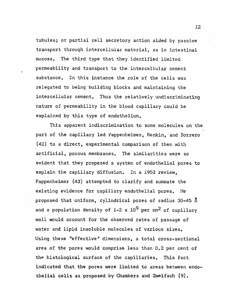
tubules; or partial cell secretory action aided by passive
transport through intercellular material, as in intestinal
mucosa. The third type that they identified limited
permeability and transport to the intercellular cement
substance. In this instance the role of the cells was
relegated to being building blocks and maintaining the
intercellular cement. Thus the relatively undiscriminating
nature of permeability in the blood capillary could be
explained by this type of endothelium.
This apparent indiscrimination to some molecules on the
part of the capillary led Pappenheimer, Renkin, and Borrero
(42) to a direct, experimental comparison of them with
artificial, porous membranes. The similarities were so
evident that they proposed a system of endothelial pores to
explain the capillary diffusion. In a 1953 review,
Pappenheimer (43) attempted to clarify and summate the
existing evidence for capillary endothelial pores. He
proposed that uniform, cylindrical pores of radius 30-45 A
and a population density of 1-2 x 10^ per cm^ of capillary
wall would account for the observed rates of passage of
water and lipid insoluble molecules of various sizes.
Using these "effective" dimensions, a total cross-sectional
area of the pores would comprise less than 0.2 per cent of
the histological surface of the capillaries. This fact
indicated that the pores were limited to areas between endo-
thelial cells as proposed by Chambers and Zweifach (9).

13
Pappenheimer made the additional distinction that most or
all of the capillary endothelial surface was available for
the passage of oxygen, carbon dioxide, and other lipid
soluble molecules. These substances were not dependent on
pores for transcapillary exchanges.
The transport of larger molecules proved to be a weak
point in Pappenheimer's (43) reasoning. Previous investi-
gators had reported decreased diffusion rates for the serum
proteins and other large molecules. To explain these
decreases, Pappenheimer developed the concept of restricted
diffusion or molecular sieving. According to this theory
the effective pore size in the capillary wall was
sufficiently great to allow even the plasma protein molecules
to penetrate the pores. The degree of molecular sieving
of any given solute depended upon the ratio of its
restricted diffusion coefficient to the filtration rate
through the capillary wall. Thus the degree of transport
for large molecules was dependent upon their restricted
filtration rates through the pores. This explanation left
many investigators in doubt, and as a result, experimentation
and hypotheses became abundant in this area.
One group of investigators headed by H. S. Mayerson
initiated some experimentation in 1955 that dominated the
thought in the area of-capillary permeability for the next
few years. Prior to this time Wasserman and Mayerson (58)
had studied the effects of the polysaccharide dextran on
14
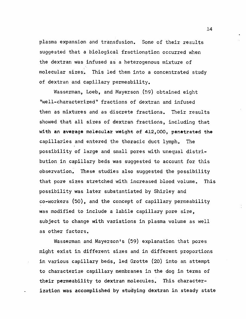
plasma expansion and transfusion. Some of their results
suggested that a biological fractionation occurred when
the dextran was infused as a heterogenous mixture of
molecular sizes;. This led them into a concentrated study
of dextran and capillary permeability.
Wasserman, Loeb, and Mayerson (59) obtained eight
"well-characterized" fractions of dextran and infused
then as mixtures and as discrete fractions. Their results
showed that all sizes of dextran fractions, including that
with an average molecular weight of 412,000, penetrated the
capillaries and entered the thoracic duct lymph. The
possibility of large and small pores with unequal distri-
bution in capillary beds was suggested to account for this
observation. These studies also suggested the possibility
that pore sizes stretched with increased blood volume. This
possibility was later substantiated by Shirley and
co-workers (50), and the concept of capillary permeability
was modified to include a labile capillary pore size,
subject to change with variations in plasma volume as well
as other factors.
Wasserman and Mayerson's (59) explanation that pores
might exist in different sizes and in different proportions
in various capillary beds, led Grotte (20) into an attempt
to characterize capillary membranes in the dog in terms of
their permeability to dextran molecules. This character-
ization was accomplished by studying dextran in steady state
15
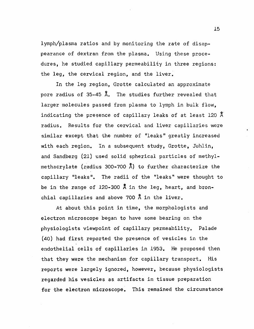
lymph/plasma ratios and by monitoring the rate of disap-
pearance of dextran from the plasma. Using these proce-
dures, he studied capillary permeability in three regions:
the leg, the cervical region, and the liver.
In the leg region, Grotte calculated an approximate
pore radius of 35-45 A. The studies further revealed that
larger molecules passed from plasma to lymph in bulk flow,
indicating the presence of capillary leaks of at least 120 A
radius. Results for the cervical and liver capillaries were
similar except that the number of "leaks" greatly increased
with each region. In a subsequent study, Grotte, Juhlin,
and Sandberg (21) used solid spherical particles of methyl-
methacrylate (radius 300-700 A ) to further characterize the
capillary "leaks". The radii of the "leaks" were thought to
be in the range of 120-300 A in the leg, heart, and bron-
chial capillaries and above 700 A in the liver.
At about this point in time, the morphologists and
electron microscope began to have some bearing on the
physiologists viewpoint of capillary permeability. Palade
(40) had first reported the presence of vesicles in the
endothelial cells of capillaries in 1953. He proposed then
that they were the mechanism for capillary transport. His
reports were largely ignored, however, because physiologists
regarded his vesicles as artifacts in tissue preparation
for the electron microscope. This remained the circumstance
16
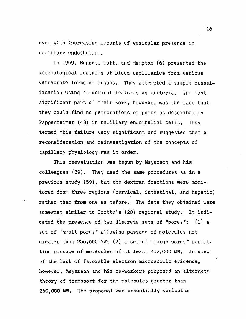
even with increasing reports of vesicular presence in
capillary endothelium.
In 1959, Bennet, Luft, and Hampton (6) presented the
morphological features of blood capillaries from various
vertebrate forms of organs. They attempted a simple classi-
fication using structural features as criteria. The most
significant part of their work, however, was the fact that
they could find no perforations or pores as described by
Pappenheimer (43) in capillary endothelial cells. They
termed this failure very significant and suggested that a
reconsideration and reinvestigation of the concepts of
capillary physiology was in order.
This reevaluation was begun by Mayerson and his
colleagues (39). They used the same procedures as in a
previous study (59), but the dextran fractions were moni-
tored from three regions (cervical, intestinal, and hepatic)
rather than from one as before. The data they obtained were
somewhat similar to Grotte's (20) regional study. It indi-
cated the presence of two discrete sets of "pores": (1) a
set of "small pores" allowing passage of molecules not
greater than 250,000 MW; (2) a set of "large pores" permit-
ting passage of molecules of at least 412,000 MW. In view
of the lack of favorable electron microscopic evidence,
however, Mayerson and his co-workers proposed an alternate
theory of transport for the molecules greater than
250,000 MW. The proposal was essentially vesicular
17
transport, but they called it "cytopempsisThis descrip-
tion was employed to convey the idea that substances were
being transported through the cytoplasm rather than being
engulfed for cell use as in "pinocytosis
As the physiologists began to reconcile their position,
the morphologists also had to change their views. With
improved techniques in electron microscopy, Luft (37)
reported some of the same observations as Chambers and
Zweifach (9) had seen with a light microscope.. He used
ruthenium red with the electron microscope and confirmed
for the first time the presence of the "endocapillary layer"
and its continuation between the cells as the "intercellular
cement". Luft further observed that the tight junction of
the intercellular cement at the region of endothelial
contact could still remain porous. In.this case, Starling's
hypothesis had a plausible foundation in structure without
the need to invoke vesicular transport of fluid across the
capillary wall. This fact did not account for the transport
of large molecules, however.
A survey of the current literature does not resolve
this question any more satisfactorily than earlier reports.
Numerous vesicular transport models based on electron micro-
scope studies are available. Shea and Karnovsky (49) seem
to have the most prominent model. It employs the use of
horseradish peroxidase, and it is based on Brownian movement
of vesicles through the endothelial cells. The work of
18
Casley-Smith (8) in measuring, numbering, and calculating
the rate of transport of these vesicles supports the model
fairly well.
Garlick and Renkin (15) on the other hand, have
attacked the question from an experimental aspect. Using
classical infusion and monitoring techniques of dextran,
they study the permeability characteristics of capillaries
in the dog's paw. With the addition of mathematical cal-
culations to their experimentation, they favor a system of
small pores of 40 X radius for transport of small molecules,
In order to carry the larger molecules, Garlick and Renkin
propose either a few large pores of approximately 800 X
radius or pinocytotic vesicles, of approximately 250 A
radius. It is obvious that much knowledge has been gained
since Starling set forth his hypothesis in 1896, but it is
also evident from the lack of decisive agreement of these
and other investigators that much more knowledge still
remains to be attained.
Altitude Studies
The effects of high altitude were probably first
noticed by the early explorers as they scaled the mountains
in search of various goals. Indeed, their reports of some
of these effects stimulated some of the initial scientific
expeditions. Douglas and his associates (12) reported one
of these classical scientific adventures in 1913. They

19
traveled to the top of Pike's Peak and described how hypoxia
or oxygen deficiency progressively stimulated their venti-
lation. As a result, their alveolar CC>2 tension fell as
they ascended to the summit. Such reports relating to
hypoxia and its associated effects have dominated the
literature in the area of altitude physiology.
Alberto Hurtado (29), who with his research group in
Lima, Peru, has been the most active investigator in this
area, related the rationale behind the predominance of
hypoxia-related reports. At reduced barometric pressure,
he explained that the partial pressure of oxygen in the
inspired air was low. As a result, the hemoglobin of the
blood circulating through the lungs became less saturated
with this gas. This fact, together with the decreased
tension of the fraction physically dissolved in the plasma,
made its diffusion and utilization at a tissue level more
difficult. A variety of coordinated mechanisms compen-
sated in an attempt to relieve the stress placed on the
tissues.
Reports of these compensatory mechanisms, both long
and short term, have covered a myriad of physiological
phenomena. Shifts in oxygen-hemoglobin dissociation curves,
changes in pH of body fluids, increases in hemopoeitic
activity, and various aspects of pulmonary edema are a few
of the areas in which altitude research has flourished. The
primary concern of the present study, however, involves
20
cardiovascular responses to short-term, simulated altitude
with an attempt to eliminate the effects of hypoxia.
To eliminate the effects of hypoxia, the most natural
answer was to administer a supply of oxygen to the experi-
mental subject during the test period. Whitehorn, Edelman,
and Hitchcock (62) examined this solution in 1946. Allowing
their subjects to breathe 100 per cent oxygen at normal
barometric pressure, they determined resulting cardiac out-
puts from ballistocardiograms. Their results indicated a
maintainance of the blood pressure level in spite of reduced
cardiac output. This was interpreted as evidence of an
increase in the general peripheral vascular resistance. The
investigators suggested that the effects be remembered in
conditions of hyperoxygenation of the blood, such as denitro-
genation, and that their presence may play a part in the
physiological responses to changes in barometric pressure.
A direct application of these suggestions was presented
by Girling and Maheux (16) in 1952. They studied the
peripheral circulation of rabbits at 10,000, 20,000, and
30,000 feet simulated altitude. Animals taken above this
level demonstrated fatal signs of anoxia, but animals
supplied with oxygen could safely withstand the additional
decreased pressure. The peripheral resistance exhibited
the same response with or without the supplemental oxygen;
small change in resistance to 20,000 feet, but an appreci-
able increase in resistance from ground to 30,000 feet. In

21
order to eliminate the possibility of nitrogen bubbles
forming in the blood and increasing peripheral resistance,
a subsequent study (17) by the same investigators employed
preoxygenation periods of up to four hours. The results
remained the same, however, and they concluded that the
observed vasomotor response was due to reduced barometric
pressure causing decreased critical closing pressures of
peripheral blood vessels.
In an attempt to observe the effects of simulated
altitudes of 60;,000 and 80,000 feet, Sullivan and De Gennaro
(54) studied the peripheral circulation in the web of a
frog's foot. Their observations suggested that the extent
of decrease in blood flow was dependent on the altitude,
the length of time the animal was subjected to this altitude,
and whether or not air was supplied to the animal during
decompression. These results were thought to be primarily
due to the hypoxia accompanying decompression, but the
investigators also suggested that the initial, reduced
venous flow was due to stasis following increased capillary
endothelial permeability under the influence of hypoxia.
At any rate, the effects could be alleviated by reducing
hypoxia.
In 1966 Marotta and Boon (38) extended the studies of
oxygenation at simulated altitudes to dogs. They monitored
femoral arterial blood flow and related cardiopulmonary
parameters during exposure to altitude (9,000, 17,000, and

22
25,000 feet simulated altitude) while breathing 100 per cent
oxygen or a mixture of oxygen and nitrogen. One series of
experimental animals were subjected to per-altitude denitro-
genation with 100 per cent oxygen at ground level. A decrease
in femoral arterial blood flow was observed in all animals
except those denitrogenated prior to ascent. Initial and
final blood flows in the latter animals, however, were all
less than in the other animals. To this extent, denitro-
genation exhibited some slowing effect in all of the animals
examined. It thus appeared that increased peripheral
resistance to blood flow was an unavoidable legacy in the
attempt to overcome the more severe effects of hypoxia.
Experimentation has thus proceeded with and without the aid
of supplemental oxygen depending on the nature of the
experiment involved.
Another cardiovascular response that interested
investigators in recent years has been the fluid shifts and
the blood component imbalances that developed with the
encounter of low barometric pressure. A general plasma
volume decrease had long been accepted as a part of the
altitude acclimatization process, but it had been thought
this loss was due to body dehydration. In 1969 Hannon,
Chinn, and Shields (25) used a new, more reliable method
for determining total body water. As a result they reported
a decreased plasma volume with a static or slightly raised
total body water. Further investigation (26) of the extra-
23
vascular compartments showed decreases in interstitial and
extracellular spaces and an increase in intracellular space.
This was thought to be caused by osmotic effects associated
with a transfer of electrolytes, principally, sodium,
chloride, and bicarbonate. Krzywicki and associates (31)
recently reported, however, that their experimentation
indicated that the loss in body weight was still due to loss
in total body water. They maintained that this hypo-
hydration was an adaptive mechanism to altitude. At this
point in experimentation, the purpose and destination of
fluid shifts in subjects maintained at altitude remains
unresolved.
Another baffling question of recent years has been the
shift in components of the blood with the advent of altitude,
Surks (55) reported an initial albumin shift from intra-
vascular to extravascular compartments with an increased
degredation of albumin during initial stages of altitude.
After six days at altitude, however, the shift was reversed
from the extravascular to the intravascular compartments.
This latter shift was accompanied by a decrease in the rate
of albumin synthesis and net transfer. Westergaard and his
associates (61) also observed the initial shift in albumin
from intravascular to extravascular compartments. They
suggested that this might be due to increased capillary
permeability, but a direct measurement was not performed.
Independent attempts were recently made by Parker (44) and
24
Reaves (47) to directly measure the capillary permeability
of dogs at altitude. Their results both indicated an
increase in capillary permeability possibly due to stretched
pores. As in the case of fluid shifts, however, the exact
purpose and mechanism of blood component shifts at high
altitude levels still remains to be determined.
Molecular Weight Determination by Gel Filtration
The class of polysaccharides called dextrans consists
of a great variety of a-polyglucosans produced by the coccus
Leuconostoc mesenteroides and closely related bacteria under
suitable environmental conditions. In their native state,
the dextran chains are composed of about 200,000 glucose
units, corresponding to a molecular weight of about 40 mil-
lion. Partial hydrolysis of these chains yields smaller
molecular weight molecules that exhibit desirable character-
istics for experimental use. They appear to be electrically
neutral, metabolically inert (no mammalian tissue extract is
known to break them down), apparently non-toxic in high
concentration, and can be assayed accurately and easily (51).
Their one drawback in most experimental endeavors is that
they are very difficult to obtain in narrow-range molecular
weight fractions. Thus, experimentors employing their use
must in some way lessen the effects of the molecular weight
dispersity, or they must acknowledge the limitations placed
25
on the validity of their results by the use of the wide
range of molecular weight molecules.
One of the methods by which the molecular weight range
of dextrans can be narrowed, or at least estimated, is gel
filtration. In this process, a mixture of molecules is
passed through a column of porous gel granules. The
molecules appear in the effluent in order of decreasing
size. Fractionation is believed to occur when diffusion of
the molecules into the pores is restricted but not prevented
because of their size. As a result of this restriction, the
molecules pass through the column at rates that are related
inversely to the fluid volume accessible to them within the
column. This molecular sieving is the basis by which
different and somewhat narrow-range dextran fractions can
be obtained. An estimation of their molecular weight may
aldo be determined as a result of this basic principle.
An early-application of gel filtration to large
molecules was attempted by Lathe and Ruthven (36) in 1956.
Using columns of starch grains, they were able to achieve
some separation of polypeptides and proteins. They proposed
that their method be used as a means of molecular weight
determination, and they reported some interesting results
in this connection. Their starch columns, however, did not
effect enough separation between proteins for further
development of the idea with this particular gel.
2 6
Numerous other types of porous gels were introduced,
however, and the idea of molecular weight determination was
applied to them. One of the more successful gels was a
cross-linked dextran gel introduced by Porath (46) in 1960.
He reported that highly cross-linked dextran gels separated
amino acids from proteins and large peptides. Gels
possessing a low degree of cross-linking even retained
proteins for fractionation. This dextran gel was later used
by Granath and Flodin (18) to fractionate low molecular
weight dextrans. These two investigators were able to
establish the limits of the molecular weight sizes of the
gels involved. With further use and development a series of
cross-linked dextran gels was marketed under the trade name
of Sephadex.
By using G-75 and G-100 of the Sephadex series, Andrews
(l) was able to' correlate the elution volumes, Ve, and
molecular, weights of proteins of known molecular weight.
Within the limits of certain ranges, the gels gave optimum
separation of proteins according to molecular weight, but
their useful working ranges extended only to about 70,000
and 150,000 respectively. The complete exclusion limits
for proteins was estimated somewhat higher, however. As a
final test, Andrews used his columns to estimate the molec-
ular weights of various enzymes, and he found that these
values were in agreement with those reported in the litera-
ture. He had essentially calibrated Sephadex columns within

27
prescribed limits for the determination of molecular weights
of proteins.
In 1967 Granath and Kvist (19) were able to apply the
same basic procedures to calibration of dextrans. This was
more difficult considering the polydispersity characteristics
of dextrans, and certain physical and mathematical modifi-
cations had to jbe employed. The results were comparable to
those obtained for proteins. Thus, the calibration of
porous gel columns for molecular weight determination of
proteins and dextrans had been accomplished. They were only
useful, however, for molecular weights up to approximately
800,000 for compact, globular molecules like proteins, or
approximately 200,000 for randomly coiled chain molecules
like dextran (30). The fractionation and molecular weight
determination of larger molecules and particles required a
gel filtration medium of much higher porosity.
In 1961, Poison (45) had described a method in which
granulated agar was used for separating protein compounds
of different molecular weights. The method demonstrated
that molecules ranging in molecular weights from 13,000 to
several millions could be distinguished. Hence, Poison
proposed that his gel be used in conjunction with the linked
dextran gels for a powerful fractionation of substances of
very low to very high molecular weights. The use of granu-
lated agar, however, was severely limited due to variable
gelling and adsorption properties encountered. These
28
variations were due to the poorly defined composition of
commercially available agar which varied from batch to
batch.
Araki (2) reported that the unsatisfactory character-
istics of granulated agar as a medium for gel filtration
was due to the fact that it consisted of two different
polysaccharides, agaropectin and agarose. It was determined
that agaropectin was a typical pectic substance containing
a high percentage of sulfate and carboxyl groups. These
constituents seemed to impart the undesirable adsorption
effects seen in gel filtration. Agarose, on the other hand,
was classified as an unchanged polysaccharide that consisted
of alternating D-galactose and 3,6-anhydro-L-galactose units,
It had the same good gelling properties as agar and was thus
a superior medium for gel filtration.
Even with this discovery, the use of agarose in gel
filtration was not widespread until Hjerten (27) developed
a simple technique to remove the agaropectin from agar
solutions by precipitation with cetylpyridinium chloride. %
A further development by Hjerten (28) and Bengtsson and
Philipson (5) established it as a bonafide gel filtration
medium. These investigators independently developed
procedures for bead-gelling of agarose solutions. Photo-
micrographs of these beads showed a granular structure, but
the nature of the granules was not readily explainable (30).
The beaded agarose gels have been marketed in series,
29
similar to the Sephadex series, under the trade name of
Sepharose. Their fractionation characteristics are similar
to those of Sephadex, but their molecular weight exclusion
limits extend into the millions.
In a recent article, Arturson and Granath (4) used
Sepharose 4B as the gel medium for molecular weight cali-
bration of narrow-range, high molecular weight dextrans.
Their method was similar to that used by Granath and Kvist
(19), who used Sephadex as the gel medium. The procedure
thus yielded the first gel filtration columns capable of
fractionating and detecting extremely high molecular weight
dextrans. Its use was demonstrated in the study of biolog-
ical membranes, and it was evident that its future use in
this area could aid in the explanations of capillary
dynamics and permeability.
Statement of the Problem
In view of its varied concepts and interpretations,
and because of the discrepancies produced by the previous
utilization of polydispersed dextrans, a study using
extremely narrow-range molecular weight dextran fractions
was initiated to reevaluate and consolidate some of the
aspects of capillary permeability. A portion of the study
was performed under decreased barometric pressure in order
to clarify further some of the mechanisms involved in
particulate transfer across the capillary endothelial
30
membranes. Gel filtration procedures augmented the study
as an assessment of the polydispersity effects of the
dextrans employed.
CHAPTER BIBLIOGRAPHY
1. Andrews, P, 1964. Estimation of the molecular weights of proteins by sephadex gel-filtration. Biochem. J. 91: 222-233.
2. Araki, C. 1956. Structure of the agarose constituent of agar-agar. Bull. Chem. Soc. Japan. 29: 543-544.
3. Arnold, R. M. and L. B. Mendel. 1926. Interrelation-ships between the chemical composition of the blood and the lymph of the dog. J. Biol. Chern. 72: 189-211.
4. Arturson, G. and K. Granath. 1972. Dextrans as test molecules in studies of the functional ultra-structure of biological membranes. Clin. Chim. Acta. 37: 309-322.
5. Bengtsson, S. and L. Philipson. 1964, Chromatography of animal viruses on pearl-condensed agar. Biochim. Biophys. Acta. 79: 399-406.
6. Bennett, H. S., J. H. Luft? and J. C. Hampton. 1959. Morphological classifications of vertebrate blood capillaries. Am. J. Physiol. 196: 381-390.
7. Bowditch, H. P. 1873. The lymph spaces in fasciae. Proc. Am. Acad. Sci. 8: 508-510.
8. Casley-Smith, J. R. 1969. The dimensions and numbers of small vesicles in cells, endothelial and meso-thelial and the significance of these for endo-thelial permeability. J. Microscopy 90: 251-268.
9. Chambers, R. and B. W. Zweifach. 1947. Intercellular cement and capillary permeability. Physiol. Rev. 27: 436-463.
10. Churchill, E. D., F. Nakazawa, and C. K. Drinker. 1927. The circulation of body fluids in the frog, J. Physiol. (London). 63: 304-308.
11. Conklin, R. E. 1930. The formation and circulation of lymph in the frog. III. The permeability of the capillaries to protein. Am, J. Physiol. 95: 98-110,
31

32
12. Douglas, C. G., J. S. Haldane, Y. Henderson, and E. C. Schneider. 1913. VI. Physiological obser-vations made on Pike's Peak, Colorado, with special reference to adaptation to low barometric pressure. Phil. Trans. Hoy. Soc. B 203: 185-318.
13. Drinker, C. K. and M. E. Field. 1931. The protein content of mammalian lymph and the relation of lymph to tissue fluid. Am. J. Physiol. 97: 32-39.
14. Field, M. E. and C. K. Drinker. 1931. The permeability of the capillaries of the dog to protein. Am. J. Physiol. 97: 40-51.
15. Garlick, D. G. and E. M. Renkin. 1970. Transport of large molecules from plasma to interstitial fluid and lymph in dogs. Am. J. Physiol. 219: 1595-1605.
16. Girling, F. and C. Maheux. 1952. Peripheral circu-lation and simulated altitude. J. Aviation Med. 23: 216-217, 270.
17. Girling, F. and C. Maheux. 1953. Peripheral circu-lation and simulated altitude. Part II. J. Aviation Med. 24: 446-448.
18. Granath, K. A. and P. Flodin. 1961. Fractionation of dextran by the gel filtration method. Makromol. Chem. 48: 160-171.
19. Granath, K. A. and B. E. Kvist. 1967. Molecular weight distribution analysis by gel chromatography on sephadex. J. Chromatogr. 28: 69-81.
20. Grotte, G. 1956. Passage of dextran molecules across the blood-lymph barrier. Acta Chir. Scand. Suppl. 211: 1-84.
21. Grotte, G., L. Juhlin, and N. Sandber. I960. Passage of solid spherical particles across the blood-lymph barrier. Acta Physiol. Scand. 50: 287-293.
22. Guyton, A. C. 1963. A concept of negative interstitual pressure based on pressures in implanted perforated capsules. Circulation Res. 12: 399-414.
23. Guyton, A. C. 1965. Interstitial fluid pressure. II. Pressure-volume curves of interstitial space. Circulation Res. 16: 452-460.
33
24. Guyton, A. C., J. Prather, K. Scheel, and J. McGehee. 1966. Interstitial fluid pressure. IV. Its effect on fluid movement through the capillary wall. Circulation Res. 19: 1022-1030.
25. Hannon, J. R., K. S. K. Chinn, and J. L. Shields. 1969. Effects of acute high-altitude exposure on body fluids. Fed. Proc. 28: 1178-1184.
26. Hannon, J. P., K. S. K. Chinn, and J. L. Shields. 1971. Alterations in serum and extracellular electrolytes during high-altitude exposure. J. Appl. Physiol. 31: 266-273.
27. Hjerten, S. 1962. A new method for preparation of agarose for gel electrophoresis. Biochim. Biophys. Acta. 62: 445-449.
28. Hjerten, S. 1964. The preparation of agarose spheres for chromatography of molecules and particles. Biochim. Biophys. Acta. 79: 393-398.
29. Hurtado, A. 1971. The influence of high altitude on physiology, p. 3-13. In Porter and Knight, High Altitude Physiology: Uardiac and respiratory aspects. Churchill Livingstone, Edinburgh and London.
30. Joustra, M. K. 1969. Gel filtration on agarose gels, p. 183-198. In J. Gerritsen, Modern separation methods of macromolecules and particles. Wiley-Inter science, New York.
31. Krzywicki, H. J., C. F. Consolazio, H. L. Johnson, W. C. Nielsen, Jr., and R. A. Barnhart. 1971. Water metabolism in humans during acute high-altitude exposure (4,300 m). J. Appl. Physiol. 30: 806-809.
32. Landis, E. M. 1926. The capillary pressure in frog mesentery as determined by micro-injection methods. Am. J. Physiol. 75: 548-570.
33. Landis, E. M. 1927. Micro-injection studies of capillary permeability. II. The relation between capillary pressure and the rate at which fluid passes through the walls of single capillaries. Am. J. Physiol. 82: 217-238.
34
34. Landis, E. M. 1928. Micro-injection studies of capillary permeability. III. The effect of lack of oxygen on the permeability of the capillary wall to fluid and to the plasma proteins. Am. J. Physiol. 83: 528-542.
35. Landis, E. M. 1930. The capillary blood pressure in mammalian mesentery as determined by the micro-injection method. Am. J. Physiol. 93: 353-362.
36. Lathe, G. H. and C. R. J. Ruthven. 1956. The separation of substances and estimation of their relative molecular sizes by the use of columns of starch in water. Biochem. J. 62: 665-674.
37. Luft, J. H. 1966. Fine structure of capillary and endocapillary layer as revealed by ruthenium red. Fed. Proc. 25: 1773-1783.
38. Marotta, S. F. and D. J. Boon. 1966. Femoral arterial circulation in nonhypoxic dogs at reduced baro-metric pressures. Am. J. Physiol. 210: 953-956.
39. Mayerson, H. S., C. G. Wolfrom, H. H. Shirley, Jr., and K. Wasserman. 1960. Regional differences in capillary permeability. Am. J. Physiol. 198: 155-160.
40. Palade, G. E. 1953. Fine structure of blood capillar-ies. J. Appl. Phys. 24: 1424.
41. Pappenheimer, J. R. and A. Soto-Rivera. 1948. Effective osmotic pressure of the plasma proteins and other quantities associated with the capillary circulation in the hindlimbs of cats and dogs. Am. J. Physiol. 152: 471-491.
42. Pappenheimer, J. R., E. M. Renkin, and L. M. Barrero. 1951. Filtration, diffusion, and molecular sieving through peripheral capillary membranes. A contribution to the pore theory of capillary permeability. Am. J. Physiol. 167: 13-46.
43. Pappenheimer, J. R. 1953. Passage of molecules through capillary walls. Physiol. Rev. 33: 387-423.
44. Parker, P. E. 1969. Capillary permeability to macro-molecules at normal and hypobaric pressure. M. S. Thesis, North Texas State Univ., Denton, Texas. 45 p.
35
45. Poison, A. 1961. Fractionation of protein mixtures on columns of granulated agar. Biochim. Biophys. Acta. 50: 565-567.
46. Porath, J. 1960. Gel filtration of proteins, peptides, and amino acids. Biochim. Biophys. Acta. 39: 193-207.
47. Reaves, T. A., Jr. 1971. Effects of high altitude exposure on capillary permeability. M. S. Thesis, North Texas State Univ., Denton, Texas. 62 p.
48. Renkin, E. M. 1952. Capillary permeability to lipid-soluble molecules. Am. J. Physiol. 168: 538-545.
49. Shea, S., M. and M. J. Karnovsky. 1969. Vesicular transport across endothelium: simulation of a diffusion model. J. Theoret. Biol. 24: 30-42.
50. Shirley, H. H., Jr.., C. G. Wolfram, K. Wasserman, and H. S. Mayerson. 1957. Capillary permeability to macro-molecules: stretched pore phenomenon. Am. J. Physiol. 190: 189-193.
51. Squire, J. R. 1951. Background to biological studies with dextran. Proc. Roy. Soc. Med. 44: 557-558. (Abstr.)
52. Starling, E. H. and A. H. Tubby, 1894. On absorption from and secretion into the serous cavities. J. Physiol. (London). 16: 140-155.
53. Starling, E. H. 1896. On the absorption of fluids from the connective tissue spaces. J. Physiol, (london). 19: 312-326.
54. Sullivan, B. J. and L. D. DeGennaro. 1953. Micro-scopical observations of peripheral circulation at simulated high altitudes. J. Aviation Med. 24: 131-137.
55. Surks, M. I. 1966. Metabolism of human serum albumin in man during acute exposure to high altitude (14,100 feet). J. Clin. Invest. 45: 1442-1451.
56. Swann, H. G.j A. V. Montgomery, J. C. Davis, Jr., and E. R. Mickle. 1950. A method for rapid measure-ment of intrarenal and other tissue pressures. J. Exp. Med. 92: 625-636.
36
57. Wassexman, K. and H. S. Mayerson. 1952. Dynamics of lymph and plasma protein exchange. Cardiologica. 21: 296-307.
58. Wasserman, K. and H. S. Mayerson.* 1954. Relative importance of dextran molecular size in plasma volume expansion. Am. J. Physiol. 176: 104-112.
59. Wasserman, K., L. Loeb, and H. S. Mayerson. 1955. Capillary permeability to macromolecules. Circulation Res. 3: 594-603.
60. Wasserman, K., J. D. Joseph, and H. S. Mayerson. 1956. Kinetics of vascular and extravascular protein exchange in unbled and bled dogs. Am, J. Physiol. 184: 175-182.
61. Westergaard, H., S. Jarnum, R. Preisig, K. Rams0e, J. Tauber, and N. Tugstrup. 1970. Degredation of albumin and IgG at high altitude. J. Appl. Physiol. 28: 728-732.
62. Whitehorri, W. V., A. Edelmann, and F. A. Hitchcock. 1946. The cardiovascular responses to the breathing of 100 per cent oxygen at normal barometric pressure. Am. J. Physiol. 146: 61-65.
CHAPTER II
METHODS AND MATERIALS
Experimental Design
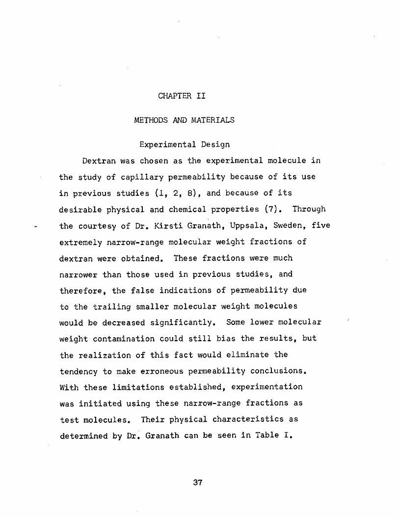
Dextran was chosen as the experimental molecule in
the study of capillary permeability because of its use
in previous studies (l, 2, 8), and because of its
desirable physical and chemical properties (7). Through
the courtesy of Dr. Kirsti Granath, Uppsala, Sweden, five
extremely narrow-range molecular weight fractions of
dextran were obtained. These fractions were much
narrower than those used in previous studies, and
therefore, the false indications of permeability due
to the trailing smaller molecular weight molecules
would be decreased significantly. Some lower molecular
weight contamination could still bias the results, but
the realization of this fact would eliminate the
tendency to make erroneous permeability conclusions.
With these limitations established, experimentation
was initiated using these narrow-range fractions as
test molecules. Their physical characteristics as
determined by Dr. Granath can be seen in Table I.
37
38
TABLE I
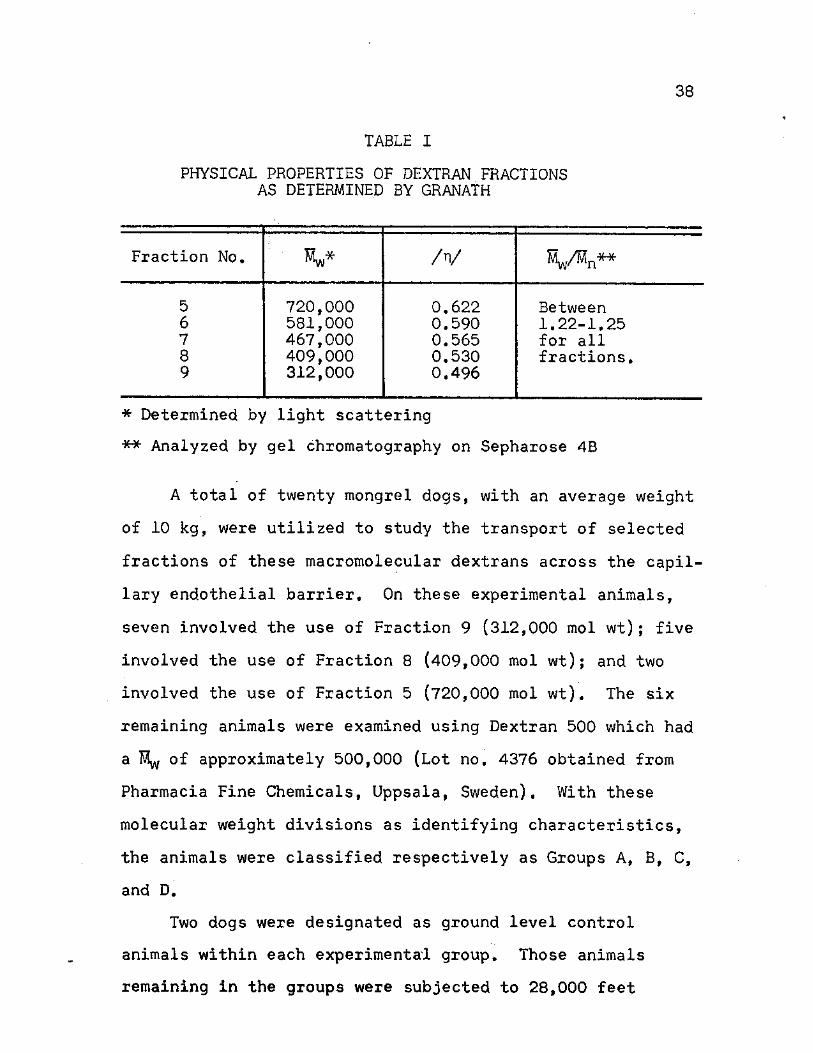
PHYSICAL PROPERTIES OF DEXTRAN FRACTIONS AS DETERMINED BY GRANATH
Fraction No. Mw* /V V^n**
5 720,000 0.622 Between 6 581,000 0.590 1.22-1.25 7 467,000 0.565 for all 8 409,000 0.530 fractions. 9 312,000 0.496
* Determined by light scattering
** Analyzed by gel chromatography on Sepharose 4B
A total of twenty mongrel dogs, with an average weight
of 10 kg, were utilized to study the transport of selected
fractions of these macromolecular dextrans across the capil-
lary endothelial barrier. On these experimental animals,
seven involved the use of Fraction 9 (312,000 mol wt); five
involved the use of Fraction 8 (409,000 mol wt); and two
involved the use of Fraction 5 (720,000 mol wt). The six
remaining animals were examined using Dextran 500 which had
a Myy of approximately 500,000 (Lot no. 4376 obtained from
Pharmacia Fine Chemicals, Uppsala, Sweden). With these
molecular weight divisions as identifying characteristics,
the animals were classified respectively as Groups A, B, C,
and D.
Two dogs were designated as ground level control
animals within each experimental group. Those animals
remaining in the groups were subjected to 28,000 feet
39
(247 mm Hg) simulated altitude. The period at altitude
lasted 2 hr, and it was preceded by a 2 >-hr equilibration
period. A 1-hr, post-altitude stabilization period
concluded the experimental run. The rate of ascent and
descent during the run was 4,000 ft/min. The animals
were denitrogenated by breathing 100% oxygen 30 min prior
to altitude, and their respiration was continually sup-
ported by this oxygen supply until 30 min after the period
at altitude. Ground level control animals were studied
under the same experimental parameters, except they
remained at ground level during the period designated for
simulated altitude. These altitude studies were deleted
in Group C.
Under these criteria established for experimental
study, each individual animal was examined under identical
surgical and analytical procedures. Every attempt was
made to reduce the occurance of procedural variations.
This experimental format was thus designed to test
capillary permeability under the individual and collective
influences of dextran molecular weight polydispersity and
decreased barometric pressure.
Preparation of the Animals
It was necessary to monitor both the circulatory
system and the lymphatic system in order to accurately
observe the permeability of the capillary walls. Since
it has been established that the permeability of

40
capillaries may vary in different regions of the body (3)
it was also desirable to monitor more than one lymphatic
drainage region. The following surgery and cannulation
procedures were developed to satisfy these monitoring
requirements. These procedures involve the thoracic
lymph duct, which collects lymph primarily from the liver,
intestines, and spleen; the popliteal lymph "network,"
which collects lymph from the subcutaneous and super-
ficial muscles of the lower leg; and the femoral artery,
which was used as the common monitor of the circulatory
system as a whole.
Surgery Procedures
Approximately 30 min before administration of anes-
thesia, the dog was fed 50 ml of condensed milk. The
absorption of this fatty substance into the intestinal
lymph subsequently rendered the' thoracic lymph a milky
white. As a result, the thoracic lymph duct could be
easily identified in later dissection. The animal was
the anesthetized by an intravenous injection of sodium
pentobarbital, 30 mg/kg body weight, in the cephalic vein.
A glass endotracheal cannula was inserted in the trachea,
and the proposed surgical areas were shaved.
A 3-inch incision, beginning at the base of the neck,
was made directly above the left external jugular vein.
The external jugular, subclavian, and transverse scapular
veins were exposed by blunt dissection. The entrance of
41
the thoracic lymph duct into the venous system was generally
located in an area under the external jugular and around
the junction of this vein with the subclavian vein.
Anatomical variations were observed due to developmental
anomalies and the use of mongrel dogs. Once it was
identified, however, approximately 2 inches of the duct
were dissected free of adipose and connective tissue for
cannulation. A small portion of the transverse scapular
vein was also cleared for subsequent bypass cannulation.
At this point the incision was packed with moist, saline
pads, and the remaining surgery was performed.
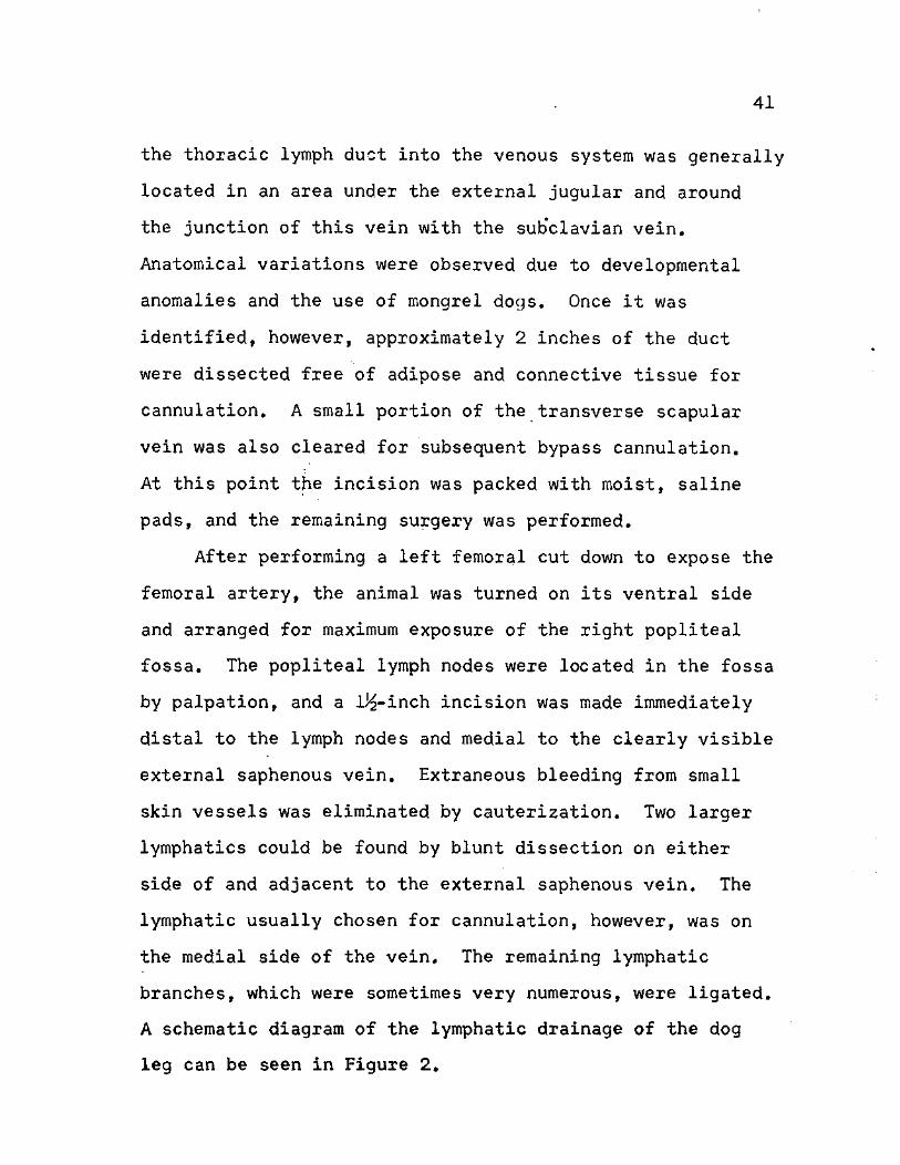
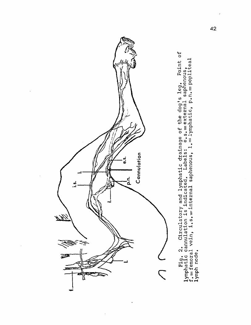
After performing a left femoral cut down to expose the
femoral artery, the animal was turned on its ventral side
and arranged for maximum exposure of the right popliteal
fossa. The popliteal lymph nodes were located in the fossa
by palpation, and a lJ£-inch incision was made immediately
distal to the lymph nodes and medial to the clearly visible
external saphenous vein. Extraneous bleeding from small
skin vessels was eliminated by cauterization. Two larger
lymphatics could be found by blunt dissection on either
side of and adjacent to the external saphenous vein. The
lymphatic usually chosen for cannulation, however, was on
the medial side of the vein. The remaining lymphatic
branches, which were sometimes very numerous, were ligated.
A schematic diagram of the lymphatic drainage of the dog
leg can be seen in Figure 2,
42
J B W V W
<4H O
- P C
•H O
03 cd •»-p
_ 0) »H P h 3 rH
o a c o
• a) a . CT*rC II a) a II
rH co • co a
w • •—I Q>
cn co o c -
Tf N CD
CD - P - P X X a j -p a>
a £
0) rH CD *11 D > 0 II 05 • C »H
O •H
Mh O
•H • • CO <0 «*
H rH (0 T5 CD 3
JQ o O CO c
•H 1—5 CD - P x : CO a
r C • (0 a x * (0 £ CD £>h-P rH
rH CO CO O c
" d - H H C T3 CD CO C - P
•H C •H
JH <0 H O »H II - P •
CO c rH O •
D *H •H O - P H (0 • *
•H rH C O D •H
CD > a c • CO
CM O rH • CO CD
• a H TJ o v h o o
•H - P £ C CO CD
CL it CL a II £ pN « >H
H C H H
43
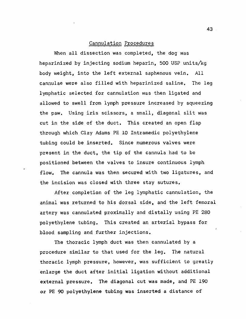
Cannulation Procedures
When all dissection was completed, the dog was
heparinized by injecting sodium heparin, 500 USP units/kg
body weight, into the left external saphenous vein. All
cannulae were also filled with heparinized saline. The leg
lymphatic selected for cannulation was then ligated and
allowed to swell from lymph pressure increased by squeezing
the paw. Using iris scissors, a small, diagonal slit was
cut in the side of the duct. This created an open flap
through which Clay Adams PE 10 Intramedic polyethylene
tubing could be inserted. Since numerous valves were
present in the duct, the tip of the cannula had to be
positioned between the valves to insure continuous lymph
flow. The cannula was then secured with two ligatures, and
the incision was closed with three stay sutures.
After completion of the leg lymphatic cannulation, the
animal was returned to his dorsal side, and the left femoral
artery was cannulated proximally and distally using PE 280
polyethylene tubing. This created an arterial bypass for
blood sampling and further injections.
The thoracic lymph duct was then cannulated by a
procedure similar to that used for the leg. The natural
thoracic lymph pressure, however, was sufficient to greatly
enlarge the duct after initial ligation without additional
external pressure. The diagonal cut was made, and PE 190
or PE 90 polyethylene tubing was inserted a distance of
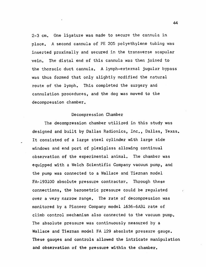
44
2-3 cm. One ligature was made to secure the cannula in
place. A second cannula of PE 205 polyethylene tubing was
inserted proximally and secured in the transverse scapular
vein. The distal end of this cannula was then joined to
the thoracic duct cannula. A lymph-external jugular bypass
was thus formed that only slightly modified the natural
route of the lymph. This completed the surgery and
cannulation procedures, and the dog was moved to the
decompression chamber.
Decompression Chamber
The decompression chamber utilized in this study was
designed and built by Dallas Radionics, Inc., Dallas, Texas.
It consisted of a large steel cylinder with large side
windows and end port of plexiglass allowing continual
observation of the experimental animal. The chamber was
equipped with a Welch Scientific Company vacuum pump, and
the pump was connected to a Wallace and Tiernan model
FA-193100 absolute pressure contractor. Through these
connections, the barometric pressure could be regulated
over a very narrow range. The rate of decompression was
monitored by a Pioneer Company model 1636-6AB1 rate of
climb control mechanism also connected to the vacuum pump.
The absolute pressure was continuously measured by a
Wallace and Tiernan model FA 129 absolute pressure gauge.
These gauges and controls allowed the intricate manipulation
and observation of the pressure within the chamber.
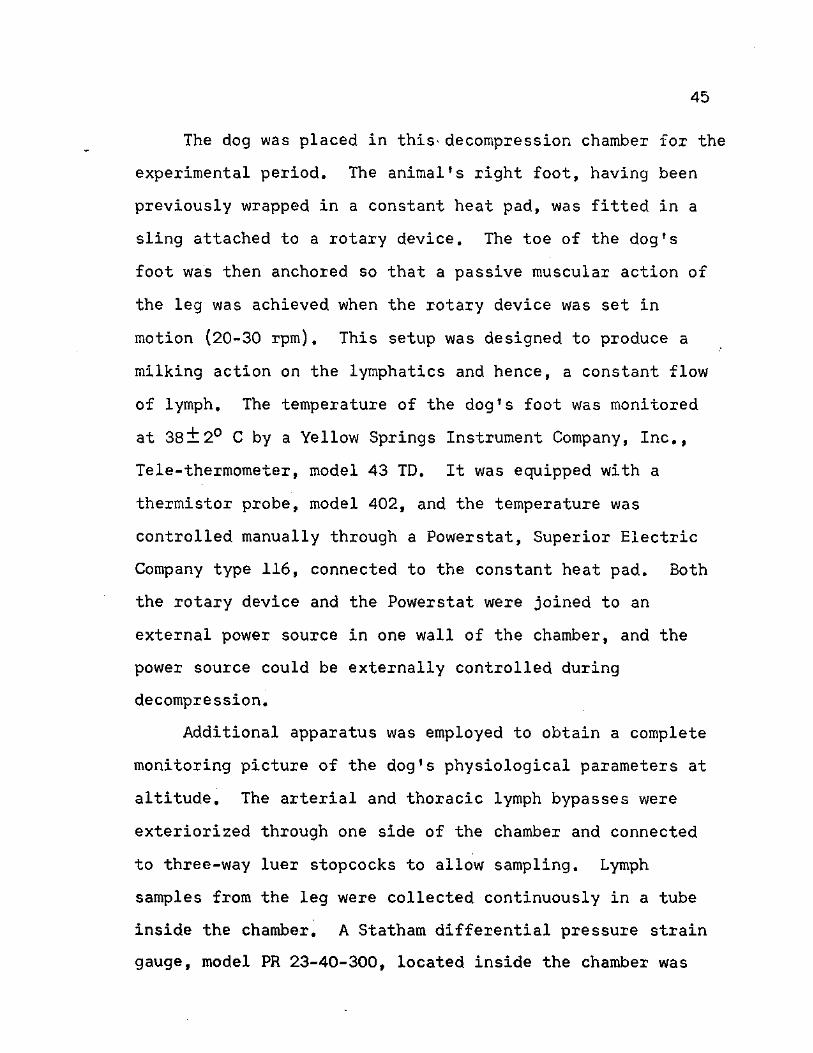
45
The dog was placed in this- decompression chamber for the
experimental period. The animal's right foot, having been
previously wrapped in a constant heat pad, was fitted in a
sling attached to a rotary device. The toe of the dog's
foot was then anchored so that a passive muscular action of
the leg was achieved when the rotary device was set in
motion (20-30 rpm). This setup was designed to produce a
milking action on the lymphatics and hence, a constant flow
of lymph. The temperature of the dog's foot was monitored
at 38lt20 C by a Yellow Springs Instrument Company, Inc.,
Tele-thermometer, model 43 TD. It was equipped with a
thermistor probe, model 402, and the temperature was
controlled manually through a Powerstat, Superior Electric
Company type 116, connected to the constant heat pad. Both
the rotary device and the Powerstat were joined to an
external power source in one wall of the chamber, and the
power source could be externally controlled during
decompression.
Additional apparatus was employed to obtain a complete
monitoring picture of the dog's physiological parameters at
altitude. The arterial and thoracic lymph bypasses were
exteriorized through one side of the chamber and connected
to three-way luer stopcocks to allow sampling. Lymph
samples from the leg were collected continuously in a tube
inside the chamber. A Statham differential pressure strain
gauge, model PR 23-40-300, located inside the chamber was
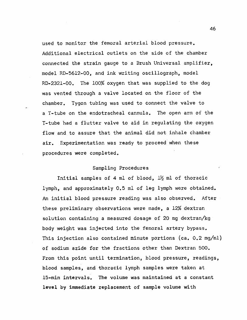
46
used to monitor the femoral arterial blood pressure.
Additional electrical outlets on the side of the chamber
connected the strain gauge to a Brush Universal amplifier,
model RD-5612-00, and ink writing oscillograph, model
RD-2321-00. The 100% oxygen that was supplied to the dog
was vented through a valve located on the floor of the
chamber. Tygon tubing was used to connect the valve to
a T-tube on the endotracheal cannula. The open arm of the
T-tube had a flutter valve to aid in regulating the oxygen
flow and to assure that the animal did not inhale chamber
air. Experimentation was ready to proceed when these
procedures were completed.
Sampling Procedures
Initial samples of 4 ml of blood, 1% ml of thoracic
lymph, and approximately 0.5 ml of leg lymph were obtained.
An initial blood pressure reading was also observed. After
these preliminary observations were made, a 12% dextran
solution containing a measured dosage of 20 mg dextran/kg
body weight was injected into the femoral artery bypass.
This injection also contained minute portions (ca. 0.2 mg/ml)
of sodium azide for the fractions other than Dextran 500.
From this point until termination, blood pressure, readings,
blood samples, and thoracic lymph samples were taken at
15-miri intervals. The volume was maintained at a constant
level by immediate replacement of sample volume with
47
isotonic saline. The leg lymph samples were collected as
flow would allow, preferably at 30- or 45-min intervals.
After the samples were acquired, several preparatory
steps occurred before analysis could proceed. Hematocrits
were obtained from each fresh blood sample using an Adams
Autocrit centrifuge, model CT 2905. The remainder of each
blood sample and the lymph samp.les were then centrifuged
in an International Equipment Company clinical centrifuge,
model CL 46417M-6, for 10 min at 3,000 rpm. The plasma
and cell-free lymph thus obtained were pipetted to sterile
plastic refrigerator tubes^ The samples were then stored
at 4° C until analysis procedures could be performed.
Dextran Analysis
The detection and quantitation of dextran in plasma
and lymph was accomplished using the combined, modified
procedures of Semple (5) and Scott and Melvin (6). A 1-ml
portion of each experimental sample was deproteinized with
5 ml of 5% trichloroacetic acid (J, T. Baker Chemical Co.).
Each was mixed on a Vortex shaker and allowed to stand for
10 min. They were then centrifuged for 20 min at 3,000 rpm
in the IEC centrifuge. Volumetric portions of 4 ml of the
resulting deproteinized supernatants were placed in 3/4-inch
dialysis bags. The bags had been previously soaked in
distilled water for 12 hr, washed thoroughly, tied at one
end, and fitted with a short piece of tygon tubing which
was held tightly in place by a rubber band. The open end
48
of the tygon tubing was securely plugged with a glass hook,
and the whole assemblage was tied to a metal dialysis rack.
When all of the deproteinized plasma ox lymph samples from
one dog were tied on the rack, they were dialyzed overnight
against distilled, deionized water to remove the blood
glucose. Dialysis was carried out for an initial minimum
period of 2 hr at 40° C in a 17-liter water bath equipped
with a Precision Scientific Company heater and circulator,
model 66590. During this period, an additional 21 liters
of distilled, deionized water was passed through the bath
by continuous flow. The samples were transfered to 5 ml
volumetric flasks when dialysis was completed, and the flasks
were filled to mark with distilled, deionized water.
Volumetric samples of 2 ml were removed from the flasks
and placed in test tubes in an ice water bath. Anthrone
reagent (200 mg anthrone/100 ml of concentrated Reagent
ACS sulfuric acid) was added to each tube in 4-ml quantities.
A truncated 4-ml pipette was used for fast, uniform reagent
addition. The tubes were then mixed by the Vortex shaker,
topped with a marble, and placed in an Electric Hotpack
Company water bath, model 106. The samples were thus
incubated at 90 — 2° C for 16 min to develop the color.
Immediately after removal from the hot water bath, the
samples were returned to the ice water bath. They were
removed from the ice water bath after a short period of
time and allowed to return to room temperature.
49
Each sample was then transferred to a quartz cuvette
and read in a Perkin-Elmer double beam spectrophotometer,
Coleman model 124. The reference cell contained a distilled
water-anthrone blank, and the wavelength was set at 625 nm.
The optical density readings for the samples were converted
to dextran concentration in mg/100 ml according to the
following formula:
OD..-OD. Cy = x C s x 6f x k,
0DS
where Cu = concentration of dextran in unknown sample
mg/100 ml; 0DU and 0DC = the optical densities of extracts
from unknown and control (initial) samples respectively;
ODs = optical density of the standard; C s = concentration
of the standard (always 5 mg/100 ml); 6f = the dilution
factor resulting from deproteinization and dilution to 5 ml
(f = 1.25); and k = a hydrolysis constant (l.O for dextran
standards). Calculated in this manner, the converted
concentrations were then presented as data for evaluation
and statistical analysis.
Molecular Weight Determination by Gel Filtration
The gel filtration procedures employed in this study
were used to compare the polydispersity of the individual
dextran fractions. The column used for the study was an
50
Ace Glass, Inc. stacked column, composed of two 600 x 25 mm
columns joined by a column connector. An 18-inch flow
adaptor was placed on the lower end of the column, and the
column was packed with Sepharose 6B (Sigma Chemical Company)
to a height of 93 cm. A double-vented plug was inserted
into the top of the column to allow sample addition and
fluid flow from the reservoir. The reservoir was filled with
0.85% sodium chloride for the liquid phase. In addition,
0.02% sodium azide was added as a bacteriostatic agent. A
bubble trap was also incorporated between the reservoir and
the top of the column. The column was allowed to run by
descending flow for a few days until the gel bed had
stabilized at a height of 91.5 cm. At that time, a 2.5 cm
diameter filter paper disc was placed on top of the gel
bed, and the void volume was determined.
Determination of void volume (VQ) was accomplished
using Blue Dextran 2000 (10 mg/ml in 0.2 M NaCl) was layered
on the filter paper disc. The vents were closed, and the
column was run at a hydrostatic pressure of 10-20 cm H2O.
The samples were collected by an Instrumentation Specialties
Company fraction collector, model 327. Equipped with an
ISCO drop counter, model 600, the fraction collector was
programed to allow 150 drops/collection tube. The volume
was checked by graduated cylinder measurement. As the Blue
Dextran began to appear in the collection tubes, the samples
were transferred to cuvettes and read in the double beam
51
spectrophotometer at 623 nm. A plot of the optical density
vs. volume yielded the void volume as the extrapolated high
point of the curve.
The dextran fractions described in Table I were
individually applied to the column. The procedure employed
was identical to that used for determining V0, except ml
were applied to the column instead of 3 ml. After a volume
equal to V0 had been collected, the collections were saved
and diluted 1:4 with distilled water. A 2-ml sample of
each dilution was analyzed using the anthrone reaction
described previously. The optical density vs. volume curve
was plotted for each fraction, and the extrapolated high
point was determined to be the elution volume (Ve) for that
particular fraction.
The total volume (V .) of the column was determined in
the same manner as VQ and Ve# L-Tryptophan (lJ£ ml at 1 mg/ml
in 0.2 M NaCl) was the molecule used to determine V^, and
the optical density was read at 280 nm. The extrapolated
high point of the optical density vs. volume curve was used
as the determining point of V ., After this value was
established the void volume was rerun to see if any shifts
had occurred in the gel bed.
Calibration of the Sepharose 6B column was completed
by calculating the partition coefficient (Kav) between the
liquid phase and the gel phase for each Dextran fraction used.
This value was obtained by using the following formula:
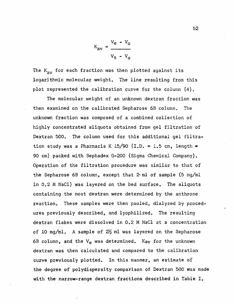
52
Kav = Ve - V0
V-t - V,
The Kav for each fraction was then plotted against its
logarithmic molecular weight. The line resulting from this
plot represented the calibration curve for the column (4).
The molecular weight of an unknown dextran fraction was
then examined on the calibrated Sepharose 6B column. The
unknown fraction was composed of a combined collection of
highly concentrated aliquots obtained from gel filtration of
Dextran 500. The column used for this additional gel filtra-
tion study was a Pharmacia K 15/90 (I.D. = 1.5 cm, length »
90 cm) packed with Sephadex G-200 (Sigma Chemical Company).
Operation of the filtration procedure was similar to that of
the Sepharose 6B column, except that 2* ml of sample (5 mg/ml
in 0.2 M NaCl) was layered on the bed surface. The aliquots
containing the most dextran were determined by the anthrone
reaction. These samples were then pooled, dialyzed by proced-
ures previously described, and lyophilized. The resulting
dextran flakes were dissolved in 0.2 M NaCl at a concentration
of 10 mg/ml. A sample of 2)£ ml was layered on the Sepharose
6B column, and the Ve was determined. Kav for the unknown
dextran was then calculated and compared to the calibration
curve previously plotted. In this manner, an estimate of
the degree of polydispersity comparison of Dextran 500 was made
with the narrow-range dextran fractions described in Table I.
CHAPTER BIBLIOGRAPHY
1. Garlick, D. G. and E. M. Rankin. 1970. Transport of large molecules from plasma to interstitial fluid and lymph in dogs. Am. J. Physiol. 219: 1595-1605,
2. Grotte, G. 1956. Passage of dextran molecules across the blood-lymph barrier. Acta Chir. Scand. Suppl. 211: 1-84.
3. Mayerson, H. S., C. G. Wolfram, H. H. Shirley, Jr., and K. Wasserman. 1960. Regional differences in capillary permeability. Am. J. Physiol. 198: 155-160.
4. Pharmacia Fine Chemicals AB. 1970. Sephadex. Gel filtration in theory and practice. Pharmacia Fine Chemicals, Inc., Uppsala, Sweden. 63 p.
5. Semple, R. E. 1957. An accurate method for the estimation of low concentrations of dextran in plasma. Can. J. Biochem. Physiol. 35: 383-390.
6. Scott, T. A., Jr. and E. H. Melvin. 1953. Determina-tion of dextran with anthrone. Anal. Chem. 25: 1656-1661.
7. Squire, J. R. 1951. Background to biological studies with dextran. Proc. Roy. Soc. Med. 44: 557-558. (Abstr.)
8. Wasserman, K., L. Loeb, and H. S. Mayerson. 1955, Capillary permeability to macromolecules. Circulation Res. 3: 594-603.
53
CHAPTER III
RESULTS
Capillary Permeability
The movement of macromolecular dextran fractions across
the capillary endothelial membrane was monitored by periodic
concentration determinations of dextran in plasma and lymph.
These determinations were organized and averaged according
to their specific molecular weight group and barometric
pressure treatment. The dextran concentrations in thoracic
and leg lymph were separately compared to the dextran
concentration in plasma.
Plasma vs. Thoracic Lymph Concentrations
A constant decrease in plasma dextran was observed in
all animals. This decline was somewhat enhanced by decreased
barometric pressure in some groups, but no definite dextran
loss was observed with the change in pressure. The dextran
levels gradually approached zero as the experiments proceeded
to termination.
Small concentrations of dextran generally appeared in
the thoracic lymph within 15 min. This concentration grad-
ually rose to a constant plateau after 1 hr, and then it
began to decline slowly until termination of the experiments.
Administration of decreased barometric pressure halted this
54
55
decline and significantly increased the dextran concentra-
tions in thoracic lymph. These concentrations decreased to
values that approached zero when the barometric pressure was
returned to normal.
The general trends cited above for dextran concentration
in plasma and thoracic lymph can be seen in the combined
group means. These graphs are presented in Figures 3 and 4.
Group A (321,000 mol. wt.).—The general decline in plasma
concentration of dextran was observed in both the ground
level control and the altitude experimental runs. A rather
erratic pattern was exhibited by the plasma means of the
control animals. This was contrasted by the relatively
smooth decline of the altitude mean plasma concentrations.
The control plasma values reached zero after 4^ hr, and the
altitude values were leveled off at 10 mg/100 ml at the end
of experimentation.
Only a small amount of dextran was identified in the
thoracic lymph of the control animals. A slight rise in
concentration was observed later in those experimental
animals, but the dextran concentration was essentially zero
after 3 3/4 hr. On the other hand, the mean thoracic lymph
concentrations of the altitude runs demonstrated the general,
characteristic pattern described for them. The concen-
tration rise with decreased barometric pressure was not as
dramatic as observed in some of the other groups, but it was

56
w O *H •H O -P
C •H O OuJQ
(0 H O JC2 -P
XI«—I Tf O (0 £ 03 ro m
o •H -P H 0) >
m S CO# 05 Q r~l p-t a/dx* c c 05 -o •h a>
*4J if) O 03 C O 03 «**H d) CQ T5 S C
•**rl c <
•H »H -P if) 03 a s " 3U4
O CO N
-P a a> a> o c. o u
if) •H CO e
£ 03
O H •P
C C 03 O H O •p X TJ +» 0 (DXl Q C O )
•H *H Xt CD • £
CO O^H o o
cn^ •H O P-i
x: a S a> >^x; H -P
c 03 0) 6

57
Hrtfci
»n xf o CN ( % Bui) NOI1VU1N3DNOD

58
CO o H >H CO CO O . H X I CO H - P r H O C CO
H O O - H a - p
N
- p
T f c x ; CO o
CO C0PL3 £ t o CO •
rH«"—N a n
a> >
jQ
TS Q)
- P CO a rH
c * a • H C
CO * 0 CO c C * * « H c o m a> co £ * # H <
o • H - P CO H
- P C 0 ) a a> c T J o 3 a - p
• H C - P CO H
• P X T * 0 ) Q)
Q C • H <4-1 £* O • £ ^ o
o
U ) CO CO a p o • N CO CT>rH
CO
• H C CO
a> >
H
I - P
CO i
c n H - i • H O PU
<D crs CO H a> >
CO j C a 6 <l> p n x i
r H - P

59
(%6u i ) NOI1VHJLN3DNOD

60
still significant. The mean concentration values fox control
and altitude runs can be seen in Figures 5 and 6.
Group B (409,000 mol. wt.).--Mean plasma dextran concen-
trations for both the control and altitude groups were similar.
Each of them exhibited the same slow decline, and they both
had a final dextran concentration around 2.5 mg/100 ml. The
average dextran concentrations of the altitude animals,
however, were initially much higher than the averages of the
control animals. This resulted in a more rapid rate of
decline during the first few hours for the altitude runs.
Administration of oxygen and decompriession created an erratic
pattern in thses animals that eventually developed into
dignificant decreases in mean plasma dextran concentrations.
The dextran concentrations in thoracic lymph demon-
strated exaggerated rises in the initial dextran concen-
trations of both the ground level control and the altitude
runs. These values decreased, however, to constant levels
which, in the case of the control concentration means,
stayed around 5 mg/100 ml. The dextran concentrations of
thoracic lymph were increased when the animals were subjected
to decompression. To a limited degree, these increased con-
centrations were mirrored by the decreases in the plasma
dextran concentrations. The thoracic lymph dextran decreased
to zero after the barometric pressure was returned to normal.
Graphs of these results can be seen in Figures 7 and 8.
61
0) x:
o -P •H O CO fl5 *rl H O -P • x: c (o •P*H H
O 03 "0 a-O c CO XI «HI
O CO 03 ctJ O e w -H 0) -p CO N HI • <D a co >
r—I c co >. •H 6 rQ
•rl « CTJ C CO <1> (0 +> <D H (0 e o o
H -H C 4-> T3 O C C •H O »H -t-> O (0 0) H i—I *rl +» <1) c a) <D yj OHt/3 C O TJ O C •
3 V) C O t-1 CO fH CO H cn S 4-> .H X »C <D < CO Q
a o 3 5
• o +> m H
a m o
a>M-t •h o a> (jli cn
jC co a n S a> > >
r-4 (0
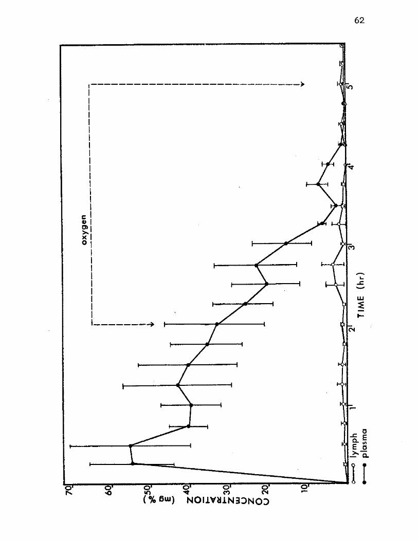
62
o ' o ' <Sr o » o r
«© «0 CO CN ( % 6 U I ) N O I 1 V U 1 N 3 D N O D
63
o o •H O <D co cn H CO O H
JC 0 -P >
CO TJ C CP CO jC -P
CO 6 to <O.H • CO CO H + J H O.C CO
•H XI C O •H 0*1—I
CO CO JC O C O.H CO CO -P CD U4 H S CD
c • ^ O CO > •H «-H J3 •P (0 (0 S T5 H n H CD -P C -P C CO CO 0) O O CD «H C T * T * O 3 C O - P t 4
•rl C -P <0 (OrH .H H CO -P S CD < CO P
a •
• o CO \0 N H
CD CO • S
• H O C tt* CO
,c a <d S > >h«H f-l H-i
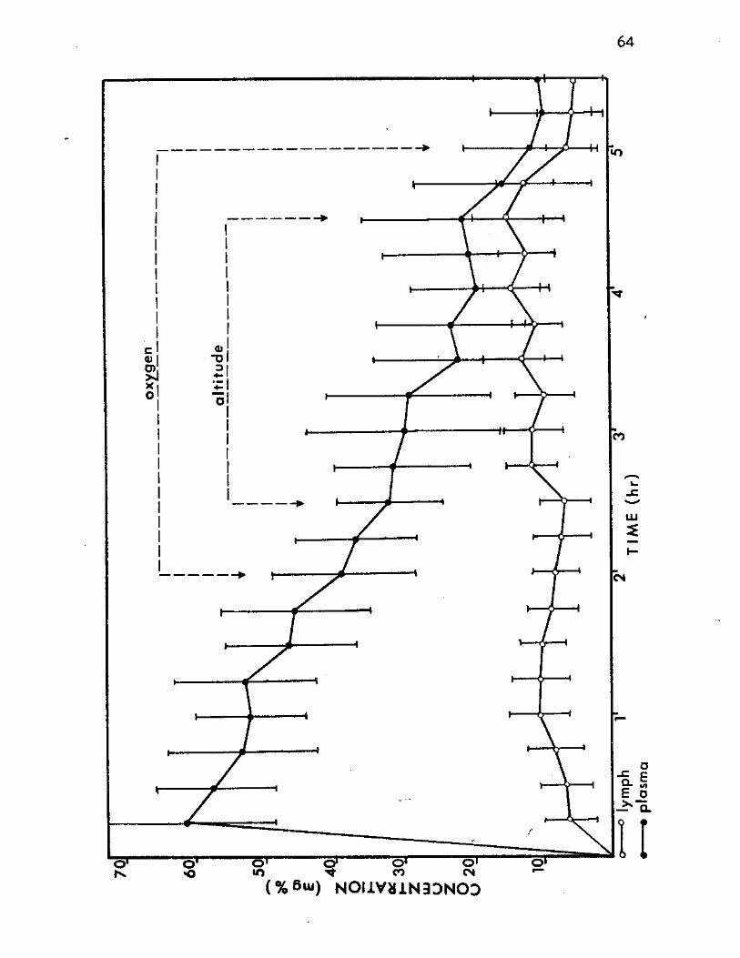
64
(%6u i ) N0I1VJI1N3DN03
65
CD x:
o -P •H U if) <0 *H H O -P • x: c co HP *H H
O CO -o ajQ c (OXJ rH
O <0 fO CO o S W d (0 -P CO H rH • 0) a w >
1 - 4 C fO > •H £ JQ
•H W C T 5 G CO <D
<dh nj g o o
H »H C -P T5 O C C •H O *H -P O CO <0 HtH *H •p a) c > S ( D O W O H CO c o"d o c: •
p (0 C O H <o H co H CJ> S -P »H X - C a> CQ co Q
a o D 5
• O -P r- N o
o o*H •HOC) Pu cn
x: <o a h £ o ^ > rH C0

66
{% 6ui) NOI1VU1N3DNOD

67
<+4 o o
•H O <1) co cn H f0 O N
x : a) - p > CO
T* C <D 05 j C
-P CO 5 <0 • 0 ) . H 0) CO H
rH -P CO a c x i
•H C O rH
•H 0 . <0 O
C O -P CO CO H <u PJ a> 6 > £ • > . O (OX*
•H rH -P CO TJ CO g <D H - H -P
• p a co C (0 o 0) *H u o n C t 3 C O O -P
•H (0 C -P *H CO rH H COS •P W X *CO O PQ Q
a • 2 <0
• O rH 00 N CO
O 6 • *H
C •H O CO PL,
jC 0) a < D 6 H
H *P
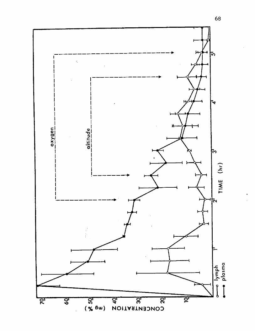
68
7
( % 6ui) NOI1VU1N33NOD
69
Group C (720,000 moi. wt.).—The ground level control
animals were the only animals examined in this molecular
weight group. Although the initial plasma dextran concen-
tration did not reach that of the other groups, it did show
the same general decline. After 3i hr the mean plasma
value was zero. Dextran of this molecular weight did appear
in the lymph, but it was present in only small quantities.
The mean concentration of dextran in thoracic lymph was
rarely above 2 mg/100 ml after 1% hr of experimentation.
Graphic analysis of these results can be seen in Figure 9.
Group D (Dextran 500).—The results obtained in this
group were similar to the general trend already set forth.
Plasma dextran concentrations in both control and altitude
groups demonstrated a slow decline. The values reached zero
in the control means and 6 mg/100 ml in the altitude means.
No noticeable change in altitude concentrations was present
in the plasma.
Dextran mean values presented in the thoracic lymph also
appeared similair to those expressed in the general trend.
The main exception, however, comes in the last samples of
the control thoracic lymph values. They show a definite
increase over both the previous thoracic lymph values and
the compared values for plasma dextran. The differences in
the concentrations of dextran in plasma and thoracic lymph
at this point seem to be significant. Another less obvious
exception to the general trend was the failure of the thoracic

70
o •H O (T5
O jC 4->
0) JC -p
•H
-P £
O
CO H CO
ax> T3 c rOXlH
O 03 CO CO e w CO co
a <o
o •H •p
N <D >
c •H
(0 C CO CD 6 a o •H -P CO fH -P C Q)
CO >* 6 X> •H C "0> CO CD
-P r-H CO o o H *H •P "0 C C 0«H O
40 H *H Q) o §
O rH CO c C O o c •
P CO O H H CO cr> g •H
* c CO
C CO H -P X <D O Q
ON
cr><+4 •H O [X,
a o 3 5 O *P H O
O
<D cr>
jc co a h S 0) > > H CO
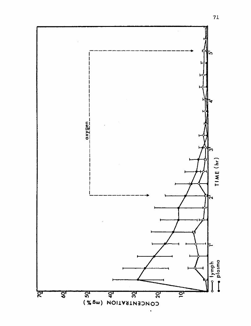
71
(%6ui) NOI iVa iN3DNOD
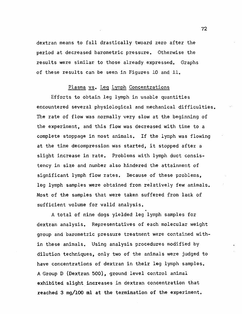
72
dextran means to fall drastically twoard zero after the
period at decreased barometric pressure. Otherwise the
results were similar to those already expressed. Graphs
of these results can be seen in Figures 10 and 11.
Plasma vs. Leg Lymph Concentrations
Efforts to obtain leg lymph in usable quantities
encountered several physiological and mechanical difficulties,
The rate of flow was normally very slow at the beginning of
the experiment, and this flow was decreased with time to a
complete stoppage in most animals. If the lymph was flowing
at the time decompression was started, it stopped after a
slight increase in rate. Problems with lymph duct consis-
tency in size and number also hindered the attainment of
significant lymph flow rates. Because of these problems,
leg lymph samples were obtained from relatively few animals.
Most of the samples that were taken suffered from lack of
sufficient volume for valid analysis. *
A total of nine dogs yielded leg lymph samples for
dextran analysis. Representatives of each molecular weight
group and barometric pressure treatment were contained with-
in these animals. Using analysis procedures modified by
dilution techniques, only two of the animals were judged to
have concentrations of dextran in their leg lymph samples.
A Group D (Dextran 500), ground level control animal
exhibited slight increases in dextran concentration that
reached 3 mg/100 ml at the termination of the experiment.

73
a> ox:
•H - p o CO (0
o x : - P • • p c
•H N T J O CO c a x * (0
r C «--4 CO O 03 g <o u CO P4 *H CO - P
r—t H Q* * <U
CO > C •—l
•H CO >* 6 x>
CO C C T5 CO CO 0 0) - P g r-4 CO o a
C N - H O - P "D
•H C £ - P 0 » H CO O H 0)
• P H *ri C <P CD > S o a> w c to o o - d
c • C 3 « (0 O I-H H H <0
• p D i e X "H <D - G
O Q (0
Cu O , O S
O O - P H H
O O
crm-t •H O <D VLi CT>
x : co a h 6 <u > >
r - i 10
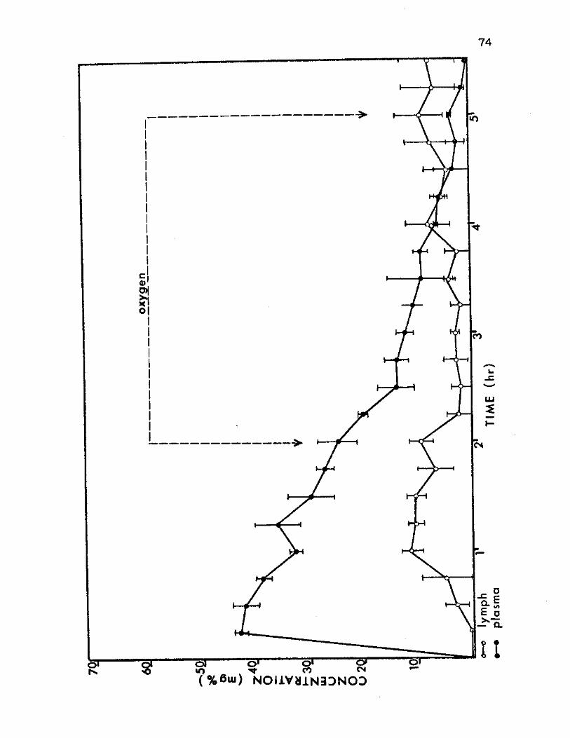
74
"SI o» o» cT
? % 6 m ) ^ N O I l V H i N B D N O D
75
OQ-I •H O O CO H O •P
T3 C CO
CD D> CO N 0) > <0
0) X
CO -P S CO <0 CO «H
A-P c c •H
CO H (0
CO
CO O <D CO G W
H JQ O O-RH
CO O •H -P N 0 > C
O •H CO •PHXI CO CO N -P«H 0) C G +•> CD CO CO O O C 0) OTF-0 O P G -P «H
C-H <0 -P N < -P CO X s OJI *JJ4 Q Q CO
a • 3 •
•HOC/) H N H
O CO
CO •rl
S •H C CO
•H O P-I .C a N E ^
R-I <+*
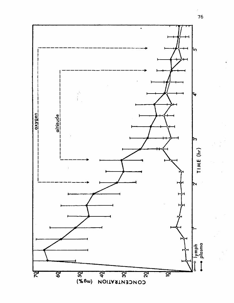
76
(%6ui) NOI1VU1N3DNOD
77
Also, a Group A (321,000 mol. wt.), altitude animal showed
inconsistent increases in dextran concentration, expecially
in the last sample collected after decreased barometric
pressure. The remaining animals revealed no measurable
amounts of dextran in leg lymph. Because of this lack, no
comparison could be made between leg lymph concentrations
and plasma concentrations of dextran.
Statistical Significance
Statistical analysis was applied to the plasma vs.
thoracic lymph results to determine their significance.
Standard errors of the mean (SEM) were calculated for each
sample of every experimental run. The results of these
calculations were presented as vertical bars on the graphs
in Figures 3-11. Loss of significant differences between
plasma and thoracic lymph concentration means were readily
observed by the overlap of the SEM vertical bars. This
overlap occurred in the combined means after 4 hr for the
control animals and after 3% hr for th'e altitude animals.
A further statistical evaluation was necessary to
establish the significance of lymph changes and plasma
changes occurring between ground level control and altitude
animals. A calculated Student's t was obtained for the thor-
acic lymph vs. thoracic lymph and the plasma vs. plasma
values of each barometric pressure treatment. These calcu-
lations were then compared to a table of t values (tQ 0 5 ) to
determine probability (P). The combined control runs were
78
compared to individual group altitude runs for this analysis.
In addition, combined control runs were compared to combined
altitude runs. Table II gives the results of these compared
calculations with their levels of significance.
The calculated t values obtained for the combined
control and altitude groups gave a general picture of the
overall significance of the experimental runs when they were
compared to the tabled t values. The thoracic lymph compar-
isons that were determined during the decompression period
revealed significant defferences in dextran concentrations.
The 2:45- through 3:45-hr samples were significant at the
0.01 percent level, and the 4:00- through 4:30-hr samples
were significant at the 0.05 percent level. These values
and two other initial values in the plasma were the only
comparisons that indicated any significant differences in
the combined control vs. altitude groups.
Group A (321,000 mol. wt.).—In both the ground level
controls and the altitude runs, the dextran concentration
differences between plasma and thoracic lymph became insig-
nificant after 3% hr. The intercomparison of lymph and
plasma values under different barometric pressure treatments
yielded values of a 0.05 percent level of significance for
all comparisons involving thoracic lymph at altitude. This
significance level was extended to one sample of thoracic
lymph in the post-altitude stabilization period. Other
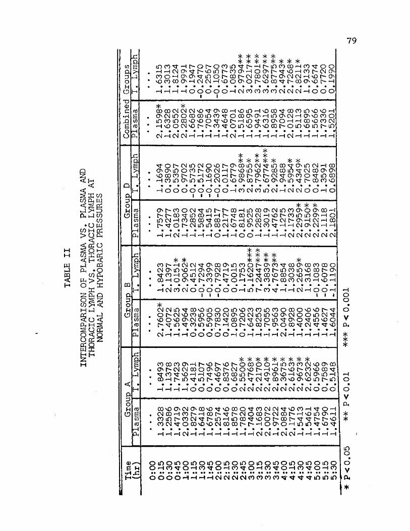
79
H
U4 $
o
CO OJ 3 O N O
x: QJ B > »-4
T5 <1> C •H XI S o o
x: oj 6 4
Q Oj 3 o M CD
0Q Ol o H CD
a o H CD
XI 04 6 kJ
0-
6 N •H X?
iooo^T'Hr-or~-oooin^t^Hi>ir)cocx)'Hco'^oo H'HOlO'^h-vOiOh-COO'HOOO'crvOHOOD-CNO coo-HooN'srioor-oot^cMoo(Ni>ON<N<M'Hvor--o ^nCDOHOJCMHvOOOOhvOCOth-OOOvOh-H HHHHOOOOOHlNCOOCOWOJCSlOlHOOO I I 5j< 5§£ OOOOOlCMOJvO tOCO'HvOlO'HvOOO rOOCOinvOvO'H OCMtOOOOOOiOCO fOOOONO-HiOOCM-HONvOCOO mcOlfiCOvDvOO^vOh-Hin^OOvOH'-I CO vO 00 CM ^voocMvDf-r-co^oiOvoovocof-oiOvOiot^in • • • • • • • • • • • • • • • • • • • • • • CM '—1 CM CM r"H 1—I'—f •—1 ""H CM '—i 'H •—i •—i •—1 1—I CM 1—I •—I 1—! •—! *—I
% sjt 5 5 * * * ^•or-cMmcMOvor-oooincM-sriooo^roinoj-ioo ooMnocot^-ooJ-Hh-voiovoh-cocoio^roJoooo vOOOOr-h-HvOOHhODh-Oh-OJ^OCOO^iOCO HCOinOiniO-HCMOvOOOOr-vOOCMO t OOCOvO -HOOOOOOOO-HCOOJOOmCMHCMOJOO-HO
I I I I *{< >|c >fc
o^coocM^inr-t^-ooHinooocMmcoooocO'H r--t^cD\rioco—i-H^-^-cocMcM^ivOh-coiniooN-Ho lOOJ-HCOOOOO^OOHr-'HLnOOOr-OJh-O'HCM'HOO h-' OIXNininoOCMvOOOOCMCO 'H'HCSlOCM-H'H • • • • • • • • • • • • • • • • • • • • a *
HHOJHHHHOHHOOHHHHCMCJOJCJOlOl
COf-HOlCM^OOOOiOCOOt -OCO^OOOCOOOOOO CMOinvOHONOCMH-HunCM^COh-iOCOiOvOOOt O ^COHOlOCMCOOhONvO^ODvOOOOvOHOOH OOHOO^h-OOI^^OHHCMCOhOOCOOlCOHvOH HCJCOOJOOdOOOHinhCOfHHOlHOOH I I I I I I
CMCMin^COvOiOOOiOvOCOCOiDCOOOOOvOvOI ' Or*-CM\OOOinOOOCMOOCMiOLO\QONCMOOifiCM<tf VOOVO OVCM ONOOO fOOCMM-OJOiO^-OO iOvOO r-^rin'st-ooiOior-'HOh-voaDr-ooco'^fCM^^rvo • • • • • • • " • • • • • • • • • • • • • • • ( M H H H O O O O O H O H H H H l N H H H H W H
COOOCOOHt-vOt vOr OCOOOHiOCOCOCMvOONOO Ot-CMCMOOOOONr CMOvQl HvOf-vOf-OOvQCXW ^CO^vDHH^vOCOOOiDl^HONOvOHvOCMC^iOH coHr-uosrior-^cx)voir)^rcM^-oocovoc>vOin^io HHHHOOOOOOOIOJOI CM'CM CM CM CM CM O O O
OOvOOCMOCWvO vOCOO -OOCMCM- vOCO-H^OH csicO'Hoot^--Hoor--'3-i>csiooot^cMoor~-—tvoino—i coiOh-cocM^f-iO'HincD^voor-oor^^^T-r-vo cocM^oco^vocMcocor-r-HoooHininKi-vo^r HHHCMHHHHHHHHOlCJHOiCMHHHHH
o i o o i n o m o i o o m o i o o i n o i o o i o o i o o i o o OHCO^OHCO^OHCO^OHCO^rOHCO^OHCO
m m m mm mm mm ££ skm mm mm mm mm mm m* mm mm mm mm ** mm mm mm
• • • • • • • • • • • • • •
OOOOHHHHCMCMOKNCOCOCOCO^^r^^iOiOin
•H o o • o V a* %
•H o <1 o V a*
in o • o V a. *
80
comparisons in this group were not significant at the
prescribed level.
Group B (409,000 mol. wt.).—Insignificant differences
between plasma and thoracic lymph concentrations of dextran
became evident after 4 hr in the control animals and after
3'4 hr in the altitude animals. The plasma differences and
thoracic lymph differences demonstrated a varied pattern of
significance. The thoracic lymph samples taken at altitude
showed significant levels of from 0.001 to no significance.
Additional levels of significance (0.05) were found in the
thoracic lymph at the 0:45-hr and 1:00-hr samples. A
significant value was also found in the plasma at the 0:15-
hr sample. Other comparisons did not give any significant
values at this level.
Group C (700,000 mol. wt.).—In this molecular weight
group, differences between plasma and lymph were not
significant after 2/4 hr. No plasma and thoracic lymph
intercomparisons could be made since there were no altitude
runs.
Group D (Dextran 500).—The concentration means of
dextran in plasma and thoracic .lymph showed some patterns
that varied from the general trend, especially toward the end
of the control animals. Insignificant differences between
the two, however, could be established after 4 hr in the
control animals and after 2 3/4 hr in the altitude animals.
81 J
In the comparisons between the thoracic lymph values of con-
trol and altitude animals, all but one of the samples invol-
ving altitude exhibited significant levels of 0.05 percent
or better. The 3:30-hr sample was significant at a 0.001
percent level. Plasma comparisons demonstrated significance
in the 4:30-hr, 4:45-hr, and 5:00-hr samples. None of the
other calculations was significant at the prescribed level.
Physiological Parameters
Pulse Pressure
The pulse pressure of the experimental animals in
Groups A, B, and C were affected by the presence of minute
quantities of sodium azide (ca. 0.02 percent NaN3) present
in the injected dextran solution. These animals experienced
a mean pulse pressure drop of 30 mm Hg immediately after
injection. The pulse pressure then slowly rose until it was
approximately normal after 3 hrs. Animals in Group D did
not have sodium azide in their dextran injections, and they
did not demonstrate the drastic drop in pulse pressure. A
0.02$ sodium azide concentration was added to the dextran
injection of one animal in Group D. The pulse pressure of
this animal responded exactly like those pulse pressures of
animals in Group A, B, and C. Animals not having sodium
azide in the dextran injection showed a slight decrease in
pulse pressure throughout the experimental run. The use of
oxygen and decreased barometric pressure did not have drastic
82
effects on the pulse pressures. Comparisons of the pulse
pressures of azide and non-azide animals can be seen in
Figure 12.
Hematocrit
Average hematocrit values of the experimental animals
changed very little during the course of experimentation.
A maximum range of from 41 to 45 percent was observed, and
the tendency was for the hematocrit to rise slightly as the
experimentation period progressed. Some individual animals
showed small decreases after injection of dextran, and other
individual animals showed slight increases with the intro-
duction of decompression. No definite trends could be
established from these animals, however. The hematocrit
means for all animals can be seen in Figure 13.
Molecular Weight Determination by Gel Filtration
Column Calibration
Calibration of the Sepharose 6B column was accomplished
by determining the elution volumes (Ve) for each molecular
weight fraction and the void volume (VQ) and internal volume
(V^) of the column. Using these values, the partition coef-
ficient (Kav) was calculated. The void volume and the
internal volume of the column were 158.5 ml and 518.3 ml
respectively. Elution patterns of the dextran fractions
were symmetrical, and they allowed easy estimation of
83
c • CO (0 0) CD
<u £ D -C •—l +-> 0) CO
tJ > C »H O N C CO CO cd a> "O <u g •H x: N-p J) CO TJ
14 «H S o n 3 <+H CO •H I TJ TS C o a> o w cn c
CO 'H R 0) o a> sx
> 4-> tO CO -P H o a> o <u Mm •+-< a) <H g TS <1) <D •
w cnw OH (0 N Xi CO H CO +> £ <1> .Q
•H > 'H C (OH O ro CO
<D O C C H-H O <1) <D -P « H 5 H •H <D H W > to • cn a o o >• £ HT3XJ O P O M UK
to p aj <D o +>
• H Mh co cm a o H TS *H
<U CO • 0) CO C
D>rH -H •H p " (i< aw <o
a) »H CD CO H S CD CO UJ £ > W
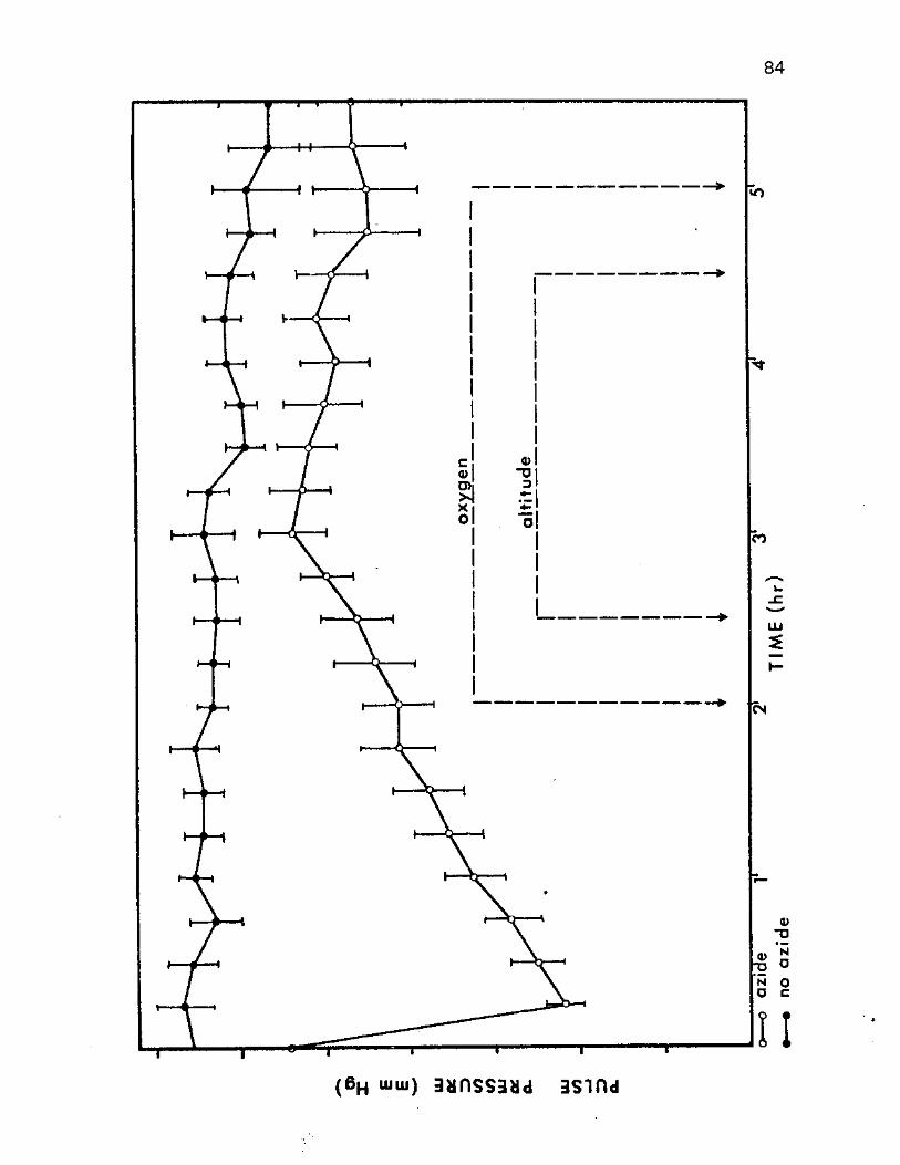
84
1 * 1
( 6 H m m ) 3 u n S S 3 M d I S i n d
a> u
JJ 'jj "D ° N O O C
II
85
CO i—! CO S
•H C CO
CO -p
c 0) £
•H N 0) a X <D •
CO r-4 r—< co re e
•H 4H C O fO
<0 C <tf Q) D <D r-i +J ro x : > CD
•rl +> <u •H H a o o +> a> CO D> S <0 <D H x ; a> > £ CO <0 a) Q) S X -p
• <o CO -H H
4-> • C
cn-H •rl O ^ a
XI a CO (XI
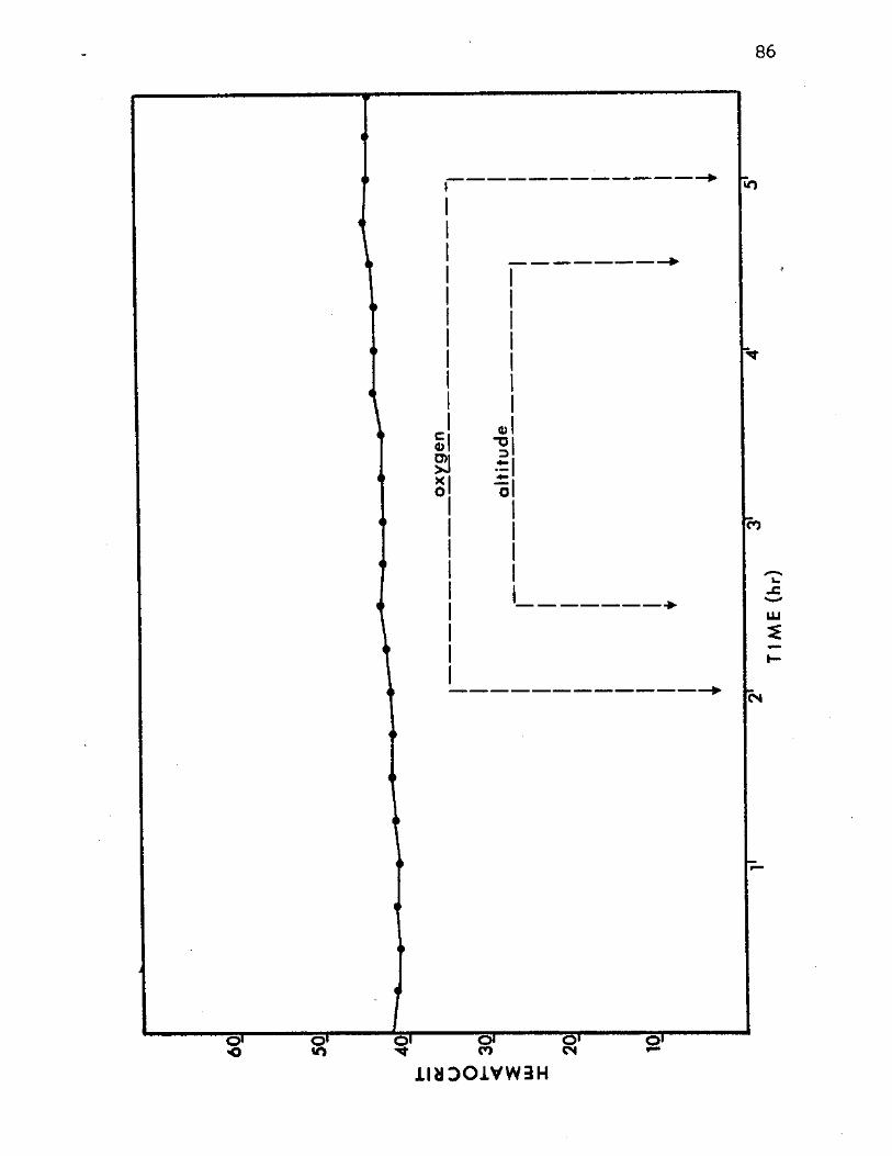
86
JLIHDOJ.VW3H
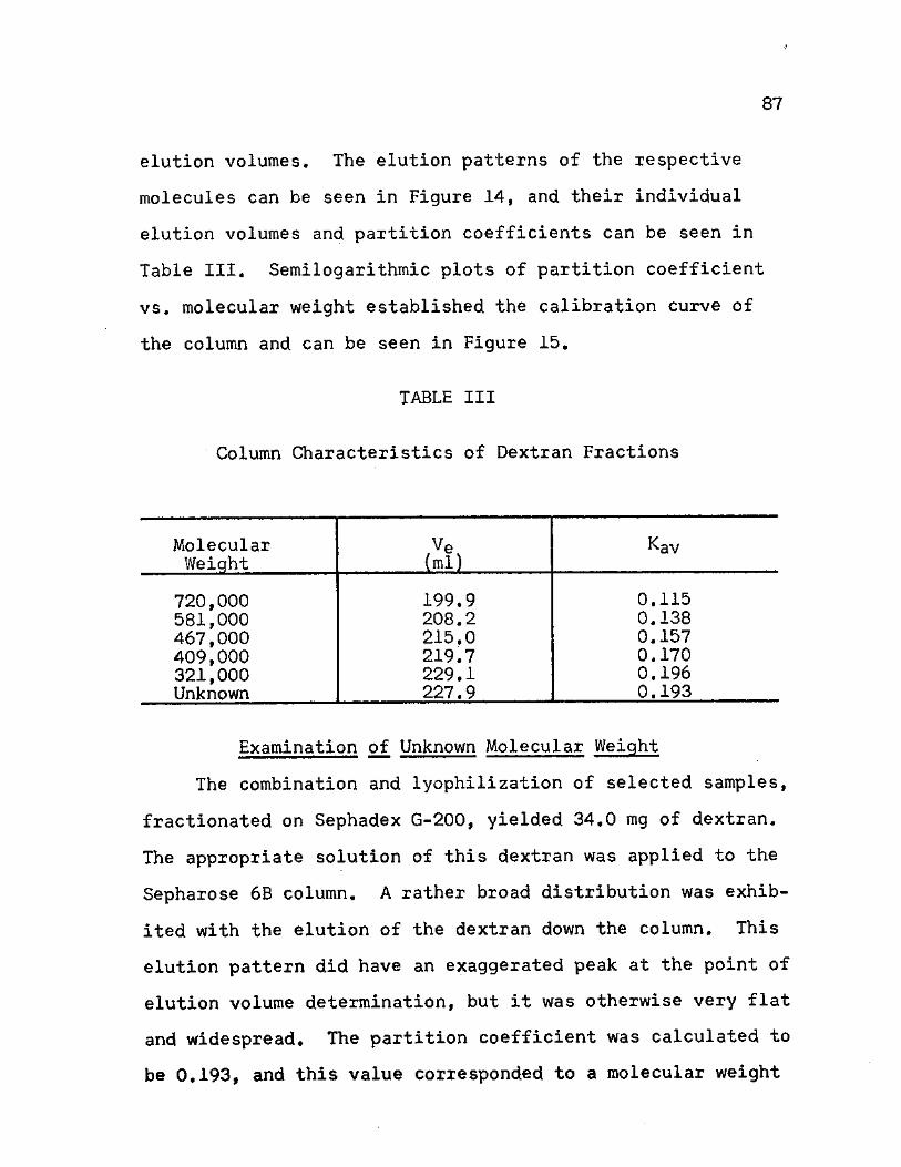
87
elution volumes. The elution patterns of the respective
molecules can be seen in Figure 14, and their individual
elution volumes and partition coefficients can be seen in
Table III. Semilogarithmic plots of partition coefficient
vs. molecular weight established the calibration curve of
the column and can be seen in Figure 15.
TABLE III
Column Characteristics of Dextran Fractions
Molecular Weiqht
Ve (ml)
Kav
720,000 199.9 0.115 581,000 208.2 0.138 467,000 215.0 0.157 409,000 219.7 0.170 321,000 229.1 0.196 Unknown 227.9 0.193
Examination of Unknown Molecular Weight
The combination and lyophilization of selected samples,
fractionated on Sephadex G-200, yielded 34.0 mg of dextran.
The appropriate solution of this dextran was applied to the
Sepharose 6B column. A rather broad distribution was exhib-
ited with the elution of the dextran down the column. This
elution pattern did have an exaggerated peak at the point of
elution volume determination, but it was otherwise very flat
and widespread. The partition coefficient was calculated to
be 0.193, and this value corresponded to a molecular weight
88
CO C o •H -P O Ctf M <+•<
C (0 H •P X TJ m o CO c N <l> -P CO a c o •H -P 3
0
O c o CO •H H 03 a 6 o o
m vD 0)
H CO O
• H CP <0 •H ,C mu a <D tn c o
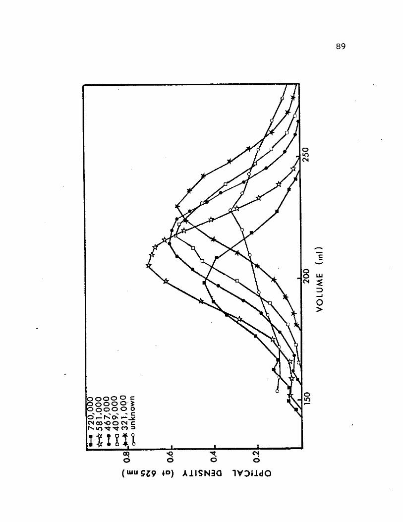
89
o o o O c ooo o £ o o o o o
K o o C O O O CN C 1 0 ^ ^ CO 3
uni t
% Z> mmJ
o >
( U I U 5 J 9 * ° ) A 1 I S N 3 Q I V D I i d O
90
Fig. 15. Calibration curve for Sepharose 6B column.

91
MOLECULAR WEIGHT
92
of 330,000 on the calibration curve (Figure 15). The
elution pattern of this dextran fraction can be compared
to the elution patterns of the previous dextran fractions
in Figure 14. The column characteristics of the unknown
can also be compared in Table III.
CHAPTER IV
DISCUSSION
Capillary Permeability
The Starling hypothesis depicts the direction and rate
of fluid transfer between plasma and tissue fluids as being
determined by three factors: (a) the hydrostatic pressures
on each side of the capillary membranes, (b) the colloid
osmotic pressures of plasma and tissue fluids acting across
the capillary membranes, and (c) the physical, chemical,
and histological properties of the capillary membranes them-
selves (8, 37, 42). If an inert, lipid-insoluble molecular
species is suddenly added to arterial blood, a disturbance
is created in the osmotic balance that initiates two related
processes to reestablish the Starling equilibrium (36).
The first process involves the diffusion of the added
molecule from the plasma across the capillary membrane to
the interstitial fluid, and the second process involves the
withdrawal of fluid from the interstitial compartment into
the capillary blood due to the concentration gradient formed
by the added molecules. Both processes continue until an
equilibrium is attained according to the new osmotic
pressures. The rate of these two processes and the time
required for them to reach equilibrium are dependent on the
93
94
hindrance to diffusion at the endothelial wall. This
membrane hinderance is primarily related to molecular size,
and through mathematical applications of the progressive
plasma and lymph concentrations, the larger molecular
species can be used to observe and describe some of the
characteristics unique to the capillary endothelial wall
(13, 35, 38). Likewise, changes in the hydrostatic
pressures can induce shifts in the transcapillary fluid
flow and/or in the osmotically active molecules to return
the system to the equilibrium portrayed by the Starling
hypothesis. Monitoring the shifts of the large, membrane-
selected molecular species can reflect additional properties c
of the capillary endothelial wall and the surrounding inter-
stitial compartment (25, 26). In essence, all three of the
factors experssed in the Starling hypothesis must be
considered in discussing the properties of capillary perme-
ability.
The macromolecular dextran fractions utilized in this
study were chosen because of their similarities with the
above hypothetical molecular species, both in physiological
inertness (41) and in biological fractionation according to
molecular size (21, 45, 46). The capillary permeability
characteristics of the fractions were studied both during
the initial equilibrium adjustments and during barometric
pressure changes after equilibrium had been reached. These
characteristics studies were facilitated by monitoring the

95
concentrations of dextran disappearing from the plasma and
appearing in the lymph obtained from.two different lymphatic
drainage areas. This was in response to the reports of
varied histological (3) and permeability (31) characteristics
of capillary endothelial membranes in different anatomical
regions. Within the limits of certain qualifications, the
results obtained from the two different regions substan-
tiated the reported variations in capillary permeability.
Plasma vs. Thoracic Lymph Concentrations
Molecular Weight Effects—The size of the molecular
species, as reflected by molecular weight, has been pointed
out as one of the primary considerations in capillary
membrane selectivity. This fact was especially evident in
the studies evaluating dextran as a plasma expander (4, 41,
45). In those studies, increased efficiency in rapidity
and duration of plasma expansion was noted with increased
molecular weights of dextran. Larger molecular weight
dextrans were simply retained longer by the restrictive
endothelial barrier. The increasing molecular weights in
the present study, however, did not appreciably alter the
permeability characteristics as they were monitored in the
thoracic duct drainage regions. With two exceptions that
will be explained later, there were no significant deviations
in the progressive dextran concentration patterns in either
96
the plasma or the thoracic lymph. This observation can
be explained by several factors.
The first explanatory factor involved, and probably
the most important one, is the region from which the
thoracic lymph is primarily derived, the spleen, intestine,
and liver. Several investigators (13, 21, 31, 46) have
shown the presence of dextran in thoracic lymph, and their
reported molecular weight average is within the range of
Groups A, B, and D. These workers have suggested the
presence of large "leaks", especially in the liver, to
account for this presence of macromolecular dextran. The
estimated size of these "leaks" is 250 to 300 A, and
electron microscope studies by Bennett and co-workers (3)
have suggested their existence. Other workers (34, 39) have
indicated that macromolecular transport may be by vesicles,
but no evidence has been offered to support the vesicular
transport of non-proteinaceous, lipid-insoluble molecules
like dextran. At this point, the endothelial "leaks"
attributed to the liver appear to be the most acceptable
theory of macromolecular transfer of dextran to the thoracic
lymph. The "leaks" must be larger than the effective
molecular size of dextrans in Groups A, B, and D, because
there are no selective differences between them in their
thoracic lymph concentrations.
A second factor that must be considered is related to
the dextran molecule itself. It has already been pointed
97
out that its molecular weight polydispersity has been a
major drawback in its use in capillary permeability studies.
Even though the dextran fractions used in this study were
much less polydispersed than those used in previous studies,
some lower molecular weight contamination could still be
expected. An example of contamination can be seen where the
thoracic lymph concentrations of dextran become greater than
the plasma concentrations in the latter samples of Group D.
This group is much more polydispersed than the other three
groups. In addition to the polydispersity effects, the
dextran molecule has been described as a flexible, linear
coil that can be subject to shear deformation in smaller
passageways (2, 22). These two molecular characteristics
of dextran indicate that some limited transfer could occur
within the proposed small pore system (13, 31, 35) or at
least through smaller "leaks". This indication is especially
significant when the comparatively small concentration
dosages are considered. The effects of the increasing
molecular weight of dextran may have been partially hidden
by the false permeability characteristics of the dextran
molecules.
Either one or combinations of both of the above factors
can explain the lack of observed concentration changes with
the use of increased molecular weight dextran fractions of
Groups A, B, and D. Molecular weight averages within these
groups present no drastic change. The molecular weight
98
average of Group C, however, is much greated than the
averages of dextrans used in previous studies. Although
only two ground level control animals are examined in
Group C, the results obtained indicate that the dextran
transfer is restricted. The average plasma concentrations
approach zero much sooner than the other groups, and very
small amounts of dextran appear in the thoracic lymph.
Conclusions based on two experimental runs are questionable,
but the tendency of the endothelial membranes in the thoracic
duct drainage regions to restrict dextrans of much higher
molecular weight averages seems to be present. Renkin (38)
has said that it would take much higher molecular weight
dextrans to establish the maximum permeability limits of the
capillary endothelial membranes, and this may be true if
the above tendency proves to be correct with further
experimentation.
Since the ground level control runs were somewhat
similar, their dextran concentrations were combined and
averaged for comparison with the altitude runs. This is
justified in the light of the above explanations for their
similarities. The statistical significance of the later
comparisons is also increased by doing this.
Loss of Dextran—One of the more puzzling aspects of
this study centers around the constant decline of dextran
concentration in both the plasma and the thoracic lymph of
all groups. At first glance, elimination by the kidneys

99
might be suspected for the decrease, but this is difficult
to support after further examination. Wasserman and his
associates (46) have shown that dextran molecular weights
greater than 40,000 are generally retained by the kidney
and returned to the circulation. This limit is far below
any of the average molecular weights used in this study,
and all of the fractions except Group D were sufficiently
narrow to believe that few if any molecules of this size
were present. Even Group D showed no change in concen-
tration patterns that would lead one to suspect significant
kidney elimination of dextran.
Metabolism could also be suspected for the decline of
dextran concentrations. Gray (19) reported that dextran
was broken down by the animal, incorporated into the carbon
pool, and distributed throughout the body in the form of
normal biochemical constituents. In this instance, the
experimental animals were mich, and the metabolic half-life
of the carbon in dextran was approximately 6.1 days. This
is contrasted to a metabolic half-life on dogs of 27 hr as
reported by Wasserman and Mayerson (44). The latter inves-
tigators also calculated the slope of exponential disap-
pearance to be -0.00043. When the 5J£-hr period of experi-
mentation for the present studies is considered, it is
unlikely that metabolism alone could have caused the dextran
decreases that were observed.

100
The factors involved in establishing an osmotic equi-
librium with the low concentration dosage is one of the more
probable causes for the decline in dextran concentration.
Previous reports (40, 43, 44, 46) give conflicting periods
of time (ranging 1-13 hr) that are required to establish an
osmotic balance, and the time periods involved depend some-
what on whether a true equilibrium or a steady state condi-
tion is acquired. All of these studies, however, used
dextran concentrations that were many times greated than the
concentrations used in the present investigation, and some
of the studies (40, 43) used the additional somotic effector
of radioactive iodinated albumen. This means that the small
quantities of dextran fractions used in this investigation
should have reached a steady state condition much sooner
than these previous studies. When the lymph to plasma con-
centration ratios are examined, it can be seen that they do
indeed become somewhat static after one hour of experimen-
tation, and even with the constant decline in respective
concentrations, this ratio remains almost constant until the
values begin to approach zero and become invalid indicators.
These facts suggest that the plasma and lymph concentrations
of the dextran fractions are basically in a steady state.
However, there is either an additional steady state equili-
brium that must be satisfied for constant concentration
values, or, as hinted previously, the metabolic activity is
accentuated by the low concentration values present.
101
The accentuated metabolic activity with small dextran
concentrations is a distinct possibility, because dextran
disappearance would be more noticeable in smaller total
quantities. In light of the reappearance of dextran in the
thoracic lymph with the advent of decreased barometric
pressure, however, metabolism seems highly unlikely. The
anabolic processes required under these circumstances are
physically impossible. Thus, metabolism must again be
discounted as a major cause for the dextran decline, and
other dextran-consuming processes or secondary equilibria
must be examined.
There are three mechanisms by which dextran could be
consumed or in which a secondary equilibrium might be
established. The first of these involves cirtain organs
that perform specific functions for the animal's circu-
latory system. In this instance, the organs are the liver
and the spleen. Both of these organs have storage and
phagocytic properties that could account for the disap-
pearance of dextran. Reports (16, 20) concerning these
properties do not indicate that dextran concentrations
would be altered from those seen in the plasma and thoracic
lymph, but no real evaluations are presented. The sinu-
soidal spaces and phagocytic characteristics of one or
both organs could cause the disappearance of dextran that
was observed.
102
The second mechanism deals with a peculiar erythrocyte
reaction in the presence of dextran. Bull and his collab-
orators (6) observed formation of rouleaux in smears of
blood previously infused with dextran. Formation was
inhanced by concentration and molecular weight increases.
Although the concentrations used in the present study were
below those reported, the molecular weights were much higher.
There is no way to know now if significant quantities of
rouleaux were formed or not, but if they were, they too might
be an explanation for the dextran decline, especially in
conjunction with the phagocytic properties suggested in the
first mechanism.
The third mechanism encompasses the theories of the
tissue spaces within the interstitial compartment. Histolo-
gists, biochemists, and physiologists (12, 14, 32) have all
pointed out that almost all normal tissue spaces are filled
with a gel or gel-like substance in addition to free inter-
stitial fluid. The gel is composed primarily of mucopoly-
saccharides, and in most tissues, the most important one is
hyaluronic acid. This substance is present in the form of
long coiled filaments, and it exists in concentrations
usually between 0.3 and 1.2 percent (33). Since the coiled
filaments are oriented in all directions, the resulting
network has been described by Ogston and Sherman (33) as a
"brush pile" that, with its entrapment effects, impedes
bulk movements of fluid and solutes within the fluid.
103
Guyton and co-workers (25) have shown that the degree of this
impedence by. this gel reticulum is inversely proportional to
the quantity of free fluid that is also in the interstitial
spaces. Under these conditions, it is conceivable that
dextran could be in a quasi-equilibrium between the plasma
and free fluid while still undergoing diffusion or entrap-
ment within the gel reticulum. This mechanism could easily
explain the disappearing pattern of the dextran fractions.
Any one of the three mechanisms cited above could have
had a part in the decline in dextran concentrations. Inter-
action between the mechanisms could also have had some part.
In view of the later results showing a reappearance of
dextran in the thoracic lymph, the latter mechanism seems
to be the most favorable. The first two mechanisms involve
a somewhat irretrievable loss in dextran that would not
allow for the observed increases in thoracic lymph dextran
at altitude. These increases can be rationalized by gel-
free fluid features present in the third mechanism. It must
be pointed out, nonetheless, that no definite answer can be
realized with the present data. Unitl further experimen-
tation involving radioisotopically tagged dextran is
performed, no conclusive statements about the disappearance
of dextran can be made.
Effects of Pressure Changes—The changes observed in
the thoracic lymph concentration of dextran at reduced
barometric pressures were the most significant results of
104
the entire study. The thoracic concentrations of the ground
level control animals continued their slow decline toward
zero throughout the experimentation period, but the thoracic
concentrations of the altitude animals demonstrated a *
dramatic increase in dextran concentration for the period
of reduced barometric pressure. In both instances, however,
the plasma dextran concentration sustained its slow decrease
without significant change during the periods at altitude.
These results could be due to one or combinations of all of
the following physiological explanations: 1) an actual
increase in the permeability characteristics of the capil-
lary endothelial membrane changes, 2) an increase in central
blood pressure that alters the dynamics of the Starling
equilibrium, and/or 3) a change in the gel consistency and
pressures in the interstitial compartment that shifts the
steady state dynamics and increases the thoracic lymph
dextran concentrations.
The stretched pore phenomenon elaborated by Shirley and
co-workers (40) is an example of a change that could take
place in the endothelial membrane itself. In their illus-
tration of the phenomenon, pore distortion was created by
enormous volumes of normal saline infused into the experi-
mental animals after a large quantity of dextran had reached
steady state conditions. The drastically increased plasma
volume supposedly stretched the small pore system to
stimulate transport of increased dextran molecular weight
105
sizes. Variations of this theory come from Grotte's system
of large pore "leaks" (21) and from Arturson and Granath's
"safety valve" system of increased large pore radii (l).
The present study, however, did not allow for the possibility
of increased pore size or leaks because of plasma volume
expansion. On the contrary, dextran concentration dosages
were intentionally kept low and no more than sample-replace-
ment volumes of normal saline were added to avoid signifi-
cant plasma expansion in the animals. Blood pressure was
also decreased substantially by the addition of minute quan-
tities of sodium azide. In this manner, any membrane chan-
ges that occurred, such as the stretched pores or "leaks",
had to be related to the decreased barometric pressure.
Under these experimental conditions, membrane changes
like those cited above, are difficult to support for the
capillary endothelial system as a whole. Decreased baro-
metric pressure, however, does have some profound effects
on the hemodynamics of experimental animals, and they may
be related to the present observations. Investigators
have identified increases in peripheral tonicity and
resistance (10, 15) and decreases in venous compliance (47)
with exposure to high altitude. The general conclusion
derived from these observations is that the central blood
volume is increased and the extreme peripheral circulation
receives a limited blood flow (10, 47). With these facts
in mind the question in the present study involves the
106
degree to which the central blood volume and peripheral
resistance is increased. If these increases are sufficient
to raise the capillary hydrostatic pressures significantly,
then stretched pores or "leaks" may indeed have caused the
the rise in thoracic lymph concentration of dextran. The
liver and spleen would again play an important part in this
supposition. Because of the large amount of supposition
imposed at this point, the stretched pore hypothesis must
remain plausible but not fully substantiated by the present
facts of this study.
The hydrostatic forces that purportedly allow the
stretched pore phenomena to operate come from inside the
capillaries. Since the conditions established in the
experimental design would affect the forces surrounding
the capillaries, the interstitial compartment and its
related forces must be examined. Guyton (25) has described
the interstitial compartment as being composed of the fol-
lowing three constituents: 1) the interstitial fluid within»
the interstitial spaces, 2) the cells and fibers of the
stroma, and 3) the mucopolysaccharide gel. The first con-
stituent forms the interstitial fluid pressure, and the last
two make up the solid tissue pressure. Accordingly, the
total tissue pressure is determined by the algebraic averages
of the interstitial fluid and solid tissue pressures that
occur on the surfaces of the capillaries.
107
Interstitial fluid pressure is commonly reported as
a negative pressure (23) that pulls the surface of the skin
toward the cell surfaces, as well as pulling the adjacent
cell surfaces together. In turn, the solid fibers in the
spaces are holding the surfaces apart. Hence, the solid
tissue pressures exerted by the solid fibers would counter-
act the pressures exerted by the negative interstitial fluid
pressure, and the total tissue pressure would be zero or
slightly positive (25). This reasoning involves a simple
application of physics that says that "at any single plane
in space in a nonaccelerating system, the forces acting in
one direction must be exactly opposed by forces acting in
the opposite direction." Within the limits of fiber strength,
skin turgor, and additional structural forces, this law
should apply both to increasing and decreasing interstitial
fluid pressures (25).
Assuming that the skin would be flaccid enough to
transmit pressures faithfully, the total tissue pressure on
the under surface of the skin is equal to zero with respect
to atmospheric pressure. A decreased atmospheric pressure,
like that presented in the present study, would cause a
decreased pressure under the skin. This same amount of
pressure is transmitted through the fluid and solid struc-
tural to the surface of the cells and capillaries because
of the "equal but opposite" physical principal stated
above. The total pressure in this instance would
108
still be around zero with respect to the reduced atmospheric
pressure, but the fluid pressure in the space between the
capillary and the surrounding tissues could be very negative
(23, 24). Thus, bulk movement and diffusion of fluid and
solute molecules would be increased in an outward direction
through the capillary pores which, in effect, is the
stretched pore phenomena in reverse. The solid forces
involved in this mechanism would reach a point of compaction
in the peripheral and cutaneous regions, and the blood ves-
sels supplying these regions would be closed or severely
restricted (7). This is in accord with the increased peri-
pheral resistance and decreased venous compliance obser-
vations noted earlier (10, 15, 47). In deeper and more
centrally located tissues, the compaction tendency would
not be as great, and the forces causing bulk fluid and
solute movement would predominate.
The influx of fluid into the interstitial spaces
increases the lymphatic flow (29) which in turn would
remove the dextran and other osmotic effectors that were
present in the tissue spaces. The dextrans would be returned
to the circulatory system in an attempt to reestablish the
balance of the Starling equilibrium, and this fact could
explain the lack of noticiable decline in plasma dextran
concentration with concomitant increases in thoracic lymph
dextran. The mechanism just explained is very similar to
the "safety valve" system proposed by Arturson and Granath
109
(1) fox the large pore system. Although their system was
set forth to explain the mechanism involved in the relief
of increased venous pressure, it also fits very well into
the explanation for the relief of unbalanced forces coming
from the interstitial compartment and decreased barometric
pressure.
In light of the above discussion, it appears that all
of the explanations initially presented combine to give a
whole picture of the mechanism involved. This could have
been expected because of the number of physiological vari-
ables that are inherent to capillary dynamics and capillary
permeability. Certainly additional work needs to be done
to establish the trends and reasoning presented in this
study.
Plasma vs. Leg Lymph Concentrations
The lack of sufficient data severely hinders the eval-
uation of capillary permeability as monitored in the leg.
The surgical procedures involving the leg lymphatics were
aborted only once due to impossible morphological formation
of the ducts, and in the remaining experimental runs,
successful cannulation was performed with lymph flow observed
at the onset of experimentation. However, only a few of
these leg cannulations continued to flow for a significant
period of time. Those cannulations which did continue to
flow for the entire experimental period were handicapped by
110
volumes insufficient for accurate analysis, especially near
the end of experimentation. Any discussion of the obser-
vations and general tendencies must be made with the real-
ization that much additional work must be done to substan-
tiate and explain them.
Initial responses of the leg lymph flow coincided with
those reported by Irisawa and Rushmer (29). The steady
state pressures appeared to be relatively low, but they
could be raised to extremely high levels by squeezing the
paw or otherwise manipulating the region distal to the
lymphatic cannulation. Although these initial results were
favorable to the success of the procedure, the subsequent
lymph flow exhibited a general decrease in rate as the
experiment progressed. The flow completely stopped in some
instances shortly after the experimentation period had
begun. These decreases and stoppages occurred in spite of
warming the paw and passively rotating the leg to stimulate
lymph flow. Similar difficulties have been indicated by
other investigators (11, 13, 48), and they are thought to
be primarily due to the overall inactivity of the anesthe-
tized animal. In addition, fibrin clots were formed in the
collecting tubes of some of the experimental animals in the
present study, and this may have been a factor in the flow
stoppage of some animals, even though thorough precautions
were taken to eliminate clotting problems. Other additional
physiological phenomena that could have slowed or stopped

1X1
the flow rate were as follows: 1) washout of the inter-
stitial osmotic effectors causing increased fluid reabsorp-
tion (11, 25), and 2) compaction of the cutaneous region
and occlusion of peripheral blood and lymph vessels with
altitude (7, 15, 25). All of these factors and probably
some that were undetected could have stopped or contrib-
uted to the slowing of the leg lymph flow.
It was additionally difficult to analyze and evaluate
the results of the few experimental animals that did yield
samples for the entire period. The dilution procedures
necessary to analyze the samples demanded that only qual-
itative results could possibly be considered valid. In
this light, only two dogs revealed the presence of dextran
in the leg lymph, and one of these seemed dubious. A ground
level control animal (Group D) showed the definite presence
of dextran and an altitude animal (Group A) showed sporadic
appearances of the test molecule. Previous reports (13, 21)
indicate that dextran molecules of this size have passed
through the capillary endothelial membranes, but the
present study cannot lend any evidence to support these
reports.
The results obtained from this portion of the study
indicate that it may be of little value to continue to use
leg lymphatics located in the popliteal fossa. It is very
difficult to obtain significant flow rates from this area,
especially with decreased barometric pressure studies. The
112
lymph obtained from these ducts is primarily derived from
subcutaneous areas (9, 30) that are dubject to compaction
and closing pressures of vessels (7, 25). As an alternate
position for the monitoring of leg lymph, the lymphatics in
the inguinal fossa should yield more satisfactory results.
Their lymph is derived from the deep muscles of the leg, and
as a result most of the problems involving consistent flow
rate and decreased barometric pressure should be diminished
(9, 30).
Statistical Significance
The standard error of the mean (SEM) was calculated
for the average of each sample of both barometric pressure
treatments within each molecular weight group. These
calculations presented rather wide standard errors of the
mean, especially in the ground level control averages, that
could have been reduced somewhat by an increased number of
runs. Additional variation from the mean could have come
from the fact that the experimental animals utilized were
mongrel dogs. Bulgin and associates (5) attributed vari-
ations in protein concentrations in purebred beagle dogs
to age differences, nutritional deficiencies, and/or disease.
Although an attempt was made to uniformly select the animals,
there was no real way of judging the above conditions in
mongrels. Consequently the variation had to be increased
by these factors.
113
Calculated Student's t values demonstrated significant
variations primarily in the thoracic lymph comparisons during
the altitude period. The significance of the thoracic lymph
increases in dextran is more consistently expressed in the
combined group comparisons. This is because more experimental
animals were evaluated, and a true expression of the sta-
tistical tool could be expressed. Individual group compar-
isons in the thoracic lymph showed essentially the same
period of significance, but the level of significance vari-
ation within the individual groups could be diminished by
increased numbers of animals. A general trend toward
decreasing levels of significance during the latter samples
of the altitude period could possibly be interpreted as the
beginning stages of a steady state equilibrium at the new
barometric pressure. No explanation other than the mongrel
variations and the small number of experimental animals can
be given for the spot appearances of significant t values
in initial samples. The obvious need is for more experiments
to be run, but the important trends can be seen from the
experimental runs presented in this study.
Physiological Parameters
Pulse Pressure
The pulse pressure drop observed in the experimental
animals of Groups A, B, and C were due to the presence of
minute quantities of sodium azide (ca. 0.02 percent Na^).

114
Graham (17) has described this chemical as a neutral salt
of hydrazoic acid that acts peripherally in small concentra-
tion dosages to relax the smooth muscle of blood vessels and
bronchi while increasing the force of cardiac contraction.
As was seen in Figure 12, the result of this action was a
drastic drop in systemic blood pressure. Graham also
reported other physiological reactions, including death,
could occur with further increases in azide concentration.
These reactions, however, required much higher concentra-
tions than were employed in this experiment.
Other than the hemodynamic effects discussed above,
there appeared to be no significant differences in the
results of the animals with or without the sodium azide.
No apparent difference was present in the dextran concen-
trations monitored. The general trend in animals not
injected with dodium azide was to show a slight decrease
in pulse pressure throughout the experiment, and the
treatment of oxygen and decreased barometric pressure had
no drastic effects on the pulse pressure. Including the
effects of sodium azide, the pulse pressure averages
demonstrated no phenomena that would suggest physiological
abnormalities in the capillary permeability studies.
Hematocrit
Average hematocrit values demonstrated no significant
changes during the pcsriod of experimentation. The individual

115
variations could have been responses to fluid shifts between
the vascular and extravascular compartments with the intro-
duction of dextran and decreased barometric pressure. These
changes were not uniform, however, and were therefore not
significant. Because of the relatively short experimental
period involved, no attempts were made to thoroughly examine
fluid shifts and hemopoietic activity reported in longer
stays under the influence of reduced atmospheric pressure
(27, 28). The hematocrit, as used in 'this study, was
simply employed as an indicator of the physiological status
of the experimental animals, and this status remained
constant throughout the period of experimentation.
Molecular Weight Determination by Gel Filtration
The method employed to calibrate the Sepharose 6B
column in this study was sufficient to reveal the molecular
weight characteristics pertinent to the involved dextran
fractions. The semilogarithmic plot revealed a fairly
straight line for molecular weight comparisons. If more
exact molecular weight determinations had been desired,
however, the polydispersity effects of the dextran calibra-
tion fractions would have had to be considered using
additional methods of calibration (1, 18).
In examining the unknown molecular weight, the most
significant result has to be the comparison of elution
patterns seen in Figure 14. The patterns of the calibrating
dextran fractions are symmetrical and somewhat concentrated
116
over a comparatively narrow volume range. The unknown
dextran demonstrated a broad, flat elution pattern. Compar-
ison of these two patterns suggests a much higher degree of
polydispersity for the unknown dextran fraction, even though
it had been previously fractionated on Sephadex columns.
Since the steps to consider the polydispersity of the
calibrating fractions were not taken, the molecular weight
determined for the unknown dextran cannot be viewed with a
great deal of exactness. It does appear, however, that the
average molecular weight for Dextran 500 is less than
500,000 and that it is extremely polydispersed in this
commercial form.
Summary
The outstanding aspects and conclusions derived from
this study can be summarized as follows:
1. The concentrations of macromolecular, narrow-range
dextran fractions were increased during exposure to a
simulated altitude of 28,000 feet. The increases were
suggested to originate from a combination of chanqes in the
capillary endothelial membranes, increased central blood
volume, and changes in the gel reticulum and pressure
balances of the interstitial compartment.
2. Further investigations should be made to determine
the pooling places of dextran and the maximum molecular
weight limits of dextran for capillary transport.
117
3. The lymphatic network of the popliteal fossa is
unsatisfactory for use in studying capillary permeability
at decreased barometric pressure. Future studies should
examine the possibility of using the lymphatics in the
inguinal fossa for monitoring leg lymph.
4. Gel filtration procedures reveal substantial
polydispersity differences between the acquired, narrow-range
dextran fractions and commercially prepared dextran. However,
no significant differences are evident in their capillary
permeability.
5. Increased significance can be achieved by
increasing the number of experimental animals in both the
control and treatment areas.
CHAPTER BIBLIOGRAPHY
1. Arturson, G. and K. Granath. 1972. Dextran as test molecules in studies of the functional ultra-structure of biological membranes. Clin. Chim. Acta. 37: 309-322.
2. Baker, R. W. and H. Strathmann. 1970. Ultra-filtration of macromolecular solutions with high-flux membranes. J. Appl. Polym. Sci. 14: 1197-1214.
3. Bennett, H. S., J. H. Luft? and J. C. Hampton. 1959. Morphological classifications of vertebrate blood capillaries. Am. J. Physiol. 196: 381-390.
4. Brewer, D. B. 1951. Renal clearances of dextrans of varying molecular weights. Proc. Roy. Soc. Med. 44: 561-563.
5. Bulgin, M. S., M. Shifrine, and S. J. Galligan. 1971. Electrophoretic analyses of normal beagle serum proteins. Lab. Anim. Sci. 21: 275-279.
6. Bull, J. P., C. Ricketts, J. R. Squire, W. d'A. Maycock, S. J. L. Spooner, P. L. Mollsion, and J. C. S. Paterson. 1949. Dextrin as a plasma substitute. Lancet 256 (l): 134-143.
7. Burton, A. C., J. T. Nichol, F. Girling, and W. Jerrard. 1950. A new view of relation between pressure and flow in a vascular bed, and validity of "peripheral resistance". Fed. Proc. 9: 20.
8. Chambers, R. and B. W. Zweifach. 1947. Intercellular cement and capillary permeability. Physiol. Rev. 27: 436-463.
9. Drinker, C. K., M. E. Field, and J. Homans. 1934. The experimental production of edema and elephantiasis as a result of lymphatic obstruction. Am. J. Physiol. 108: 509-520.
118
119
10. Field, M. E. and C. K. Drinker. 1931. The rapidity of interchanges between the blood and lymph in the dog. Am. J. Physiol. 98: 378-386.
11. Durand, J., J. Verpillat, M. Pradel, and J. Martineaud. 1969. Influence of altitude on the cutaneous circulation of residents and newcomers. Fed. Proc. 28: 1124-1128.
12. Fund, Y. C., B. W. Zweifach, and M. Intaglietta. 1966. Elastic environment of the capillary bed. Circulation Res. 19: 441-461.
13. Garlick, D. G. and E. M. Renkin. 1970.# Transport of large molecules from plasma to interstitial fluid and lymph in dogs. Am. J. Physiol. 219: 1595-1605.
14. Gersh, I. and H. R. Catchpole. 1960.# The nature of ground substance of connective tissue. Perspectives Biol. Med. 3: 282-319.
15. Girling, F. and C. Maheux. 1952. Peripheral circu-lation and simulated altitude. J. Aviat. Med. 23: 216-217, 270.
16. Godart, S. J. and W. F. Hamilton. 1963. Lymphatic drainage of the spleen. Am. J. Physiol. 204: 1107-1114.
17. Graham, J. D. P. 1949. Actions of sodium azide. Brit. J. Pharmacol. 4: 1-6.
18. Granath, K. A. and B. E. Krist. 1967. Molecular weight distribution analysis by gel chroma-tography on sephadex. J. Chromatog. 28: 69-81.
19. Gray, I. 1953. .Metabolism of plasma expanders studied with carbon -labeled dextran. Am. J. Physiol. 174: 462-466.
20. Greenway, C. V. and R. D. Stark. 1971. Hepatic vascular bed. Physiol. Rev. 51: 23-65.
21. Grotte, G. 1956. Passage of dextran molecules across the blood-lymph barrier. Acta Chir. Scand. Suppl. 211: 1-84.
22. Grotte, G., L. Juhlin, and N. Sandberg. J-^O. Passage of solid spherical particles across the blood-lymph barrier. Acta Physiol. Scand. 50: 287-293.
120
23. Guyton, A. C. 1963. A concept of negative inter-stitial pressure based on pressures in implanted perforated capsules. Circulation Res. 12: 399-414.
24. Guyton, A. C. 1965. Interstitial fluid pressure: II. Pressure-volume curves of interstitial space. Circulation Res. 16: 452-460.
25. Guyton, A. C., H. J. Granger, and A. E. Taylor. 1971. Interstitial fluid pressure. Physiol. Rev. 51: 527-563.
26. Guyton, A. C., J. Prather, K. Scheel, and J. McGehee. 1966. Interstitial fluid pressure. IV. Its effect on fluid movement through the capillary wall. Circulation Res. 19: 1022-1030.
27. Hannon, J. P., K. S. K. Chinn, and J. L. Shields. 1971. Alterations in serum and extracellular electrolytes during high altitude exposure. J. Appl. Physiol. 31: 266-273.
28. Hurtado, A., C. Merion, and E. Dalgado. ,1945. Influence of anoxemia on the hemopoietic activity. Arch. Intern. Med. 75: 284-323.
29. Irisawa, A. and R. F. Rushmer. 1959. Relationship between lymphatic and venous pressure in leg of dog. Am. J. Physiol. 196: 495-498.
30. Jacobsson, S. and I. Kjellmer.^ 1964. Flow and protein content of lymph in resting and exercising skeletal muscle. Acta Physiol. Scand. 60: 278-285.
31. Mayerson, H. S., C. G. Wolfrom, H. H. Shirley, Jr., and K. Wasserman. 1960. Regional differences in capillary permeability. Am. J. Physiol. 198: 155-160.
32. Meyer, K., E. Davidson, A. Lindker, and P. Hoffman.^ 1956. The acid mucopolysaccharides of connective tissue. Biochim. Biophys. Acta. 21: 506-518.
33. Ogston, A. G. and T. F. Sherman. 1961. Effects of hyaluronic acid upon diffusion of solutes and flow of solvent. J. Physiol. (London). 156: 67-74.
121
34. Palade, G. E. 1953. Fine structure of blood capil-laries. J. Appl. Phys. 24: 1424.
35. Pappenheimer, J. R. 1953. Passage of molecules through capillary walls. Physiol. Rev. 33: 387-423.
36. Pappenheimer, J. R., E. M. Renkin, and L. M. Borrero. 1951. Filtration, diffusion, and molecular sieving through peripheral capillary membranes. A contribution to the pore theory of capillary permeability. Am. J. Physiol. 167: 13-46.
37. Pappenheimer, J. R. and A. Soto-Rivera. 1948. Effective osmotic pressure of the plasma proteins and other quantities associated with the capillary circulation in the hindlimbs of cats and dogs. Am. J. Physiol. 152; 471-491.
38. Renkin, E. M. 1964. Eighth Bowditch Lecture. Transport of large moleaies across capillary walls. Physiologist. 7: 13-28.
39. Shea, S. M. and M. J. Karnovsky. 1969. Vesicular transport across endothelium: simulation of a diffusion model. J. Theoret. Biol. 24: 30-42.
40. Shirley, H. H., Jr., C. G. Wolfrom, K. Wasserman, and H. S. Mayerson. 1957. Capillary permeability to macromolecules; stretched pore phenomenon. Am. J. Physiol. 190: 189-193.
41. Squire, J. R. 1951. Background to biological studies with dextran. Proc. Roy. Soc. Med. 44: 557-558 (Abstr.)
42. Starling, E. H. 1896. On the absorption of fluids from the connective tissue spaces. J. Physiol. (London). 19: 312-326.
43. Wasserman, K. and H. S. Mayerson. 1951. Exchange of albumen between plasma and lymph. Am. J. Physiol. 165: 15-26.
44. Wasserman, K. and H. S. Mayerson. 1952. Plasma, lymph, and urine studies after dextran infusions. Am. J. Physiol. 171: 218-232.
45. Wasserman, K. and H. S. Mayerson. 1954. Relative importance of dextran molecular size in plasma volume expansion. Am. J. Physiol. 176: 104-112.
122
46. Wasserman, K., L. Loeb, and H. S. Mayerson. 1955. Capillary permeability to macromolecules. Circulation Res. 3: 594-603.
47. Weil, J. V., E. Byrne-Quinn, D. J. Battock, R. F. Grover, and C. A. Chidsey. 1971. Forearm circulation in man at high altitude. Clin, Sci. 40: 325-246.
48. White, J. C., M. E. Field, and C. K. Drinker. 1933. On the protein content and normal flowof lymph from the foot of the dog. Am. J. Physiol. 103: 34-44.
BIBLIOGRAPHY
Andrews, P. 1964. Estimation of the molecular weights of proteins by sephadex gel-filtration. Biochem. J. 91: 222-233.
Araki, C. 1956. Structure of the agarose constituent of agar-agar. Bull. Chem. Soc. Japan. 29: 543-544.
Arnold, R. M. and L. B. Mendel. 1926. Interrelationships between the chemical composition of the blood and the lymph of the dog. J. Biol. Chem. 72: 189-211.
Arturson, G. and K. Granath. 1972. Dextrans as test molecules in studies of the functional ultra-structure of biological membranes. Clin. Chim. Acta. 37: 309-322.
Baker, R. W. and H. Strathmann. 1970. Ultrafiltration of macromolecular solutions with high-flux membranes. J. Appl. Polym. Sci. 14: 1197-1214.
Bengtsson, S. and L. Philipson. 1964. Chromatography of animal viruses on pearl-condensed agar. Biochim. Biophys. Acta. 79: 399-406.
Bennett, H. S., J. H. Luft? and J. C. Hampton. 1959. Morphological classifications of vertebrate blood capillaries. Am. J. Physiol. 196: 381-390.
Bowditch, H. P. 1873. The lymph spaces in fasciae. Proc. Am Acad. Sci. 8: 508-510.
Brewer, D. B. 1951. Renal clearances of dextrans of varying molecular weights. Proc. Roy. Soc. Med. 44: 561-563.
Bulgin, M. S., M. Shifrine, and S. J. Galligan. 1971. Electrophoretic analysis of normal beagle serum proteins. Lab. Anim. Sci. 21: 275-279.
Bull, J. P., C. Ricketts, J. R. Squire, W. d'A. Maycock, S. J. L. Spooner, P. L. Mollsion, and J. C. S. Paterson. 1949. Dextran as a plasma substitute. Lancet 256 (1): 134-143.
123
124
Burton, A. C., J. T. Michol, F. Girling, and W. Jerrard. 1950. A new view of relation between pressure and flow in a vascular bed, and validity of "peripheral resistance". Fed. Proc. 9: 20.
Casley-Smith, J. R. 1969. The dimensions and numbers of small vesicles in cells, endothelial and mesothelial and the significance of these for endothelial perm-eability. J. Microscopy 90: 251-268.
Chambers, R. and B. W. Zweifach. 1947. Intercellular cement and capillary permeability. Physiol. Rev. 27: 436-463.
Churchill, E. D., F. Nakazawa, and C. K. Drinker. 1927. The circulation of body fluids in the frog. J. Physiol. (London). 63: 304-308.
Conklin, R. E. 1930. The formation and circulation of lymph in the frog. III. The permeability of the capillaries to protein. Am. J. Physiol. 95: 98-110.
Douglas, C. G., J. S, Haldane, Y. Henderson, and E. C. Sch-neider. 1913. VI. Physiological observations made on Pike's Peak, Colorado, with special reference to adaptation to low barometric pressure. Phil. Trans. Roy. Soc. B 203: 185-318.
Drinker, C. K. and M. E. Field. 1931. The protein content of mammalian lymph and the relation of lymph to tissue fluid. Am. J. Physiol. 97: 32-39.
Drinker, C. K., M. E. Field, and J. Homans. 1934. The experimental production of edema and elephantiasis as a result of lymphatic obstruction. Am. J. Physiol. 108: 509-520.
Durand, J., JT. Verpillat, M. Pradel, and J. Martineaud. 1969. Influence of altitude on the cutaneous circu-lation of residents and newcomers. Fed. Proc. 28: 1124-1128.
Field, M. E. and C. K. Drinker. 1931. The permeability of the capillaries of the dog to protein. Am. J. Physiol. 97: 40-51.
Field, M. E., and C. K. Drinker. 1931. The rapidity of interchanges between the blood and lymph in the dog. Am. J. Physiol. 98: 378-386.
125
Fund, Y. C., B. W. Zweifach, and M. Intaglietta. 1966. Elastic environment of the capillary bed. Circulation Res. 19: 441-461.
Garlick, D. G. and E. M. Renkin. 1970. Transport of large molecules from plasma to interstitial fluid and lymph in dogs. Am, J. Physiol. 219; 1595-1605.
Gersh, I. and H. R. Catchpole. 1960. The nature of ground substance of connective tissue. Perspectives Biol, Med. 3: 282-319.
Girling, F. and C. Maheux. 1952. Peripheral circulation and simulated altitude. J. Aviation Med. 23: 216-217, 270.
Girling, F. and C. Maheux. 1953. Peripheral circulation and simulated altitude. Part II. J. Aviation Med. 24: 446-448.
Godart, S. J. and W. F. Hamilton. 1963. Lymphatic drainage of the spleen. Am, J. Physiol. 204: 1107-1114.
Graham, J. D. P. 1949. Actions of sodium azide. Brit. J. Pharmacol. 4: 1-6.
Granath, K. A. and P. Flodin. 1961. Fractionation of dextran by the gel filtration method. Makromol. Chem. 48: 160-171.
Granath, K. A. and B. E. Kvist. 1967. Molecular weight distribution analysis by gel chromatography on spehadex. J. Chromatogr. 28: 69-81.
Gray, I. 1953. Metabolism of plasma expanders studied with carbon14-labeled dextran. Am. J. Physiol. 174: 462-466.
Greenway, C. V. and R. P. Stark. 1971. Hepatic vascular bed. Physiol. Rev. 51: 23-65. .
Grotte, G. 1956. Passage of dextran molecules across the blood-lymph barrier. Acta Chir. Scand. Suppl. 211: 1-84.
Grotte, G., L. Juhlin, and N. Sandberg. 1960. Passage of solid spherical particles across the blood-lymph barrier. Acta Physiol. Scand. 50: 287-293.
126
Guyton, A. C. 1963. A concept of negative interstitial pressure based on pressures in implanted perforated capsules. Circulation Res. 12: 399-414.
Guyton, A. C. 1965. Interstitial fluid pressure: II. Pressure-volume curves of interstitial space. Circulation Res. 16: 452-460. ,
Guyton, A. C., H. J. Granger, and A. E. Taylor. 27 "* Interstitial fluid pressure. Physiol. Rev. 51: 527-563.
Guyton, A. C., J. Prather, K. Scheel, and J. McGehee. 1966. Interstitial fluid pressure. IV. Its effect on fluid movement through the capillary wall. Circulation Res. 19: 1022-1030.
Hannon, J. R., K. S. K. Chinn, and J. L. Shields. 1969. Effects of acute high-altitude exposure on body fluids. Fed. Proc. 28: 1178-1184.
Hannon, J. P., K. S. K. Chinn, and J. L. Shields. ^ 7 1 . Alterations in serum and extracellular electrolytes during high-altitude exposure. J. Appl. Physiol. 31:. 266-273.
Hjerten,- S. 1962. A new method for perparation of agarose for qel electrophoresis. Biochim. Biophys. Acta. 62: 445-449.
Hjerten,. S. 1964. The preparation of agarose spheres for chromatography of molecules and particles. Biochim. Biophys, Acta. 79: 393-398.
Hurtado, A. 1971. The influence of high altitude on physiology, p. 3-13. In Porter and Knight, High Altitude Physiology: Uardiac and respiratory aspects. Churchill Livingstone, Edinburgh and London.
Hurtado, A., C. Merino, and E. Delgado.^ 1945. Influence of anoxemia on the hemopoietic activity. Arch. Intern. Med. 75: 284-323.
Irisawa, A. and R. F. Rushmer. 1959. Relationship between lymphatic and venous pressure in leg of dog. Am. J. Physiol. 196: 495-498.
Jacobsson, S. and I. Kjellmer. 1964. Flow and protein content of lymph in resting and exercising skeletal muscle. Acta Physiol. Scand. 60: 278-285.
127
Joustra, M. K. ;1969. Gel filtration on agarose gels, p. 183-198. In J. Gerritsen, Modern separation methods of macromolecules and particles. Wiley-Inter science, New York.
Krzywicki, H. J., C. F. Consolazio, H. L, Johnson, W. C. Nielsen, Jr., and R. A. Barnhart. 1971. Water metabolism in humans during acute high-altitude exposure (4,300 m). J. Appl. Physiol. 30: 806-809.
Landis, E. M. 1926. The capillary pressure in frog mesentery as determined by micro-injection methods. Am. J. Physiol. 75: 548-570.
Landis, E. M'. 1927. Micro-injection studies of capillary permeability. II. The relation between capillary pressure and the rate at which fluid passes through the walls of single capillaries. Am. J. Physiol. 82: 217-238.
Landis, E. M. 1928. Micro-injection studies of capillary permeability. III. The effectoof lack of oxygen on the permeability of the capillary wall to fluid and to the plasma proteins. Am. J. Physiol. 83: 528-542.
Landis, E. M. 1930. The capillary blood pressure in mammalian mesentery as determined by the micro-injection method. Am. J. Physiol 93: 353-362.
Lathe, G. H. and C. R. J. Ruthven. 1956. The separation of substances and estimation of their relative molecular sizes by the use of columns of starch in water. Biochem. J. 62: 665-674.
Luft, J. H. 1966. Fine structure of capillary and endo-capillary layers as revealed by ruthenium red. Fed. Proc. 25: 1773-1783.
Marotta, S. F. and D. J. Boon. 1966. Femoral arterial^ circulation in nonhypoxic dogs at reduced barometric pressures. Am. J. Physiol. 210: 953-956.
Mayerson, H. S., C. G. Wolfram, H. H. Shirley, Jr., and K. Wasserman. 1960. Regional differences in capillary permeability. Am. J. Physiol. 198: 155-160.
Meyer, K., E. Davidson, A. Lindker, and P. Hoffman. 1956. The acid mucopolysaccharides of connective tissue. Biochim. Biophys. Acta. 21: 506-518.
128
Ogston, A. G. and T. F. Sherman. 1961. Effects of hyaluronic acid upon diffusion of solutes and flow of solvent. J. Physiol (London). 156: 67-74.
Palade, G. E. 1953. Fine structure of blood capillaries. J. Appl. Phys. 24: 1424.
Pappenheimer, J. R. 1953. Passage of molecules through capillary walls. Physiol. Rev. 33: 387-423.
Pappenheimer, J. R., B. M. Renkin,and L. M. Borrero. 1951. Filtration, diffusion, and molecular sieving through peripheral capillary membranes. A contribution to the pore theory of capillary permeability. Am. J. Physiol. 167: 13-46.
Pappenheimer, J. R. and A. Soto-Rivera. 1948. Effective osmotic pressure of the plasma proteins and other quantities associated with the capillary circulation in the hindlimbs of cats and dogs. Am. J. Physiol. 152: 471-491.
Parker, P, E. 1969. Capillary permeability to macro-molecules at normal and hypobaric pressure. M. S. Thesis, North Texas State Univ., Denton, Texas. 45p.
Pharmacia Fine Chemicals AB. 1970. Sephadex. Gel filtration in theory and practice. Pharmacia Fine Chemicals, Inc., Uppsala, Sweden. 63 p.
Poison, A. 1961. Fractionation of protein mixtures on columns of granulated agar. Biochem. Biophys. Acta. 50: 565-567.
Porath, J. I960. Gel filtration of proteins, peptides, and amino acids. Biochim. Biophys. Acta. 39: 193-207.
Reaves, T. A., Jr. 1971. Effects of high altitude exposure on capillary permeability. M. S. Thesis, North Texas State Univ., Denton, Texas. 62 p.
Renkin, E. M. 1952. "Capillary permeability to lipid-soluble molecules. Am, J. Physiol. 168: 538-545.
Renkin, E. M. 1964. Eighth Bowditch Lecture. Transport of large molecules across capillary walls. Physiologist, 7: 13-28.
Semple, R. E. 1957. An accurate method for the estimation of low concentrations of dextran in plasma. Can. J. Biochem. Physiol. 35: 383-390.
129
Scott, T. A., Jr. and E. H. Melvin. 1953. Determination of dextran with anthrone. Anal. Chem. 25: 1656-1661.
Shea, S. M. and M. J. Karnovsky. 1969. Vesicular transport across endothelium: simulation of a diffusion model. J. Theoret. Biol. 24: 30-42.
Shirley, H. H., Jr., C. G. Wolfram, K. Wasserman, and H. S. Mayerson. 1957. Capillary permeability to macromolecules.: stretched pore phenomenon. Am. J. Physiol. 190: 189-193.
Squire, J. R. 1951. Background to biological studies with dextran. Proc. Roy. Soc. Med. 44: 557-558.
Starling, E. H. and A. H. Tubby. 1894. On absorption from and secretion into the serous cavities. J. Physiol. (London). 16: 140-155.
Starling, E. H. 1896. On the absorption of fluids from the connective tissue spaces. J. Physiol. (London). 19: 312-326.
Sullivan, B. J. and L. D. DeGennaro. 1953. Microscopical observations of peripheral circulation at simulated high altitudes. J. Aviation Med. 24: 131-137.
Surks, M. I. 1966. Metabolism of human serum albumin in man during acute exposure to high altitude (14,100 feet). J. Clin. Invest. 45: 1442-1451.
Swann, H. G., A. V. Montgomery, J. C. Davis? Jr., and E. R. Mickle. 1950. A method for rapid measurement of intrarenal and other tissue pressures. J. Exp. Med. 92: 625-636.
Wasserman, K. and H. S. Mayerson. 1951. Exchange of albumen between plasma and lymph. Am. J. Physiol. 165: 15-26.
Wasserman, K. and H. S. Mayerson. 1952. Dynamics of lymph and plasma protein exchange. Cardiologica. 21: 296-307.
Wasserman, K. and H. S. Mayerson. 1952. Plasma, lymph, and urine studies after dextran infusions. Am. J, Physiol. 171: 218-232.
130
Wasserman, K. and H. S. Mayerson. 1954. Relative importance of dextran molecular size in plasma volume expansion. Am. J. Physiol. 176: 104-112.
Wasserman, K., J. D. Joseph, and H. S. Mayerson. 1956. Kinetics of vascular and extravascular protein exchange in unbled and bled dogs. Am. J. Physiol. 184: 175-182.
Wasserman, K., L. Loeb, and H. S. Mayerson. 1955. Capillary permeability to macromolecules. Circulation Res. 3: 594-603.
Weil, J. V., E. Byrne-Quinn, D. J. Battock, R. F. Grover, and C. A. Chidsey. 1971. Forearm circulation in man at high altitude. Clin. Sci. 40: 235-246.
Westergaard, H., S. Jarnum, R. Preisig, K. Ramstfe, J. Tauber, and N. Tugstrup. 1970. Degredation of albumin and IgG at high altitude. J. Appl. Physiol. 28: 728-732.
White, J. C., M. E. Field, and C. K. Drinker. 1933. On the protein content and normal flow of lymph from the foot of the dog. Am. J. Physiol. 103: 34-44.
Whitehorn, W. V., A. Edelmann, and F. A. Hitchcock. 1946. The cardiovascular responses to the breathing of 100 per cent oxygen at normal barometric pressure. Am. J. Physiol. 146: 61-65.