Embed Size (px)
Citation preview

7/04/2015
1
Preventing and reversing peri-bracket white spot
lesionsLaurence J. Walsh
BDSc PhD DDSc FFOP(RCPA) GCEd FICD FADI FIADFE
© 2015
Acknowledgements
• Prof Eric Reynolds and CRC OHS collaborators Profs David Manton and Stuart Dashper
• Prof Kim Seow and UQ Centre for Paediatric Dentistry
• UQ academic clinicians and collaborators
• Research students: Lei Chai, Hui Sing Chang, Jason Yap, Carol Tran, Kathryn Plonka, Margaret Pukallus, Teresa Huynh, Annetta Tsang, Jasyn Randall, Sepehr Tabatatee, Alana Evans
• Funding:
– CRC Oral Health
– NHMRC
– Qld Health
– ADRF
Disclosure 1: Interactions with GC Australasia
• Saliva Check Buffer kit
• Plaque Check + pH kit (single site caries diagnostic)
• Tri-Plaque ID gel (multisite caries diagnostic)
• G-Coat Plus surface sealant
• Fluorescence imaging camera
• X-ray contrast paste for proximal caries
• Clinical protocols for using Tooth Mousse
Online resources: Recaldent
• http://www.gcasia.info/download-australia.asp
• White spots book
• Tooth Mousse Cookbook
• A World of Proof
• Usage guide
• Nothing else comes close
Disclosure 2:
• KOL, consultant and adviser to
– Colgate (oral health consumer products)
– Oral-B (powered brushes)
– Johnson & Johnson (mouthrinses)
Decalcification during orthodontic treatment• Common complication of fixed
orthodontic treatment
• Reported prevalence up to 96%
• The teeth most commonly affected are MD molars, MX lateral incisors, MD premolars
• Limited contact with saliva
• Rosenbloom & Tinanoff. Am J Orthod Dentofac Orthop 1991.
• Chang et al. Aust Dent J 1997.
• Chang et al. Aust Orth J 1999.

7/04/2015
2
Worse on MX anteriors
(c) LJ Walsh 9
• PREVENT
• Tooth surface protection
– GIC vs resins
– Photonic conversion
• Non-cariogenic plaque
– Ecological modification
• Non-cariogenic diet
– Non-fermentable substrates
• REVERSE
• Initial experience with TM (2002)
• Current WSL clinical trial
• Concept of surface pre-treatment
• Science of remin
Tooth surface protection options
• Short term effect (days – thus need to
repeat)
» F varnish (various brands)
» CPP-ACFP varnish (GC MI Varnish)
• Surface coverage (years)
» Unfilled resin (e.g. sealant)
» Filled resin (micro or nanofillers)
» GIC with high F release (Fuji VII)
» GIC with CPP-ACP (Fuji VII EP)
Orthodontic labial protective veneer
material placed around brackets at the
time of bonding
• Aesthetic (Translucent or tooth shaded)
• Resists brushing
• Paint on application – thin layer
• Easy/practical to remove
• Yap et al. ADJ 2014

7/04/2015
3
Smooth Surface Sealant (SSS)
SSS adjacent to
orthodontic bracket
Silverstone 1974
• “Enamel sealant”
• Transparent
• Low wear rate from toothbrushing
• GC G Coat Plus
• The only light-cured, protective clear coating formulated with adhesive monomer and evenly dispersed nanofillers. One thin coat protects the surface and fills in scratches and small voids.
• MMA containing
GIC for Tooth surface protection
CURRENT:
GC Fuji VII for partially
erupted teeth
GC Fuji VII EP (containing
10% Recaldent CPP-ACP),
e.g. to protect exposed roots
Both have high F release and
F recharging (market leading)
Fuji VII / Triage (and EP)

7/04/2015
4
Buffering lactic acid from dental
plaque fermentation• GIC have an ability to buffer lactic acid.
• Lactic acid at the pH of active caries (4.5) can be
buffered to the pH of arrested caries (5.5) within
less than 30 seconds, and with negligible erosion of
the GIC.
• This effect is likely to be beneficial, and would
inhibit the development of caries around a GIC.
Hypothesis
• GIC with CPP-ACP smooth surface sealant
provides superior protection than resins
against enamel demineralisation adjacent to
orthodontic brackets due to the combined
physical and chemical protective effects.
Materials
• Resin fissure sealant (Conseal Clear)
• Nano-filled self-adhesive (G-Coat Plus)
• Resin infiltrant (Icon)
• Autocure GIC containing CPP-ACP
(FUJI VII EP)
Quantitative Light-induced Fluorescence
(QLF)
• QLF has been shown in laboratory studies to
be a useful technique that may be applied to
orthodontic patients. It has been validated
against several other methods of quantifying
demineralization.
(Aljehani 2004, Shi 2001)
• QLF can be used in both laboratory and
clinical settings.
Measures mineral loss
(Surface analysis)

7/04/2015
5
EPBackscatter SEM analysis (lesion depth)
Bracket
Dentine
Enamel
Subsurface lesion
Lesion depth
FUJI VII EP Surface sealant for inhibiting enamel demineralisation.
Yap et al, Aust Dent J
59:70-80 (2014)
Less demin
(no lesions
visible)
Tukey post-hoc test, P
< 0.01
S. mutans biofilm formation after 16 hours
inoculation at 37ºC with Fuji VII GIC as the
substratum.
S. mutans biofilm formation after 16 hours
inoculation at 37ºC with Fuji VII EP containing
3% RecaldentTM as the substratum.
Fuji VII EP releases CPP-ACP to discourage biofilm growth
A/Prof S Dashper, CRC Oral Health
Future purpose-built materials for
peri-bracket protection
• Tooth-coloured GIC with CPP-ACP
• Tooth-coloured GIC with CHX release
• CPP-ACFP varnish (applied in multiples)
• Smart resin
» Avoid TEGDMA and EDGMA
Resin components with advantages

7/04/2015
6
• Protocol: MI Varnish (CPP-ACP/F) versus Duraphat varnish (F) for preventing enamel demineralization around bonded brackets, assessed using Optical Coherence Tomography
• Results: “MI varnish was shown to be more effective in diminishing caries lesion depth compared with Duraphat, irrespective of brushing and F mouthwash use”
Release of CHX Release of Fluoride
“Photonic conversion”
• Light-activated fluoride (LAF) therapy using neutral NaF gel or APF then 20 seconds/tooth light 15 J/cm2
• Previously shown with 488 nm blue laser
• Works across visible and NIR wavelengths to protect dental enamel from cariogenic challenges (488, 532, 633, 670, 830, 1064 nm). Works with LEDs or lasers.
• Surface conversion to FAp. Increased Vicker's hardness. Increased resistance of enamel to acid dissolution in laboratory models of dental caries.
• Vlacic J, Meyers IA, Kim J, Walsh LJ. Laser-activated fluoride treatment of enamel against an artificial caries challenge: comparison of five wavelengths. Aust Dent J. 2007;52(2):101-5
Less cariogenic plaque
• Reduce plaque biomass and maturity – regular
mechanical disruption by brushing/interdental
cleaning; Use toothpaste - Effect of toothpastes on
cariogenic bacteria (more on this in Lecture 2)
• Lower plaque acid production – less dietary
substrate, lower levels of aciduric and acidogenic
species
• Alter plaque ecology – using CHX or CPP-
ACP/ACFP

7/04/2015
7
37
2 weeks in rats
Coca Cola and ACCC• Reduce frequency of sucrose
and other substrates between
meals
• Replace substrates with
non-cariogenic alternatives
• Twice daily brushing with
fluoride toothpaste
» 5000 ppm if high risk
• Interdental cleaning
» Floss
» Interdental brushes
» Philips AirFloss
7/04/2015
8
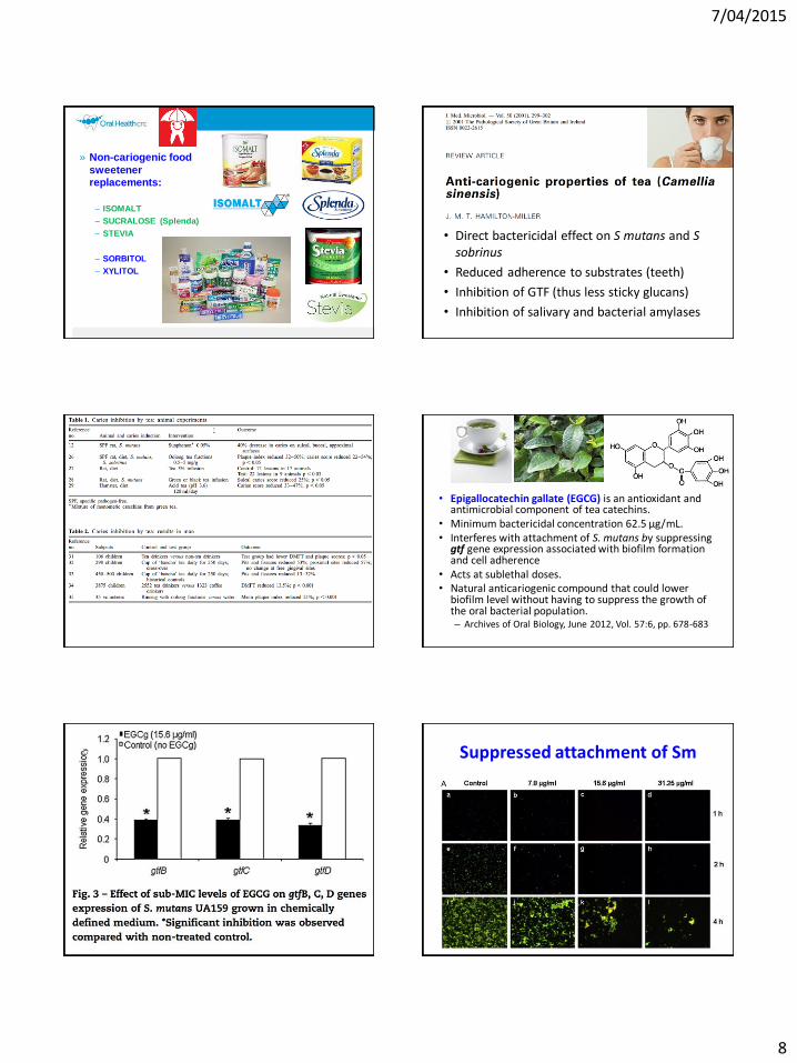
» Non-cariogenic food
sweetener replacements:
– ISOMALT
– SUCRALOSE (Splenda)
– STEVIA
– SORBITOL
– XYLITOL
• Direct bactericidal effect on S mutans and S sobrinus
• Reduced adherence to substrates (teeth)
• Inhibition of GTF (thus less sticky glucans)
• Inhibition of salivary and bacterial amylases
• Epigallocatechin gallate (EGCG) is an antioxidant and antimicrobial component of tea catechins.
• Minimum bactericidal concentration 62.5 µg/mL.• Interferes with attachment of S. mutans by suppressing gtf gene expression associated with biofilm formation and cell adherence
• Acts at sublethal doses.• Natural anticariogenic compound that could lower
biofilm level without having to suppress the growth of the oral bacterial population.– Archives of Oral Biology, June 2012, Vol. 57:6, pp. 678-683
Suppressed attachment of Sm

7/04/2015
9
Ecological therapy with CPP-ACP
• Lower adherence of key
pathogens to tooth surfaces, pellicle and existing biofilms,
e.g. by blocking adhesins
• Elevate plaque fluid pH by buffering plaque acids (protein
and phosphate)
• Decreased fermentation from
bio-available Ca and fluoride
ions
• Large bio-available calcium ion
reservoir slows diffusion of free
calcium.
• 73-80% lower S. sobrinus in oral cavity of rats with crude CPP.Guggenheim, et al. Powdered milk micellar casein prevents oral colonisation by Streptococcus sobrinus and dental caries in rats: a
basis for the caries-protective effect of dairy products. Caries Res 33:446-454 (1999).
• 75-83% lower binding of MS to pellicle-coated apatite discs. Schüpbach P, Neeser JR, Golliard M, Rouvet M, Guggenheim B. Incorporation of caseinoglycomacropeptide and
caseinophosphopeptide into the salivary pellicle inhibits adherence of mutans streptococci. J Dent Res 75:1779-1788 (1996).
• Impaired adherence of S. sobrinus to saliva-coated apatite beads.Neeser JR, Golliard M, Woltz A, Rouvet M, Dillmann ML, Guggenheim B. In vitro modulation of oral bacterial adhesion to saliva-coated
hydroxyapatite beads by milk casein derivatives. Oral Microbiol Immunol 9:193-201 (1994).
• Impaired formation of dental plaque in situ.Rahiotis C, Vougiouklakis G, Eliades G. Characterization of oral films formed in the presence of a CPP-ACP agent: An in situ study. J Dent 36:272-280 (2008).
• Lower Streptococcus mutans levels in biofilms.Erdem AP, Sepet E, Avshalom T, Gutkin V, Steinberg D. Effect of CPP-ACP and APF on Streptococcus mutans biofilm: A laboratory study. Am J Dent 24:119-123 (2011).
• RCT showing lower MS with CPP-ACP in 24 month old ECC children.Pukalllus ML, Plonka KA, Holcombe TF, Barnett AG, Walsh LJ, Seow WK. A randomized controlled trial of a 10 percent CPP-ACP cream to reduce mutans streptococci colonization. Pediatr Dent 35:550-555 (2013).
Inhibition of mutans streptococci and plaque by CPP-ACP
• Using TM one child with one carious tooth
• MS presence reduced P = 0.02 † †
• 90% of TM 1+ group had zero presence of MS, with none >105
Tooth Mousse Results: MS* *
*** *
**
††††
†††
†
TM eliminates high MS levels in dental plaque in 2 yr old children
Tooth Mousse Results: LB
TM no effect on LB levels
Effects of CPP-ACP on biofilms
• Effect of 2% CPP-ACP spray against
Cariogenic & Non-Cariogenic Bacteria
7/04/2015
10
Before and after TMP for 4 weeks
S. mutans biofilm testing using the flow cell model
Confocal Microscopy
• S. mutans incubated for one hour to
encourage cell attachment formation
• 25% ASM was pumped through the
system at 0.2 mL/min over 15 h (total
running time = 16 h).
• Biofilm in the flow cell stained with
Live/Dead® BacLightTM
• Biofilms imaged using Confocal
Laser Scanning Microscopy.
• Images analysed using the
COMSTAT software.
Stuart Dashper
Anti-MS biofilm effect of CPP-ACP
Control
Treated with 1 mL water for 10 min
1% CPP ACP treated
Treated with 1 mL 1% CPP-
ACP for 10 min
A/Prof S Dashper, CRC Oral Health58
The following results can be drawn from this randomized controlled trial.
1.MI Paste Plus not only had a preventive action of white spot development during orthodontic treatment, but also decreased the number of white spot lesions.2.The placebo had no preventive action on white spot development during orthodontic treatment; the number of lesions actually increased.3.MI Paste Plus had an impact on reducing white spots on the gingival surfaces, whereas the placebo group had the opposite effect.

7/04/2015
11
Orthodontic WSL Reversal:
Clinical aspects
Map the full extent of white spots
•Clean and dry the tooth
»Visual inspection of the dry tooth with
magnification
•Fluorescence using
orange filter
64
Active white spot caries: porous surface
Active WSL
• Active white-spot lesions on the buccal surfaces
= dull, rough and chalky white in a plaque
stagnation area with adjacent mild gingivitis.• Zandona & Zero, 2006
Arrested WSL
• Arrested WSL = shiny, smooth, translucent and
having healthy adjacent gingiva.

7/04/2015
12
• Arrested WSL = shiny, smooth, translucent and having healthy adjacent gingiva.
Prof. Edwina Kidd
J Dent Res. 2004; 83 Spec Iss C: C35-38
• Active enamel lesions involve surface erosion and
subsurface porosity.
• Inactive or arrested lesions have an abraded
surface, but subsurface mineral loss remains, and
a true subsurface remineralization is rarely
achievable, because the surface zone acts as a
diffusion barrier.
Requirements for remin
• Diffuse into the subsurface
• Overcome the challenge of delivery Ca and P into the subsurface zone
• Not delivery an excess of Ca
• Work at lower pH than neutral range
• Remove Ca pptsand protein plugs from enamel surface pores
• Remove pellicle and enamel proteins from the surface, e.g. with NaOCl, alkaline or acid etching
DT Zero, BMC Oral Health 2006;6(Suppl 1)
First report of post-orthodontic WSL reversal using Tooth Mousse(Walsh 2004)
Key issue: ensuring surface penetration for arrested lesions !Various methods for this:Short acid etchPeroxideProteolytic agent(Tran et al. 2013)
No need to etch ACTIVE lesions prior to CPP-ACP as their surface is porous
Need to surface treat arrestedlesions to make the surface permeable
26.5.04
After Tooth Mousse 13.10.0426.8.04
26.5.04
After Tooth Mousse 13.10.04

7/04/2015
13
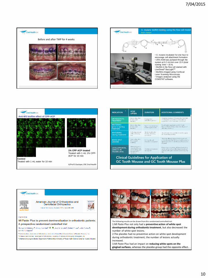
http://www.kiheiorthodonticsmaui.com/Treatments/Tooth-Stain-Removal.aspx
http://www.orthocare.co.uk/acatalog/info_9000_002522.html
http://vutooth.smugmug.com/Dental/MI-Paste-White-Spot-Removal/9005404_Cxw74w/599020519_EDsvH
Remin model
using slabs in ortho brackets
» 5 subjects
» 4 mounted slabs per subject
» TM vs TMP vs TMPME vs vehicle vs vehicle with F
» 2 week washouts between runs
77
Clinical protocol for treating ‘active’ white spot lesions
1. Clean teeth thoroughly using a soft brush and low-medium concentration fluoride tooth paste
2. Immediately follow with an application of GC Tooth Mousse Plus, either directly to the surface. Alternatively load into the patient’s orthodontic retainer in the region of the white spot lesions.
3. Repeat twice daily.
4. Continue the treatment until the tube of Tooth Mousse Plus is finished.
http://www.dentistryiq.com/articles/wdj/print/volume-5/issue-4/you-and-your-practice/minimal-intervention-dentistry.html
Correct application methods

7/04/2015
14
Don’t use in trays – less saliva contact !
No surface treatment was done of the lesions. Key factor !!
Intervention was delayed so some natural arrest of WSL would have occurred.
Duration of Tx only 8 weeks – too short
Digital analysis not corrected for reflections (or polarizing effects)
Hypomineralized enamel – developmental defect, treated at the end of fixed ortho immediately after debanding
Trauma-induced enamel hypoplasia
83
Tx of fluorosis after debanding
• 1. Check for and remove any surface bonding residues after orthodontic treatment
– Diagnostic etch 10 seconds
• 2. Enamel microabrasion
– Etch 37% phosphoric acid for 2 min with agitation
– Gentle abrasion with fine pumice 1500 rpm
– Repeat (X2) until surface contour is even
• 3. Apply Tooth Mousse Plus
– immediately, and then each night before bed
– Review at 4-6 weeks84
7/04/2015
15
85 86
87
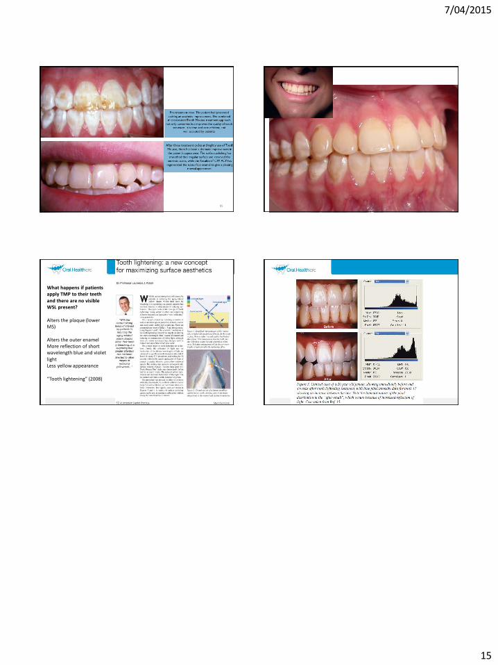
What happens if patients apply TMP to their teeth and there are no visible WSL present?
Alters the plaque (lower MS)
Alters the outer enamelMore reflection of short wavelength blue and violet
lightLess yellow appearance
“Tooth lightening” (2008)