Embed Size (px)
Citation preview
Case ReportA Rare Case of Spontaneous Pneumocephalus Associated withNontraumatic Cerebrospinal Fluid Leak
Murad Baba, Omer Tarar, and Amer Syed
Department of Internal Medicine, Jersey City Medical Center, 355 Grand Street, Jersey City, NJ 07302, USA
Correspondence should be addressed to Murad Baba; [email protected]
Received 13 November 2015; Accepted 14 April 2016
Academic Editor: Abbass Amirjamshidi
Copyright © 2016 Murad Baba et al. This is an open access article distributed under the Creative Commons Attribution License,which permits unrestricted use, distribution, and reproduction in any medium, provided the original work is properly cited.
Introduction. Spontaneous nontraumatic pneumocephalus (PNC) and cerebrospinal fluid (CSF) leaks are both very uncommonconditions. We report a rare case of spontaneous pneumocephalus associated with CSF leak secondary to right sphenoid sinusbony defect without history of trauma. Case Description. 51-year-old Hispanic female with past medical history of hypertensionand idiopathic intracranial hypertension (Pseudotumor Cerebri) presented to the emergency room complaining of headache andclear discharge from the right nostril. Physical examination was significant for right frontal sinus tenderness and clear dischargefrom right nostril. Computed Tomography (CT) scan of the brain showed moderate amount of extra-axial air within the rightcerebral hemisphere indicative of pneumocephalus. CT scan of facial bones showed bony defect along the right sphenoid sinuswith abnormal CSF collection. The patient was started on intravenous antibiotics for meningitis prophylaxis and subsequentlyunderwent transsphenoidal repair of cerebrospinal fluid leak with abdominal fat graft. CSF rhinorrhea stopped completely after thesurgery with near complete resolution of pneumocephalus before discharge. Conclusions. Early identification of pneumocephalusand surgical intervention can help decrease the morbidity and avoid possible complications. Idiopathic intracranial hypertension,although rare, can lead to CSF leak and pneumocepahlus.
1. Introduction
Pneumocephalus (PNC) is defined as pathological collectionof gas within the cranial cavity accumulating in the epidural,subdural, subarachnoid, intraventricular, or intraparenchy-mal compartments [1]. The main cause of PNC is headinjury, trauma accounts for 74% of all cases followed byintracranial neoplasms, infections, neurosurgery, paranasalsinus surgery, and diagnostic or neurosurgical interventionssuch as pneumoencephalography or lumbar puncture [2,3].
Chiari was the first to describe PNC in 1884 [4]. Thedevelopmental mechanism of pneumocephalus is mainlybased on two factors: a reduction in intracranial pressure andthe presence of a defect in the dura. It is caused by eithera ball-valve mechanism that allows air to enter but not toexit or by CSF leakage which creates a negative pressure withsubsequent air entry [2].
2. Case Description
51-year-old Hispanic female with past medical history ofhypertension and idiopathic intracranial hypertension (Pseu-dotumor Cerebri) presented to the emergency room com-plaining of headache for three weeks. Her headache wasprogressively worsening more on the right side, associatedwith clear discharge from right nostril which was aggravatedby bending forward and straining. She denied any historyof trauma. She reported having flu with frequent sneezingepisodes around the same time. She reported similar symp-toms six years ago; at that time further workup was doneincluding Computed Tomography (CT) scan of the brainwhich was unremarkable and lumbar puncture (LP) whichshowed high opening pressure. She was diagnosed with idio-pathic intracranial hypertension and treated conservatively.
Physical examination was significant for right frontalsinus tenderness and clear discharge from right nostril. Initiallaboratory workup did not reveal any significant results.
Hindawi Publishing CorporationCase Reports in Neurological MedicineVolume 2016, Article ID 1828461, 3 pageshttp://dx.doi.org/10.1155/2016/1828461
2 Case Reports in Neurological Medicine
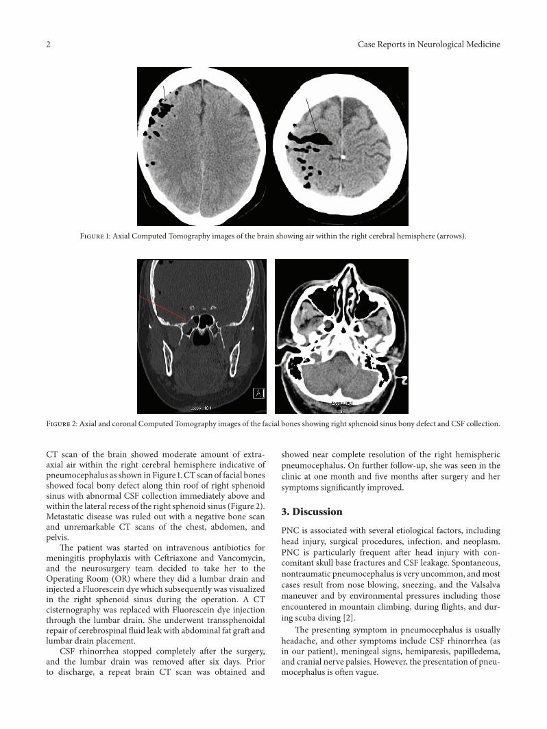
Figure 1: Axial Computed Tomography images of the brain showing air within the right cerebral hemisphere (arrows).
Figure 2: Axial and coronal Computed Tomography images of the facial bones showing right sphenoid sinus bony defect and CSF collection.
CT scan of the brain showed moderate amount of extra-axial air within the right cerebral hemisphere indicative ofpneumocephalus as shown in Figure 1. CT scan of facial bonesshowed focal bony defect along thin roof of right sphenoidsinus with abnormal CSF collection immediately above andwithin the lateral recess of the right sphenoid sinus (Figure 2).Metastatic disease was ruled out with a negative bone scanand unremarkable CT scans of the chest, abdomen, andpelvis.
The patient was started on intravenous antibiotics formeningitis prophylaxis with Ceftriaxone and Vancomycin,and the neurosurgery team decided to take her to theOperating Room (OR) where they did a lumbar drain andinjected a Fluorescein dye which subsequently was visualizedin the right sphenoid sinus during the operation. A CTcisternography was replaced with Fluorescein dye injectionthrough the lumbar drain. She underwent transsphenoidalrepair of cerebrospinal fluid leak with abdominal fat graft andlumbar drain placement.
CSF rhinorrhea stopped completely after the surgery,and the lumbar drain was removed after six days. Priorto discharge, a repeat brain CT scan was obtained and
showed near complete resolution of the right hemisphericpneumocephalus. On further follow-up, she was seen in theclinic at one month and five months after surgery and hersymptoms significantly improved.
3. Discussion
PNC is associated with several etiological factors, includinghead injury, surgical procedures, infection, and neoplasm.PNC is particularly frequent after head injury with con-comitant skull base fractures and CSF leakage. Spontaneous,nontraumatic pneumocephalus is very uncommon, andmostcases result from nose blowing, sneezing, and the Valsalvamaneuver and by environmental pressures including thoseencountered in mountain climbing, during flights, and dur-ing scuba diving [2].
The presenting symptom in pneumocephalus is usuallyheadache, and other symptoms include CSF rhinorrhea (asin our patient), meningeal signs, hemiparesis, papilledema,and cranial nerve palsies. However, the presentation of pneu-mocephalus is often vague.

Case Reports in Neurological Medicine 3
Idiopathic intracranial hypertension most commonlyaffects women of child bearing age and is usually a self-limiting condition, and the association between idiopathicintracranial hypertension and spontaneous CSF rhinorrhoeais rare [5].
Persistent or tension PNC may cause headache, lethargy,and neurological deterioration. It reflects an abnormal com-munication between the intradural space and external envi-ronment, creating a risk factor for central nervous systeminfection [1]. The Mount Fuji sign on CT scans of the brainis a characteristic radiological finding defined as compres-sion of frontal lobes and widening of the interhemisphericspace between the tips of the frontal lobes; it is useful indiscriminating tension pneumocephalus from nontensionpneumocephalus.
Tension pneumocephalus occurs most commonly afterthe neurosurgical evacuation of a subdural hematoma; itcan be a neurosurgical emergency. The prevalence of ten-sion pneumocephalus following the evacuation of chronicsubdural hematomas has been reported from 2.5% to 16%.Tension pneumocephalus can also occur as a result of skullbase surgery, paranasal sinus surgery, posterior fossa surgeryin the sitting position, or head trauma [6, 7].
Treatment of PNC is either conservative or surgical. Thisshould be decided based on the type, severity, and presenceof increased intracranial pressure. PNC has been treatedby ventilating the patient with normobaric 100% oxygen,resulting in the reabsorption of nitrogen into the bloodstream and reduction of the volume of the intracranial air[1, 8].When tension PNC is suspected by clinical and imagingfindings, treatment consists of emergent decompression toalleviate pressure on the brain parenchyma, as air is toxicto neurons and can cause further damage to the alreadycompromised parenchyma, and that leads to cerebral edemasurrounding the air evolving into encephalomalacia [9].
Surgical options for tension pneumocephalus includedrilling of burr holes, craniotomy, needle aspiration, ven-triculostomy placement, and closure of dural defect. Inaddition, patient education to avoid Valsalva’s maneuvers ornose blowing can possibly contribute to reducing recurrence.
4. Conclusion
Few cases were described in the literature and most weretreated surgically. Our rare case presented with nontraumaticCSF leak and spontaneous pneumocephalus; early identifica-tion of such cases and surgical intervention can help decreasethe morbidity and the complications that can happen.
CSF rhinorrhoea is a rare complication of idiopathicintracranial hypertension; if it happens, it can lead to menin-gitis and PNC; therefore surgical repair is always recom-mended.
Abbreviations
PNC: PneumocephalusCSF: Cerebrospinal fluidCT: Computed TomographyLP: Lumbar puncture.
Competing Interests
The authors have no competing interests to declare relevantto this paper.
References
[1] W. S. Paiva, A. F. de Andrade, E. G. Figueiredo, R. L. Amorim,M. Prudente, andM. J. Teixeira, “Effects of hyperbaric oxygena-tion therapy on symptomatic pneumocephalus,” Therapeuticsand Clinical Risk Management, vol. 10, pp. 769–773, 2014.
[2] J.-S. Lee, Y.-S. Park, J.-T. Kwon, and J.-S. Suk, “Spontaneouspneumocephalus associated with pneumosinus dilatans,” Jour-nal of Korean Neurosurgical Society, vol. 47, no. 5, pp. 395–398,2010.
[3] J.W.Markham, “The clinical features of pneumocephalus basedupon a survey of 284 cases with report of 11 additional cases,”Acta Neurochirurgica, vol. 16, no. 1-2, pp. 1–78, 1967.
[4] H. Chiari, “A case of acummulation of air in the ventricles ofhuman brain,” Zeitschrift fur Heilkunde, vol. 5, pp. 383–390,1884.
[5] D. Clark, P. Bullock, T. Hui, and J. Firth, “Benign intracranialhypertension: a cause of CSF rhinorrhoea,” Journal of Neurology,Neurosurgery and Psychiatry, vol. 57, no. 7, pp. 847–849, 1994.
[6] C. B. Dabdoub, G. Salas, N. Silveira Edo, and C. F. Dabdoub,“Review of the management of pneumocephalus,” SurgicalNeurology International, vol. 6, article 155, 2015.
[7] Y. Ishiwata, K. Fujitsu, T. Sekino et al., “Subdural tension pneu-mocephalus following surgery for chronic subdural hematoma,”Journal of Neurosurgery, vol. 68, no. 1, pp. 58–61, 1988.
[8] H. Yates, M. Hamill, C. O. Borel, and T. J. K. Toung, “Incidenceand perioperative management of tension pneumocephalusfollowing craniofacial resection,” Journal of Neurosurgical Anes-thesiology, vol. 6, no. 1, pp. 15–20, 1994.
[9] S. K. Venkatesh and V. Bhargava, “Clinics in diagnostic imaging(119). Post-traumatic intracerebral pneumatocele,” SingaporeMedical Journal, vol. 48, no. 11, pp. 1055–1059, 2007.

Submit your manuscripts athttp://www.hindawi.com
Stem CellsInternational
Hindawi Publishing Corporationhttp://www.hindawi.com Volume 2014
Hindawi Publishing Corporationhttp://www.hindawi.com Volume 2014
MEDIATORSINFLAMMATION
of
Hindawi Publishing Corporationhttp://www.hindawi.com Volume 2014
Behavioural Neurology
EndocrinologyInternational Journal of
Hindawi Publishing Corporationhttp://www.hindawi.com Volume 2014
Hindawi Publishing Corporationhttp://www.hindawi.com Volume 2014
Disease Markers
Hindawi Publishing Corporationhttp://www.hindawi.com Volume 2014
BioMed Research International
OncologyJournal of
Hindawi Publishing Corporationhttp://www.hindawi.com Volume 2014
Hindawi Publishing Corporationhttp://www.hindawi.com Volume 2014
Oxidative Medicine and Cellular Longevity
Hindawi Publishing Corporationhttp://www.hindawi.com Volume 2014
PPAR Research
The Scientific World JournalHindawi Publishing Corporation http://www.hindawi.com Volume 2014
Immunology ResearchHindawi Publishing Corporationhttp://www.hindawi.com Volume 2014
Journal of
ObesityJournal of
Hindawi Publishing Corporationhttp://www.hindawi.com Volume 2014
Hindawi Publishing Corporationhttp://www.hindawi.com Volume 2014
Computational and Mathematical Methods in Medicine
OphthalmologyJournal of
Hindawi Publishing Corporationhttp://www.hindawi.com Volume 2014
Diabetes ResearchJournal of
Hindawi Publishing Corporationhttp://www.hindawi.com Volume 2014
Hindawi Publishing Corporationhttp://www.hindawi.com Volume 2014
Research and TreatmentAIDS
Hindawi Publishing Corporationhttp://www.hindawi.com Volume 2014
Gastroenterology Research and Practice
Hindawi Publishing Corporationhttp://www.hindawi.com Volume 2014
Parkinson’s Disease
Evidence-Based Complementary and Alternative Medicine
Volume 2014Hindawi Publishing Corporationhttp://www.hindawi.com