Embed Size (px)
Citation preview

Zacarias Föhrding et al. Journal of Cardiothoracic Surgery 2014, 19:172http://www.cardiothoracicsurgery.org/content/19/1/172
CASE REPORT Open Access
A case of lethal spontaneous massive hemothoraxin a patient with neurofibromatosis 1Luisa Zacarias Föhrding1, Timur Sellmann2, Sebastian Angenendt1, Detlef Kindgen-Milles2, Stefan A Topp1,Bernhard Korbmacher3, Artur Lichtenberg3 and Wolfram T Knoefel1*
Abstract
Neurofibromatosis type 1 is an autosomal dominant disease characterized by multiple dermatological disordersamongst others. Among the less frequent manifestations are vascular abnormalities. Here, we present a case ofspontaneous massive hemothorax in a 39-year-old Caucasian woman with neurofibromatosis 1 and a thoracicmeningocele with a lethal outcome despite extensive surgical intervention as well as intensive care measures.Spontaneous hemothorax is a rare, but potentially lethal complication of neurofibromatosis type 1, which necessitatesquick and decisive intervention; endovascular embolization where possible, otherwise aggressive surgical interventionin unstable patients.
Keywords: Neurofibromatosis type 1, Spontaneous hemothorax, Meningocele, Endovascular embolization
BackgroundNeurofibromatosis 1 (NF1) is an autosomal dominant dis-ease characterized by café-au-lait macules, neurofibromasof any type, axillary and inguinal freckling and Lisch nod-ules in the iris. Additionally, numerous vascular manifesta-tions have been reported [1-3]. Intrathoracic meningoceleshave also been described in NF1 several times [4-6]. Spon-taneous hemothorax is a rare but potentially lethal compli-cation [7-9]. Here, we present a case of a lethal spontaneoushemothorax associated to an intrathoracic meningocele ina patient with NF 1.
Case presentationA 39-year old Caucasian female with known NF1 pre-sented herself in the emergency department of an outsidehospital with acute symptoms of thoracic pain and dys-pnea. Her history was otherwise significant for an exten-sive thoracic myelomeningocele and status post-surgicalcorrection of a thoracic scoliosis using a tibia bone graft27 years earlier.On physical examination at the outside hospital, she was
awake, afebrile, blood pressure of 80/40 mmHg and a heart
* Correspondence: [email protected] of General, Visceral and Paediatric Surgery, Heinrich HeineUniversity Düsseldorf, Düsseldorf, GermanyFull list of author information is available at the end of the article
© 2014 Zacarias Föhrding et al.; licensee BioMCreative Commons Attribution License (http:/distribution, and reproduction in any mediumDomain Dedication waiver (http://creativecomarticle, unless otherwise stated.
rate of 100 beats per minute. The initial haemoglobin was12.5 g/dl, quickly deteriorating to 8.4 g/dl in the followinghour. Chest x-ray (Figure 1) was performed 40 mins afteradmission and revealed an extensive mediastinal shift. Sub-sequent computer tomography (Figure 2) was performed1h later at the outside hospital and revealed a massive he-mothorax on the left side. The patient was intubated oro-tracheally because of hemorrhagic shock and imminentcardiopulmonary arrest and received seven packed redblood cell concentrates. A thoracic drainage was insertedprior to transfer. 4 h 10 min after admission to the outsidehospital the patient was transferred to our institution andarrived there 1h later.On admission, she was highly catecholamine-dependent.
We found one left-thoracic suction drainage installedusing Monaldi technique with 50 ml of blood in the drain-age bottle. During placement of venous and arterial linesshe suffered a cardiocirculatory arrest. Resuscitation wasimmediately initiated and we proceeded with performingan emergency exploratory left antero-lateral thoracot-omy in the intensive care unit. Thereupon 2 liters ofblood exsanguinated spontaneously. Despite massive bloodtransfusions (16 packed red blood cells, 2 thrombocyte con-centrates, and 4 fresh frozen plasmas) and intravenousfluid administration no sufficient circulation could beestablished. Due to massive bleeding and very difficultoperative conditions it was not possible to perform
ed Central Ltd. This is an Open Access article distributed under the terms of the/creativecommons.org/licenses/by/4.0), which permits unrestricted use,, provided the original work is properly credited. The Creative Commons Publicmons.org/publicdomain/zero/1.0/) applies to the data made available in this
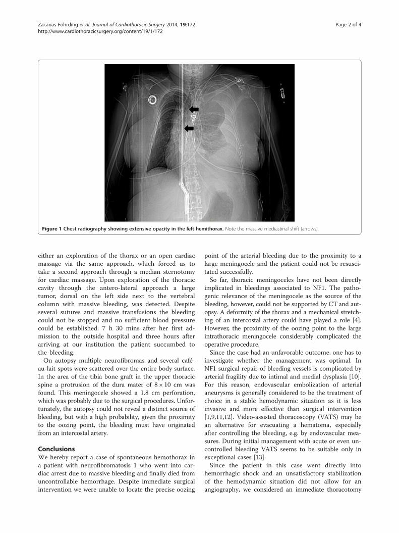
Figure 1 Chest radiography showing extensive opacity in the left hemithorax. Note the massive mediastinal shift (arrows).
Zacarias Föhrding et al. Journal of Cardiothoracic Surgery 2014, 19:172 Page 2 of 4http://www.cardiothoracicsurgery.org/content/19/1/172
either an exploration of the thorax or an open cardiacmassage via the same approach, which forced us totake a second approach through a median sternotomyfor cardiac massage. Upon exploration of the thoraciccavity through the antero-lateral approach a largetumor, dorsal on the left side next to the vertebralcolumn with massive bleeding, was detected. Despiteseveral sutures and massive transfusions the bleedingcould not be stopped and no sufficient blood pressurecould be established. 7 h 30 mins after her first ad-mission to the outside hospital and three hours afterarriving at our institution the patient succumbed tothe bleeding.On autopsy multiple neurofibromas and several café-
au-lait spots were scattered over the entire body surface.In the area of the tibia bone graft in the upper thoracicspine a protrusion of the dura mater of 8 × 10 cm wasfound. This meningocele showed a 1.8 cm perforation,which was probably due to the surgical procedures. Unfor-tunately, the autopsy could not reveal a distinct source ofbleeding, but with a high probability, given the proximityto the oozing point, the bleeding must have originatedfrom an intercostal artery.
ConclusionsWe hereby report a case of spontaneous hemothorax ina patient with neurofibromatosis 1 who went into car-diac arrest due to massive bleeding and finally died fromuncontrollable hemorrhage. Despite immediate surgicalintervention we were unable to locate the precise oozing
point of the arterial bleeding due to the proximity to alarge meningocele and the patient could not be resusci-tated successfully.So far, thoracic meningoceles have not been directly
implicated in bleedings associated to NF1. The patho-genic relevance of the meningocele as the source of thebleeding, however, could not be supported by CT and aut-opsy. A deformity of the thorax and a mechanical stretch-ing of an intercostal artery could have played a role [4].However, the proximity of the oozing point to the largeintrathoracic meningocele considerably complicated theoperative procedure.Since the case had an unfavorable outcome, one has to
investigate whether the management was optimal. InNF1 surgical repair of bleeding vessels is complicated byarterial fragility due to intimal and medial dysplasia [10].For this reason, endovascular embolization of arterialaneurysms is generally considered to be the treatment ofchoice in a stable hemodynamic situation as it is lessinvasive and more effective than surgical intervention[1,9,11,12]. Video-assisted thoracoscopy (VATS) may bean alternative for evacuating a hematoma, especiallyafter controlling the bleeding, e.g. by endovascular mea-sures. During initial management with acute or even un-controlled bleeding VATS seems to be suitable only inexceptional cases [13].Since the patient in this case went directly into
hemorrhagic shock and an unsatisfactory stabilizationof the hemodynamic situation did not allow for anangiography, we considered an immediate thoracotomy
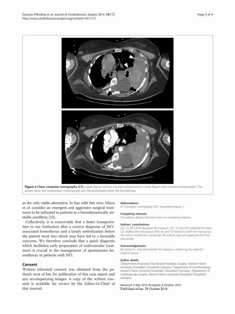
Figure 2 Chest computer tomography (CT). Upper figure: without contrast enhancement. Lower figure: with contrast enhancement. Thearrows show the intrathoracic meningocele and the arrowheads show the hemothorax.
Zacarias Föhrding et al. Journal of Cardiothoracic Surgery 2014, 19:172 Page 3 of 4http://www.cardiothoracicsurgery.org/content/19/1/172
as the only viable alternative. In line with this view, Miuraet al. consider an emergent and aggressive surgical treat-ment to be indicated in patients in a hemodynamically un-stable condition [14].Collectively, it is conceivable that a faster transporta-
tion to our institution after a correct diagnosis of NF1-associated hemothorax and a timely embolization beforethe patient went into shock may have led to a favorableoutcome. We therefore conclude that a quick diagnosiswhich facilitates early preparation of endovascular treat-ment is crucial in the management of spontaneous he-mothorax in patients with NF1.
ConsentWritten informed consent was obtained from the pa-tient’s next of kin for publication of this case report andany accompanying images. A copy of the written con-sent is available for review by the Editor-in-Chief ofthis journal.
AbbreviationsCT: Computer tomography; NF1: Neurofibromatosis 1.
Competing interestsThe authors declare that they have no competing interests.
Authors’ contributionsLZF, TS, BK, DK-M designed the research; LZF, TS and SAT collected the data;LZF drafted the manuscript; WTK, AL and TS helped to draft the manuscript.All authors revised the manuscript. All authors read and approved the finalmanuscript.
AcknowledgementsWe thank Dr. Jörg Stammkötter for helping in obtaining the patient’smedical history.
Author details1Department of General, Visceral and Paediatric Surgery, Heinrich HeineUniversity Düsseldorf, Düsseldorf, Germany. 2Department of Anesthesiology,Heinrich Heine University Düsseldorf, Düsseldorf, Germany. 3Department ofCardiovascular Surgery, Heinrich Heine University Düsseldorf, Düsseldorf,Germany.
Received: 4 May 2014 Accepted: 8 October 2014
Zacarias Föhrding et al. Journal of Cardiothoracic Surgery 2014, 19:172 Page 4 of 4http://www.cardiothoracicsurgery.org/content/19/1/172
References1. Kipfer B, Lardinois D, Triller J, Carrel T: Embolization of a ruptured
intercostal artery aneurysm in type I neurofibromatosis. Eur JCardiothorac Surg 2001, 19:721–723.
2. Oderich GS, Sullivan TM, Bower TC, Gloviczki P, Miller DV, Babovic-Vuksanovic D,Macedo TA, Stanson A: Vascular abnormalities in patients withneurofibromatosis syndrome type I: clinical spectrum, management,and results. J Vasc Surg 2007, 46:475–484.
3. Salyer WR, Salyer DC: The vascular lesions of neurofibromatosis.Angiology 1974, 25:510–519.
4. Kaneda H, Saito T, Konobu T, Saito Y: Chest wall bleeding with giantintrathoracic meningocele in neurofibromatosis type 1. Interact CardiovascThorac Surg 2011, 12:328–330.
5. Miles J, Pennybacker J, Sheldon P: Intrathoracic meningocele. Its developmentand association with neurofibromatosis. J Neurol Neurosurg Psychiatr 1969,32:99–110.
6. Rainov NG, Heidecke V, Burkert W: Thoracic and lumbar meningocele inneurofibromatosis type 1. Report of two cases and review of theliterature. Neurosurg Rev 1995, 18:127–134.
7. Nopajaroonsri C, Lurie AA: Venous aneurysm, arterial dysplasia, andnear-fatal hemorrhages in neurofibromatosis type 1. Hum Pathol1996, 27:982–985.
8. Pezzetta E, Paroz A, Ris HB, Martinet O: Spontaneous hemothoraxassociated with von Recklinghausen's disease. Eur J Cardiothorac Surg2003, 23:1062–1064.
9. Teitelbaum GP, Hurvitz RJ, Esrig BC: Hemothorax in type Ineurofibromatosis. Ann Thorac Surg 1998, 66:569–571.
10. Greene JF, Fitzwater JE, Burgess J: Arterial lesions associated withneurofibromatosis. Am J Clin Pathol 1974, 62:481–487.
11. Arai K, Sanada J, Kurozumi A, Watanabe T, Matsui O: Spontaneoushemothorax in neurofibromatosis treated with percutaneousembolization. Cardiovasc Intervent Radiol 2007, 30:477–479.
12. Sandhu C, Sabharwal T: Spontaneous hemothorax in patients withneurofibromatosis treated with percutaneous embolization.Cardiovasc Intervent Radiol 2008, 31:1260–1261.
13. Mydin Muhammad Izanee M, Sharma A, Zia Z, Hawari M, Jadoon M,Majewski A: A novel approach in managing right-sided haemothoraxin neurofibromatosis type 1. Asian Cardiovasc Thorac Ann 2014,doi:10.1177/0218492314522636.
14. Miura T, Kawano Y, Chujo M, Miyawaki M, Mori H, Kawahara K: Spontaneoushemothorax in patients with von Recklinghausen's disease. Jpn J ThoracCardiovasc Surg 2005, 53:649–652.
doi:10.1186/s13019-014-0172-yCite this article as: Zacarias Föhrding et al.: A case of lethal spontaneousmassive hemothorax in a patient with neurofibromatosis 1. Journal ofCardiothoracic Surgery 2014 19:172.
Submit your next manuscript to BioMed Centraland take full advantage of:
• Convenient online submission
• Thorough peer review
• No space constraints or color figure charges
• Immediate publication on acceptance
• Inclusion in PubMed, CAS, Scopus and Google Scholar
• Research which is freely available for redistribution
Submit your manuscript at www.biomedcentral.com/submit