Embed Size (px)
Citation preview
Hindawi Publishing CorporationCase Reports in MedicineVolume 2013, Article ID 956849, 4 pageshttp://dx.doi.org/10.1155/2013/956849
Case ReportSpontaneous Rapid Resolution of Acute EpiduralHematoma in Childhood
Ismail GülGen,1 Hakan Ak,2 Enver Sösüncü,1 Alpaslan Yavuz,1 and Nejmi Kiymaz3
1 Yuzuncu Yıl University, School of Medicine, Van, Turkey2 Bozok University, School of Medicine, Yozgat, Turkey3 Akdeniz University, School of Medicine, Antalya, Turkey
Correspondence should be addressed to Hakan Ak; [email protected]
Received 3 October 2013; Revised 19 October 2013; Accepted 10 November 2013
Academic Editor: Di Lazzaro Vincenzo
Copyright © 2013 Ismail Gulsen et al. This is an open access article distributed under the Creative Commons Attribution License,which permits unrestricted use, distribution, and reproduction in any medium, provided the original work is properly cited.
Acute epidural hematoma is a critical emergency all around the world, and its aggressive diagnosis and treatment are of vitalimportance. Emergent surgical evacuation of the hematoma is known as standard management; however, conservative proceduresare also used for small ones. Spontaneous rapid resolution of these hematomas has also been reported in eight pediatric cases.Various theories have been proposed to explain the underlying pathophysiology of this resolution. Herein, we are reporting a newpediatric case with spontaneously resolving acute epidural hematoma 12 hours after admission to the emergency room.
1. Introduction
Acute epidural hematoma is a serious complication of headinjury; rapid diagnosis and early surgical evacuation of itare considered as the standard management [1, 2]. However,conservative followup also has made its place in the literature[1, 3, 4]. Since 1981, reported cases of spontaneous rapidresolution encourage the opinion of conservative followup[5]. Various theories have been proposed to explain theunderlying mechanism of these cases [2, 5–7].
Herein, we are presenting an acute epidural hematomacase which resolved 12 hours after admitting to emergencydepartment in a 4-year-old child and we are discussing theunderlying mechanisms for our case in the light of theliterature.
2. Case
Four-year-old boy was evaluated in the emergency depart-ment about one hour after a fall from a height of abouttwo meters. His history was unremarkable except for mildheadache. In the physical examination, there was edema,abrasion, and erythema on the right temporoparietal region.Neurological examination was normal. Computed tomog-raphy of the patient in the emergency department revealed
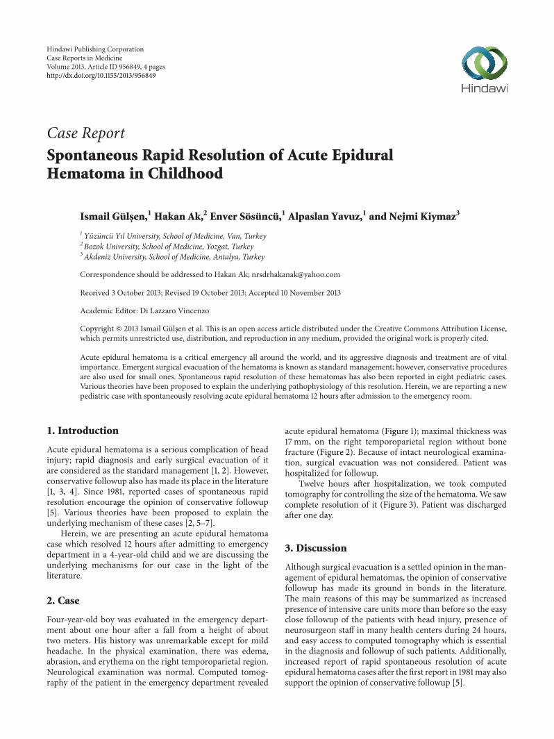
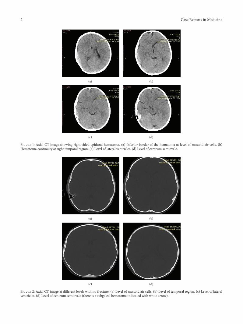
acute epidural hematoma (Figure 1); maximal thickness was17mm, on the right temporoparietal region without bonefracture (Figure 2). Because of intact neurological examina-tion, surgical evacuation was not considered. Patient washospitalized for followup.
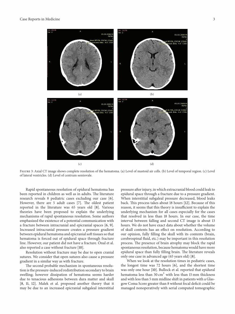
Twelve hours after hospitalization, we took computedtomography for controlling the size of the hematoma.We sawcomplete resolution of it (Figure 3). Patient was dischargedafter one day.
3. Discussion
Although surgical evacuation is a settled opinion in the man-agement of epidural hematomas, the opinion of conservativefollowup has made its ground in bonds in the literature.The main reasons of this may be summarized as increasedpresence of intensive care units more than before so the easyclose followup of the patients with head injury, presence ofneurosurgeon staff in many health centers during 24 hours,and easy access to computed tomography which is essentialin the diagnosis and followup of such patients. Additionally,increased report of rapid spontaneous resolution of acuteepidural hematoma cases after the first report in 1981may alsosupport the opinion of conservative followup [5].
2 Case Reports in Medicine
(a) (b)
(c) (d)
Figure 1: Axial CT image showing right sided epidural hematoma. (a) Inferior border of the hematoma at level of mastoid air cells. (b)Hematoma continuity at right temporal region. (c) Level of lateral ventricles. (d) Level of centrum semiovale.
(a) (b)
(c) (d)
Figure 2: Axial CT image at different levels with no fracture. (a) Level of mastoid air cells. (b) Level of temporal region. (c) Level of lateralventricles. (d) Level of centrum semiovale (there is a subgaleal hematoma indicated with white arrow).
Case Reports in Medicine 3
(a) (b)
(c) (d)
Figure 3: Axial CT image shows complete resolution of the hematoma. (a) Level of mastoid air cells. (b) Level of temporal region. (c) Levelof lateral ventricles. (d) Level of centrum semiovale.
Rapid spontaneous resolution of epidural hematoma hasbeen reported in children as well as in adults. The literatureresearch reveals 8 pediatric cases excluding our case [6].However, there are 5 adult cases [7]. The oldest patientreported in the literature was 65 years old [8]. Varioustheories have been proposed to explain the underlyingmechanisms of rapid spontaneous resolution. Some authorsemphasized the existence of a potential communication witha fracture between intracranial and epicranial spaces [6, 9].Increased intracranial pressure creates a pressure gradientbetween epidural hematoma and epicranial soft tissues so thathematoma is forced out of epidural space through fractureline. However, our patient did not have a fracture. Onal et al.also reported a case without fracture [10].
Resolution without fracture may be due to open cranialsutures. We consider that open sutures also cause a pressuregradient in a similar way as with fracture.
The second probable mechanism in spontaneous resolu-tion is the pressure-induced redistribution secondary to brainswelling; however dissipation of hematoma seems harderdue to tenacious adhesions between dura matter and skull[8, 11, 12]. Malek et al. proposed another theory that itmay be due to an increased epicranial subgaleal interstitial
pressure after injury, inwhich extracranial blood could leak toepidural space through a fracture due to a pressure gradient.When interstitial subgaleal pressure decreased, blood leaksback. This process takes about 18 hours [12]. Because of thisreason, it seems that this theory is insufficient to explain theunderlying mechanism for all cases especially for the casesthat resolved in less than 18 hours. In our case, the timeinterval between falling and second CT image is about 13hours. We do not have exact data about whether the volumeof skull contents has an effect on resolution. According toour opinion, fully filling the skull with its contents (brain,cerebrospinal fluid, etc.) may be important in this resolutionprocess. The presence of brain atrophy may block the rapidspontaneous resolution, because hematomawould havemoreepidural space than fully filling brain. The literature revealsonly one case in advanced age (65 years old) [8].
When we look at the resolution times in pediatric cases,the longest time was 72 hours [6], and the shortest timewas only one hour [10]. Bullock et al. reported that epiduralhematoma less than 30 cm3 with less than 15mm thicknessand with less than 5mmmidline shift in patients with a Glas-gow Coma Score greater than 8 without focal deficit could bemanaged nonoperatively with serial computed tomographic
4 Case Reports in Medicine
scanning and close observation in a neurosurgical center [13].In our case thickness was 17mm; however, total blood volumewas less than 30 cm3, midline shift was less than 5mm, andGCSwas 15.We decided tomanage the patient conservativelywith serial CT. In our case, we detected the resolution after 12hours. If we took the computed tomography sooner, perhapswe would see the resolution earlier. Onal et al. saw theresolution just after one hour, because of the deteriorationof the general status of their patient. They retook CT andsaw the resolution of acute epidural hematoma.Deteriorationwas due to hepatic artery injury in their patient [10]. We didnot take computed CT earlier, because of the intact status ofpatient.
4. Conclusion
The underlying pathophysiology of rapid spontaneous res-olution of acute epidural hematomas has not yet been fullyclarified, despite being more frequently reported cases, espe-cially in children. We think that whether the skull contentsfully fill the skull may be important in the pathophysiologyof spontaneous rapid resolution. We believe that futureexperimental studies will shed light on our way.
Consent
The authors are able to provide a written confirmation thatthe patient described in the case report has given them aninformed consent for the case report to be published when-ever editor requests it.
Conflict of Interests
The authors have no conflict of interest.
References
[1] K. S. Bhau, S. S. Bhau, S. Dhar, S. L. Kachroo, M. L. Babu,and R. K. Chrungoo, “Traumatic extradural hematoma—role ofnon-surgical management and reasons for conversion,” IndianJournal of Surgery, vol. 72, no. 2, pp. 124–129, 2010.
[2] L. F. Ugarriza, J. M. Cabezudo, and I. Fernandez-Portales,“Rapid spontaneous resolution of an acute extradural hae-matoma: case report,” British Journal of Neurosurgery, vol. 13,no. 6, pp. 604–605, 1999.
[3] D. Pang, J. A. Horton, and J. M. Herron, “Nonsurgical man-agement of extradural hematomas in children,” Journal ofNeurosurgery, vol. 59, no. 6, pp. 958–971, 1983.
[4] T. P. Sullivan, J. G. Jarvik, and W. A. Cohen, “Follow-up ofconservatively managed epidural hematomas: implications fortiming of repeat CT,” American Journal of Neuroradiology, vol.20, no. 1, pp. 107–113, 1999.
[5] D. Weaver, L. Pobereskin, and J. A. Jane, “Spontaneous reso-lution of epidural hematomas. Report of two cases,” Journal ofNeurosurgery, vol. 54, no. 2, pp. 248–251, 1981.
[6] Z. Tataryn, B. Botsford, R. Riesenburger et al., “Spontaneousresolution of an acute epidural hematomawith normal intracra-nial pressure: case report and literature review,” Child’s NervousSystem, vol. 29, no. 11, pp. 2127–2130, 2013.
[7] H. Dolgun, E. Turkoglu, H. Kertmen, E. R. Yılmaz, B. R. Ergun,and Z. Sekerci, “Rapid resolution of acute epidural hematoma:case report and review of the literature,” Turkish Journal ofTrauma & Emergency Surgery, vol. 17, no. 3, pp. 283–285, 2011.
[8] F. Servadei, G. Staffa, E. Pozzati, and G. Piazza, “Rapid spon-taneous disappearance of an acute extradural hematoma: casereport,” Journal of Trauma, vol. 29, no. 6, pp. 880–882, 1989.
[9] N. Aoki, “Rapid resolution of acute epidural hematoma. Reportof two cases,” Journal of Neurosurgery, vol. 68, no. 1, pp. 149–151,1988.
[10] M. B. Onal, E. Civelek, A. Kırcelli et al., “Re-formation ofacute parietal epidural hematoma following rapid spontaneousresolution in a multitraumatic child: a case report,” TurkishJournal of Trauma & Emergency Surgery, vol. 18, no. 6, pp. 524–526, 2012.
[11] S. H. . Kang, Y. G. Chung, and H. K. Lee, “Rapid disappear-ance of acute posterior fossa epidural hematoma,” NeurologiaMedico-Chirurgica, vol. 45, pp. S462–S463, 2005.
[12] A.M.Malek, F.H. Barnett,M. S. Schwartz, andR.Michael Scott,“Spontaneous rapid resolution of an epidural hematoma asso-ciated with an overlying skull fracture and subgaleal hematomain a 17-month-old child,” Pediatric Neurosurgery, vol. 26, no. 3,pp. S160–S165, 1997.
[13] M. R. Bullock, R. Chesnut, J. Ghajar et al., “Surgical manage-ment of acute epidural hematomas,” Neurosurgery, vol. 58, no.3, pp. S7–S15, 2006.

Submit your manuscripts athttp://www.hindawi.com
Stem CellsInternational
Hindawi Publishing Corporationhttp://www.hindawi.com Volume 2014
Hindawi Publishing Corporationhttp://www.hindawi.com Volume 2014
MEDIATORSINFLAMMATION
of
Hindawi Publishing Corporationhttp://www.hindawi.com Volume 2014
Behavioural Neurology
EndocrinologyInternational Journal of
Hindawi Publishing Corporationhttp://www.hindawi.com Volume 2014
Hindawi Publishing Corporationhttp://www.hindawi.com Volume 2014
Disease Markers
Hindawi Publishing Corporationhttp://www.hindawi.com Volume 2014
BioMed Research International
OncologyJournal of
Hindawi Publishing Corporationhttp://www.hindawi.com Volume 2014
Hindawi Publishing Corporationhttp://www.hindawi.com Volume 2014
Oxidative Medicine and Cellular Longevity
Hindawi Publishing Corporationhttp://www.hindawi.com Volume 2014
PPAR Research
The Scientific World JournalHindawi Publishing Corporation http://www.hindawi.com Volume 2014
Immunology ResearchHindawi Publishing Corporationhttp://www.hindawi.com Volume 2014
Journal of
ObesityJournal of
Hindawi Publishing Corporationhttp://www.hindawi.com Volume 2014
Hindawi Publishing Corporationhttp://www.hindawi.com Volume 2014
Computational and Mathematical Methods in Medicine
OphthalmologyJournal of
Hindawi Publishing Corporationhttp://www.hindawi.com Volume 2014
Diabetes ResearchJournal of
Hindawi Publishing Corporationhttp://www.hindawi.com Volume 2014
Hindawi Publishing Corporationhttp://www.hindawi.com Volume 2014
Research and TreatmentAIDS
Hindawi Publishing Corporationhttp://www.hindawi.com Volume 2014
Gastroenterology Research and Practice
Hindawi Publishing Corporationhttp://www.hindawi.com Volume 2014
Parkinson’s Disease
Evidence-Based Complementary and Alternative Medicine
Volume 2014Hindawi Publishing Corporationhttp://www.hindawi.com


![A Traumatic Cervical Epidural Hematoma that Showed Rapid · Cervical spinal epidural hematoma is rare, and most cases are caused by spontaneous bleeding [1]. Traumatic cervical spinal](https://img.pdfslide.net/doc/110x75/5d1b365088c993dc468c7296/a-traumatic-cervical-epidural-hematoma-that-showed-rapid-cervical-spinal-epidural.jpg)