Embed Size (px)
Citation preview
1/6https://jkms.org
ABSTRACT
Alternating hemiplegia of childhood (AHC) is a rare neurodevelopmental disorder characterized by recurrent paroxysmal hemiplegic attacks that affect one or the other side of the body. Up to 74% of patients with AHC have a pathologic variant in the ATP1A3 gene. After the introduction of next-generation sequencing, intermediate cases and atypical cases have expanded the clinical spectrum of ATP1A3-related disorders. Herein, we report the first case of AHC in Korea. A 33-year-old man visited our hospital with recurrent hemiplegic and dystonic episode after his first birthday. He was completely normal between episodes and did not have any ataxia, but brain magnetic resonance imaging showed cerebellar atrophy. He also had pes planovalgus deformity. Whole exome sequencing revealed a heterozygous G947R variant in the ATP1A3 gene (c.2839G > C, rs398122887), which is a known pathologic variant. This atypical case of AHC demonstrates the importance of the clinical approach in diagnosing ATP1A3-related disorders.
Keywords: Alternating Hemiplegia of Childhood; ATP1A3 Gene; Cerebellar Atrophy; Pes Planovalgus
INTRODUCTION
Alternating hemiplegia of childhood (AHC) is a rare neurodevelopmental disorder characterized by recurrent paroxysmal hemiplegic attacks that affect one or the other side of the body.1 A wide range of other symptoms can also accompany the hemiplegic episodes, such as ipsilateral dystonic spells, abnormal ocular movements (ocular deviation, monocular nystagmus, disconjugated gaze, and up-rolling of the eyes), dyspnea, and autonomic dysfunction, which can persist from minutes to days or even weeks. Up to 74% of patients with AHC have a pathologic variant in the ATP1A3 gene, which encodes the α3 subunit of the neuronal specific Na+/K+ adenosine triphosphatase (ATPase) 1 protein (ATP1α3).2 Most cases of AHC are sporadic, but families with autosomal dominant inheritance have been documented.2,3
The ATP1A3 gene pathologic variant is detected in other distinct clinical syndromes, such as rapid-onset dystonia parkinsonism and cerebellar ataxia, areflexia, pes cavus, optic atrophy,
J Korean Med Sci. 2020 Jul 6;35(26):e203https://doi.org/10.3346/jkms.2020.35.e203eISSN 1598-6357·pISSN 1011-8934
Case Report
Received: Dec 31, 2019Accepted: May 7, 2020
Address for Correspondence:Beomseok Jeon, MD, PhDDepartment of Neurology, MRC and Movement Disorder Center, Seoul National University Hospital, Seoul National University College of Medicine, 101 Daehak-ro, Jongno-gu, Seoul 03080, Korea.E-mail: [email protected]
© 2020 The Korean Academy of Medical Sciences.This is an Open Access article distributed under the terms of the Creative Commons Attribution Non-Commercial License (https://creativecommons.org/licenses/by-nc/4.0/) which permits unrestricted non-commercial use, distribution, and reproduction in any medium, provided the original work is properly cited.
ORCID iDsChaewon Shin https://orcid.org/0000-0002-4157-492XDallah Yoo https://orcid.org/0000-0002-9736-6118Han-Joon Kim https://orcid.org/0000-0001-8219-9663Beomseok Jeon https://orcid.org/0000-0003-2491-3544
FundingThis study was supported by the grant of the International Cooperation Program from the National Research Foundation of Korea (2015K2A1A2070274) and the Research Fund of the Seoul National University Hospital (3020160040).
Chaewon Shin ,1,2 Dallah Yoo ,3 Han-Joon Kim ,4 and Beomseok Jeon 4
1Department of Neurology, Chungnam National University Sejong Hospital, Sejong, Korea2 Department of Neurology, Chungnam National University Hospital, Chungnam National University College of Medicine, Daejeon, Korea
3Department of Neurology, Kyung Hee University Hospital, Seoul, Korea4 Department of Neurology, MRC and Movement Disorder Center, Seoul National University Hospital, Parkinson Study Group, Seoul National University College of Medicine, Seoul, Korea
Alternating Hemiplegia of Childhood in Korea: a Case Report
Neuroscience
DisclosureThe authors have no potential conflicts of interest to disclose.
Author ContributionsConceptualization: Jeon B. Data curation: Shin C, Yoo D, Kim HJ, Jeon B. Writing - original draft: Shin C. Writing - review & editing: Shin C, Yoo D, Kim HJ, Jeon B.
and sensorineural hearing loss (CAPOS) syndrome. After the introduction of next-generation sequencing, intermediate cases and atypical cases, such as severe infantile epilepsy, severe hypotonia, relapsing encephalopathy and cerebellar ataxia, fever-induced weakness and encephalopathy, and rapid onset ataxia, have expanded the clinical spectrum of ATP1A3-related disorders.1,4
Herein, we report an atypical case of AHC, which has not yet been reported in Korea.
CASE DESCRIPTION
A 33-year-old man was admitted to our hospital on December 21th, 2009 with recurrent dyskinesia episodes. He had not had any problems during the perinatal period, but after his first birthday, his limbs had twisted intermittently. Symptoms occurred on either the right or left side limbs, starting from the leg to the arm, and occasionally occurred in the arm and leg simultaneously, or were only experienced on the arm or leg. The symptoms lasted from hours to days. He reported having frequent symptoms that were more than four times per week, and for almost every instance he reported crying. He was unable to move during the hemiplegic episodes and also had a history of convulsions that had occurred several times before starting elementary school. Without the episodes, his developmental milestones were normal in the childhood.
After he entered an elementary school, hemiplegic events recurred if he was nervous or encountered a new environment although it was not provoked by his crying anymore. Symptoms occurred on alternating sides and occasionally in all four extremities. However, once the symptoms presented on one side, the symptoms did not progress to the other side. His symptoms still lasted hours to days and recurred several times a week. Because of these frequent episodes, he could not complete the elementary school although he did not have mental retardation. However, he had not had growth or cognitive abnormality until he became an adult.
Before visiting our outpatient clinic, he reported that the symptoms manifested once a month, and lasted for three days to two weeks. The characteristics of the episodes were similar to the episodes during his elementary school age. When the attacks were not present, he could live normally and did not have cognitive dysfunction. He did not have any loss of consciousness during the attack. The family history revealed that his cousin was diagnosed with cerebral palsy and died in her twenties.
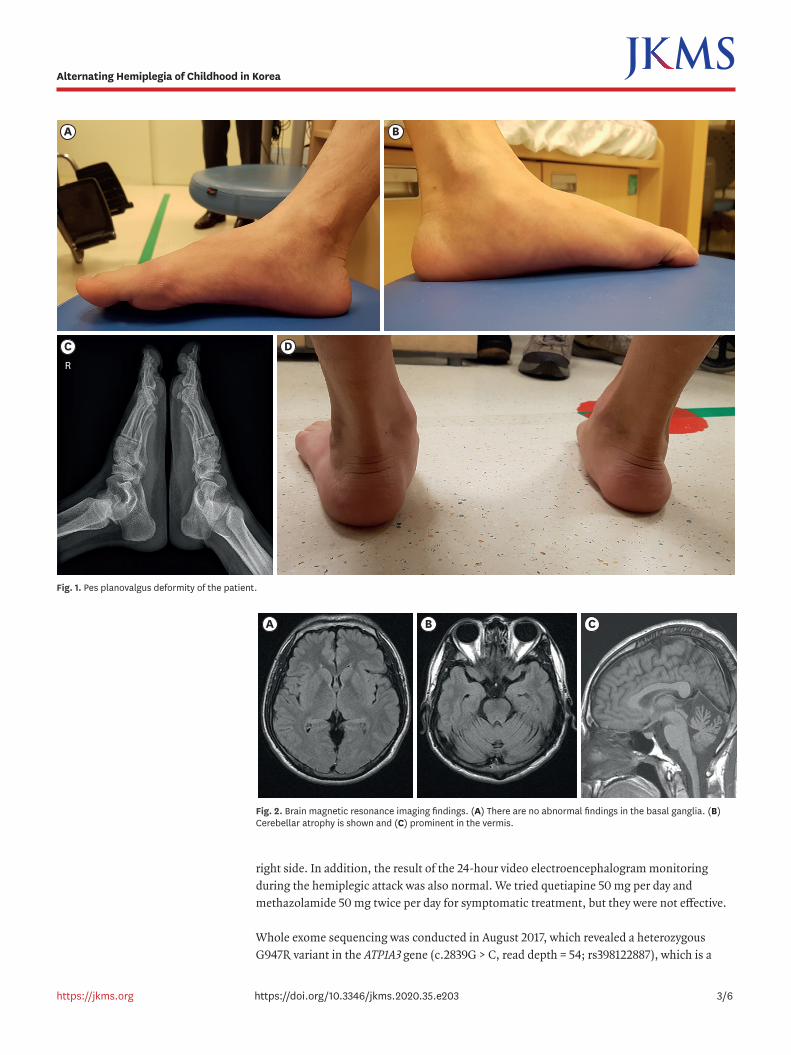
An initial neurological examination revealed motor weakness (motor power on upper and lower extremities: Medical Research Council grade IV- and III, respectively) and dystonia on the right side and peripheral type facial palsy (Supplementary Video 1). There was no hearing loss or ataxic features, and other examination findings were normal. His mini-mental status exam score was 24/30 (−1 point in memory recall, −4 in attention and calculation, and −1 in interlocking pentagon drawing). In addition, the pes planovalgus deformity was identified in his feet (Fig. 1).
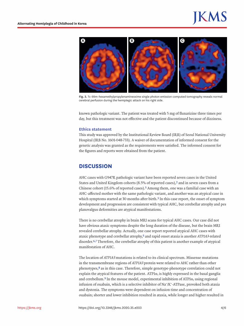
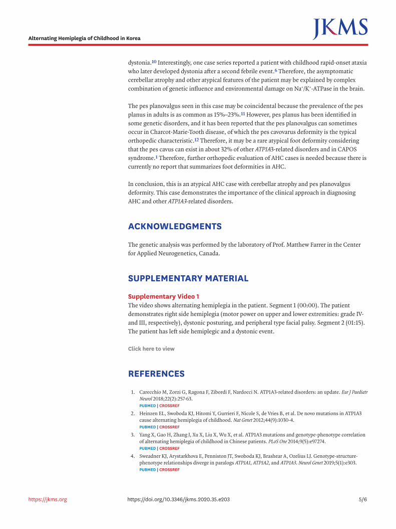
Brain magnetic resonance imaging (MRI) showed cerebellar atrophy (Fig. 2) without any vascular lesions. There was no abnormal finding on Tc-99m-hexamethylpropylenamineoxime single photon emission computed tomography (Fig. 3) during the hemiplegic attack on the
2/6https://jkms.org https://doi.org/10.3346/jkms.2020.35.e203
Alternating Hemiplegia of Childhood in Korea
right side. In addition, the result of the 24-hour video electroencephalogram monitoring during the hemiplegic attack was also normal. We tried quetiapine 50 mg per day and methazolamide 50 mg twice per day for symptomatic treatment, but they were not effective.
Whole exome sequencing was conducted in August 2017, which revealed a heterozygous G947R variant in the ATP1A3 gene (c.2839G > C, read depth = 54; rs398122887), which is a
3/6https://jkms.org https://doi.org/10.3346/jkms.2020.35.e203
Alternating Hemiplegia of Childhood in Korea
BA
DCR
Fig. 1. Pes planovalgus deformity of the patient.
BA C
Fig. 2. Brain magnetic resonance imaging findings. (A) There are no abnormal findings in the basal ganglia. (B) Cerebellar atrophy is shown and (C) prominent in the vermis.
known pathologic variant. The patient was treated with 5 mg of flunarizine three times per day, but this treatment was not effective and the patient discontinued because of dizziness.
Ethics statementThis study was approved by the Institutional Review Board (IRB) of Seoul National University Hospital (IRB No. 1601-048-733). A waiver of documentation of informed consent for the genetic analysis was granted as the requirements were satisfied. The informed consent for the figures and reports were obtained from the patient.
DISCUSSION
AHC cases with G947R pathologic variant have been reported seven cases in the United States and United Kingdom cohorts (8.5% of reported cases),2 and in seven cases from a Chinese cohort (15.6% of reported cases).3 Among them, one was a familial case with an AHC-affected mother with the same pathologic variant, and another was an atypical case in which symptoms started at 30 months after birth.3 In this case report, the onset of symptom development and progression are consistent with typical AHC, but cerebellar atrophy and pes planovalgus deformities are atypical manifestations.
There is no cerebellar atrophy in brain MRI scans for typical AHC cases. Our case did not have obvious ataxic symptoms despite the long duration of the disease, but the brain MRI revealed cerebellar atrophy. Actually, one case report reported atypical AHC cases with ataxic phenotype and cerebellar atrophy,5 and rapid onset ataxia is another ATP1A3-related disorder.6,7 Therefore, the cerebellar atrophy of this patient is another example of atypical manifestation of AHC.
The location of ATP1A3 mutations is related to its clinical spectrum. Missense mutations in the transmembrane regions of ATP1A3 protein were related to AHC rather than other phenotypes,8 as in this case. Therefore, simple genotype-phenotype correlation could not explain the atypical features of the patient. ATP1α3 is highly expressed in the basal ganglia and cerebellum.9 In the mouse model, experimental inhibition of ATP1α3 using regional infusion of ouabain, which is a selective inhibitor of Na+/K+-ATPase, provoked both ataxia and dystonia. The symptoms were dependent on infusion time and concentration of ouabain; shorter and lower inhibition resulted in ataxia, while longer and higher resulted in
4/6https://jkms.org https://doi.org/10.3346/jkms.2020.35.e203
Alternating Hemiplegia of Childhood in Korea
BA C
Fig. 3. Tc-99m-hexamethylpropylenamineoxime single photon emission computed tomography reveals normal cerebral perfusion during the hemiplegic attack on his right side.
dystonia.10 Interestingly, one case series reported a patient with childhood rapid-onset ataxia who later developed dystonia after a second febrile event.6 Therefore, the asymptomatic cerebellar atrophy and other atypical features of the patient may be explained by complex combination of genetic influence and environmental damage on Na+/K+-ATPase in the brain.
The pes planovalgus seen in this case may be coincidental because the prevalence of the pes planus in adults is as common as 15%–23%.11 However, pes planus has been identified in some genetic disorders, and it has been reported that the pes planovalgus can sometimes occur in Charcot-Marie-Tooth disease, of which the pes cavovarus deformity is the typical orthopedic characteristic.12 Therefore, it may be a rare atypical foot deformity considering that the pes cavus can exist in about 32% of other ATP1A3-related disorders and in CAPOS syndrome.1 Therefore, further orthopedic evaluation of AHC cases is needed because there is currently no report that summarizes foot deformities in AHC.
In conclusion, this is an atypical AHC case with cerebellar atrophy and pes planovalgus deformity. This case demonstrates the importance of the clinical approach in diagnosing AHC and other ATP1A3-related disorders.
ACKNOWLEDGMENTS
The genetic analysis was performed by the laboratory of Prof. Matthew Farrer in the Center for Applied Neurogenetics, Canada.
SUPPLEMENTARY MATERIAL
Supplementary Video 1The video shows alternating hemiplegia in the patient. Segment 1 (00:00). The patient demonstrates right side hemiplegia (motor power on upper and lower extremities: grade IV- and III, respectively), dystonic posturing, and peripheral type facial palsy. Segment 2 (01:15). The patient has left side hemiplegic and a dystonic event.
Click here to view
REFERENCES
1. Carecchio M, Zorzi G, Ragona F, Zibordi F, Nardocci N. ATP1A3-related disorders: an update. Eur J Paediatr Neurol 2018;22(2):257-63. PUBMED | CROSSREF
2. Heinzen EL, Swoboda KJ, Hitomi Y, Gurrieri F, Nicole S, de Vries B, et al. De novo mutations in ATP1A3 cause alternating hemiplegia of childhood. Nat Genet 2012;44(9):1030-4. PUBMED | CROSSREF
3. Yang X, Gao H, Zhang J, Xu X, Liu X, Wu X, et al. ATP1A3 mutations and genotype-phenotype correlation of alternating hemiplegia of childhood in Chinese patients. PLoS One 2014;9(5):e97274. PUBMED | CROSSREF
4. Sweadner KJ, Arystarkhova E, Penniston JT, Swoboda KJ, Brashear A, Ozelius LJ. Genotype-structure-phenotype relationships diverge in paralogs ATP1A1, ATP1A2, and ATP1A3. Neurol Genet 2019;5(1):e303. PUBMED | CROSSREF
5/6https://jkms.org https://doi.org/10.3346/jkms.2020.35.e203
Alternating Hemiplegia of Childhood in Korea
5. Sasaki M, Ishii A, Saito Y, Hirose S. Progressive brain atrophy in alternating hemiplegia of childhood. Mov Disord Clin Pract (Hoboken) 2017;4(3):406-11. PUBMED | CROSSREF
6. Schirinzi T, Graziola F, Nicita F, Travaglini L, Stregapede F, Valeriani M, et al. Childhood rapid-onset ataxia: expanding the phenotypic spectrum of ATP1A3 mutations. Cerebellum 2018;17(4):489-93. PUBMED | CROSSREF
7. Sweadner KJ, Toro C, Whitlow CT, Snively BM, Cook JF, Ozelius LJ, et al. ATP1A3 mutation in adult rapid-onset ataxia. PLoS One 2016;11(3):e0151429. PUBMED | CROSSREF
8. Rosewich H, Ohlenbusch A, Huppke P, Schlotawa L, Baethmann M, Carrilho I, et al. The expanding clinical and genetic spectrum of ATP1A3-related disorders. Neurology 2014;82(11):945-55. PUBMED | CROSSREF
9. Bøttger P, Tracz Z, Heuck A, Nissen P, Romero-Ramos M, Lykke-Hartmann K. Distribution of Na/K-ATPase alpha 3 isoform, a sodium-potassium P-type pump associated with rapid-onset of dystonia parkinsonism (RDP) in the adult mouse brain. J Comp Neurol 2011;519(2):376-404. PUBMED | CROSSREF
10. Calderon DP, Fremont R, Kraenzlin F, Khodakhah K. The neural substrates of rapid-onset dystonia-parkinsonism. Nat Neurosci 2011;14(3):357-65. PUBMED | CROSSREF
11. Dare DM, Dodwell ER. Pediatric flatfoot: cause, epidemiology, assessment, and treatment. Curr Opin Pediatr 2014;26(1):93-100. PUBMED | CROSSREF
12. Hoellwarth JS, Mahan ST, Spencer SA. Painful pes planovalgus: an uncommon pediatric orthopedic presentation of Charcot-Marie-Tooth disease. J Pediatr Orthop B 2012;21(5):428-33. PUBMED | CROSSREF
6/6https://jkms.org https://doi.org/10.3346/jkms.2020.35.e203
Alternating Hemiplegia of Childhood in Korea