Embed Size (px)
Citation preview
Journal of Frailty, Sarcopenia and Falls
Case Report Article
Nocardia osteomyelitis in an immunosuppressed patient
Evangelos A. Boulios1, Pinelopi P. Konstantopoulou1, Georgios D. Bailas1, Anastasia K. Klagkou1, Georgia C. Tseliou1, Ioanna Z. Gkoulia2, Athanasios I. Georgountzos1
1Department of Rheumatology, Athens General Hospital «G. Gennimatas», Athens, Greece 2Department of Pediatric Surgery, General Maternity Hospital «Elena Venizelou», Athens, Greece
Introduction
Nocardiosis is an acute, sub acute or chronic disease by Gram positive bacterium similar to fungi which occurs cutaneous, pulmonary and disseminated forms (ex. brain, meninges). It is a rare cause of osteomyelitis and was described for the first time approximately 60 years ago. Treatment of nocardia infection depends on the subtype and antimicrobial options. Includes Trimethoprim - Sulfamethoxazole (TMP-SMX), Penicillin, Quinolone, Amikacin, Cephalosporin and others1.
GPA is characterized by granulomatous necrotizing lesions of upper or/and lower respiratory tract associated with glomerulonephritis which in some cases can progress rapidly. Clinical manifestations include general symptoms (ex. fever, fatigue, weight loss), musculoskeletal symptoms (ex. polyarthritis), upper airway involvement (ex. rhinitis, sinusitis, otitis, nasal septum perforation, subglottic stenosis), pulmonary involvement (cogitated nodules, alveolar hemorrhage), renal involvement, neurological involvement (mononeuritis multiplex, peripheral neuropathy, sensor motor polyneuropathy), skin lesions (purpura, nodules, cutaneous ulcerations). The induction treatment is based on glucocorticoids and combinations of immunosuppressant agent [Rituximab (RTX) or Cyclophosphamide (CYC)] while the maintenance treatment is based on less toxic immunosuppressant agents (Azathioprine or Methotrexate)2,3.
Patients with depressed cellular immunity are at high risk
of Nocardiosis. Only a few case reports of autoimmune disease are related with Nocardia spp. Physicians must always suspect such infections in these immunosuppressed patients because early diagnosis and treatment are the key to therapy1,4.
Case report
A 68 years old Caucasian male with GPA and coronary heart disease under treatment was admitted to our department after hospitalization at internal medicine clinic for further treatment and investigation due to recurrence of the underlying disease.
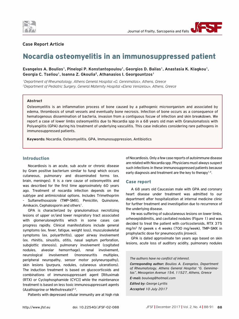
He was suffering of subcutaneous lesions on lower limbs, orheoepididimitis, and cavitated nodules (Figure 1) and was decided to treat the patient with corticosteroids, RTX 375 mg/m2 IV qweek x 4 weeks (700 mg/week), TMP-SMX in prophylactic dose for pneumocystis jirovecii.
GPA is dated approximate ten years ago based on skin lesions, acute loss of auditory acidity, pulmonary nodules
Abstract
Osteomyelitis is an inflammation process of bone caused by a pathogenic microorganism and associated by edema, thrombosis of small vessels and eventually bone necrosis. Infection of bone occurs as a consequence of hematogenous dissemination of bacteria, invasion from a contiguous focuw of infection and skin breakdown. We report a case of lower limbs osteomyelitis due to Nocardia spp in a 68 years old man with Granulomatosis with Polyangiitis (GPA) during his treatment of underlying vasculitis. This case indicates considering rare pathogens in immunosuppressed patients.
Keywords: Nocardia, Osteomyelitis, GPA, Immunosuppression, Antibiotics
The authors have no conflict of interest.
Corresponding author: Boulios A. Evangelos, Department of Rheumatology, Athens General Hospital “G. Gennima-tas”, Mesogeion Avenue 154, 11527, Athens, Greece
E-mail: [email protected]
Edited by: George Lyritis
Accepted 10 July 2017
http://www.jfsf.eu 88JFSF | December 2017 | Vol. 2, No. 4 | 88-91doi: 10.22540/JFSF-02-088
JFSF89
Nocardia osteomyelitis in an immunosuppressed patient
with cavities which was associated with the presence of ANCA specific for proteinase 3 (PR3-ANCA) and findings compatible from lung’s FNA. He was treating with glucosteroids and p.o. cyclophosphamide.
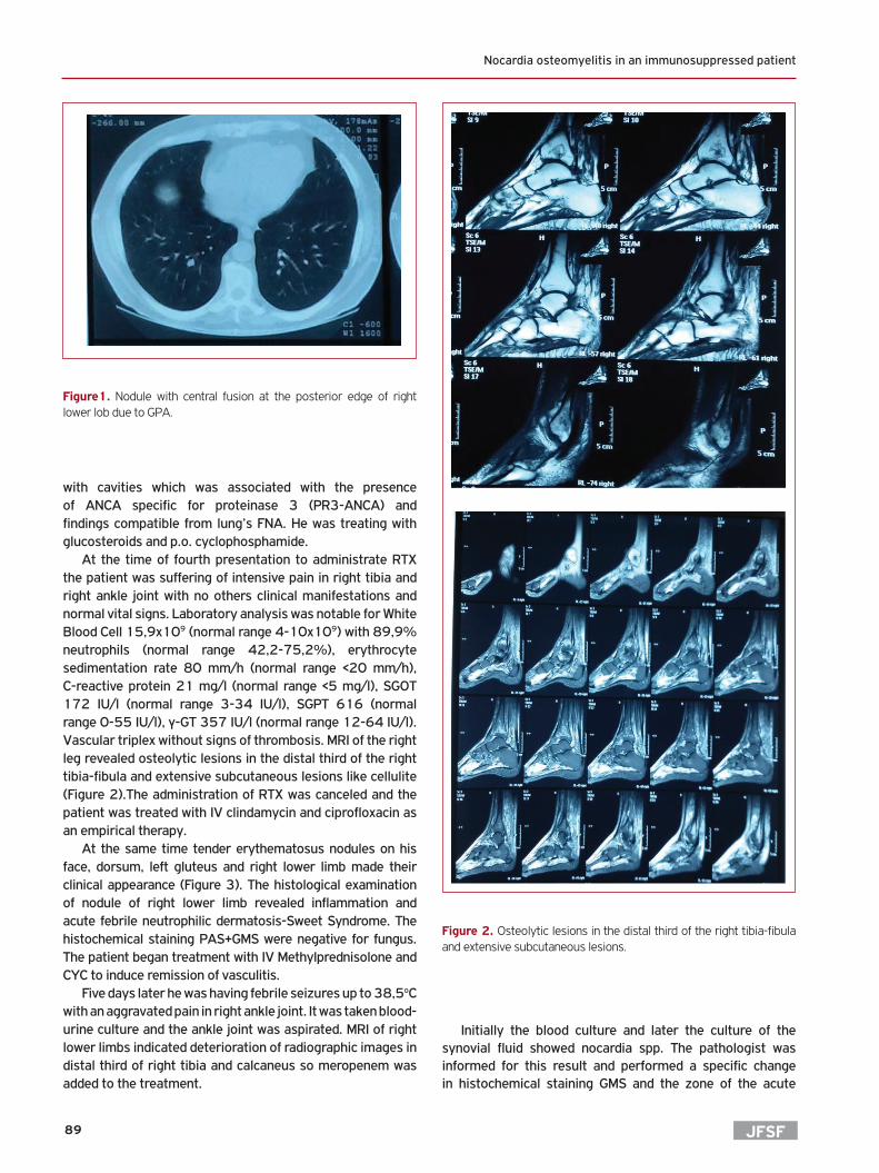
At the time of fourth presentation to administrate RTX the patient was suffering of intensive pain in right tibia and right ankle joint with no others clinical manifestations and normal vital signs. Laboratory analysis was notable for White Blood Cell 15,9x10⁹ (normal range 4-10x109) with 89,9% neutrophils (normal range 42,2-75,2%), erythrocyte sedimentation rate 80 mm/h (normal range <20 mm/h), C-reactive protein 21 mg/l (normal range <5 mg/l), SGOT 172 IU/l (normal range 3-34 IU/l), SGPT 616 (normal range 0-55 IU/l), γ-GT 357 IU/l (normal range 12-64 IU/l).Vascular triplex without signs of thrombosis. MRI of the right leg revealed osteolytic lesions in the distal third of the right tibia-fibula and extensive subcutaneous lesions like cellulite (Figure 2).The administration of RTX was canceled and the patient was treated with IV clindamycin and ciprofloxacin as an empirical therapy.
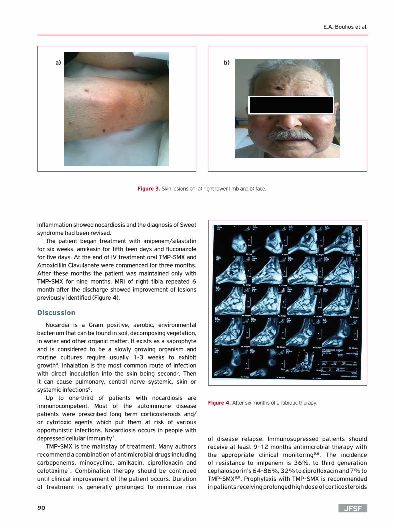
At the same time tender erythematosus nodules on his face, dorsum, left gluteus and right lower limb made their clinical appearance (Figure 3). The histological examination of nodule of right lower limb revealed inflammation and acute febrile neutrophilic dermatosis-Sweet Syndrome. The histochemical staining PAS+GMS were negative for fungus. The patient began treatment with IV Methylprednisolone and CYC to induce remission of vasculitis.
Five days later he was having febrile seizures up to 38,5oC with an aggravated pain in right ankle joint. It was taken blood-urine culture and the ankle joint was aspirated. MRI of right lower limbs indicated deterioration of radiographic images in distal third of right tibia and calcaneus so meropenem was added to the treatment.
Initially the blood culture and later the culture of the synovial fluid showed nocardia spp. The pathologist was informed for this result and performed a specific change in histochemical staining GMS and the zone of the acute
Figure1. Nodule with central fusion at the posterior edge of right lower lob due to GPA.
Figure 2. Osteolytic lesions in the distal third of the right tibia-fibula and extensive subcutaneous lesions.
JFSF90
E.A. Boulios et al.
inflammation showed nocardiosis and the diagnosis of Sweet syndrome had been revised.
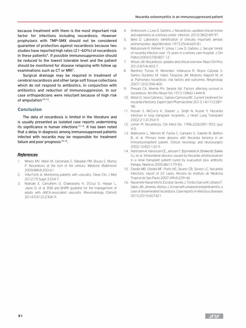
The patient began treatment with imipenem/silastatin for six weeks, amikasin for fifth teen days and fluconazole for five days. At the end of IV treatment oral TMP-SMX and Amoxicillin Clavulanate were commenced for three months. After these months the patient was maintained only with TMP-SMX for nine months. MRI of right tibia repeated 6 month after the discharge showed improvement of lesions previously identified (Figure 4).
Discussion
Nocardia is a Gram positive, aerobic, environmental bacterium that can be found in soil, decomposing vegetation, in water and other organic matter. It exists as a saprophyte and is considered to be a slowly growing organism and routine cultures require usually 1-3 weeks to exhibit growth4. Inhalation is the most common route of infection with direct inoculation into the skin being second5. Then it can cause pulmonary, central nerve systemic, skin or systemic infections6.
Up to one-third of patients with nocardiosis are immunocompetent. Most of the autoimmune disease patients were prescribed long term corticosteroids and/or cytotoxic agents which put them at risk of various opportunistic infections. Nocardiosis occurs in people with depressed cellular immunity7.
TMP-SMX is the mainstay of treatment. Many authors recommend a combination of antimicrobial drugs including carbapenems, minocycline, amikacin, ciprofloxacin and cefotaxime1. Combination therapy should be continued until clinical improvement of the patient occurs. Duration of treatment is generally prolonged to minimize risk
of disease relapse. Immunosupressed patients should receive at least 9-12 months antimicrobial therapy with the appropriate clinical monitoring5,6. The incidence of resistance to imipenem is 36%, to third generation cephalosporin’s 64-86%, 32% to ciprofloxacin and 7% to TMP-SMX8,9. Prophylaxis with TMP-SMX is recommended in patients receiving prolonged high dose of corticosteroids
Figure 3. Skin lesions on: a) right lower limb and b) face.
a) b)
Figure 4. After six months of antibiotic therapy.
JFSF91
Nocardia osteomyelitis in an immunosuppressed patient
because treatment with them is the most important risk factor for infections including nocardiosis. However prophylaxis with TMP-SMX should not be considered guarantee of protection against nocardiosis because two studies have reported high rates (21-60%) of nocardiosis in these patients4. If possible immunosuppression should be reduced to the lowest tolerable level and the patient should be monitored for disease relapsing with follow up examinations such as CT or MRI7.
Surgical drainage may be required in treatment of cerebral nocardiosis and other large soft tissue collections which do not respond to antibiotics, in conjunction with antibiotics and reduction of immunosuppresion. In our case orthopedicians were reluctant because of high risk of amputation10-12.
Conclusion
The data of nocardiosis is limited in the literature and is usually presented as isolated case reports undermining its significance in human infections13,14. It has been noted that a delay in diagnosis among immunosuppresed patients infected with nocardia may be responsible for treatment failure and poor prognosis15,16.
References
1. Minero MV, Marin M, Cercenado E, Rabadan PM, Bouza E, Munoz P. Nocardiosis at the turn of the century. Medicine (Baltimore) 2009;88(4):250-61.
2. Villa-Forte A. Monitoring patients with vasculitis. Cleve Clin J Med 2012;79 Suppl 3:S34-7.
3. Ntatsaki E, Carruthers D, Chakravarty K, D’Cruz D, Harper L, Jayne D, et al. BSR and BHPR guideline for the management of adults with ANCA-associated vasculitis. Rheumatology (Oxford) 2014;53(12):2306-9.
4. Ambrosioni J, Lew D, Garbino J. Nocardiosis: updated clinical review and experience at a tertiary center. Infection. 2010;38(2):89-97.
5. Berd D. Laboratory identification of clinically important aerobic actinomycetes. Appl Microbiol 1973;25(4):665-81.
6. Matulionyte R, Rohner P, Uckay I, Lew D, Garbino J. Secular trends of nocardia infection over 15 years in a tertiary care hospital. J Clin Pathol 2004;57(8):807-12.
7. Wilson JW. Nocardiosis: updates and clinical overview. Mayo Clin Proc 2012;87(4):403-7.
8. Martinez Tomas R, Menendez Villanueva R, Reyes Calzada S, Santos Durantez M, Valles Tarazona JM, Modesto Alapont M, et al. Pulmonary nocardiosis: risk factors and outcomes. Respirology 2007;12(3):394-400.
9. Presant CA, Wiernik PH, Serpick AA. Factors affecting survival in nocardiosis. Am Rev Respir Dis 1973;108(6):1444-8.
10. Welsh O, Vera-Cabrera L, Salinas-Carmona MC. Current treatment for nocardia infections. Expert Opin Pharmacother 2013;14(17):2387-98.
11. Husain S, McCurry K, Dauber J, Singh N, Kusne S. Nocardia infection in lung transplant recipients. J Heart Lung Transplant 2002;21(3):354-9.
12. Lerner PI. Nocardiosis. Clin Infect Dis. 1996;22(6):891-903; quiz 4-5.
13. Malincarne L, Marroni M, Farina C, Camanni G, Valente M, Belfiori B, et al. Primary brain abscess with Nocardia farcinica in an immunocompetent patient. Clinical neurology and neurosurgery 2002;104(2):132-5.
14. Hartmann A, Halvorsen CE, Jenssen T, Bjorneklett A, Brekke IB, Bakke SJ, et al. Intracerebral abscess caused by Nocardia otitidiscaviarum in a renal transplant patient--cured by evacuation plus antibiotic therapy. Nephron 2000;86(1):79-83.
15. Chedid MB, Chedid MF, Porto NS, Severo CB, Severo LC. Nocardial infections: report of 22 cases. Revista do Instituto de Medicina Tropical de Sao Paulo 2007;49(4):239-46.
16. Navarrete-Navarrete N, Escobar Sevilla J, Toribio Garcia M, Urbano F, Sabio JM, Jimenez-Alonso J. A man with unilateral endophthalmitis: a case of disseminated nocardiosis. Case reports in infectious diseases 2015;2015:607421.


![Nocardia Brain Abscess in an Immunocompetent Patient · Nocardia species are a rare cause of cerebral abscess [3]. Nocardia brain abscess appears in a gradually progressive mass lesion,](https://img.pdfslide.net/doc/110x75/5f9d9fa5c479af2f1c584bd9/nocardia-brain-abscess-in-an-immunocompetent-patient-nocardia-species-are-a-rare.jpg)