Embed Size (px)
Citation preview
322
Korean J Ophthalmol 2010;24(5):322-324DOI: 10.3341/kjo.2010.24.5.322pISSN: 1011-8942 eISSN: 2092-9382
Case Report
Complete Visual Recovery after Mycotic Aneurysm Embolization Complicated by Cavernous Sinus Thrombophlebitis
Ungsoo Samuel Kim1, Ji Soo Kim2, O-Ki Kwon3, Jeong-Min Hwang4
1Department of Ophthalmology, Kim’s Eye Hospital, Konyang University College of Medicine, Seoul, Korea Departments of 2Neurology, 3Neurosurgery, and 4Ophthalmology, Seoul National University Bundang Hospital,
Seoul National University College of Medicine, Seongnam, Korea
A 62-year-old woman has been suffered from cavernous sinus thrombophlebitis which was confirmed by four-ves-sel angiography, orbit magnetic resonance imaging, and blood culture. Three weeks after recovery of cavernous si-nus thrombophlebitis, right eye proptosis and complete third, fourth, and sixth cranial nerve palsies developed. Best-corrected visual acuity decreased to 20/70 in the right eye. Repeat magnetic resonance imaging demonstrated a 1.5-cm-sized mass in the right cavernous sinus, suspicious for mycotic aneurysm. Amphotericin B supplementa-tion was begun and was followed by successful transarterial Guglielmi detachable coil embolization. Four months later, extraocular movement was normalized, and visual acuity improved to 20/25 in the right eye.
Key Words: Cavernous sinus thrombosis, Guglielmi detachable coils embolization, Mycotic aneurysm
ⓒ2010 The Korean Ophthalmological SocietyThis is an Open Access article distributed under the terms of the Creative Commons Attribution Non-Commercial License (http://creativecommons.org/licenses/by-nc/3.0/) which permits unrestricted non-commercial use, distribution, and reproduction in any medium, provided the original work is properly cited.
Received: February 23, 2009 Accepted: May 14, 2009
Reprint requests to Jeong-Min Hwang. Department of Ophthalmology, Seoul National University Bundang Hospital, #300 Gumi-dong, Bundang-gu, Seongnam 463-707, Korea. Tel: 82-31-787-7372, Fax: 82-31-787-4057, E-mail: [email protected]
Mycotic aneurysm is a rare complication of cavernous si-nus thrombosis and may be treated with antibiotics, carotid ligation, internal carotid artery balloon occlusion, or Guglielmi detachable coil (GDC) embolization [1]. However, a complete visual recovery from a mycotic aneur-ysm associated with cavernous sinus thrombophlebitis after endovascular coiling has not been reported in the English literature. We describe a case of mycotic aneurysm compli-cated by cavernous sinus thrombosis, which was success-fully treated with coil embolization and resulted in a com-plete visual recovery.
Case Report
A 62-year-old woman presented with diplopia for a peri-od of one day. She had a continuous headache for five days and fever of 38.7℃. She was receiving medication for hy-pertension but denied any history of trauma, viral infection, or any other systemic disease.
Best corrected visual acuity (BCVA) was 20/25 in the
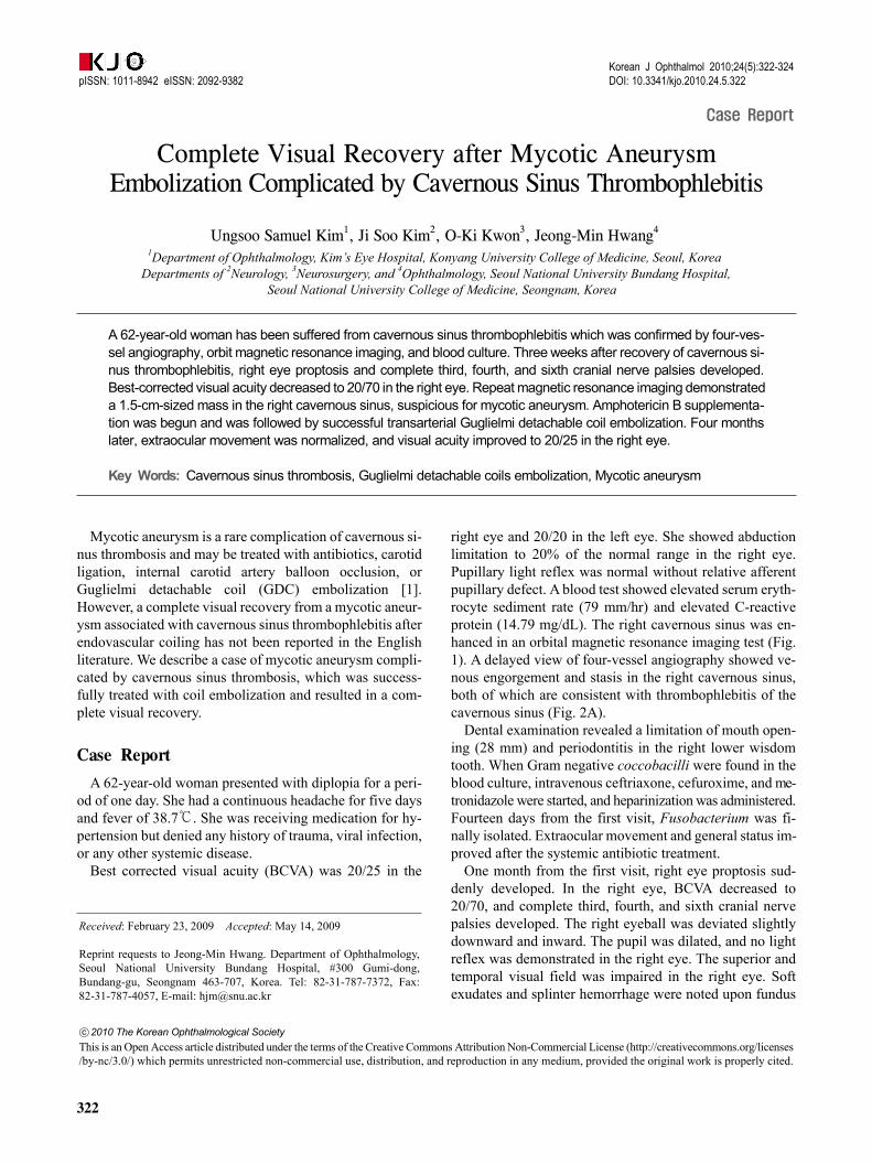
right eye and 20/20 in the left eye. She showed abduction limitation to 20% of the normal range in the right eye. Pupillary light reflex was normal without relative afferent pupillary defect. A blood test showed elevated serum eryth-rocyte sediment rate (79 mm/hr) and elevated C-reactive protein (14.79 mg/dL). The right cavernous sinus was en-hanced in an orbital magnetic resonance imaging test (Fig. 1). A delayed view of four-vessel angiography showed ve-nous engorgement and stasis in the right cavernous sinus, both of which are consistent with thrombophlebitis of the cavernous sinus (Fig. 2A).
Dental examination revealed a limitation of mouth open-ing (28 mm) and periodontitis in the right lower wisdom tooth. When Gram negative coccobacilli were found in the blood culture, intravenous ceftriaxone, cefuroxime, and me- tronidazole were started, and heparinization was administered. Fourteen days from the first visit, Fusobacterium was fi-nally isolated. Extraocular movement and general status im-proved after the systemic antibiotic treatment.
One month from the first visit, right eye proptosis sud-denly developed. In the right eye, BCVA decreased to 20/70, and complete third, fourth, and sixth cranial nerve palsies developed. The right eyeball was deviated slightly downward and inward. The pupil was dilated, and no light reflex was demonstrated in the right eye. The superior and temporal visual field was impaired in the right eye. Soft exudates and splinter hemorrhage were noted upon fundus

US Kim, et al. Mycotic Aneurysm in Cavernous Sinus Thrombosis
323
A B
Fig. 1. Gadolinium-enhanced magnetic resonance imaging shows enhancement in the right cavernous sinus in the fat-sup-pressed T1-weighted axial image (A) and T1-weighted sagittal image (B).
A B
C D
Fig. 2. (A) Four-vessel angiography reveals that the cavernous sinus is not filled in the delayed views, and venous engorgement and stasis are shown (arrow). (B) Soft exudates and splinter hemorrhage are noted upon fundus examination. (C) Mycotic aneurysm is located at the cav-ernous sinus portion upon digital four-vessel angiography (arrow). (D) Guglielmi detachable coil embolization is performed successfully with-out the remaining aneurysm (arrow).

Korean J Ophthalmol Vol.24, No.5, 2010
324
examination of the right eye (Fig. 2B). Four-vessel angio-graphy demonstrated a newly defined 1.5-cm-sized mass in right cavernous sinus, suspicious for mycotic aneurysm (Fig. 2C). The patient was treated with amphotericin B (1 mg/kg).
After systemic status improved, the mycotic aneurysm was completely occluded with transarterial GDC (Boston Scientific Corp., Fremont, CA, USA) embolization without complication. Ten days after the embolization, follow-up angiography showed no evidence of residual aneurysm in the cavernous sinus (Fig. 2D). Four months later, extra-ocular movement was normalized, and visual acuity im-proved to 20/25 in the right eye.
Discussion
The mechanism of mycotic aneurysm formation is thought to be due to hematogenous spread of infectious mi-croemboli to the vasa vasorum, occlusion by an infected embolus of a distal artery lumen, or contiguous spread from an extavascular infection, such as sinusitis, orbital in-fection, or middle ear infection [2]. However, a case com-plicated by cavernous sinus thrombophlebitis is much rarer, with about only 20 cases reported.
Mycotic aneurysm in the cavernous sinus may present as ophthalmoplegia, diplopia, orbital swelling, and/or visual disturbance. Quisling et al. [3] have reported a case of blindness which resulted from enlarging mycotic aneurysm after cavernous sinus thrombosis.
Several treatments, such as antibiotic therapy, carotid li-gation, internal carotid artery balloon occlusion, and GDC embolization have been adopted to treat mycotic aneurysms in the cavernous sinus [1,4]. The GDC embolization, first used for patients with aneurysm in 1991 [5], prevents blood flow into the aneurysmal sac by filling the aneurysm with coils and thrombus. Although GDC embolization may be
complicated by thromboembolism, intra-operative aneur-ysm rupture, coil migration, bacteremia and vasospasm, our patient showed a complete recovery of vision and motility without complication.
This is the first case of complete visual recovery after my-cotic aneurysm embolization complicated by cavernous si-nus thrombophlebitis showing a possibility of successful treatment with GDC embolization without any complications.
Conflict of Interest
No potential conflict of interest relevant to this article was reported.
Acknowledgements
This work was supported by grant No. R01-2005-000- 10875-0 from the Basic Research Program of the Korea Science & Engineering Foundation.
References 1. Hurst RW, Choi IS, Persky M, Kupersmith M. Mycotic
aneurysms of the intracavernous carotid artery: a case report and review of the literature. Surg Neurol 1992;37:142-6.
2. Wilson WR, Hawrych A, Olan W. Rapid development of bi-lateral internal carotid artery aneurysm from sphenoid sinus aspergillosis. Skull Base Surg 1998;8:211-4.
3. Quisling SV, Mawn LA, Larson TC 3rd. Blindness associated with enlarging mycotic aneurysm after cavernous sinus thrombosis. Ophthalmology 2003;110:2036-9.
4. Koebbe CJ, Veznedaroglu E, Jabbour P, Rosenwasser RH. Endovascular management of intracranial aneurysms: cur-rent experience and future advances. Neurosurgery 2006;59 (5 Suppl 3):S93-102.
5. Guglielmi G, Vinuela F, Dion J, Duckwiler G. Electrothrom- bosis of saccular aneurysms via endovascular approach. Part 2: Preliminary clinical experience. J Neurosurg 1991;75: 8-14.