Embed Size (px)
Citation preview
Dow
nloa
ded
from
http
://w
ww
.jri.i
r
Case Report
J Reprod Infertil. 2017;18(2):257-260
Maternal and Fetal Tuberous Sclerosis: Do We Know Enough as an Obstetrician? Nalini Sharma 1, Shriram Sharma 2, Jion Lalnunnem Thiek 1, Santa Singh Ahanthem 1, Arnab Kalita 3, Donboklang Lynser 3 1- Department of Obstetrics and Gynecology, North Eastern Indira Gandhi Regional Institute of Health and Medical Sciences Shillong, Meghalaya, India 2- Department of Neurology, North Eastern Indira Gandhi Regional Institute of Health and Medical Sciences Shillong, Meghalaya, India 3. Department of Radiology, North Eastern Indira Gandhi Regional Institute of Health and Medical Sciences Shillong, Meghalaya, India
Abstract Background: Tuberous sclerosis, also known as tuberous sclerosis complex (TSC), is a rare genetic condition that mainly causes hamartomas to develop in different parts of the body. TSC, an autosomal dominant trait with variable penetrance, can adversely affect maternal and fetal outcome. Case Presentation: In this paper, a case of maternal and fetal tuberous sclerosis having fetal cardiac rhabdomyoma detected in utero at 26 weeks was reported who subsequently had fetal demise at 31 weeks. Conclusion: Tuberous sclerosis is a rare genetic condition that mainly causes devel-opment of hamartomas. In tuberous sclerosis, a cardiac rhabdomyoma is the only sign that can be detected prenatally. In maternal tuberous sclerosis, fetal ECHO is advisable after 24 weeks. A pregnancy complicated by maternal or fetal tuberous sclerosis deserves careful observation and the fetus should undergo prenatal fetal Doppler echocardiography and if possible magnetic resonance imaging for evalua-tion of other fetal structures including brain and renal parenchyma, so that parents can be counseled regarding its future prognostic implications. Tuberous sclerosis can lead to poor fetal outcome including intrauterine fetal death; hence regular antenatal follow up is required. Genetic counseling is recommended for couples who have a family history of tuberous sclerosis and who want to have children. Prenatal diagno-sis is available for families with a known gene mutation or history of this condition. Keywords: Pregnancy, Tuberous sclerosis complex, Tuberous sclerosis. To cite this article: Sharma N, Sharma SR, Thiek JL, Ahanthem SS, Kalita A, Lynser D. Maternal and Fetal Tuberous Sclerosis: Do We Know Enough as an Obstetrician?. J Reprod Infertil. 2017;18(2):257-260.
Introduction uberous sclerosis, also known as tuberous sclerosis complex (TSC), is a rare genetic condition that causes mainly development of
hamartomas in different parts of the body. TSC, an autosomal dominant trait with variable pene-trance, can adversely affect maternal and fetal outcome.
This case is presented because of its rarity (tu-berous sclerosis with pregnancy) and it can ad-versely affect maternal and fetal outcome. The obstetrician should know about this entity.
Case Presentation A 23 year old primigravida was referred to the
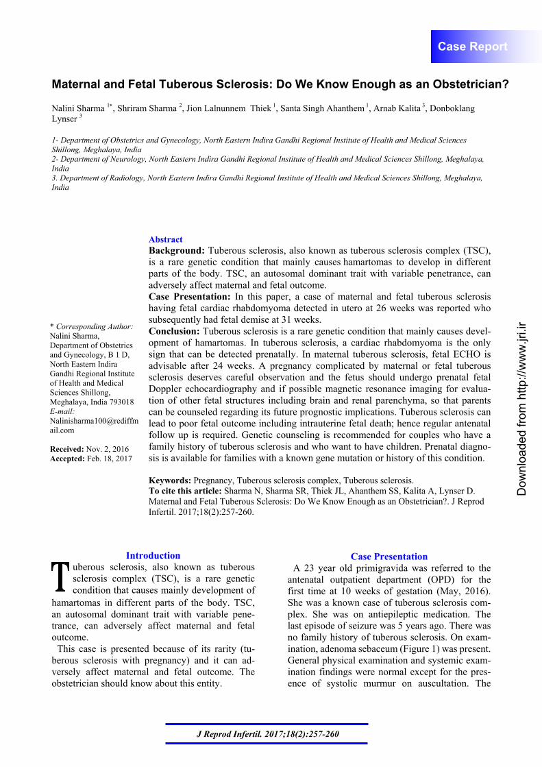
antenatal outpatient department (OPD) for the first time at 10 weeks of gestation (May, 2016). She was a known case of tuberous sclerosis com-plex. She was on antiepileptic medication. The last episode of seizure was 5 years ago. There was no family history of tuberous sclerosis. On exam-ination, adenoma sebaceum (Figure 1) was present. General physical examination and systemic exam-ination findings were normal except for the pres-ence of systolic murmur on auscultation. The
* Corresponding Author: Nalini Sharma, Department of Obstetrics and Gynecology, B 1 D, North Eastern Indira Gandhi Regional Institute of Health and Medical Sciences Shillong, Meghalaya, India 793018 E-mail: [email protected] Received: Nov. 2, 2016 Accepted: Feb. 18, 2017
Dow
nloa
ded
from
http
://w
ww
.jri.i
r
258 J Reprod Infertil, Vol 18, No 2, Apr-Jun 2017
Maternal and Fetal Tuberous SclerosisJRI
computed tomography scan (CT scan) of brain per-formed before pregnancy showed multiple paren-chymal/perinuclear subependymal calcification, focal encephalomalacia. Echocardiography (ECHO) revealed a small patent ductus arteriosus (PDA) of 2 mm with left to right shunt. Fundoscopic exami-nation revealed no abnormality. All the other rou-tine antenatal investigations were normal. She was on regular follow up with regular consultation with neurologist. The anomaly scans which were performed at 13 and 20 weeks of gestation re-vealed no gross congenital anomaly. At 27 weeks of gestation while performing ultrasound for
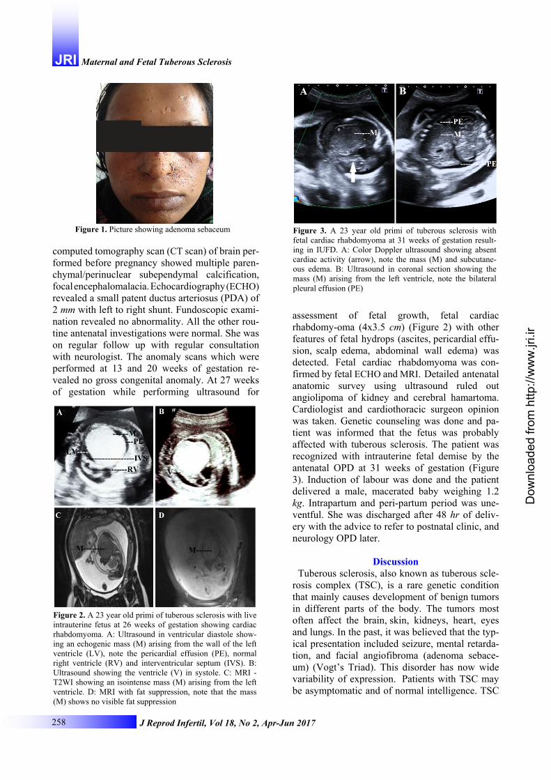
assessment of fetal growth, fetal cardiac rhabdomy-oma (4x3.5 cm) (Figure 2) with other features of fetal hydrops (ascites, pericardial effu-sion, scalp edema, abdominal wall edema) was detected. Fetal cardiac rhabdomyoma was con-firmed by fetal ECHO and MRI. Detailed antenatal anatomic survey using ultrasound ruled out angiolipoma of kidney and cerebral hamartoma. Cardiologist and cardiothoracic surgeon opinion was taken. Genetic counseling was done and pa-tient was informed that the fetus was probably affected with tuberous sclerosis. The patient was recognized with intrauterine fetal demise by the antenatal OPD at 31 weeks of gestation (Figure 3). Induction of labour was done and the patient delivered a male, macerated baby weighing 1.2 kg. Intrapartum and peri-partum period was une-ventful. She was discharged after 48 hr of deliv-ery with the advice to refer to postnatal clinic, and neurology OPD later.
Discussion
Tuberous sclerosis, also known as tuberous scle-rosis complex (TSC), is a rare genetic condition that mainly causes development of benign tumors in different parts of the body. The tumors most often affect the brain, skin, kidneys, heart, eyes and lungs. In the past, it was believed that the typ-ical presentation included seizure, mental retarda-tion, and facial angiofibroma (adenoma sebace-um) (Vogt’s Triad). This disorder has now wide variability of expression. Patients with TSC may be asymptomatic and of normal intelligence. TSC
Figure 2. A 23 year old primi of tuberous sclerosis with live intrauterine fetus at 26 weeks of gestation showing cardiac rhabdomyoma. A: Ultrasound in ventricular diastole show-ing an echogenic mass (M) arising from the wall of the left ventricle (LV), note the pericardial effusion (PE), normal right ventricle (RV) and interventricular septum (IVS). B: Ultrasound showing the ventricle (V) in systole. C: MRI -T2WI showing an isointense mass (M) arising from the left ventricle. D: MRI with fat suppression, note that the mass (M) shows no visible fat suppression
Figure 3. A 23 year old primi of tuberous sclerosis with fetal cardiac rhabdomyoma at 31 weeks of gestation result-ing in IUFD. A: Color Doppler ultrasound showing absent cardiac activity (arrow), note the mass (M) and subcutane-ous edema. B: Ultrasound in coronal section showing the mass (M) arising from the left ventricle, note the bilateral pleural effusion (PE)
Figure 1. Picture showing adenoma sebaceum
Dow
nloa
ded
from
http
://w
ww
.jri.i
r
J Reprod Infertil, Vol 18, No 2, Apr-Jun 2017 259
Sharma N, et al. JRI affects all races without a clear-cut predominance. TSC affects both sexes equally and it can present at any age. Its prevalence is around 1 in 6000-9000 individuals (1).
The inheritance of TSC is an autosomal domi-nant trait with variable penetrance. Tuberous scle-rosis is caused by mutations in either the TSC1 or TSC2 gene. These genes are involved in regulat-ing cell growth, and the mutations lead to uncon-trolled growth and multiple tumors throughout the body. Only one of the genes needs to be affected for TSC to be present. The TSC1 gene is located on chromosome 9 and is called the hamartin gene. The other gene, TSC2, is located on chromosome 16 and is called the tuberin gene (1, 2).
It can be inherited from one parent with TSC or can result from a spontaneous genetic mutation. Children have a 50 percent chance of inheriting TSC if one of their parents has this condition. At this point, only one-third of TSC cases are known to be inherited. The other two-thirds result from a spontaneous and unpredictable mutation occurring during conception or very early development of the human embryo. In infant cardiac involvement and seizure are common presenting signs where dermatological, pulmonary or renal involvement may lead to diagnosis in older individuals. It can adversely impact fetal and maternal health. Preg-nancy can be complicated by preeclampsia, in-trauterine growth retardation, preterm labour, pre-term premature rupture of membrane, oligohydr-amnios, polyhydramnios, hydrops, abruption, hae-morrhage from rupture renal tumor, renal failure and fetal demise (3, 4). Fetus may have rhabdo-myoma and/or intracranial tubers (4). However, in one of the case series, fetal outcome was good (5) because none of the fetuses were affected by tu-berous sclerosis.
In mother, complications of neurological in-volvement are the most common causes of mortal-ity and morbidity. These are due chiefly to intrac-table epilepsy, status epilepticus, and subependy-mal giant cell astrocytoma (SEGA) with associated hydrocephalus. Renal complications are the next most frequent cause of morbidity and death. Less common are cardiac arrhythmias (which can pre-sent with sudden, unexplained death), congestive heart failure, and end-stage lung disease. Renal involvement appears to be the single most im-portant prognostic factor in pregnancies with tu-berous sclerosis. Renal evaluation should be per-formed in any patient who presents for preconcep-tion counseling. In tuberous sclerosis, a cardiac
rhabdomyoma is the only sign that can be detect-ed prenatally by ultrasound. In maternal tuberous sclerosis, fetal ECHO can be advisable after 22 weeks. A pregnancy complicated by maternal or fetal tuberous sclerosis deserves careful observa-tion and the fetus should undergo prenatal fetal Doppler echocardiography and if possible an MRI for evaluation of other fetal structures including brain and renal parenchyma, so that parents can be counseled regarding its future prognostic implica-tions (6).
Fetal cardiac rhabdomyomas are often benign and have a tendency to regress. It can occasionally induce poor outcome and the need for surgery depends on the patient's clinical presentation (7). Diagnosis is usually made on an obstetric ultraso-nography between 21 to 30 weeks. In our case, it was diagnosed at 26 weeks by obstetric ultraso-nography and fetal ECHO. Tuberous sclerosis can lead to poor fetal outcome including intrauterine fetal death; hence regular antenatal follow up is required. Genetic counseling is recommended for couples who have a family history of tuberous sclerosis and who want to have children. Prenatal diagnosis is available for families with a known gene mutation or history of this condition. How-ever, tuberous sclerosis often appears as a new DNA mutation. These cases are not preventable.
Conclusion Tuberous sclerosis is a rare genetic condition
that mainly causes development of hamartomas. It can adversely affect maternal and fetal outcome. In tuberous sclerosis, a cardiac rhabdomyoma is the only sign that can be detected prenatally. In maternal tuberous sclerosis, fetal ECHO is advis-able after 22 weeks.
A pregnancy complicated by maternal or fetal tuberous sclerosis deserves careful observation and the fetus should undergo prenatal fetal Dop-pler echocardiography and if possible an MRI for evaluation of other fetal structures including brain and renal parenchyma, so that parents can be counseled regarding its future prognostic implica-tions. Tuberous sclerosis can lead to poor fetal outcome including intrauterine fetal death; hence regular antenatal follow up is required. Genetic counseling is recommended for couples who have a family history of tuberous sclerosis and who want to have children. Prenatal diagnosis is avail-able for families with a known gene mutation or history of this condition.
Dow
nloa
ded
from
http
://w
ww
.jri.i
r
260 J Reprod Infertil, Vol 18, No 2, Apr-Jun 2017
Maternal and Fetal Tuberous SclerosisJRI
Conflict of Interest Authors declare no conflict of interest.
References 1. Daroff RB, Fenichel GM, Jankovic J, Mazziotta JC.
Bradley’s Neurology in clinical practice. 6 th ed. Philadelphia: Elsevier; 2012. p. 1508-14.
2. Agrawal SN, Kulkarni YA, Deshmukh YR, Jane SD. Tuberous Sclerosis in Pregnancy. Our Dermatol On-line. 2014;5(2):160-2.
3. Gupta N, Singh N, Sarangi S, Dalmia S, Mittal S. Fe-tal cardiac rhabdomyoma with maternal tuberous scle-rosis complicating pregnancy. Arch Gynecol Obstet. 2008;278(2):169-70.
4. King JA, Stamilio DM. Maternal and fetal tuberous
sclerosis complicating pregnancy: a case report and overview of the literature. Am J Perinatol. 2005;22 (2):103-8.
5. Bonebrake L, Rai K, Yankowitz J. Outcomes of preg-nancies complicated by maternal tuberous sclerosis. Proc Obstet Gynecol. 2012;2(3):1-2.
6. Khanna PC, Godinho S, Pungavkar SA, Patkar DP. Ultrafast MRI in the prenatal diagnosis of Bourne ville's tuberous sclerosis. Neurol India. 2005;53(3): 349-50.
7. Pipitone S, Mongiovì M, Grillo R, Gagliano S, Spe-randeo V. Cardiac rhabdomyoma in intrauterine life: clinical features and natural history. A case series and review of published reports. Ital Heart J. 2002;3 (1):48-52.