Embed Size (px)
Citation preview
Case ReportMilwaukee Shoulder Syndrome
Channa Vasanth Nadarajah and Immo Weichert
Department of Acute Medicine, The Ipswich Hospital NHS Trust, Ipswich, Suffolk IP4 5PD, UK
Correspondence should be addressed to Channa Vasanth Nadarajah; channa vasanth [email protected]
Received 16 September 2013; Accepted 5 December 2013; Published 16 January 2014
Academic Editors: R. Cevik, P. Njobvu, and A. Zoli
Copyright © 2014 C. V. Nadarajah and I. Weichert. This is an open access article distributed under the Creative CommonsAttribution License, which permits unrestricted use, distribution, and reproduction in any medium, provided the original work isproperly cited.
Milwaukee shoulder syndrome (MSS) is a rare destructive, calcium phosphate crystalline arthropathy. It encompasses an effusionthat is noninflammatory with numerous aggregates of calcium hydroxyapatite crystals in the synovial fluid, associated with rotatorcuff defects.We describe a patient that presentedwith recurrent shoulder pain and swellingwith characteristic radiographic changesand MSS was confirmed on aspiration of the synovial fluid.
1. Case
An 89-year-old lady presented to themedical assessment unitwith a four-day history of melaena associated with shortnessof breath on exertion, postural dizziness, and lethargy. Thiswas secondary to esophagitis and gastric erosions. During herhospital stay she developed a rapid onset, painful swelling ofher right shoulder with a limited range of movement. Therewas no history of recent trauma to the right shoulder. Thiswas her third presentation; previous ones had been followedup by her general practitioner.
Past medical history included well-controlled asthma,hypertension, and atrial fibrillation. There was no history ofdiabetes, hyperparathyroidism, or syphilis. She was an ex-smoker with a 10-pack-year history and consumed minimalamounts of alcohol only.
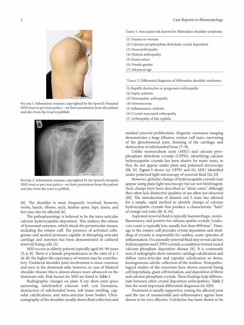
On examination, she was pale, with no rashes or bruises.There was a large right shoulder effusion. Both active andpassive movements were limited. Figure 1 shows the patient’slarge right shoulder swelling which was warm to touch andtender with no erythema.
Plain radiograph of the affected shoulder showed jointspace narrowing, subchondral sclerosis, destruction of sub-chondral bone, soft-tissue swelling, capsular calcifications,and intra-articular loose bodies (Figure 2).
Arthrocentesis was performed and 250mLs of haem-orrhagic fluid was aspirated. Analysis of the synovial fluidshowed a noninflammatory cell count with leukocytes721/mm3 which were predominantly neutrophils. Gram stain
was negative; no organisms were cultured and cytologyanalysis was negative, as was VDRL on the synovial fluid.The aspirate stained bright orange-red with alizarin red S,indicating the presence of calcium hydroxyapatite crystals.The diagnosis of Milwaukee shoulder syndrome was madeand she was treated conservatively with colchicine andphysiotherapy with a good outcome.
2. Discussion
The term Milwaukee shoulder syndrome (MSS) was firstused in 1981 to describe four elderly women in Milwaukee inthe state of Wisconsin, USA, with recurrent bilateral shoul-der effusions, radiographic evidence of severe destructivechanges of the glenohumeral joints, and massive tears ofthe rotator cuff [1–3]. The term rapid destructive arthritis ofthe shoulder was introduced in 1982 to describe six elderlyfemales with spontaneous large glenohumeral effusions, mildpain, and tears of the rotator cuff [4]. Apatite-associateddestructive arthritis and idiopathic destructive arthritis wereintroduced to illustrate rotator cuff tear arthropathy of theshoulder in 1983 [5].
MSS is a destructive, calcium phosphate crystallinearthropathy; it encompasses an effusion that is noninflam-matory with numerous aggregates of calcium hydroxyap-atite crystals in the synovial fluid, associated with rotatorcuff defects [6, 7]. Calcium hydroxyapatite crystal diseaseis characterised by recurrent painful periarticular calcificdeposits in tendons, soft tissues, or intra-articular surfaces
Hindawi Publishing CorporationCase Reports in RheumatologyVolume 2014, Article ID 458708, 4 pageshttp://dx.doi.org/10.1155/2014/458708
2 Case Reports in Rheumatology
Figure 1: Submission remains copyrighted by the Ipswich HospitalNHS trust as per trust policy—we have permission from the patientand also from the trust to publish.
Figure 2: Submission remains copyrighted by the Ipswich HospitalNHS trust as per trust policy—we have permission from the patientand also from the trust to publish.
[6]. The shoulder is most frequently involved; however,wrists, hands, elbows, neck, lumbar spine, hips, knees, andfeet may also be affected [6].
The pathophysiology is believed to be the intra-articularcalcium hydroxyapatite deposition. This induces the releaseof lysosomal enzymes, which attack the periarticular tissues,including the rotator cuff. The presence of activated colla-genase and neutral proteases capable of disrupting articularcartilage and enzymes has been demonstrated in culturedsynovial lining cells [3].
MSS occurs in elderly patients typically aged 60–90 years[3, 6, 8]. There is a female preponderance in the ratio of 4 : 1[6–8]; the higher life expectancy of women may be contribu-tory. Unilateral shoulder joint involvement is more commonand seen in the dominant side; however, in case of bilateralshoulder disease this is almost always more advanced on thedominant side. Risk factors for MSS are listed in Table 1.
Radiographic changes on plain X-ray show joint spacenarrowing, subchondral sclerosis with cyst formation,destruction of subchondral bone, soft-tissue swelling, cap-sular calcifications, and intra-articular loose bodies. Ultra-sonography of the shoulder usually shows fluid collection and
Table 1: Associated risk factors for Milwaukee shoulder syndrome.
(1) Trauma or overuse(2) Calcium pyrophosphate dehydrate crystal deposition(3) Neuroarthropathy(4) Dialysis arthropathy(5) Denervation(6) Female gender(7) Advanced age
Table 2: Differential diagnosis of Milwaukee shoulder syndrome.
(1) Rapidly destructive or progressive arthropathy(2) Septic arthritis(3) Neuropathic arthropathy(4) Osteonecrosis(5) Inflammatory arthritis(6) Crystal-associated arthropathy(7) Arthropathy of late syphilis
marked synovial proliferation. Magnetic resonance imagingdemonstrates a large effusion, rotator cuff tears, narrowingof the glenohumeral joint, thinning of the cartilage, anddestruction of subchondral bone [7–9].
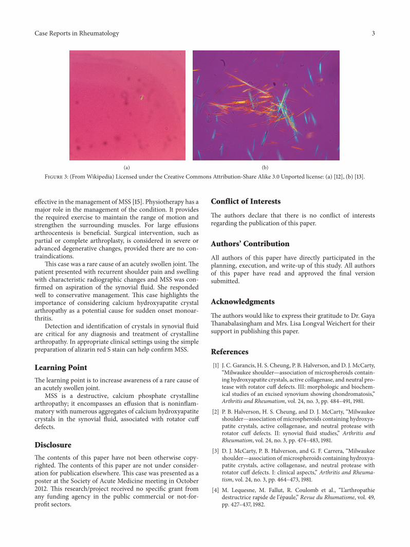
Unlike monosodium urate (MSU) and calcium pyro-phosphate dehydrate crystals (CPPD), identifying calciumhydroxyapatite crystals has been elusive for many years, asthey do not appear under plain and polarized microscopy[10, 11]. Figure 3 shows (a) CPPD and (b) MSU identifiedunder polarized light microscopy of synovial fluid [12, 13].
However, globular clumps of hydroxyapatite crystals mayappear using plain light microscopy but are not birefringent.Such clumps have been described as “shiny coins,” althoughthey often lack distinctive qualities or are often not observed[10]. The introduction of alizarin red S stain has allowedfor a simple, rapid method to identify clumps of calciumhydroxyapatite crystals that produce a characteristic “halo”of orange-red stain [10, 11, 14].
Aspirated synovial fluid is typically haemorrhagic, nonin-flammatory, and positive for calcium apatite crystals. Leuko-cyte count is typically low, usually less than 800/mm3. Dam-age to the rotator cuff precedes crystal deposition and shed-ding of crystals is responsible for sudden, acute episodes ofinflammation.Occasionally synovial fluidmay reveal calciumhydroxyapatite andCPPD crystals, a condition termedmixedcalcium phosphate deposition disease [6]. It is commonlyseen if radiographs show extensive cartilage calcification anddiffuse intra-articular and capsular calcification or dense,homogeneous calcific collection of the tendons. Histopatho-logical studies of the synovium have shown synovial liningcell hyperplasia, giant cell formation, and deposition of fibrinand calcium phosphate crystals.These findings help differen-tiate between other crystal deposition arthropathies. Table 2lists the most important differential diagnoses for MSS.
Treatment is usually supportive; resting the affected jointand the use of nonsteroidal anti-inflammatory agents haveshown to be very effective. Colchicine has been shown to be
Case Reports in Rheumatology 3
(a)
(b)
Figure 3: (FromWikipedia) Licensed under the Creative Commons Attribution-Share Alike 3.0 Unported license: (a) [12], (b) [13].
effective in the management of MSS [15]. Physiotherapy has amajor role in the management of the condition. It providesthe required exercise to maintain the range of motion andstrengthen the surrounding muscles. For large effusionsarthrocentesis is beneficial. Surgical intervention, such aspartial or complete arthroplasty, is considered in severe oradvanced degenerative changes, provided there are no con-traindications.
This case was a rare cause of an acutely swollen joint. Thepatient presented with recurrent shoulder pain and swellingwith characteristic radiographic changes and MSS was con-firmed on aspiration of the synovial fluid. She respondedwell to conservative management. This case highlights theimportance of considering calcium hydroxyapatite crystalarthropathy as a potential cause for sudden onset monoar-thritis.
Detection and identification of crystals in synovial fluidare critical for any diagnosis and treatment of crystallinearthropathy. In appropriate clinical settings using the simplepreparation of alizarin red S stain can help confirmMSS.
Learning Point
The learning point is to increase awareness of a rare cause ofan acutely swollen joint.
MSS is a destructive, calcium phosphate crystallinearthropathy; it encompasses an effusion that is noninflam-matory with numerous aggregates of calcium hydroxyapatitecrystals in the synovial fluid, associated with rotator cuffdefects.
Disclosure
The contents of this paper have not been otherwise copy-righted. The contents of this paper are not under consider-ation for publication elsewhere. This case was presented as aposter at the Society of Acute Medicine meeting in October2012. This research/project received no specific grant fromany funding agency in the public commercial or not-for-profit sectors.
Conflict of Interests
The authors declare that there is no conflict of interestsregarding the publication of this paper.
Authors’ Contribution
All authors of this paper have directly participated in theplanning, execution, and write-up of this study. All authorsof this paper have read and approved the final versionsubmitted.
Acknowledgments
The authors would like to express their gratitude to Dr. GayaThanabalasingham and Mrs. Lisa Longval Weichert for theirsupport in publishing this paper.
References
[1] J. C. Garancis, H. S. Cheung, P. B. Halverson, and D. J. McCarty,“Milwaukee shoulder—association of microspheroids contain-ing hydroxyapatite crystals, active collagenase, and neutral pro-tease with rotator cuff defects. III: morphologic and biochem-ical studies of an excised synovium showing chondromatosis,”Arthritis and Rheumatism, vol. 24, no. 3, pp. 484–491, 1981.
[2] P. B. Halverson, H. S. Cheung, and D. J. McCarty, “Milwaukeeshoulder—association of microspheroids containing hydroxya-patite crystals, active collagenase, and neutral protease withrotator cuff defects. II: synovial fluid studies,” Arthritis andRheumatism, vol. 24, no. 3, pp. 474–483, 1981.
[3] D. J. McCarty, P. B. Halverson, and G. F. Carrera, “Milwaukeeshoulder—association of microspheroids containing hydroxya-patite crystals, active collagenase, and neutral protease withrotator cuff defects. I: clinical aspects,” Arthritis and Rheuma-tism, vol. 24, no. 3, pp. 464–473, 1981.
[4] M. Lequesne, M. Fallut, R. Coulomb et al., “L’arthropathiedestructrice rapide de l’epaule,” Revue du Rhumatisme, vol. 49,pp. 427–437, 1982.
4 Case Reports in Rheumatology
[5] C. S. Neer II, E. V. Craig, and H. Fukuda, “Cuff-tear arthropa-thy,” Journal of Bone and Joint Surgery A, vol. 65, no. 9, pp. 1232–1244, 1983.
[6] M. S. Genta and C. Gabay, “Milwaukee Shoulder,”New EnglandJournal of Medicine, vol. 354, no. 2, article e2, 2006.
[7] D. J. McCarty, “Milwaukee shoulder syndrome,” Transactions ofthe American Clinical and Climatological Association, vol. 102,pp. 271–283, 1990.
[8] V. D. Nguyen, “Rapid destructive arthritis of the shoulder,” Skel-etal Radiology, vol. 25, no. 2, pp. 107–112, 1996.
[9] J. Llanger, J. Palmer, N. Roson, S. Bague, A. Camins, and R.Cremades, “Nonseptic monoarthritis: Imaging features withclinical and histopathologic correlation,” Radiographics, vol. 20,pp. S263–S278, 2000.
[10] H. Paul, A. J. Reginato, and H. R. Schumacher, “Alizarin red Sstaining as a screening test to detect calcium compounds in syn-ovial fluid,”Arthritis andRheumatism, vol. 26, no. 2, pp. 191–200,1983.
[11] E. S. Molloy and G. M. McCarthy, “Hydroxyapatite depositiondisease of the joint,” Current Rheumatology Reports, vol. 5, no.3, pp. 215–221, 2003.
[12] Wikipedia, “File:Calcium pyrophosphate crystal.jpg,” 2013,http://en.wikipedia.org/wiki/File:Calcium pyrophosphatecrystal.jpg.
[13] Wikipedia, “File:Fluorescent uric acid.JPG,” 2013, http://en.wik-ipedia.org/wiki/File:Fluorescent uric acid.JPG.
[14] L. X. Chen, G. Clayburne, and H. R. Schumacher, “Updateon identification of pathogenic crystals in joint fluid,” CurrentRheumatology Reports, vol. 6, no. 3, pp. 217–220, 2004.
[15] C. J. Forster, R. J. Oglesby, A. J. Szkutnik, and J. R. Roberts, “Pos-itive alizarin red clumps in Milwaukee shoulder syndrome,”Journal of Rheumatology, vol. 36, no. 12, p. 2853, 2009.

Submit your manuscripts athttp://www.hindawi.com
Stem CellsInternational
Hindawi Publishing Corporationhttp://www.hindawi.com Volume 2014
Hindawi Publishing Corporationhttp://www.hindawi.com Volume 2014
MEDIATORSINFLAMMATION
of
Hindawi Publishing Corporationhttp://www.hindawi.com Volume 2014
Behavioural Neurology
EndocrinologyInternational Journal of
Hindawi Publishing Corporationhttp://www.hindawi.com Volume 2014
Hindawi Publishing Corporationhttp://www.hindawi.com Volume 2014
Disease Markers
Hindawi Publishing Corporationhttp://www.hindawi.com Volume 2014
BioMed Research International
OncologyJournal of
Hindawi Publishing Corporationhttp://www.hindawi.com Volume 2014
Hindawi Publishing Corporationhttp://www.hindawi.com Volume 2014
Oxidative Medicine and Cellular Longevity
Hindawi Publishing Corporationhttp://www.hindawi.com Volume 2014
PPAR Research
The Scientific World JournalHindawi Publishing Corporation http://www.hindawi.com Volume 2014
Immunology ResearchHindawi Publishing Corporationhttp://www.hindawi.com Volume 2014
Journal of
ObesityJournal of
Hindawi Publishing Corporationhttp://www.hindawi.com Volume 2014
Hindawi Publishing Corporationhttp://www.hindawi.com Volume 2014
Computational and Mathematical Methods in Medicine
OphthalmologyJournal of
Hindawi Publishing Corporationhttp://www.hindawi.com Volume 2014
Diabetes ResearchJournal of
Hindawi Publishing Corporationhttp://www.hindawi.com Volume 2014
Hindawi Publishing Corporationhttp://www.hindawi.com Volume 2014
Research and TreatmentAIDS
Hindawi Publishing Corporationhttp://www.hindawi.com Volume 2014
Gastroenterology Research and Practice
Hindawi Publishing Corporationhttp://www.hindawi.com Volume 2014
Parkinson’s Disease
Evidence-Based Complementary and Alternative Medicine
Volume 2014Hindawi Publishing Corporationhttp://www.hindawi.com