Embed Size (px)
Citation preview
Yeditep Medical Journal 2008;(5): 33-37
33
Spontaneous Resorption of Subdural Hematoma Caused by Lumbar CSF Leak Following Spine Surgery Case Report
Omurga Cerrahisi Sonrası Meydana Gelen BOS Kaçağına Bağlı Olarak Oluşmuş Subdural Hematomun Spontan Emilimi Olgu Sunumu Salih Gulsen
Neurosurgeon Baskent University, Department of Neurosurgery
Cem Yilmaz
Associate Professor of Neurosurgery Baskent University, Department of Neurosurgery
Tarkan Calisaneller
Associate Professor of Neurosurgery Baskent University, Department of Neurosurgery
Hakan Caner
Professor of Neurosurgery Baskent University, Department of Neurosurgery
Nur Altınors
Professor of Neurosurgery Baskent University, Department of Neurosurgery
Corresponding Author Salih Gulsen MD
Neurosurgeon Baskent University, Department of Neurosurgery Faculty of Medicine Bahcelievler / Ankara / Turkey E-mail: [email protected]
ABSTRACT In the atrophic brain especially in the elderly ,development of the spontaneous subdural hematoma provoked by CSF drainage procedures, which causes intracranialhypotension. Nontraumatic spontaneous chronic subdural hematomas may occur from different pathologies in predisposed population including being older than 65 years, using an anticoagulant agent, coagulopathies, alcohol abuse and the procedures leading to CSF overdrainage, such as lumbar puncture, external ventricular drainage, ventriculoperitoneal shunt, and also iatrogenic injuries to the duramater and arachnoid during spinal surgery would lead to CSF leakage from the wound site and may result in either meningitis or intracranial subdural hematoma. In this article, we describe the occurrence of a subdural hematoma and it's spontaneous resorption following spinal surgery procedure. Key words: Lumbar surgery, Resorption, Subdural hematoma. ÖZET Atrofik beyni olan yaşlılarda beyin omurilik sıvısının (BOS) boşalmasına yol açan işlemler sonucunda ortaya çıkan intrakranial hipotansiyon spontan olarak subdural hematom oluşmasına yol açar. Travmaya bağlı olmayan ve kendiliğinden ortaya çıkan kronik subdural hematomlar BOS boşaltılması işlemlemlerinden sonra (iatrojenik duramater- araknoid yırtılması, ventriküloperitoneal şant takılması) daha çok 65 yaş üzerindeki antikoagülan kullanan, aşırı alkol alan ve koagülopatisi olan hastalarda gelişir. Bu makalede omurga cerrahisi sonrası duramater- araknoid yırtılması sonucu gelişen spontan subdural hematom ve bu hematomun kendiliğinden emilimi tanımlanmıştır.
Anahtar Kelimeler: Omurga cerrahisi, Rezorpsiyon, Subdural Hematom
Gulsen S. et al Yeditepe Medical Journal 2008;(5): 33-37
34
INTRODUCTION In the atrophic brain especially in the elderly , development of the spontaneous subdural hematoma provoked by CSF drainage procedures, which causes intracranial hypotension (1,2). Nontraumatic spontaneous chronic subdural hematomas may occur from different pathologies in predisposed population including being older than 65 years, using an anticoagulant agent, coagulopathies, alcohol abuse and the procedures leading to CSF overdrainage, such as lumbar puncture, external ventricular drainage, ventriculoperitoneal shunt, and also iatrogenic injuries to the duramater and arachnoid during spinal surgery would lead to CSF leakage from the wound site and may result in either meningitis or intracranial subdural hematoma(1,2). In this article, we describe the occurrence of a subdural hematoma and it's spontaneous resorption following spinal surgery procedure. CASE REPORT History and Examination This 80 - year – old woman presented with a 1 - mounth history of headache following lumbar spinal surgery, which were performed five weeks ago (During spinal surgery duramater and arachnoid were inadvertently injured and CSF leakage were occurred, dural tearing were small, so we did not repair it, we put a piece of adipose tissue on it and the patient were discharged uneventfully). She had back pain and left leg pain before the operation, but her complaints relieved postoperatively, but she complainted from headache about 10 days later the operation, her primary physican offering acetaminophene for her headache, but the medication relieve did not the headache. Her headache was constant and not positional, she noticed that her headaches was increasing while she was making valsalva maneuvers. Her physical examination and blood analysis revealed
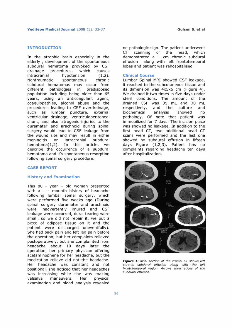
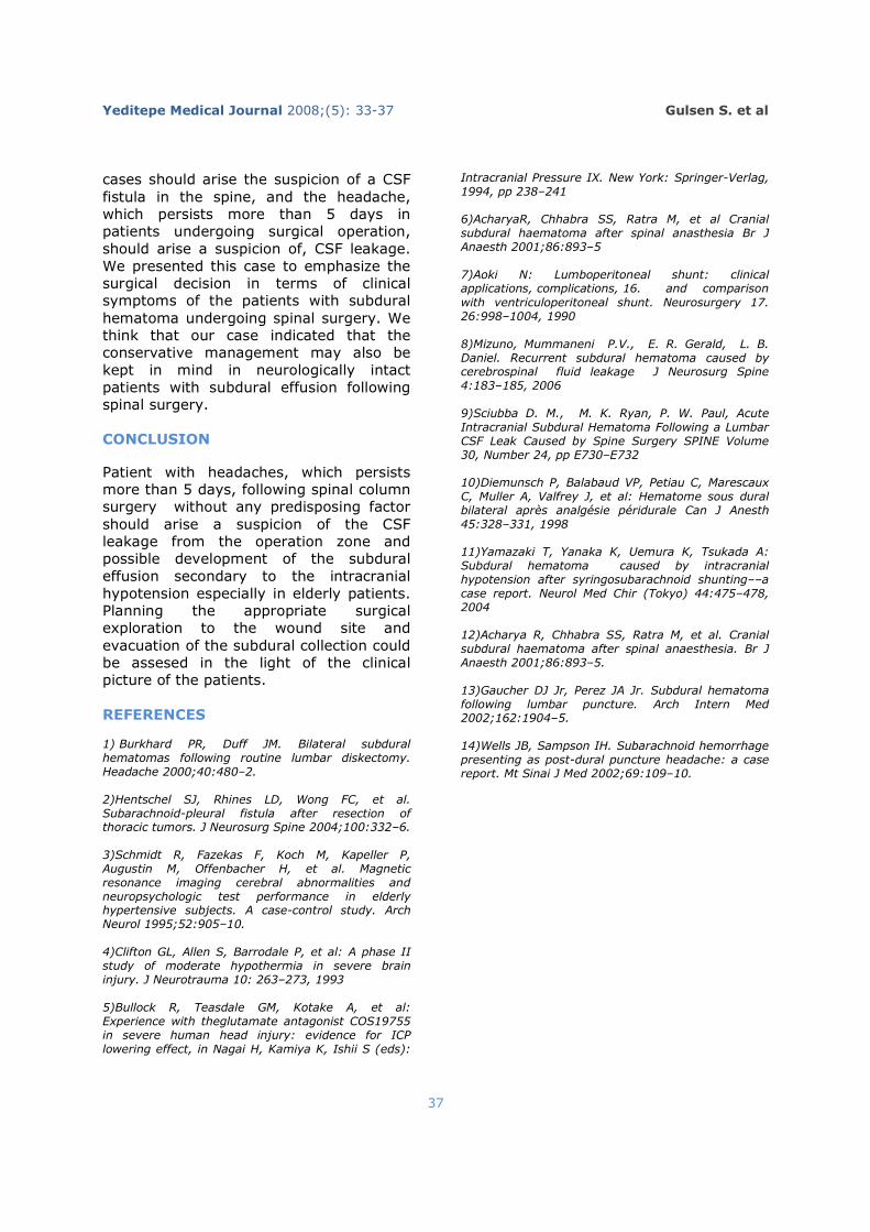
no pathologic sign. The patient underwent CT scanning of the head, which demonstrated a 1 cm chronic subdural effusion along with left frontotemporal lobes and patient was rehospitalised. Clinical Course Lumbar Spinal MRI showed CSF leakage, it reached to the subcutaneous tissue and its dimension was 4x5x6 cm (Figure 4). We drained it two times in five days under steril conditions. The amount of the drained CSF was 35 mL and 30 mL respectively, and the culture and biochemical analysis showed no pathology. Of note that patient was immobilized for 7 days. The incision place was showed no leakage. In addition to the first head CT, two additional head CT scans were performed and the last one showed no subdural effusion in fifteen days Figure (1,2,3). Patient has no complaints regarding headache ten days after hospitalization.
Figure 1: Axial section of the cranial CT shows left chronic subdural effusion along with the left frontotemporal region. Arrows show edges of the subdural effusion.
Gulsen S. et al Yeditepe Medical Journal 2008;(5): 33-37
35
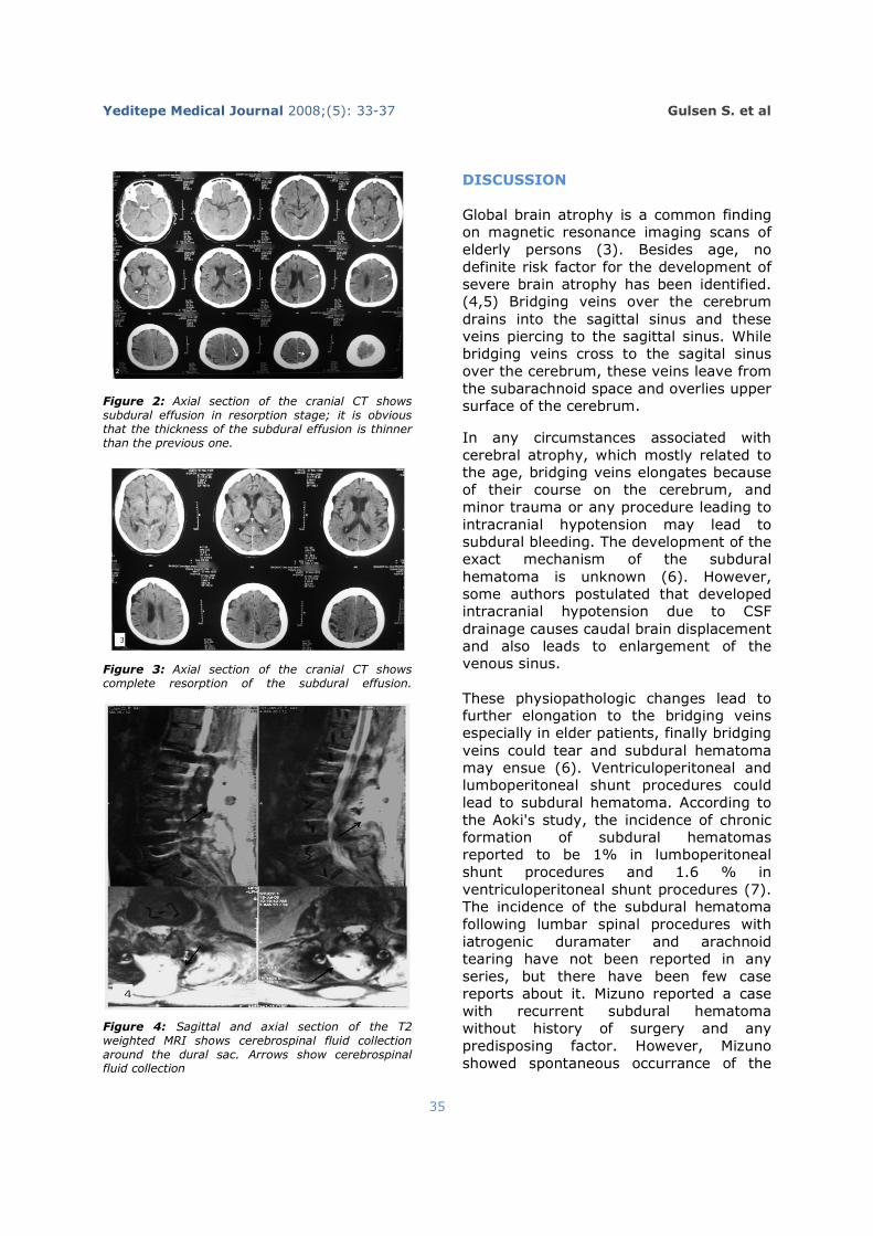
Figure 2: Axial section of the cranial CT shows subdural effusion in resorption stage; it is obvious that the thickness of the subdural effusion is thinner than the previous one.
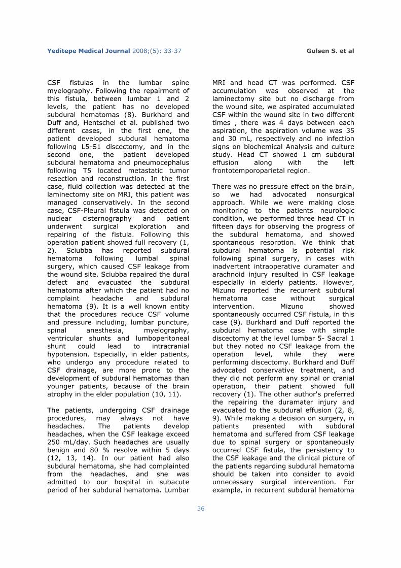
Figure 3: Axial section of the cranial CT shows complete resorption of the subdural effusion.
Figure 4: Sagittal and axial section of the T2 weighted MRI shows cerebrospinal fluid collection around the dural sac. Arrows show cerebrospinal fluid collection
DISCUSSION Global brain atrophy is a common finding on magnetic resonance imaging scans of elderly persons (3). Besides age, no definite risk factor for the development of severe brain atrophy has been identified. (4,5) Bridging veins over the cerebrum drains into the sagittal sinus and these veins piercing to the sagittal sinus. While bridging veins cross to the sagital sinus over the cerebrum, these veins leave from the subarachnoid space and overlies upper surface of the cerebrum.
In any circumstances associated with cerebral atrophy, which mostly related to the age, bridging veins elongates because of their course on the cerebrum, and minor trauma or any procedure leading to intracranial hypotension may lead to subdural bleeding. The development of the exact mechanism of the subdural hematoma is unknown (6). However, some authors postulated that developed intracranial hypotension due to CSF drainage causes caudal brain displacement and also leads to enlargement of the venous sinus. These physiopathologic changes lead to further elongation to the bridging veins especially in elder patients, finally bridging veins could tear and subdural hematoma may ensue (6). Ventriculoperitoneal and lumboperitoneal shunt procedures could lead to subdural hematoma. According to the Aoki's study, the incidence of chronic formation of subdural hematomas reported to be 1% in lumboperitoneal shunt procedures and 1.6 % in ventriculoperitoneal shunt procedures (7). The incidence of the subdural hematoma following lumbar spinal procedures with iatrogenic duramater and arachnoid tearing have not been reported in any series, but there have been few case reports about it. Mizuno reported a case with recurrent subdural hematoma without history of surgery and any predisposing factor. However, Mizuno showed spontaneous occurrance of the
Gulsen S. et al Yeditepe Medical Journal 2008;(5): 33-37
36
CSF fistulas in the lumbar spine myelography. Following the repairment of this fistula, between lumbar 1 and 2 levels, the patient has no developed subdural hematomas (8). Burkhard and Duff and, Hentschel et al. published two different cases, in the first one, the patient developed subdural hematoma following L5-S1 discectomy, and in the second one, the patient developed subdural hematoma and pneumocephalus following T5 located metastatic tumor resection and reconstruction. In the first case, fluid collection was detected at the laminectomy site on MRI, this patient was managed conservatively. In the second case, CSF-Pleural fistula was detected on nuclear cisternography and patient underwent surgical exploration and repairing of the fistula. Following this operation patient showed full recovery (1, 2). Sciubba has reported subdural hematoma following lumbal spinal surgery, which caused CSF leakage from the wound site. Sciubba repaired the dural defect and evacuated the subdural hematoma after which the patient had no complaint headache and subdural hematoma (9). It is a well known entity that the procedures reduce CSF volume and pressure including, lumbar puncture, spinal anesthesia, myelography, ventricular shunts and lumboperitoneal shunt could lead to intracranial hypotension. Especially, in elder patients, who undergo any procedure related to CSF drainage, are more prone to the development of subdural hematomas than younger patients, because of the brain atrophy in the elder population (10, 11). The patients, undergoing CSF drainage procedures, may always not have headaches. The patients develop headaches, when the CSF leakage exceed 250 mL/day. Such headaches are usually benign and 80 % resolve within 5 days (12, 13, 14). In our patient had also subdural hematoma, she had complainted from the headaches, and she was admitted to our hospital in subacute period of her subdural hematoma. Lumbar
MRI and head CT was performed. CSF accumulation was observed at the laminectomy site but no discharge from the wound site, we aspirated accumulated CSF within the wound site in two different times , there was 4 days between each aspiration, the aspiration volume was 35 and 30 mL, respectively and no infection signs on biochemical Analysis and culture study. Head CT showed 1 cm subdural effusion along with the left frontotemporoparietal region. There was no pressure effect on the brain, so we had advocated nonsurgical approach. While we were making close monitoring to the patients neurologic condition, we performed three head CT in fifteen days for observing the progress of the subdural hematoma, and showed spontaneous resorption. We think that subdural hematoma is potential risk following spinal surgery, in cases with inadvertent intraoperative duramater and arachnoid injury resulted in CSF leakage especially in elderly patients. However, Mizuno reported the recurrent subdural hematoma case without surgical intervention. Mizuno showed spontaneously occurred CSF fistula, in this case (9). Burkhard and Duff reported the subdural hematoma case with simple discectomy at the level lumbar 5- Sacral 1 but they noted no CSF leakage from the operation level, while they were performing discectomy. Burkhard and Duff advocated conservative treatment, and they did not perform any spinal or cranial operation, their patient showed full recovery (1). The other author's preferred the repairing the duramater injury and evacuated to the subdural effusion (2, 8, 9). While making a decision on surgery, in patients presented with subdural hematoma and suffered from CSF leakage due to spinal surgery or spontaneously occurred CSF fistula, the persistency to the CSF leakage and the clinical picture of the patients regarding subdural hematoma should be taken into consider to avoid unnecessary surgical intervention. For example, in recurrent subdural hematoma
Gulsen S. et al Yeditepe Medical Journal 2008;(5): 33-37
37
cases should arise the suspicion of a CSF fistula in the spine, and the headache, which persists more than 5 days in patients undergoing surgical operation, should arise a suspicion of, CSF leakage. We presented this case to emphasize the surgical decision in terms of clinical symptoms of the patients with subdural hematoma undergoing spinal surgery. We think that our case indicated that the conservative management may also be kept in mind in neurologically intact patients with subdural effusion following spinal surgery. CONCLUSION Patient with headaches, which persists more than 5 days, following spinal column surgery without any predisposing factor should arise a suspicion of the CSF leakage from the operation zone and possible development of the subdural effusion secondary to the intracranial hypotension especially in elderly patients. Planning the appropriate surgical exploration to the wound site and evacuation of the subdural collection could be assesed in the light of the clinical picture of the patients. REFERENCES 1).Burkhard PR, Duff JM. Bilateral subdural hematomas following routine lumbar diskectomy. Headache 2000;40:480–2. 2)Hentschel SJ, Rhines LD, Wong FC, et al. Subarachnoid-pleural fistula after resection of thoracic tumors. J Neurosurg Spine 2004;100:332–6.
3)Schmidt R, Fazekas F, Koch M, Kapeller P, Augustin M, Offenbacher H, et al. Magnetic resonance imaging cerebral abnormalities and neuropsychologic test performance in elderly hypertensive subjects. A case-control study. Arch Neurol 1995;52:905–10.
4)Clifton GL, Allen S, Barrodale P, et al: A phase II study of moderate hypothermia in severe brain injury. J Neurotrauma 10: 263–273, 1993
5)Bullock R, Teasdale GM, Kotake A, et al: Experience with theglutamate antagonist COS19755 in severe human head injury: evidence for ICP lowering effect, in Nagai H, Kamiya K, Ishii S (eds):
Intracranial Pressure IX. New York: Springer-Verlag, 1994, pp 238–241
6)AcharyaR, Chhabra SS, Ratra M, et al Cranial subdural haematoma after spinal anasthesia Br J Anaesth 2001;86:893–5
7)Aoki N: Lumboperitoneal shunt: clinical applications, complications, 16. and comparison with ventriculoperitoneal shunt. Neurosurgery 17. 26:998–1004, 1990
8)Mizuno, Mummaneni P.V., E. R. Gerald, L. B. Daniel. Recurrent subdural hematoma caused by cerebrospinal fluid leakage J Neurosurg Spine 4:183–185, 2006
9)Sciubba D. M., M. K. Ryan, P. W. Paul, Acute Intracranial Subdural Hematoma Following a Lumbar CSF Leak Caused by Spine Surgery SPINE Volume 30, Number 24, pp E730–E732
10)Diemunsch P, Balabaud VP, Petiau C, Marescaux C, Muller A, Valfrey J, et al: Hematome sous dural bilateral après analgésie péridurale Can J Anesth 45:328–331, 1998
11)Yamazaki T, Yanaka K, Uemura K, Tsukada A: Subdural hematoma caused by intracranial hypotension after syringosubarachnoid shunting––a case report. Neurol Med Chir (Tokyo) 44:475–478, 2004
12)Acharya R, Chhabra SS, Ratra M, et al. Cranial subdural haematoma after spinal anaesthesia. Br J Anaesth 2001;86:893–5.
13)Gaucher DJ Jr, Perez JA Jr. Subdural hematoma following lumbar puncture. Arch Intern Med 2002;162:1904–5.
14)Wells JB, Sampson IH. Subarachnoid hemorrhage presenting as post-dural puncture headache: a case report. Mt Sinai J Med 2002;69:109–10.