Embed Size (px)
Citation preview
D isc losures : L indsay: S tem line - em p loym en t, equ ity ow nersh ip ; C hen : S tem line - em p loym en t, equ ity ow nersh ip ; B rooks : S tem line - em p loym en t, equ ity ow nersh ip ; B arra t: S tem line - research fund ing
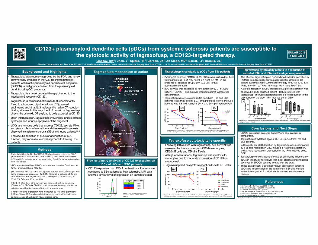
Background and Highlights• Tagraxofusp was recently approved by the FDA, and is now
commercially available in the U.S, for the treatment of patients with blastic plasmacytoid dendritic cell neoplasm (BPDCN), a malignancy derived from the plasmacytoid dendritic cell (pDC) precursor.
• Tagraxofusp is a novel targeted therapy directed to the interleukin-3 receptor (CD123).
• Tagraxofusp is comprised of human IL-3 recombinantly fused to a truncated diphtheria toxin (DT) payload engineered such that IL-3 replaces the native DT receptor-binding domain. In this way, the IL-3 domain of tagraxofusp directs the cytotoxic DT payload to cells expressing CD123.
• Upon internalization, tagraxofusp irreversibly inhibits protein synthesis and induces apoptosis of the target cell.
• pDCs are immune cells that express CD123, secrete IFNα, and play a role in inflammation and disease pathogenesis observed in systemic sclerosis (SSc) and lupus patients1,2,3.
• Therapeutic depletion of pDCs or attenuation of pDCfunction, may represent a novel approach to treating SScpatients.
Tagraxofusp mechanism of action
Methods• Patients fulfilled the 2013 ACR/EULAR classification criteria for SSc4.
peripheral blood mononuclear cells (PBMCs) from healthy volunteers (HV) and SSc patients were prepared using Ficoll-Paque density gradient from fresh blood.
• pDCs were isolated from PBMCs as previously described5 and used to further enrich additional PBMCs.
• pDC-enriched PBMCs (3-6% pDCs) were cultured at 2x10⁵ cells per well in the presence or absence of CpG-274 (0.5 μM) to activate pDCs and then incubated with tagraxofusp (0.01-100 ng/ml, 0.17pM-1.7nM) at 37°C, 5% CO2, and 95% humidity.
• After 24 h of culture, pDC survival was assessed by flow cytometry (CD14-, CD3- BDCA4+ CD123+), and supernatants were collected for cytokine quantification by a multiplexed Luminex assay.
• Changes in gene expression were measured by real-time quantitative PCR on 10ng cDNA, and calculated based on relative threshold cycle and expression of a ubiquitin housekeeping gene.
Tagroxofusp is cytotoxic to pDCs from SSc patients
• 2x10⁵ pDC enriched PBMCs (3-6% pDCs) were cultured for 24hr with tagraxofusp (0.01-100 ng/ml, 0.17 pM-1.7 nM ) in the presence or absence of CpG-274 (0.5 μM) for DC activation/maturation.
• pDC survival was assessed by flow cytometry (CD14-, CD3-BDCA4+ CD123+) and survival graphed against tagraxofusp concentration.
• Tagraxofusp was cytotoxic to pDCs from both HVs and SScpatients to a similar extent. ED50 of tagraxofusp in HVs and SScpatients was 4.3 and 3.2 ng/ml (74.4 and 55.4 pM) respectively.
Flow cytometry analysis of CD123 expression on pDCs of HVs and SSC patients
• CD123 expression on pDCs from healthy volunteers was compared to SSc patients by flow cytometry. MFI data shows a similar level of expression on samples tested.
0 102 103 104 105
CD123
0
20
40
60
80
100
% o
f Max
Sample CD123 MFIHV 16501SSc1 18852SSc2 17886
Figure 2: Ex vivo survival of pDCs from HVs and SSc patients treated with tagraxofusp. pDC enriched PBMCs were incubated with tagraxofusp for 24h and cell survival assessed by flow cytometry (n=5 HVs and n=5 SSc patients)
Figure 1: Flow cytometry analysis of CD123 expression on pDCs from HV and SScpatients
Tagraxofusp cytotoxicity results in a reduction of secreted IFNα and IFNα-induced gene expression
Tagraxofusp cytotoxicity is specific
Conclusions and Next Steps• CD123 expression on pDCs from HV and SSc patients is
comparable.• Tagraxofusp is cytotoxic against CD123+ pDCs from HVs and
SSc patients.• In SSc patients, pDC depletion by tagraxofusp was accompanied
by a 68-fold reduction in CpG-induced IFNα protein secretion, and a 3-fold reduction in expression of the IFNα-induced gene, GBP.
• Tagraxofusp concentrations effective at eliminating inflammatory pDCs in this study were lower than peak plasma concentrations observed in BPDCN patients treated with the drug.
• These data present a potentially novel approach of targeting pDCs and inflammation in the treatment of SSc and warrant further investigation. A clinical trial is planned in autoimmune disease.
References1. Ah Kioon, MD. Sci Trans Med 2018: 10(423)2. Van Bon, L. N Engl J Med 2014: 370(5):433-433. Collin, M. Immunology 140 (1) :22-304. Van den Hoogen, F. Ann Rheum Dis 2013: 72(11):1747-555. Guiducci, C. J Exp Med 2006: 203(8):1999-20086. Sun, Q. Blood 1996: 87(1): 83-92
HV SSc
HV SSc
Figure 4: A) Ex vivo IFNα secretion and B) GBP gene expression from pDC-enriched PBMCs from SSc patients cultured and treated with tagraxofusp for 24h (n=5)
• The effect of tagraxofusp on CpG-induced cytokine secretion by PBMCs from SSc patients was assessed by screening cell culture supernatant by Luminex technology for IL-1β, IL-6, IL-8, IFNα, IFNγ IP-10, TNFα, MIP-1α/β, MCP1 and RANTES.
• A 68-fold reduction in CpG-induced IFNα protein secretion was observed in pDC-enriched patient PBMCs cultured with tagraxofusp; this was accompanied by a 3-fold reduction in the expression of the type 1 IFN-induced gene, GBP.
Figure 3: Ex vivo survival of monocytes, B cells and T cells from HVs and SSc patients treated with tagraxofusp. pDC enriched PBMCs were incubated with tagraxofusp for 24h and cell survival assessed by flow cytometry (n=5 HVs and n=5 SSc patients)
monocytesB cells
CD4+ T cellsCD8+ T cells
• Following 24h culture with tagraxofusp, cell survival was assessed by flow cytometry on CD14+ monocytes, CD20+ B cells and CD4/8+ T cells.
• At high concentrations, tagraxofusp was cytotoxic to monocytes due to moderate expression of CD123 on monocytes6.
• Tagraxofusp had no cytotoxic effect on B-cells or T-cells.
CD123+ plasmacytoid dendritic cells (pDCs) from systemic sclerosis patients are susceptible to the cytotoxic activity of tagraxofusp, a CD123-targeted therapy. EULAR 2019
# SAT0284
Lindsay, RW1; Chen, J1; Spiera, RF2; Gordon, JK2; Ah Kioon, MD3; Barrat, FJ3; Brooks, CL1
1Stemline Therapeutics, Inc., New York, NY 10022; 2Scleroderma and Vasculitis Center, Hospital for Special Surgery, New York, NY 10021; 3Autoimmunity and Inflammation Program, HSS Research Institute, Hospital for Special Surgery, New York, NY 10021
0.01 0.1 1 10 1000
25
50
75
100
125
[Tagraxofusp] ng/ml
pDC
sur
viva
l (%
)
0.01 0.1 1 10 1000
50
100
150
200
[Tagraxofusp] ng/ml
pDC
sur
viva
l (%
) no CpG+CpG
0 0.01 0.1 1 10 1000
25
50
75
100
125
[Tagroxofusp] ng/ml
Cel
l sur
viva
l )%
)
0 0.01 0.1 1 10 1000
25
50
75
100
125
[Tagraxofusp] ng/ml
Cel
l sur
viva
l )%
)
contro
l0.0
1 0.1 1 10 100
0
200
400
600
[Tagraxofusp] ng/ml
[IFN
-α] p
g/m
l
contro
l0.0
1 0.1 1 10 100
0
2000
4000
6000
[Tagraxofusp] ng/ml
Rel
ativ
e C
t
A B


![PDCS Publications - Springer978-3-642-79789-7/1.pdf · PDCS Publications The following lists ... PDCS that have been published in refereed journals and technical conferences. [1]](https://img.pdfslide.net/doc/110x75/5a90baf27f8b9af27f8e0528/pdcs-publications-springer-978-3-642-79789-71pdfpdcs-publications-the-following.jpg)