Embed Size (px)
Citation preview

CASE REPORT
CD4+ CD56+ Hematodermic Neoplasm Without CutaneousInvolvement
Seema Sharma • Chanchal Rana
Received: 26 March 2012 / Accepted: 23 October 2012
� Indian Society of Haematology & Transfusion Medicine 2012
Abstract CD4?/CD56? hematodermic neoplasm is a
recently recognized highly aggressive tumor presenting in
skin, usually with lymph node and bone marrow involve-
ment, often terminating in a leukemic phase. It has a dis-
tinct clinical presentation of primary skin lesions in the
form of papules, nodules or bruise like areas. Bone marrow
and peripheral blood involvement without skin involve-
ment is a very rare phenomenon. We are reporting an
interesting case where an elderly male, a known diabetic,
when investigated for the cause of generalised weakness
was found to have CD4?/CD56? hematodermic neo-
plasm, the unusual feature being absence of any associated
cutaneous involvement.
Introduction
CD4?/CD56? natural killer hematodermic neoplasm vari-
ously known as blastic or blastoid (NK) cell lymphoma or
tumours of plasmacytoid dendritic lineage, is a highly
aggressive neoplasm with a rapidly fatal course. It was first
described in 1994 by Adachi et al. [1]. Since then around
150 cases have been documented in the literature [2].
Majority (*90 %) of patients presents with asymptomatic
solitary or multifocal skin lesions. Though low level bone
marrow and peripheral blood involvement are seen in most
of the cases but leukemic manifestation with extensive bone
marrow involvement in absence of skin lesion is a very rare
phenomenon [2].
To our knowledge there are only two such documented
cases, both been submitted in the 2005 Society for
Hematopathology/European association for pathology
workshop [2]. We are reporting one another case of CD4?/
CD56? hematodermic neoplasm with marked cytopenia
and extensive bone marrow involvement without any
associated skin lesions.
Case History
A 58-years-old male was admitted in the department of
endocrinology of our institute with chief complaint of gen-
eralised weakness with off and on fever for 2 months. This
patient was a known case of diabetes and was on strict gly-
cemic control. The physical examination was normal except
for the presence of marked pallor. There was no lymphade-
nopathy, splenomegaly or skin lesion. Peripheral blood
examination revealed severe anemia (hemoglobin 5.1 g/dL),
marked leucopenia (total leucocyte count 1.0 9 106/L) and
thrombocytopenia (platelet count 85 9 109/L). Red cell
morphology was microcytic and hypochromic. Hence a bone
marrow examination was advised. Bone marrow aspiration
was hypercellular and was predominantly composed of
immature cells with plasmacytoid morphology, high nucleo-
cytoplasmic ratio, moderate amount of cytoplasm, fine
chromatin and single prominent nucleoli. Cytoplasmic vac-
uolation was also seen in some cells. Cytochemical stain for
myeloperoxidase was negative. On immunophenotyping
these immature cells were gated and were positive for CD4,
CD56, CD45, CD33, HLA-DR. Other T and B cell markers
were negative. Immunohistochemistry performed on sections
of bone marrow biopsy revealed that the cells were strongly
positive with antibody for CD68 (Fig. 1). Hence with all
these features diagnosis of CD4?/CD56? hematodermic
S. Sharma (&) � C. Rana
Department of Pathology, Sanjay Gandhi Postgraduate Institute
of Medical Science, Type 5B/13, SGPGIMS, Raibareli Road,
Lucknow 226014, Uttar Pradesh, India
e-mail: [email protected]; [email protected]
123
Indian J Hematol Blood Transfus
DOI 10.1007/s12288-012-0212-5
neoplasm was made. The patient refused treatment and died
within few months of diagnosis.
Discussion
For a long time there has been a debate regarding the true
histogenesis of CD4?/CD56? hematodermic neoplasm
which is reflected in it’s nomenclature as these are known as
blastic or blastoid natural killer cell lymphoma, CD4?
CD56? hematodermic neoplasm or tumors of plasmacytoid
dendritic cell (pDC) lineage. Initially due to CD4 positivity a
T cell lineage was hypothesised but later in 1990s a natural
killer (NK) cell histogenesis was favoured based on CD56
expression, which was later found to be a non specific NK
cell marker [3]. With the delineation of pDC by Grouard
et al. [4] which express CD4, CD45, HLA-DR and CD123,
more recently the World Health Organisation has proposed
the term CD4?/CD56? hematodermic neoplasm or early
pDC leukemia/lymphoma for this neoplasm [5]. The number
of well documented cases of HN in literature is around 150,
with four largest series representing more than half of all
published cases [6–8].
This neoplasm predominantly arises in elderly male and
presents asymptomatic skin lesions which may be solitary
or multifocal. In 50 % cases skin lesion may be the only
extramedullary manifestation. Lymph node involvement
(40–50 %) is relatively common, however splenomegaly
(*20 %) and systemic B symptoms are very rare. Low
level bone marrow and peripheral blood involvement is
seen in almost all cases along with skin lesions [2]. But
best to our knowledge there are only two documented cases
till date showing extensive bone marrow involvement
without any cutaneous manifestation. Both of these cases
were submitted to the 2005 Society for Hematopathology/
European association for Hematopathology workshop
(cases 50 and 77) [2]. In cases with skin lesion diagnosis is
based on skin biopsy which shows immature cells with
blastic morphology. But in cases like our case diagnosis
can only be achieved by immunohistochemical or immu-
nophenotypical methods. When these immature cells
express CD4 and CD56 in absence of lineage specific
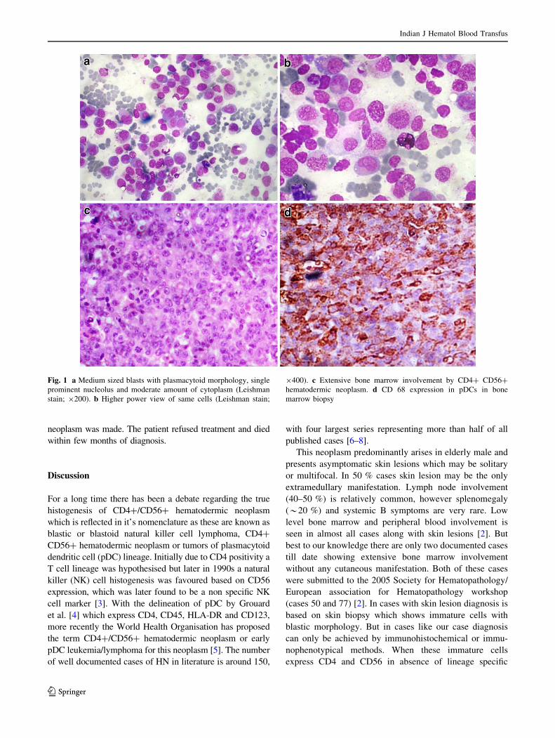
Fig. 1 a Medium sized blasts with plasmacytoid morphology, single
prominent nucleolus and moderate amount of cytoplasm (Leishman
stain; 9200). b Higher power view of same cells (Leishman stain;
9400). c Extensive bone marrow involvement by CD4? CD56?
hematodermic neoplasm. d CD 68 expression in pDCs in bone
marrow biopsy
Indian J Hematol Blood Transfus
123

markers of T cells and B cells then diagnosis of HN can be
made. The extended phenotype is CD45RA?/HLA-DR?/
CD123?/CD116. A subset of HN may also express CD68.
The recent development of pDC associated immunomar-
kers has led to improved specificity in the diagnosis of
HNs. The most important of these is CD123 (interleukin,
IL-3, receptor a chain) and TCL1 (an Akt kinase regulator
and lymphoid proto-oncogene) [4, 9].
Based on the emergence of typical myelomonocytic
leukaemia in a subset of HN cases, these tumors may also
have monocytic or multilineage potential. This is consistent
with the finding that DC2s are sometimes increased in
patients with myelomonocytic leukaemia and can be derived
from monocytes in vitro under certain conditions and may
undergo conversion in vivo to myeloid-type DCs. Hence
they can also express CD33 variably as in our case [10, 11].
The common differential diagnosis in the reported set-
ting is acute leukemia and myelodysplastic syndrome but
they can be ruled out by simple cytochemical stain like
myeloperoxidase because expression of MPO should
always rule out the diagnosis of HN.
The prognosis is poor with median survival of about
14 months with a little better prognosis in patients where
disease is localised to skin. Treatment is conventional
chemotherapy for acute myeloid leukemia which generally
shows a good response initially followed by quick and fatal
relapse in extracutaneous sites such as bone marrow.
Alternative therapies are immunomodulation or immuno-
therapy with interleukin-3 or with anti-CD123 antibody.
References
1. Adachi M, Maeda K, Takekawa M et al (1994) High expression
of CD56 (N182CAM) in a patient with cutaneous CD4-positive
lymphoma. Am J Hematol 47:278–282
2. Herling M, Jones D (2007) CD4?/CD56? hematodermic tumor:
the features of an evolving entity and its relationship to dendritic
cells. Am J Clin Pathol 127:687–700
3. Prasthofer EF, Prchal JT, Grizzle WE et al (1985) Plasmacytoid
T-cell lymphoma associated with chronic myeloproliferative
disorder. Am J Surg Pathol 9:380–387
4. Grouard G, Rissoan MC, Filgueira L et al (1997) The enigmatic
plasmacytoid T cells develop into dendritic cells with interleukin
(IL)-3 and CD40-ligand. J Exp Med 185:1101–1111
5. Willemze R, Jaffe ES, Burg G et al (2005) WHO-EORTC clas-
sification for cutaneous lymphoma. Blood 105:3768–3785
6. Reichard KK, Burks EJ, Foucar MK et al (2005) CD4(?)
CD56(?) lineage-negative malignancies are rare tumors of
plasmacytoid dendritic cells. Am J Surg Pathol 29:1274–1283
7. Petrella T, Bagot M, Willemze R et al (2005) Blastic NK-cell
lymphomas (agranular CD4?CD56? hematodermic neoplasms):
a review. Am J Clin Pathol 123:662–675
8. Feuillard J, Jacob MC, Valensi F et al (2002) Clinical and bio-
logic features of CD4(?)CD56(?) malignancies. Blood 99:1556–
1563
9. Herling M, Teitell MA, Shen RR et al (2003) TCL1 expression in
plasmacytoid dendritic cells (DC2) and the related CD4?
CD56? blastic tumors of skin. Blood 101:5007–5009
10. Bagot M, Bouloc A, Charue D et al (1998) Do primary cutaneous
non-T non-B CD4? CD56? lymphomas belong to the myelo-
monocytic lineage? J Invest Dermatol 111:1242–1244
11. Khoury JD, Medeiros LJ, Manning JT et al (2002) CD56(?)
TdT(?) blastic natural killer cell tumor of the skin: a primitive
systemic malignancy related to myelomonocytic leukemia.
Cancer 94:2401–2424
Indian J Hematol Blood Transfus
123