Embed Size (px)
Citation preview
Chapter 8 DNA Replication, Binary Fission,
and Mitosis
World’s tallest man © Frederic J. Brown/AFP/Getty Images
Copyright © McGraw-Hill Education. All rights reserved. No reproduction or distribution without the prior written consent of McGraw-Hill Education.
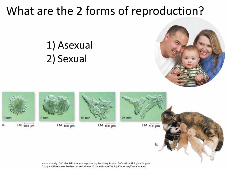
What are the 2 forms of reproduction?
1) Asexual2) Sexual
Human family: © Corbis RF; Amoeba reproducing by binary fission: © Carolina Biological Supply
Company/Phototake; Mother cat and kittens: © Jane Burton/Dorling Kindersley/Getty Images
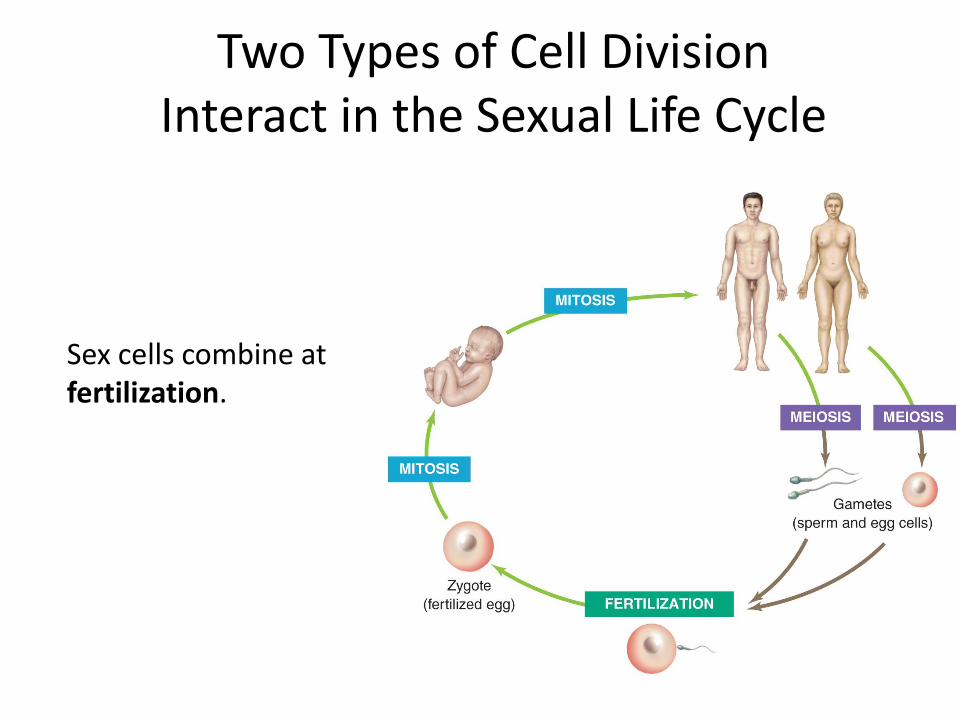
Two Types of Cell Division Interact in the Sexual Life Cycle
Sex cells combine at fertilization.

DNA Replication Precedes Cell Division
Section 8.2
Semi conservative:½ old
&½ new
Figure 8.6
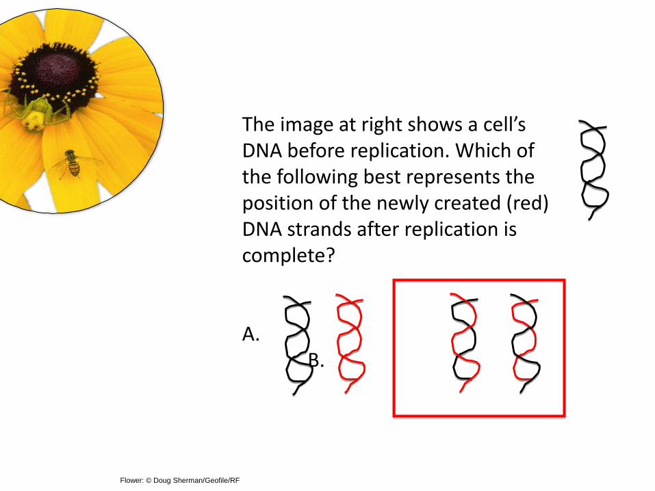
The image at right shows a cell’s DNA before replication. Which of the following best represents the position of the newly created (red) DNA strands after replication is complete?
A.B.
Flower: © Doug Sherman/Geofile/RF
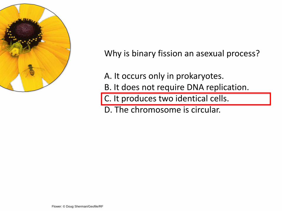
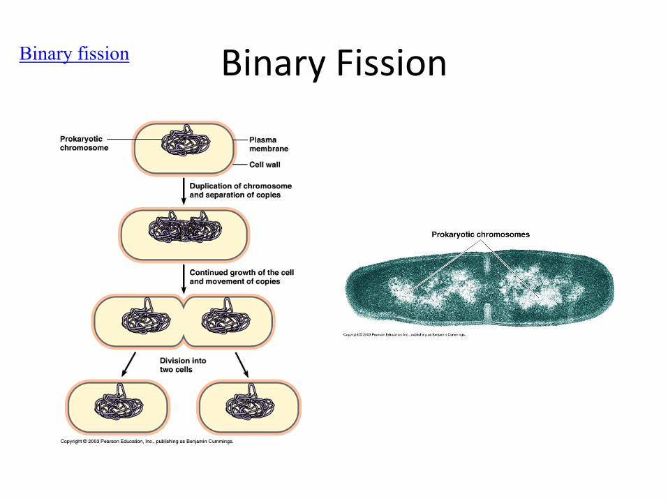
Why is binary fission an asexual process?
A. It occurs only in prokaryotes.B. It does not require DNA replication.C. It produces two identical cells.D. The chromosome is circular.
Flower: © Doug Sherman/Geofile/RF
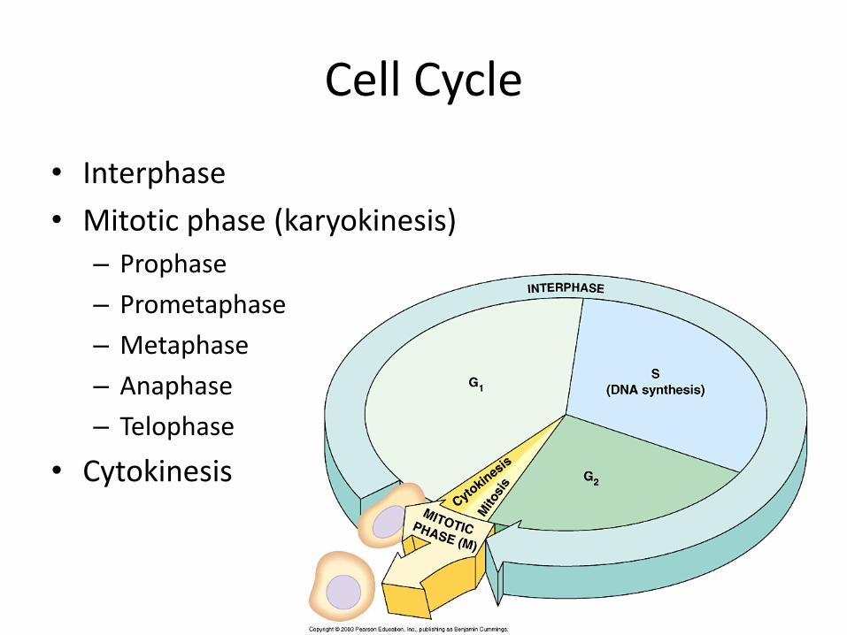
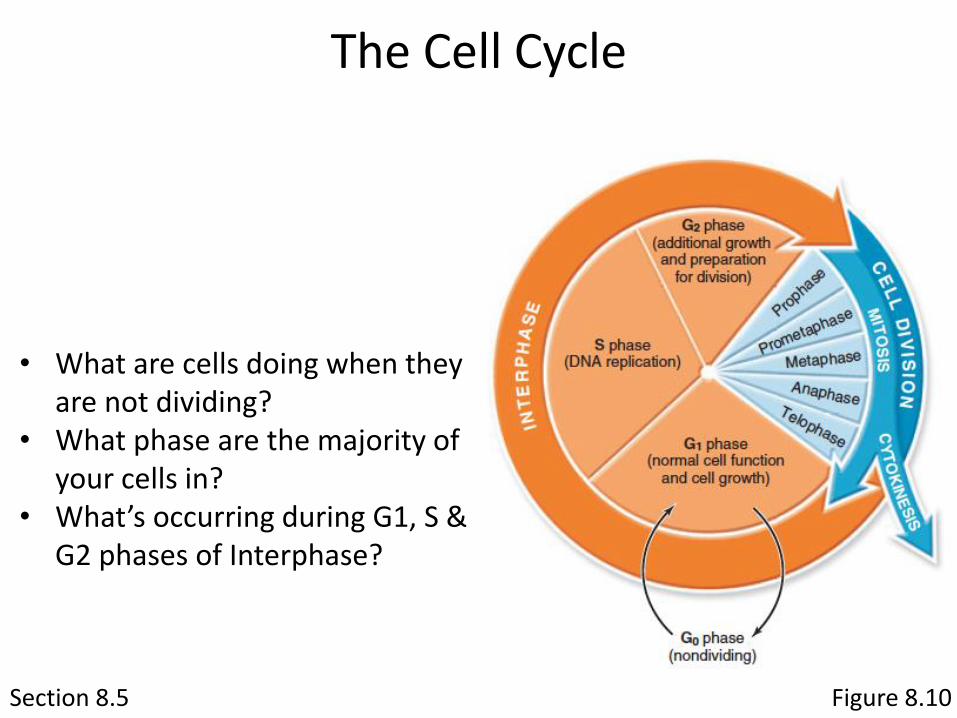
Cell Cycle
• Interphase
• Mitotic phase (karyokinesis)
– Prophase
– Prometaphase
– Metaphase
– Anaphase
– Telophase
• Cytokinesis
Section 8.5
• What are cells doing when they are not dividing?
• What phase are the majority of your cells in?
• What’s occurring during G1, S & G2 phases of Interphase?
The Cell Cycle
Figure 8.10
Section 8.4
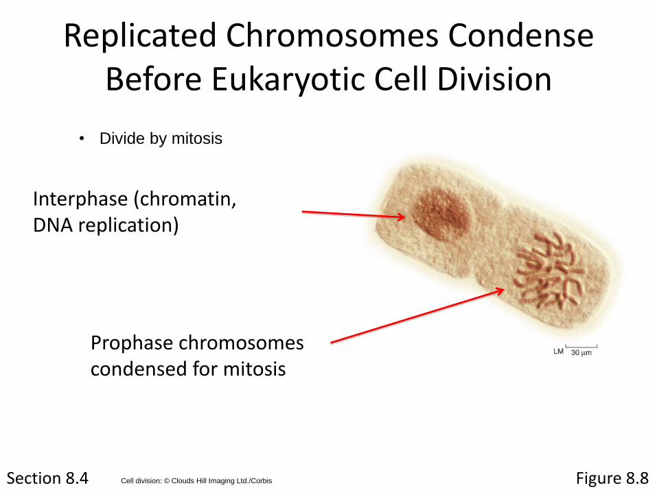
Prophase chromosomes condensed for mitosis
Replicated Chromosomes Condense Before Eukaryotic Cell Division
Figure 8.8Cell division: © Clouds Hill Imaging Ltd./Corbis
• Divide by mitosis
Interphase (chromatin, DNA replication)
Section 8.4
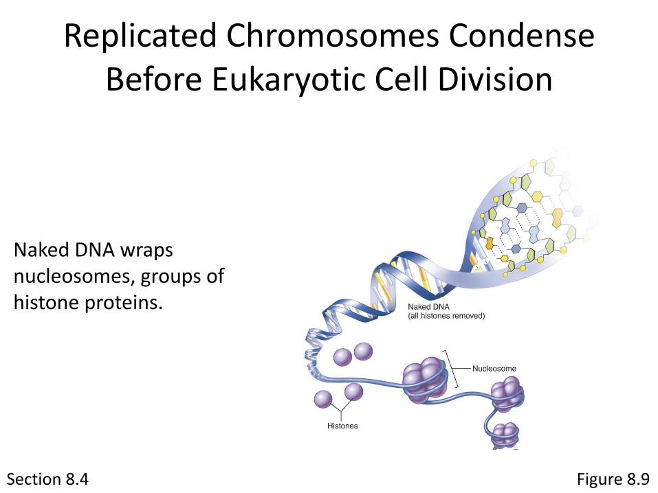
Naked DNA wraps nucleosomes, groups of histone proteins.
Figure 8.9
Replicated Chromosomes Condense Before Eukaryotic Cell Division
Section 8.4
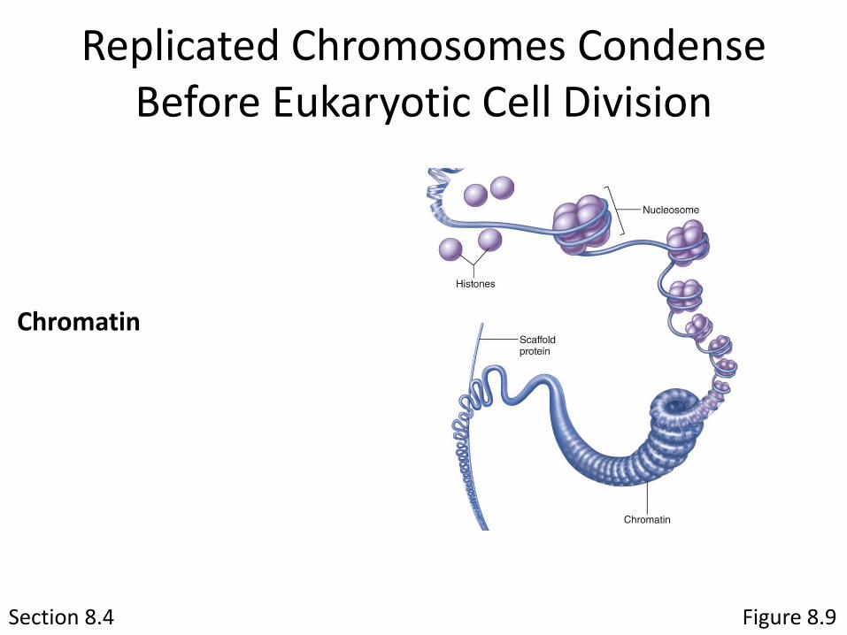
Chromatin
Replicated Chromosomes Condense Before Eukaryotic Cell Division
Figure 8.9
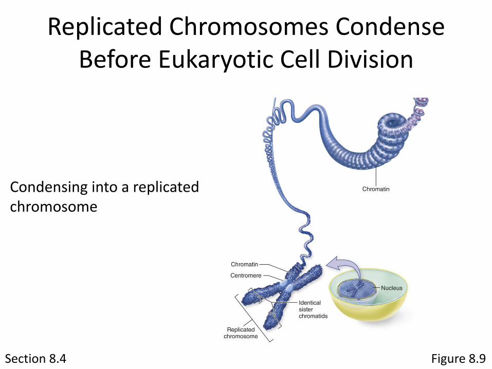
Section 8.4
Condensing into a replicated chromosome
Replicated Chromosomes Condense Before Eukaryotic Cell Division
Figure 8.9
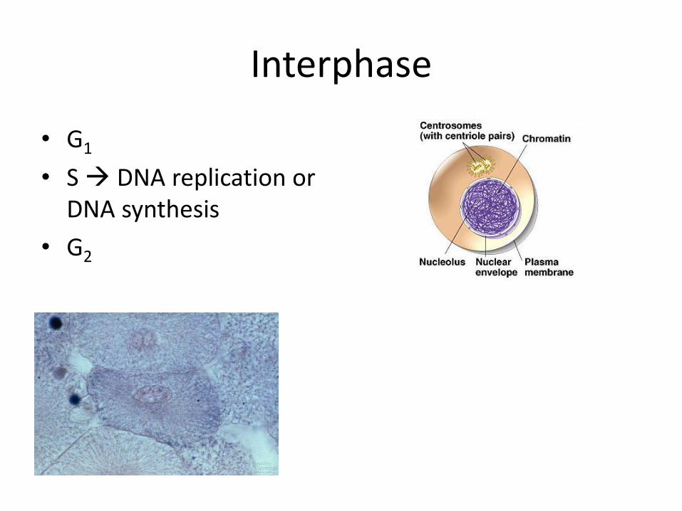
Interphase
• G1
• S DNA replication or DNA synthesis
• G2
Section 8.5 Figure 8.22
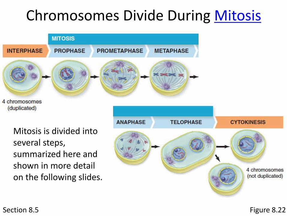
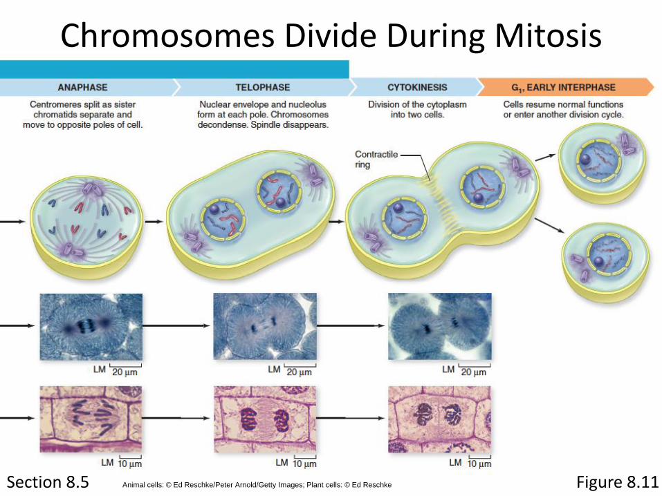
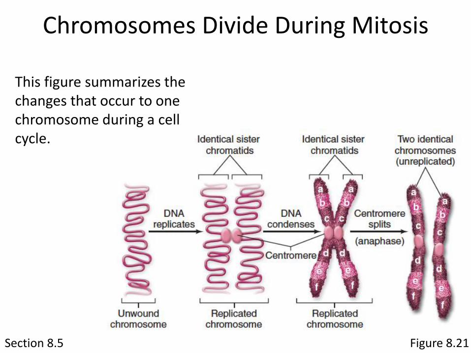
Chromosomes Divide During Mitosis
Mitosis is divided into several steps, summarized here and shown in more detail on the following slides.
Section 8.5 Figure 8.11
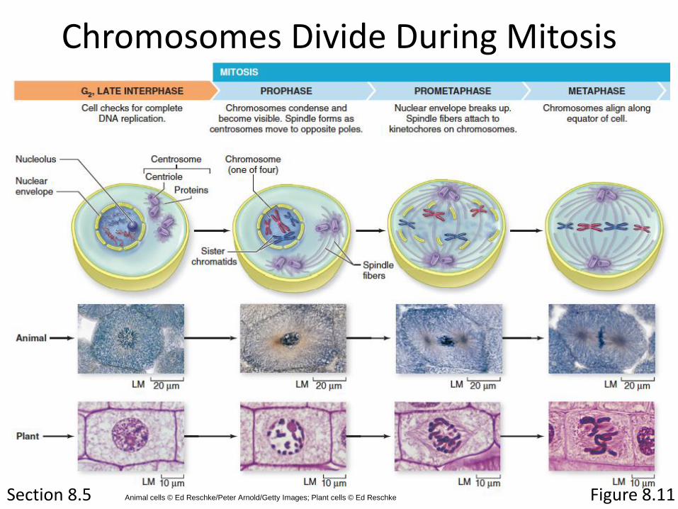
Chromosomes Divide During Mitosis
Animal cells © Ed Reschke/Peter Arnold/Getty Images; Plant cells © Ed Reschke
Section 8.5
Chromosomes Divide During Mitosis
Figure 8.11Animal cells: © Ed Reschke/Peter Arnold/Getty Images; Plant cells: © Ed Reschke
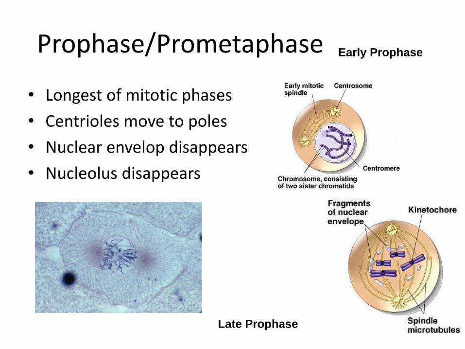
Prophase/Prometaphase
• Longest of mitotic phases
• Centrioles move to poles
• Nuclear envelop disappears
• Nucleolus disappears
Early Prophase
Late Prophase
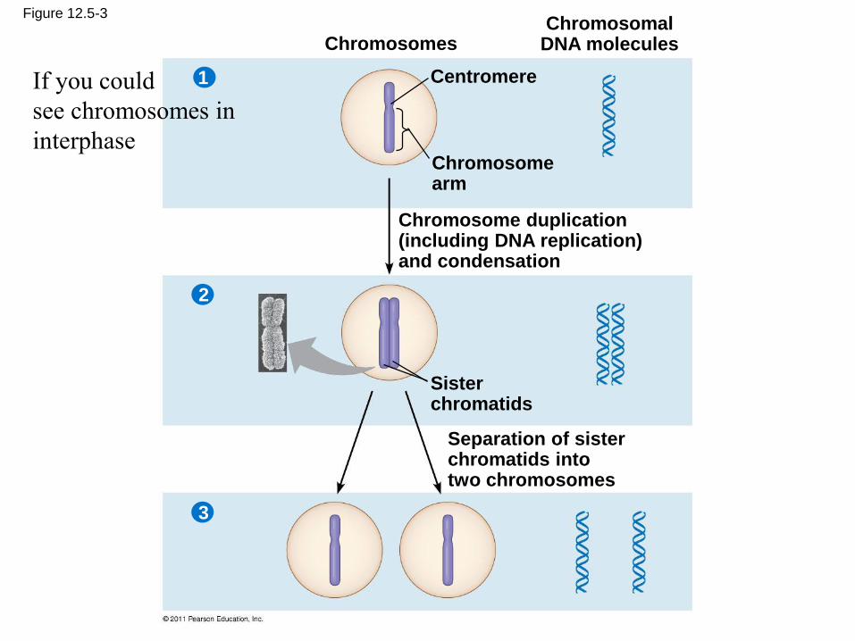
Figure 12.5-3
ChromosomesChromosomal
DNA molecules
Centromere
Chromosomearm
Chromosome duplication(including DNA replication)and condensation
Sisterchromatids
Separation of sisterchromatids intotwo chromosomes
1
2
3
If you could
see chromosomes in
interphase
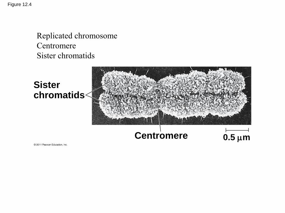
Figure 12.4
0.5 mCentromere
Sisterchromatids
Replicated chromosome
Centromere
Sister chromatids
Section 8.5
Chromosomes Divide During Mitosis
Figure 8.21
This figure summarizes the changes that occur to one chromosome during a cell cycle.
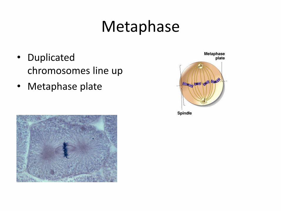
Metaphase
• Duplicated chromosomes line up
• Metaphase plate
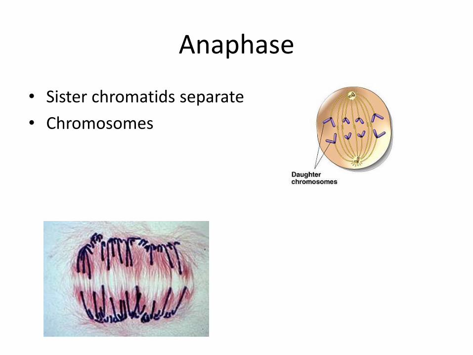
Anaphase
• Sister chromatids separate
• Chromosomes
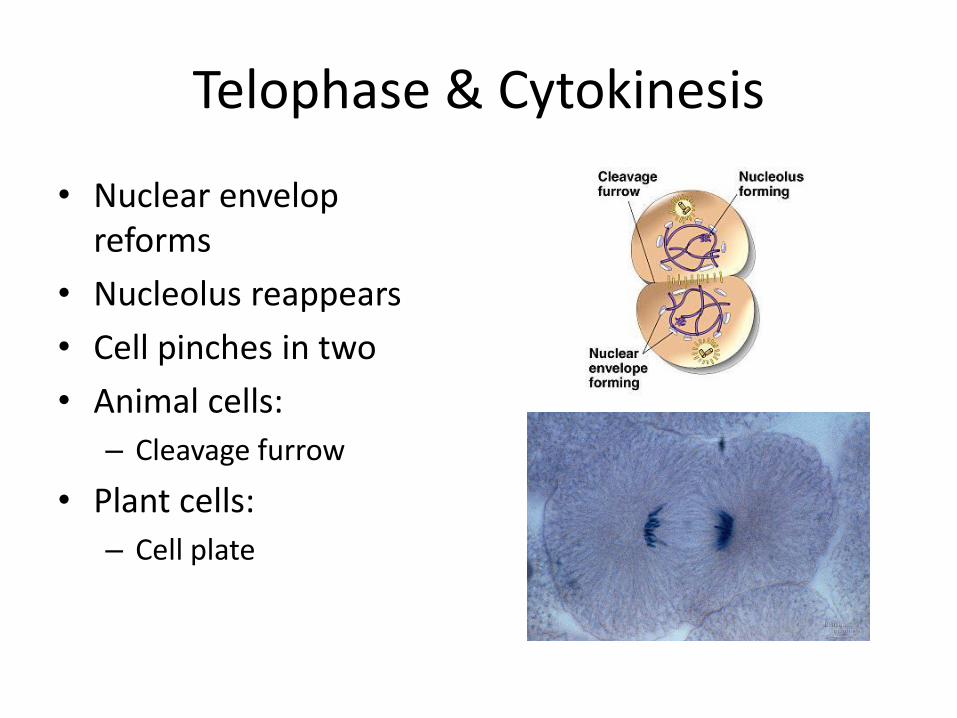
Telophase & Cytokinesis
• Nuclear envelop reforms
• Nucleolus reappears
• Cell pinches in two
• Animal cells:
– Cleavage furrow
• Plant cells:
– Cell plate
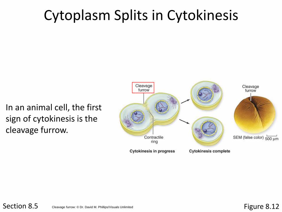
Section 8.5 Figure 8.12
Cytoplasm Splits in Cytokinesis
In an animal cell, the first sign of cytokinesis is the cleavage furrow.
Cleavage furrow: © Dr. David M. Phillips/Visuals Unlimited
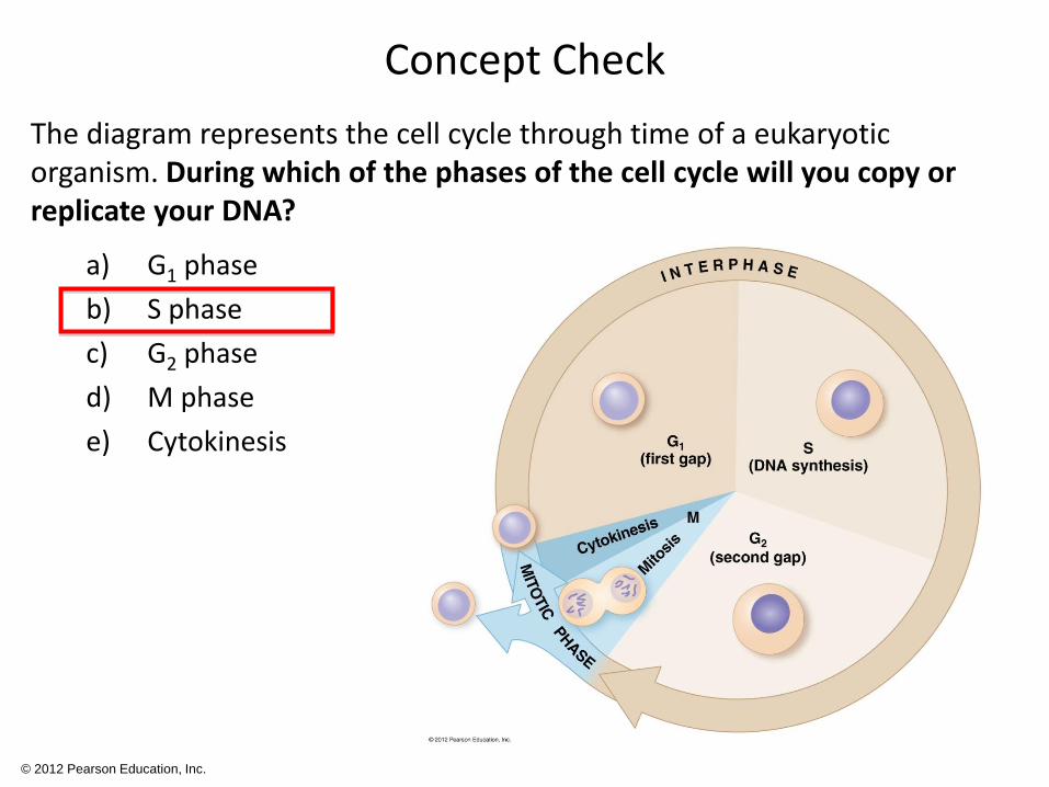
Concept Check
The diagram represents the cell cycle through time of a eukaryotic organism. During which of the phases of the cell cycle will you copy or replicate your DNA?
a) G1 phase
b) S phase
c) G2 phase
d) M phase
e) Cytokinesis
© 2012 Pearson Education, Inc.
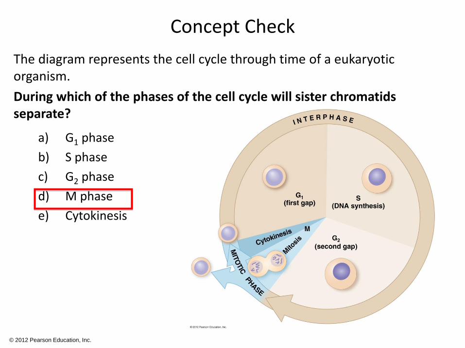
Concept Check
The diagram represents the cell cycle through time of a eukaryotic organism.
During which of the phases of the cell cycle will sister chromatids separate?
a) G1 phase
b) S phase
c) G2 phase
d) M phase
e) Cytokinesis
© 2012 Pearson Education, Inc.
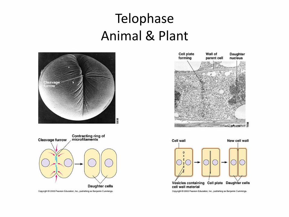
TelophaseAnimal & Plant
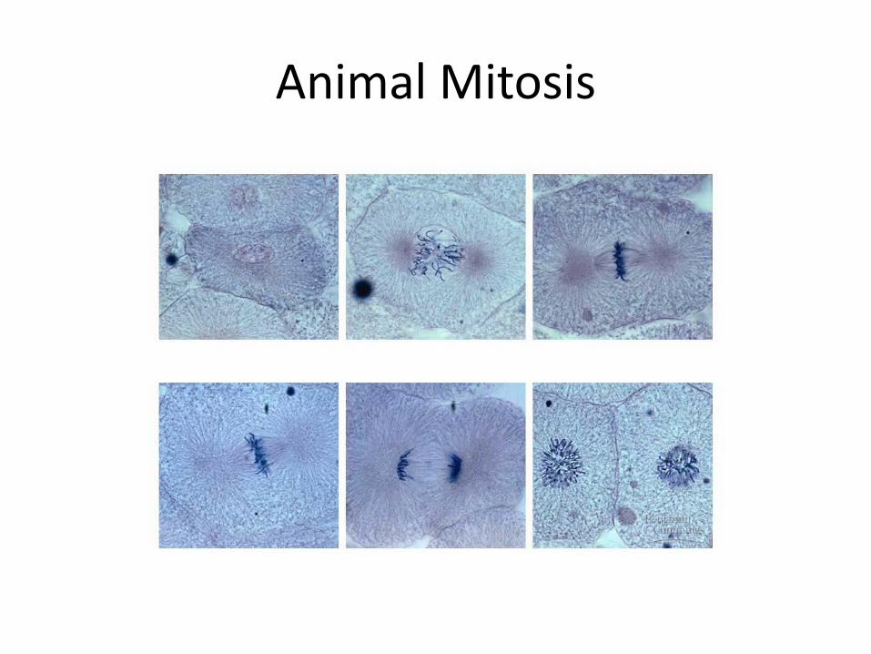
Animal Mitosis
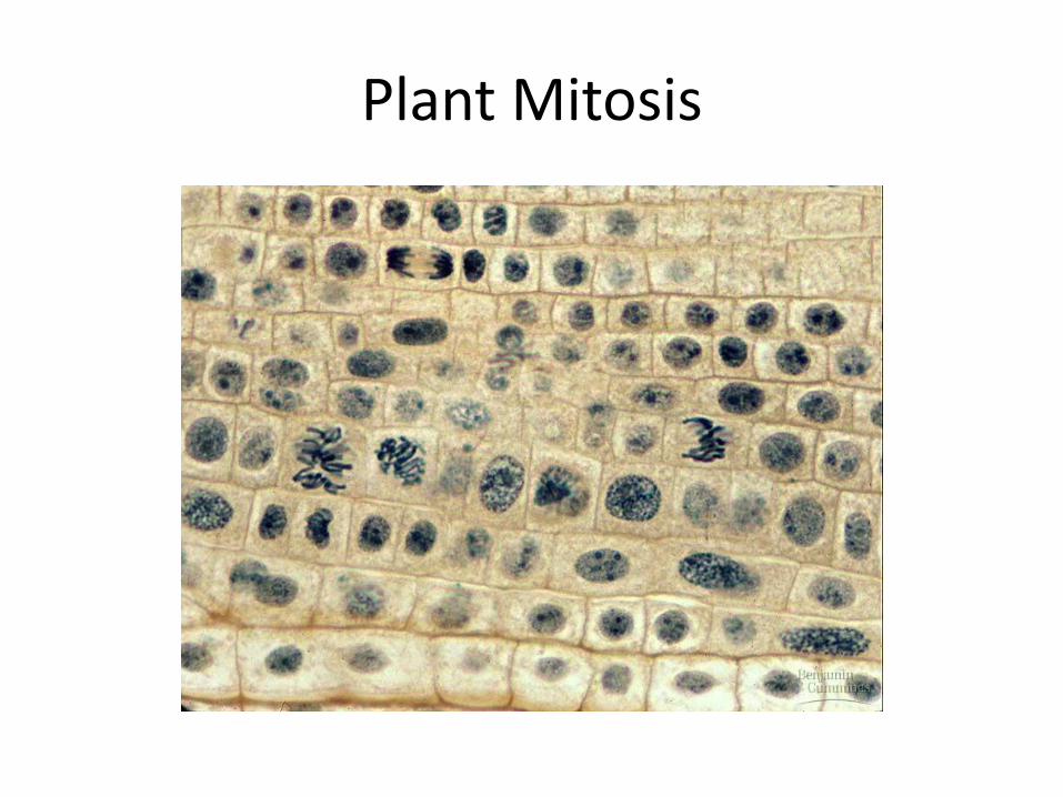
Plant Mitosis
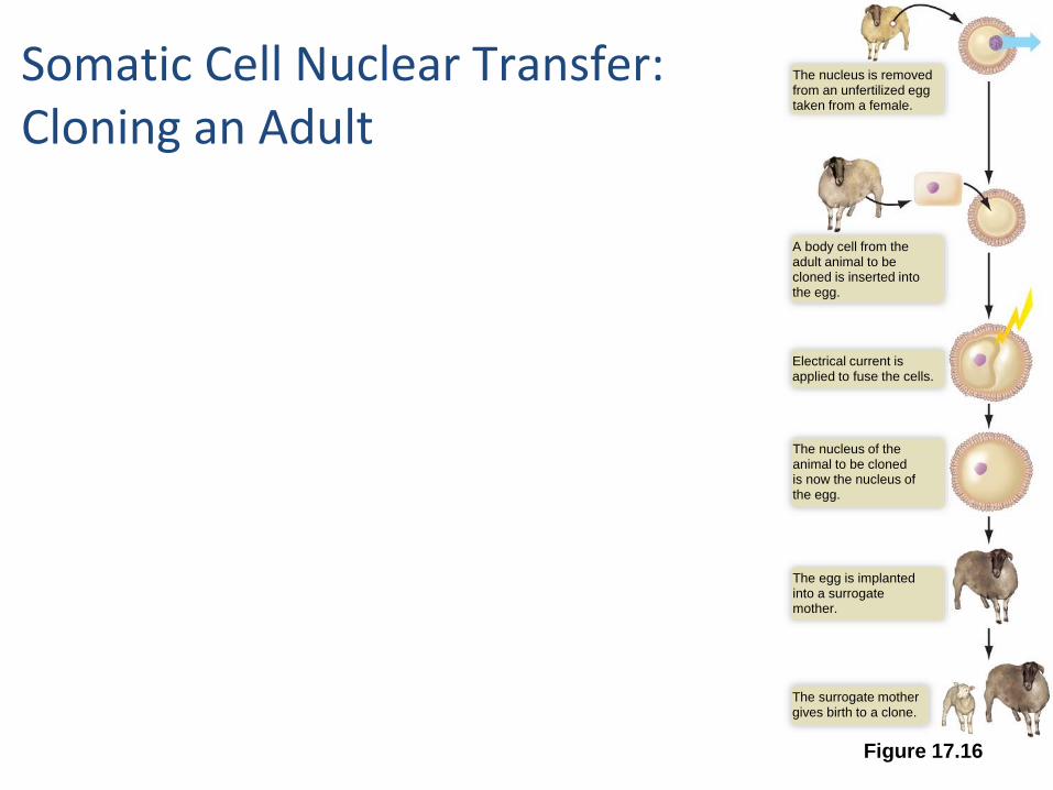
Figure 17.16
The nucleus is removedfrom an unfertilized eggtaken from a female.
A body cell from theadult animal to becloned is inserted intothe egg.
Electrical current isapplied to fuse the cells.
The nucleus of theanimal to be clonedis now the nucleus ofthe egg.
The egg is implantedinto a surrogatemother.
The surrogate mothergives birth to a clone.
Somatic Cell Nuclear Transfer: Cloning an Adult
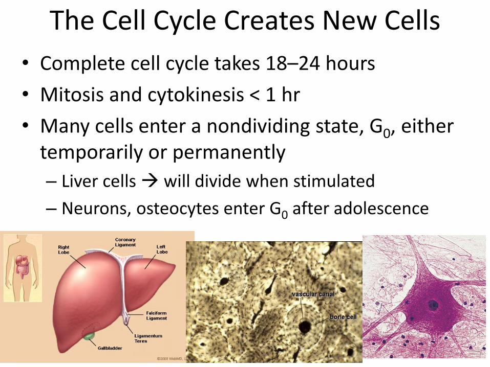
The Cell Cycle Creates New Cells
• Complete cell cycle takes 18–24 hours
• Mitosis and cytokinesis < 1 hr
• Many cells enter a nondividing state, G0, either temporarily or permanently
– Liver cells will divide when stimulated
– Neurons, osteocytes enter G0 after adolescence
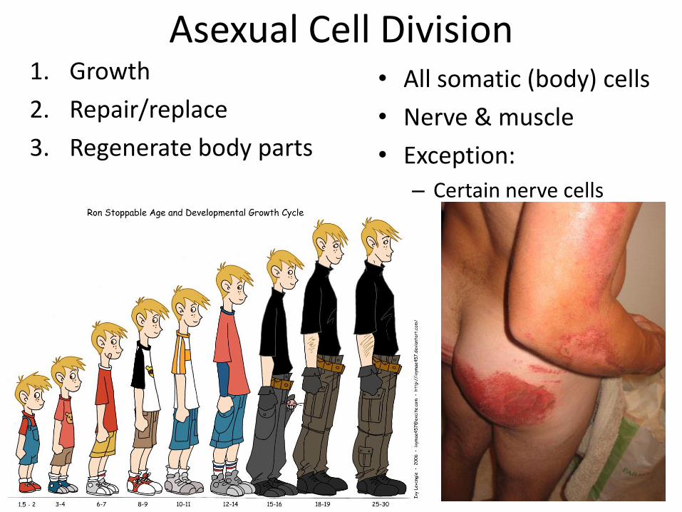
Asexual Cell Division1. Growth
2. Repair/replace
3. Regenerate body parts
• All somatic (body) cells
• Nerve & muscle
• Exception:
– Certain nerve cells
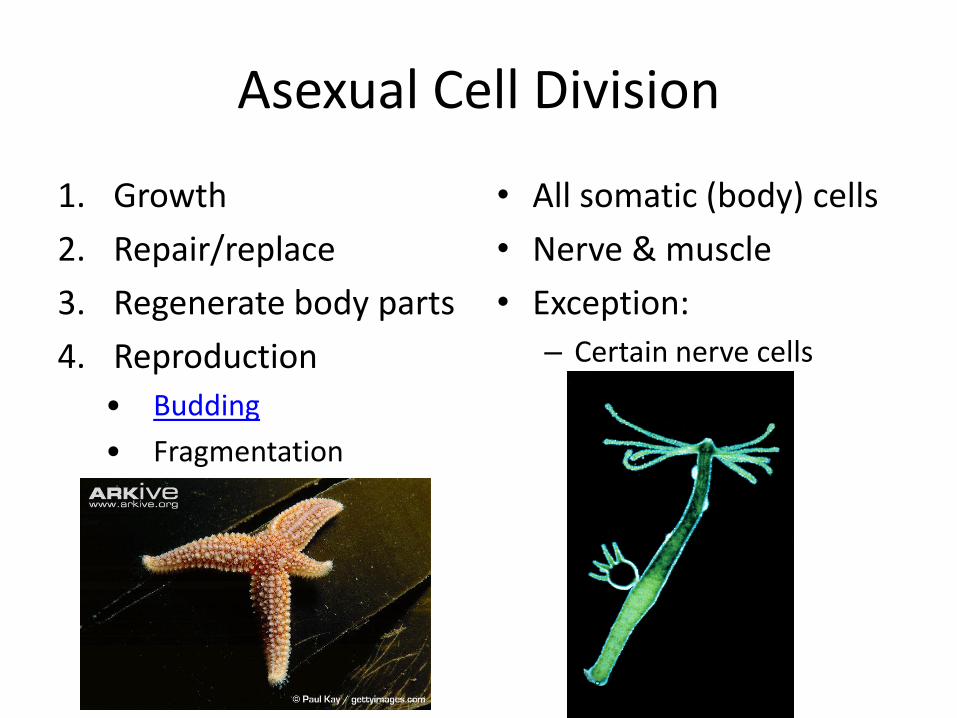
Asexual Cell Division
1. Growth
2. Repair/replace
3. Regenerate body parts
4. Reproduction
• Budding
• Fragmentation
• All somatic (body) cells
• Nerve & muscle
• Exception:
– Certain nerve cells
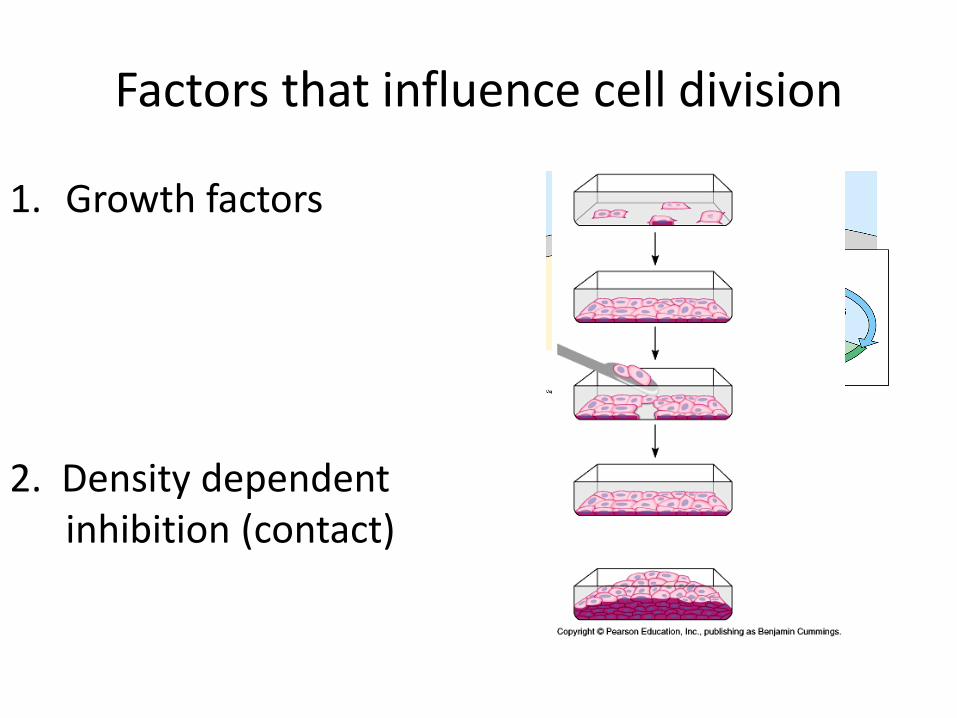
Factors that influence cell division
1. Growth factors
2. Density dependent inhibition (contact)
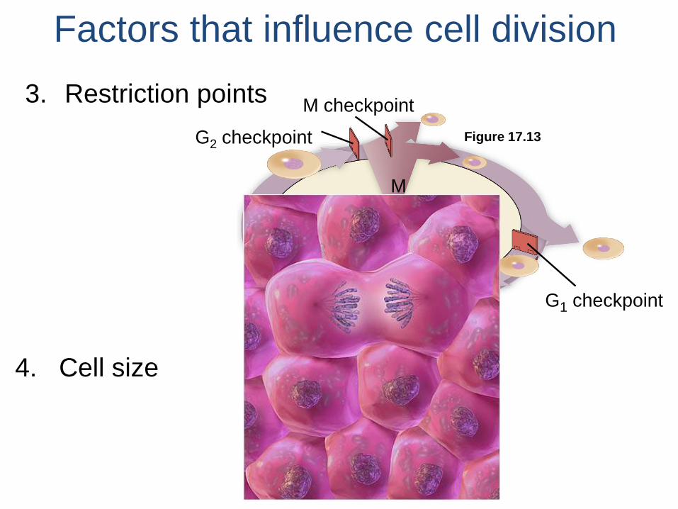
Figure 17.13
M checkpoint
G2 checkpoint
M
G2
S
G1
G1 checkpoint
Factors that influence cell division
3. Restriction points
4. Cell size
Factors that influence cell division
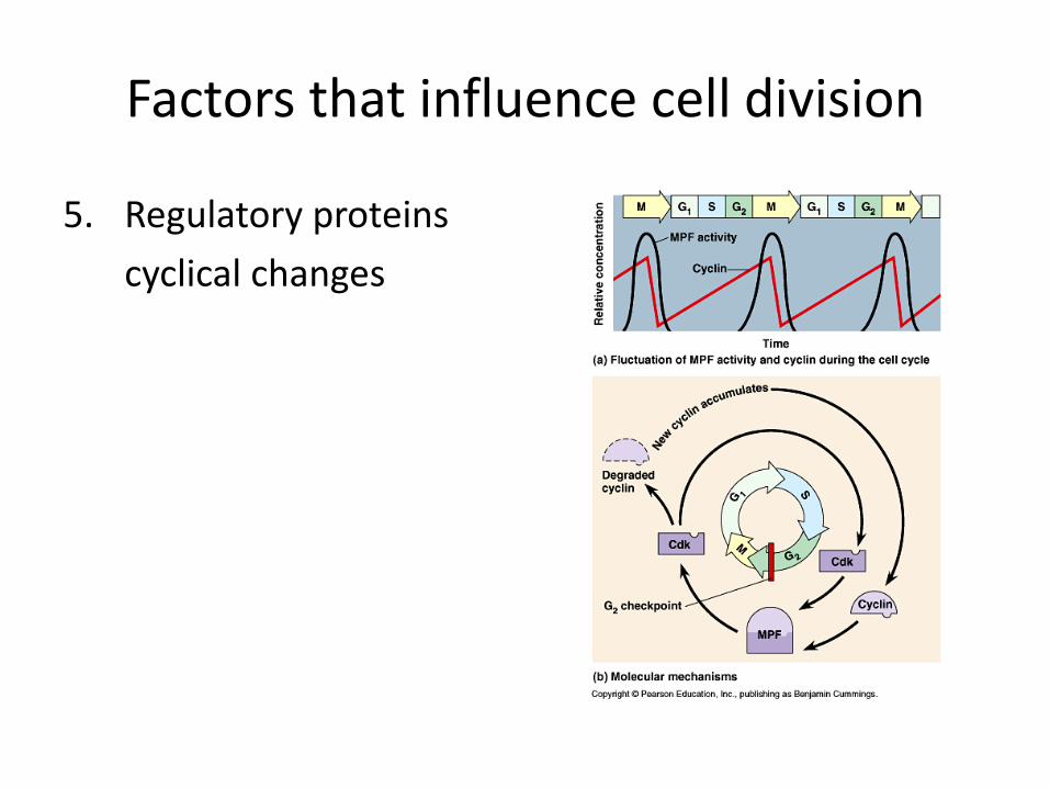
5. Regulatory proteins
cyclical changes
Section 8.6
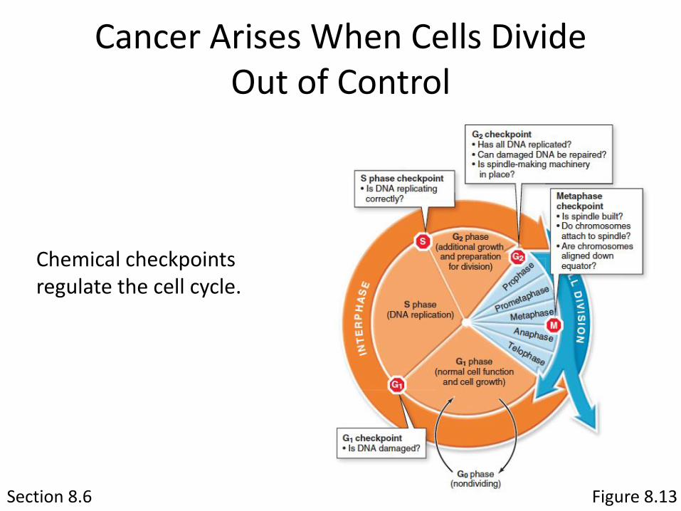
Cancer Arises When Cells Divide Out of Control
Chemical checkpoints regulate the cell cycle.
Figure 8.13
Section 8.6
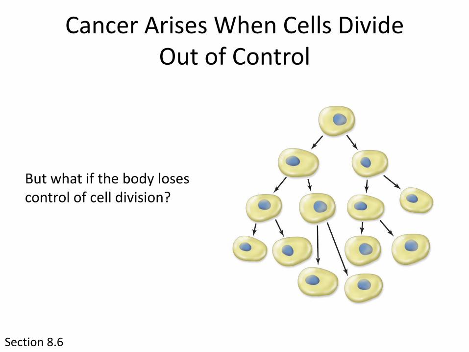
Cancer Arises When Cells Divide Out of Control
But what if the body loses control of cell division?
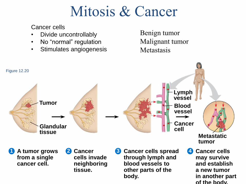
Figure 12.20
Glandulartissue
Tumor
Lymph vessel
Bloodvessel
Cancercell
Metastatictumor
A tumor growsfrom a singlecancer cell.
Cancer cells invade neighboringtissue.
Cancer cells spreadthrough lymph andblood vessels to other parts of the body.
Cancer cells may survive and establisha new tumor in another part of the body.
4321
Mitosis & CancerCancer cells
• Divide uncontrollably
• No “normal” regulation
• Stimulates angiogenesis
Benign tumor
Malignant tumor
Metastasis
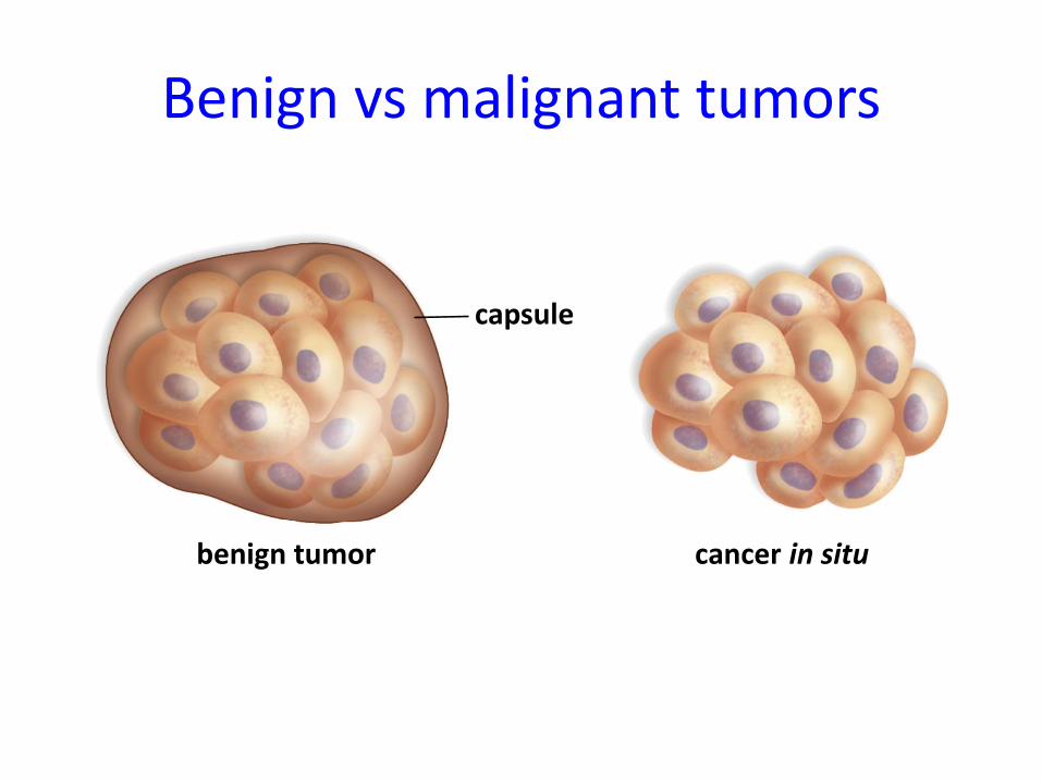
Benign vs malignant tumors
benign tumor cancer in situ
capsule
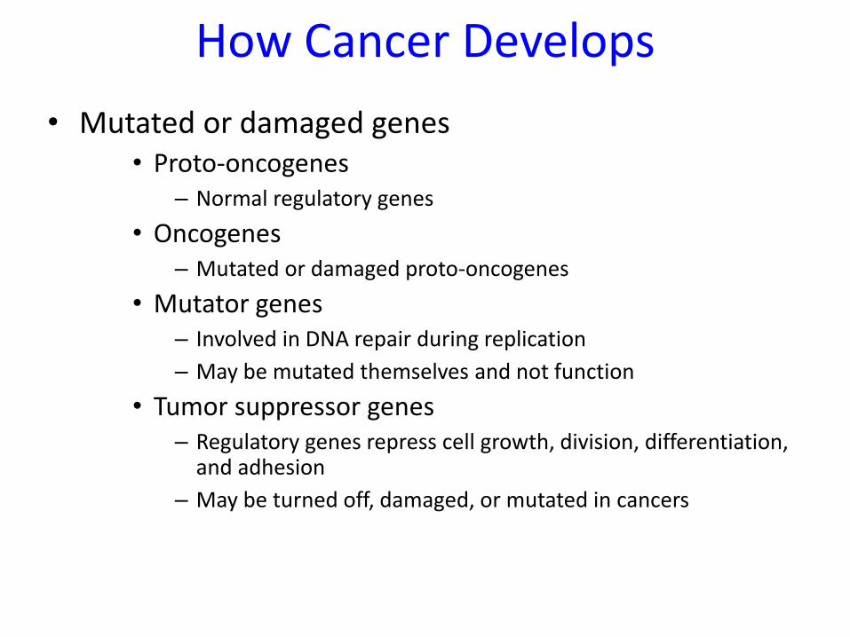
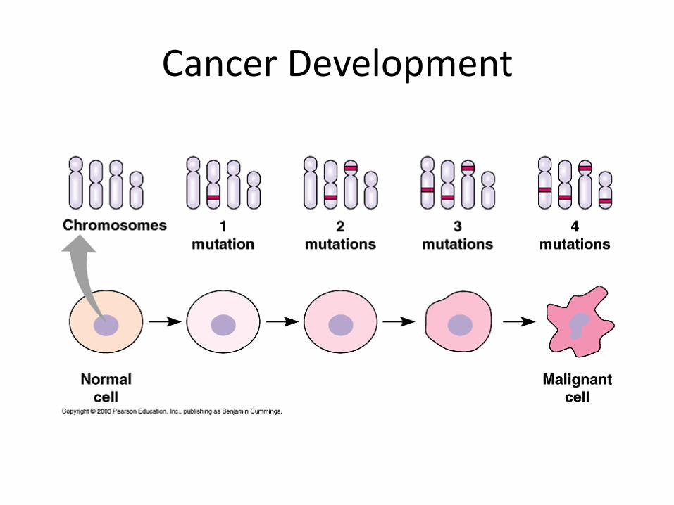
How Cancer Develops
• Mutated or damaged genes• Proto-oncogenes
– Normal regulatory genes
• Oncogenes– Mutated or damaged proto-oncogenes
• Mutator genes– Involved in DNA repair during replication
– May be mutated themselves and not function
• Tumor suppressor genes– Regulatory genes repress cell growth, division, differentiation,
and adhesion
– May be turned off, damaged, or mutated in cancers
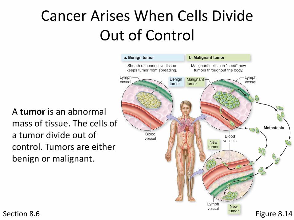
Section 8.6 Figure 8.14
Cancer Arises When Cells Divide Out of Control
A tumor is an abnormal mass of tissue. The cells of a tumor divide out of control. Tumors are either benign or malignant.
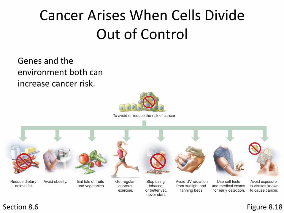
Section 8.6
Cancer Arises When Cells Divide Out of Control
Genes and the environment both can increase cancer risk.
Figure 8.18
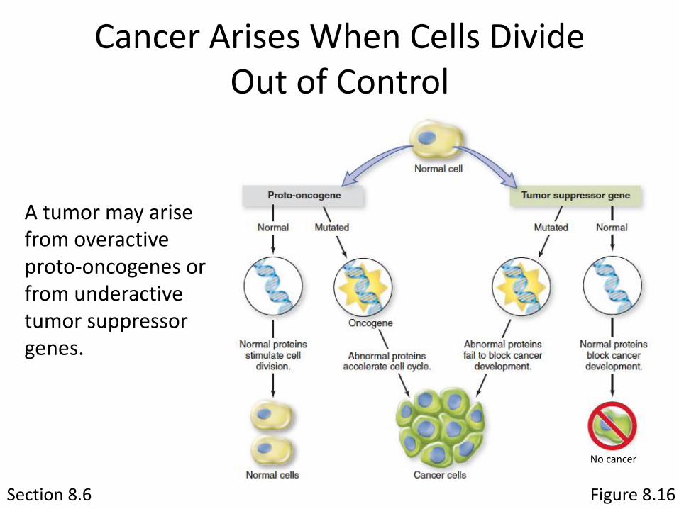
Section 8.6
Cancer Arises When Cells Divide Out of Control
A tumor may arise from overactive proto-oncogenes or from underactive tumor suppressor genes.
Figure 8.16
No cancer
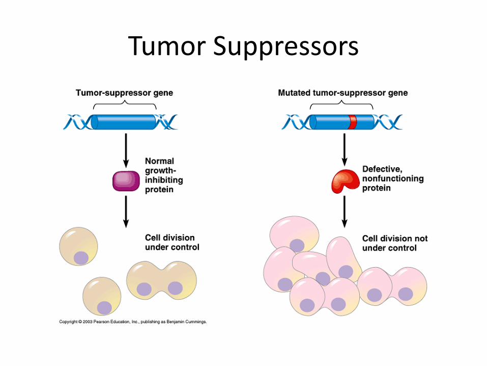
Tumor Suppressors
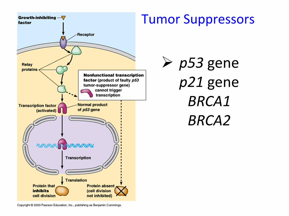
p53 genep21 geneBRCA1BRCA2
Tumor Suppressors
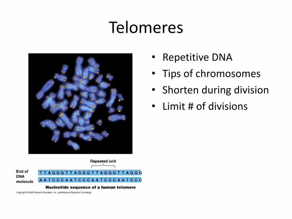
Telomeres
• Repetitive DNA
• Tips of chromosomes
• Shorten during division
• Limit # of divisions
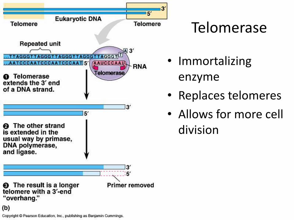
Telomerase
• Immortalizing enzyme
• Replaces telomeres
• Allows for more cell division
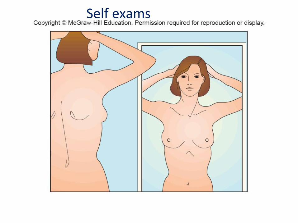
Fig. 20BSelf exams
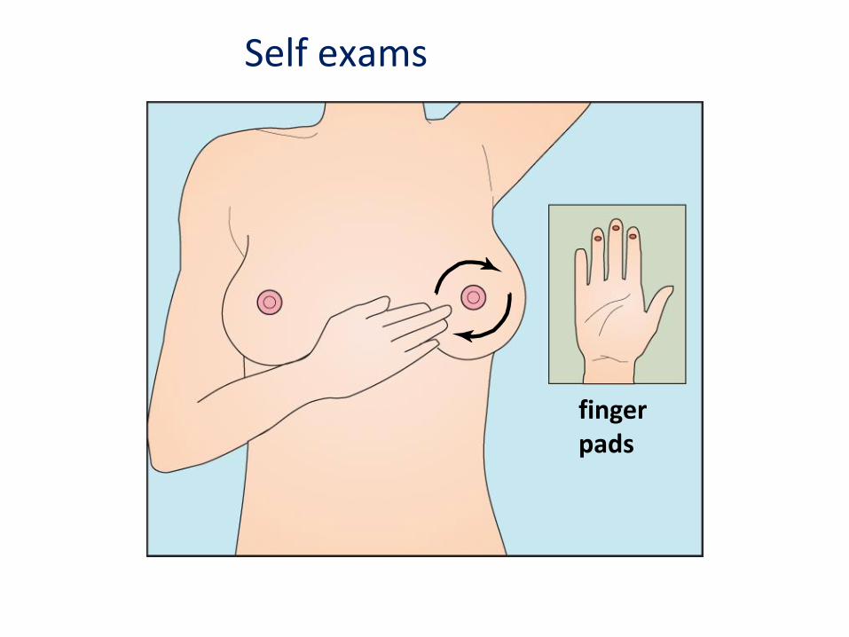
fingerpads
Self exams
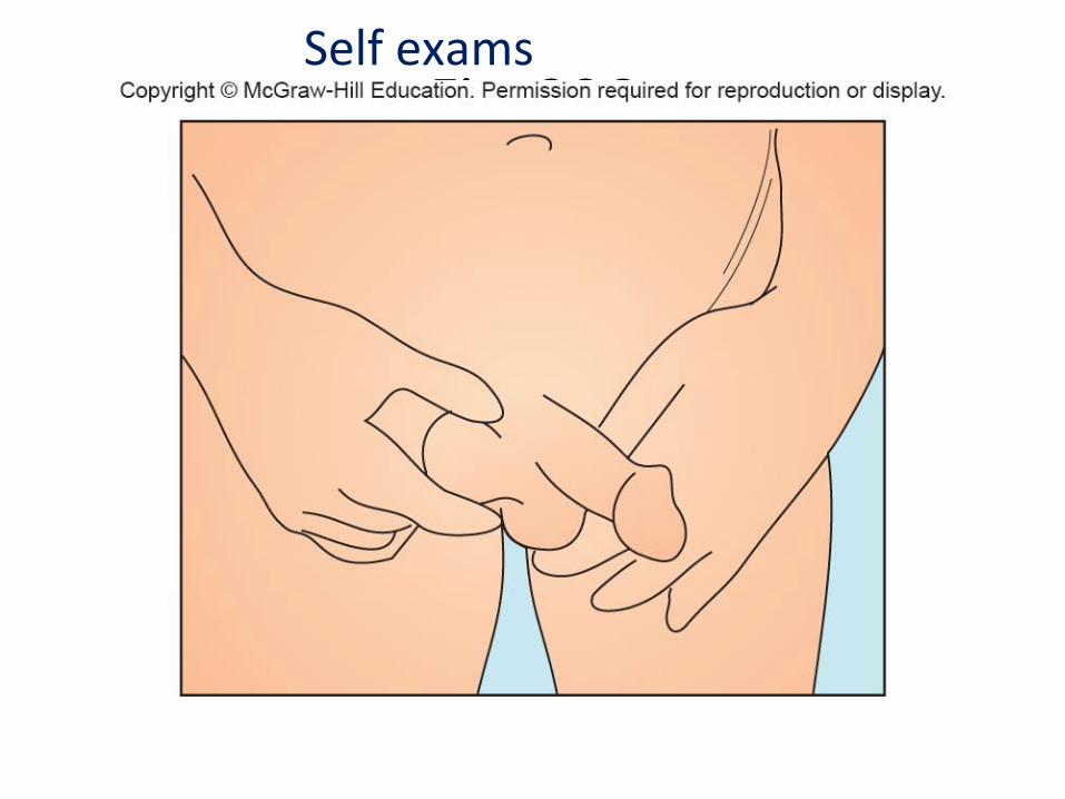
Fig. 20CSelf exams
8.6 Mastering Concepts
What keeps cells from dividing when they are not supposed to?
World’s tallest man © Frederic J. Brown/AFP/Getty Images
Cancer Development
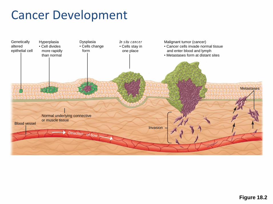
Figure 18.2
Genetically
altered
epithelial cell
Hyperplasia
• Cell divides
more rapidly
than normal
Dysplasia
• Cells change
form
In situ cancer
• Cells stay in
one place
Malignant tumor (cancer)
• Cancer cells invade normal tissue
and enter blood and lymph
• Metastases form at distant sites
Normal underlying connective
or muscle tissueBlood vessel
Invasion
Metastases
Cancer Development
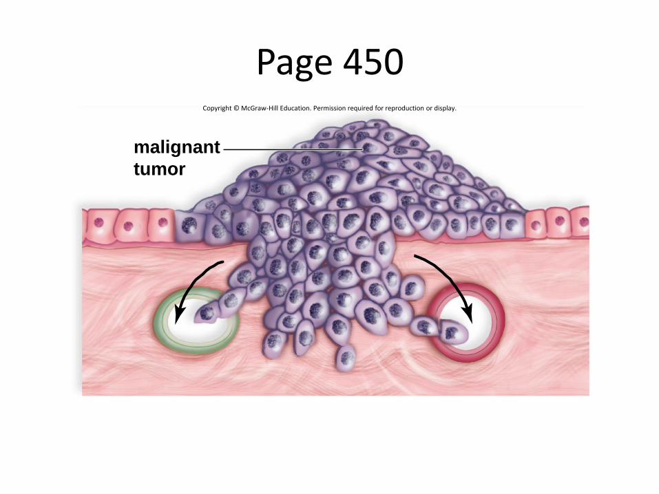
Page 450Copyright © McGraw-Hill Education. Permission required for reproduction or display.
malignant
tumor
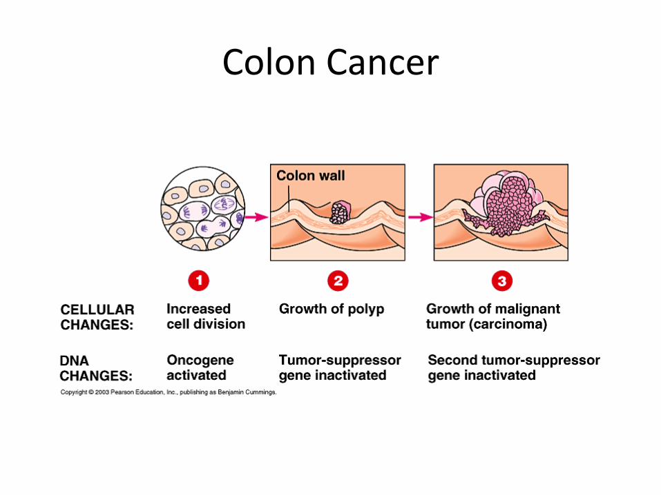
Colon Cancer
Advances in Diagnosis: Early Detection
• Tumor imaging
– X-rays
• Example: mammogram
– Positron emission tomography (PET)
– Magnetic resonance imaging (MRI)
• Genetic testing
– Identify mutated genes
– Privacy and treatment issues
• Enzyme tests for cancer markers
– Screening large numbers of people
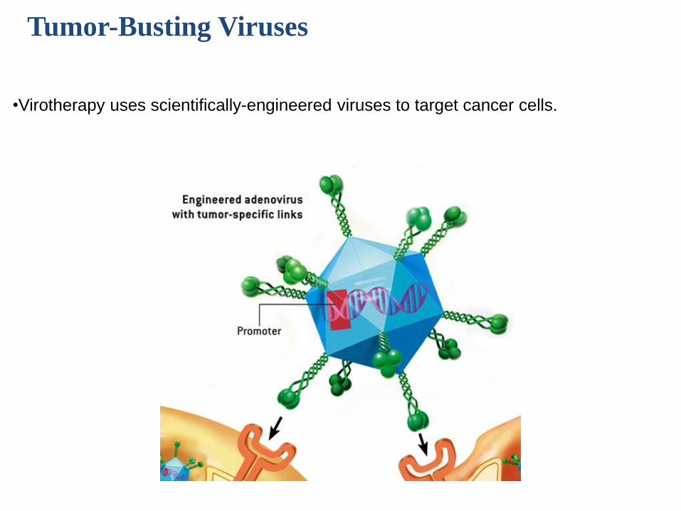
Tumor-Busting Viruses
•Virotherapy uses scientifically-engineered viruses to target cancer cells.
Tying topics together - Comprehension
Tumor-Busting Viruses
How do viruses selectively attach to target cells?
a) target cells display unique cell-surface receptors
b) target cells secrete unique identification proteins
c) viruses attach to specific materials engulfed by cells
d) viruses are not selective about target cells
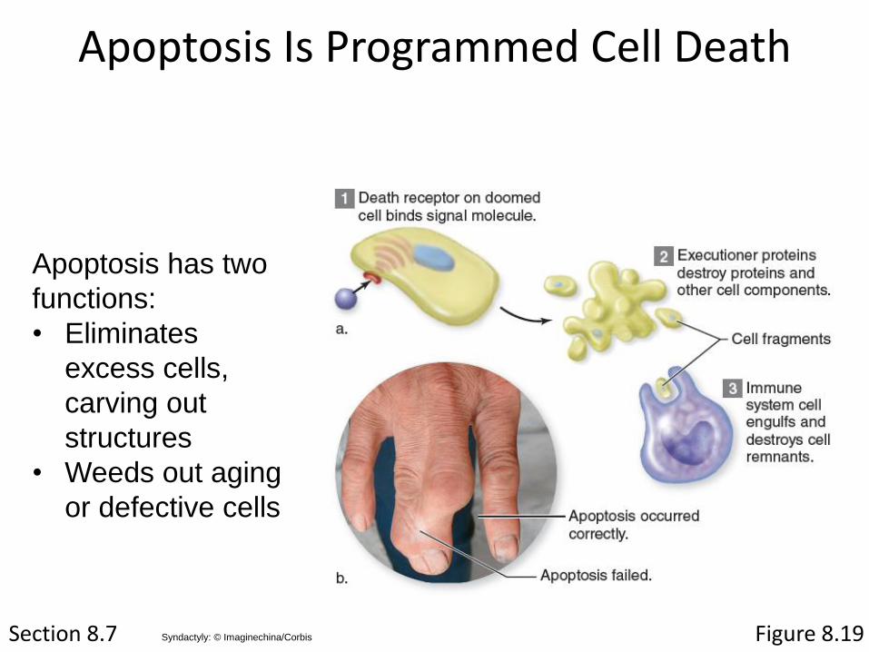
Apoptosis Is Programmed Cell Death
Section 8.7
Apoptosis has two
functions:
• Eliminates
excess cells,
carving out
structures
• Weeds out aging
or defective cells
Figure 8.19Syndactyly: © Imaginechina/Corbis