Embed Size (px)
Citation preview
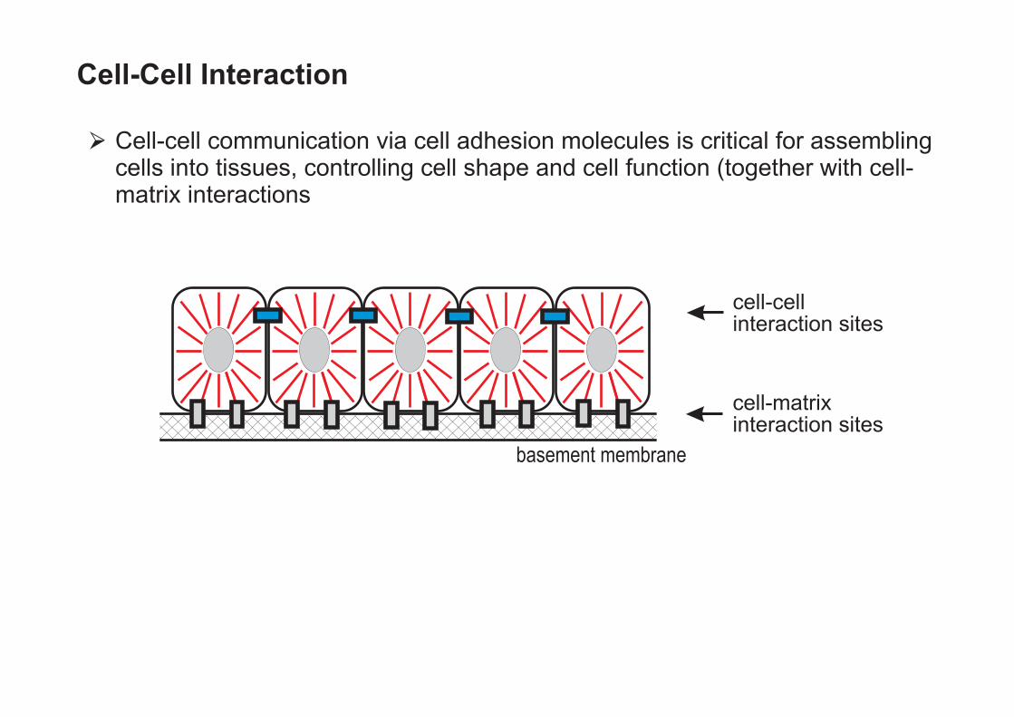
basement membrane
cell-matrixinteraction sites
cell-cellinteraction sites
Cell-Cell Interaction
Cell-cell communication via cell adhesion molecules is critical for assemblingcells into tissues, controlling cell shape and cell function (together with cell-matrix interactions

Cell-Cell InteractionCell-Cell Interaction
Major cell-cell adhesion sites can be visualized by electron microscopy
Tight junctionsAdherens junctionsDesmosomes
Hemidesmosom
Desmosom
Gapjunction
Adherensjunction
Tightjunction
Mikrovili
Basallamina

Cells can also interact without forming visible adhesion sites
most important cell adhesion molecules:
Cadherins
Ig-superfamily
Selectins
(all cells)
(ubiquitous, enriched in the nervous system)
(interaction of leukocytes with endothelial cells
Cell-Cell InteractionCell-Cell Interaction
Major cell-cell adhesion sites can be visualized by electron microscopy
Tight junctionsAdherens junctionsDesmosomes

Cadherins
• Ca2+-dependent cell-cell adhesion proteins,
epressed in almost all cells of vertebrates and invertebrates
• Transmembrane proteins
• characteristic structural feature:
- tandem repeats of homologous domains (CAD-domains): length of about 110 amino acids
-
β-sheet structure, distantly related- to immunoglobulin-fold
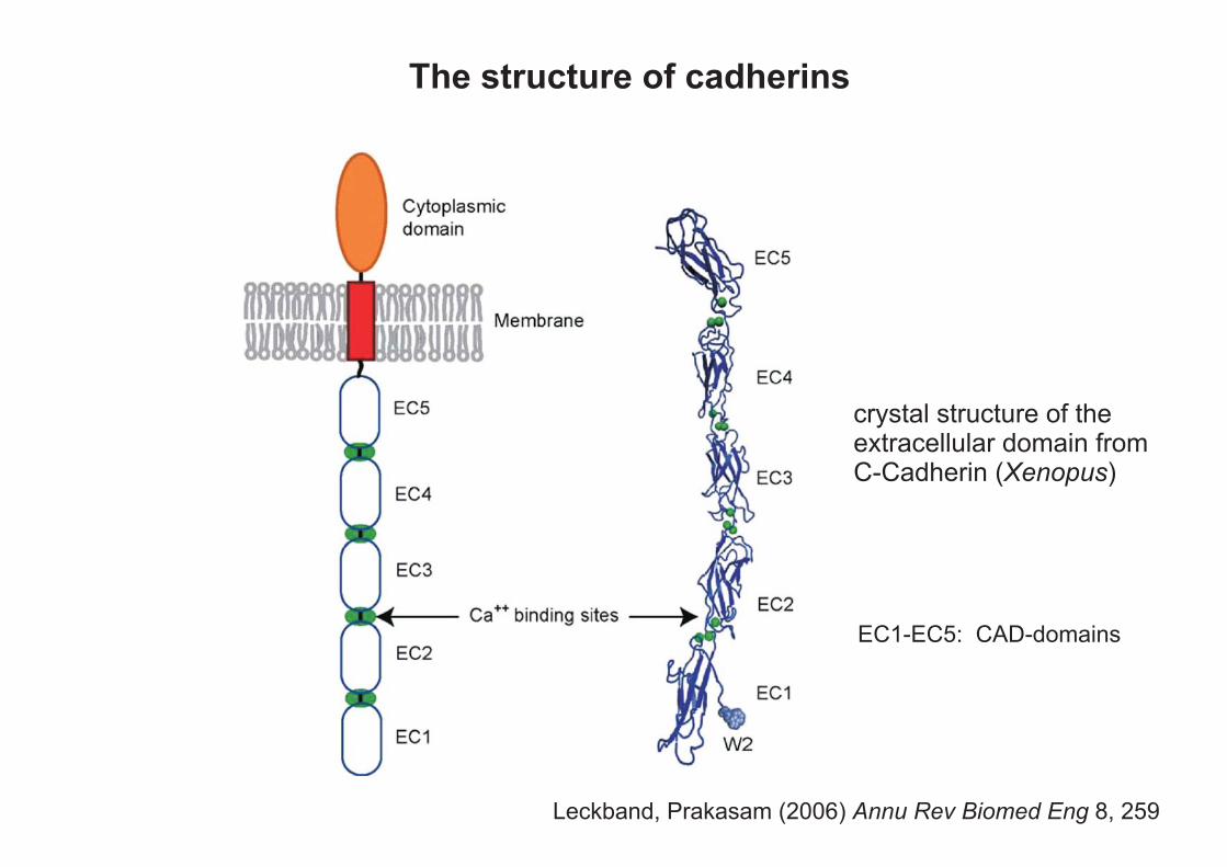
Leckband, Prakasam (2006) 8, 259Annu Rev Biomed Eng
The structure of cadherins
crystal structure of theextracellular domain fromC-Cadherin ( )Xenopus
EC1-EC5: CAD-domains
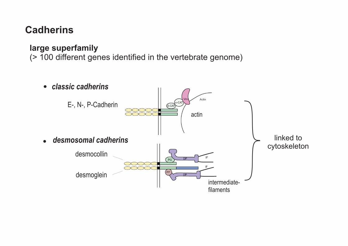
classic cadherins
actin
E-, N-, P-Cadherin
desmocollin
desmoglein
desmosomal cadherins
intermediate-filaments
linked tocytoskeleton
Cadherins
large superfamily(> 100 different genes identified in the vertebrate genome)

protocadherins
7TM-cadherin (Flamingo)
Fat-family
�-Pcdh(CNR-Cadherin)
EGF-RepeatsLaminin-G-Domäne-Repeats
in many cases notassociated with the
cytoskeleton
Cadherins
large superfamily(> 100 different genes identified in the vertebrate genome)

Classical Cadherins (Examples)
Name Main location Junction Association Phenotype when Inactivated in Mice
E-cadherin epithelia epithelial adherens Junctions (AJ)
embryonic lethal, mice die at blastocyst stage
N-cadherin neurons, heart, muscle, eye lens
AJ-like structures, synapses
embryonic lethal, heart defects
VE-cadherin Endothelzellen adherens junctions
embryonic lethal, apoptosis of endothelial cells
cadherin-11
mesenchymal cells, neural crest cells, several cancer types
bone and behavioral abnormalities, resistant to rheumatic arthritis
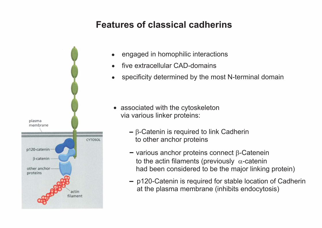
Features of classical cadherins
five extracellular CAD-domains
engaged in homophilic interactions
specificity determined by the most N-terminal domain
associated with the cytoskeletonvia various linker proteins:
various anchor proteins connect -Catenein
to the actin filaments (previously
�
-cateninhad been considered to be the major linking protein)
�
�-Catenin is required to link Cadherinto other anchor proteins
p120-Catenin is required for stable location of Cadherinat the plasma membrane (inhibits endocytosis)
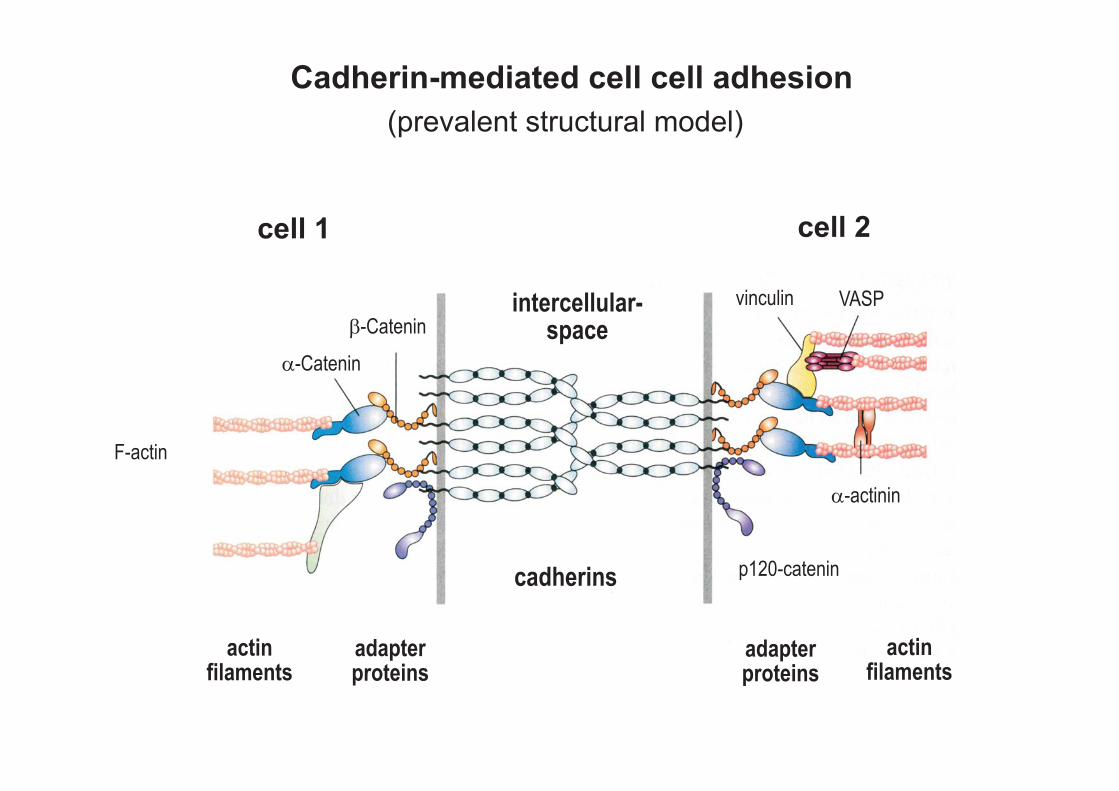
adapterproteins
actinfilaments
F-actin
cell 1 cell 2
intercellular-space
cadherins
�-Catenin
vinculin VASP
�-actinin
p120-catenin
�-Catenin
(prevalent structural model)
adapterproteins
actinfilaments
Cadherin-mediated cell cell adhesion

Major functions of classical cadherins :
cell shape, tissue architecture cell polarity
cell-sorting
signaling
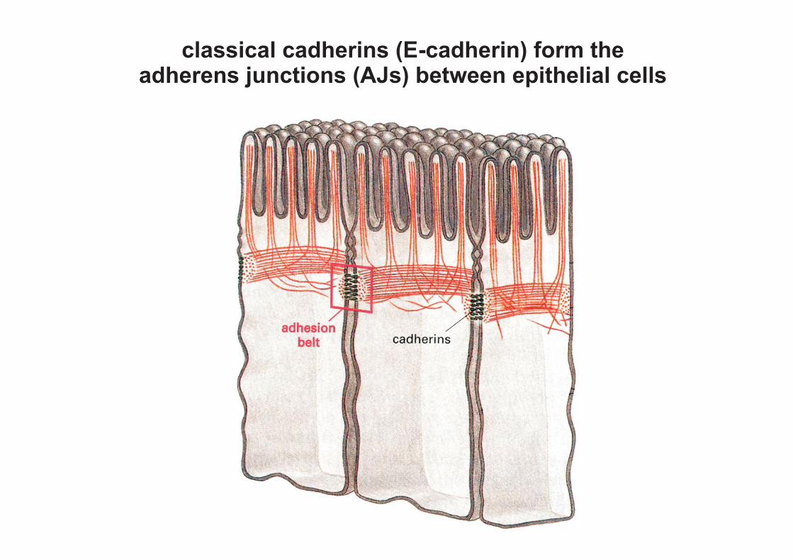
classical cadherins (E-cadherin) form theadherens junctions (AJs) between epithelial cells

Major functions of classical cadherins :
cell shape, tissue architecture cell polarity
cell-sorting
signaling
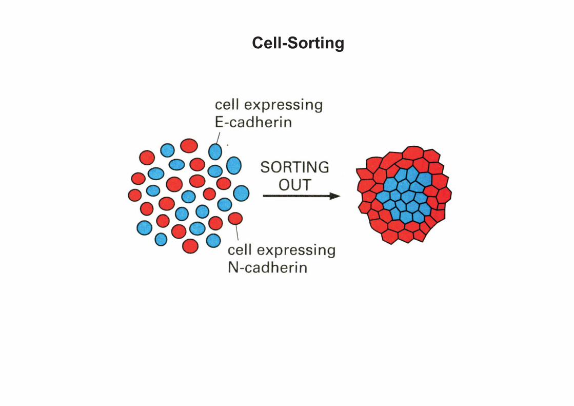
Cell-Sorting

Ektoderm
neural crestcells
infolding of theneural tube
expression of E-Cadherin
expression of N-Cadherin
expression of Cadherin-6B
expression of Cadherin-7
changes of cadherin expression during development of theneural tube and neural crest cells
neural tubeclosure

cell adhesion by cadherins is strictly regulated examples:
- migration of neural crest cells: E-cadherin expression is down-regulated (→ disassembly of AJs) and substituted by other cadherins (→ allowing cell contact during migration)
- gastrulation:
inhibited, if E-cadherin is not down-regulated (Cells cannot leave the ectoderm)
- pathological conditions: Carcinomas down-regulate the expression of E-Cadherin, which is often substituted by N-cadherin

Major functions of classical cadherins :
cell shape, tissue architecture cell polarity
cell-sorting
signaling

Signaling via Cadherins / Catenins
examples:
1. Modulation of kinase/ phosphatase activities by binding to cadherin/catenin complexes
- N-cadherin activates FGF-receptors - E-cadherin inhibits FGF- and EGF-receptors - VE-cadherin: Co-receptor for VEGF,
loss of VE-cadherin leads to apoptosis of endothelilal cells
2. Wnt-signaling
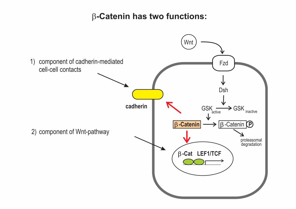
Wnt
Dsh
GSK
Fzd
active inactive
proteasomaldegradation
GSK
P-Catenin-Catenin
LEF1/TCF-Cat
cadherin
�-Catenin has two functions:
1)
2) component of Wnt-pathway
component of cadherin-mediatedcell-cell contacts

Non-Classical Cadherins (Examples)
Name Main Location Functions
Desmosomal Cadherins (they are associated with intermediate filaments)
Desmoglein, Desmocollin
skin (desmosomes) inactivation results in detachment of the skin, hyperproliferation, abnormal differentiation
Protocadherins PAPS (paraxial protocadherin)
paraxial mesoderm cell movements during gastrulation
-, -,- Pcdh´s (clustered protocadherins)
neurons (synapses) three gene clusters encoding more than 50 cadherins. All members of one cluster contain the same C-terminal domain. Not connected to cytoskeleton
Cadherin 23 Protocadherin 15
inner ear deafness, if inactivated
Others Flamingo-cadherins
epithelia, associated with junctions
planar cell polarity
FAT, Dachsous epithelia, associated with junctions
inactivation in Dros. results in overgrowth of imaginal discs, defects in differentiation and morphogenesis, formation of tumors

Desmosomes
intermediatefilaments
cadherin-familyadhesion proteins
dense plaque ofanchor proteins

Desmosomes
intermediatefilaments
cadherin-familyadhesion proteins
desmoplakin
plakoglobin
plakophilin
desmoglein
desmocollin
plasma membrane
cadherin-familyadhesion proteins
intermediatefilaments
(= -catenin)�
dense plaque ofanchor proteins

Planar cell polarity (PCP)
Polarization of cells of a cell sheetwithin the plane
PCP is best investigated in Drosophila wing epithelium
flamingo-cadherin
Seifert, Mlodzik (2007)8, 126Nat Rev Genet
mutant clone(marker: wing hairs)
wing epithelial cells

Cell-Cell Interaction the most important cell adhesion molecules:
• Cadherins (all cells)
• Ig-superfamily (ubiquitous, enriched in the nervous system)
• Selectins ( interaction of leukocytes with endothelial cells)
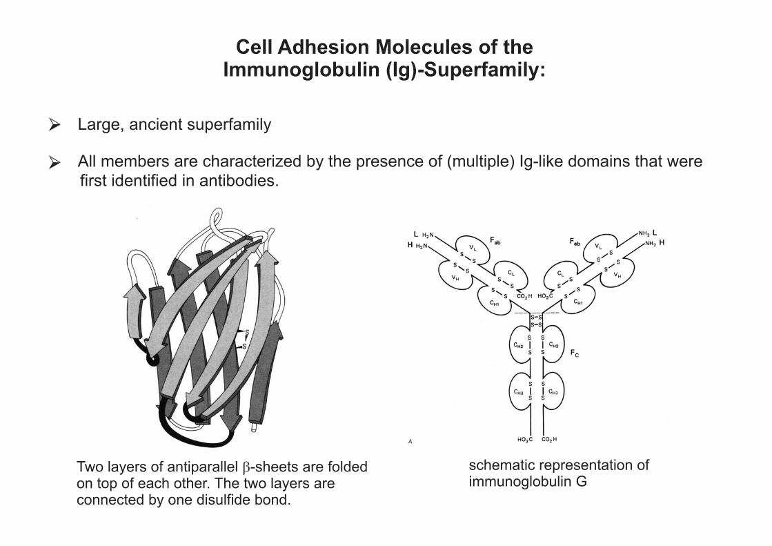
Cell Adhesion Molecules of theImmunoglobulin (Ig)-Superfamily:
Large, ancient superfamily
All members are characterized by the presence of (multiple) Ig-like domains that were
first identified in antibodies.
Two layers of antiparallel -sheets are foldedon top of each other. The two layers areconnected by one disulfide bond.
� schematic representation ofimmunoglobulin G

Cell Adhesion Molecules of the Immunoglobulin (Ig)-Superfamily: In addition to cell adhesion molecules and antibodies, Ig-like domains are found in
many proteins. Ig-like domains are encoded in 765 human genes!
Examples are: - T-cell receptor, CD4, MHC I, MHC.II - FGF-receptor

Cell Adhesion Molecules of the Immunoglobulin (Ig)-Superfamily: Cell adhesion is not Ca2+ -dependent (in contrast to the cadherins). Cell adhesion can be
- homophilic (between like molecules) or - heterophilic (between different molecules)
Binding is more stable than binding between cadherins!
plasmamembrane
plasmamembrane
intercellularspace
Possible binding pattern between two Ig-CAMs

Some Types of Ig-Superfamily Cell Adhesion Molecules
Name Main Location Typical Function
neuronal CAM´s
NCAM L1 (NgCAM)
nerves cell adhesion, -migration, axonal growth and guidance
non-neuronal CAM´s
ICAM´s (intercellular adhesion molecules)
leukocytes, lymphocytes, endothelial cells
VCAM´s (vascular cell adhesion molecules) endothelial cells
PECAM (platelet endothelial cell adhesion Molecule)
platlets; endothelial cells
adhesion of lymphocytes and leukocytes to endothelial and epithelial cells (inflammation response)
Nectins adherens junctions together with cadherins essential for adherens junctions assembly

cc
cc
cc
cc
cc
cc
cc
cc
cc
cc
cc
cc
Schematic Structure of Some Ig-CAMs
cc
cc
cc
cc
cc
cc
cc
cc
cc
cc
cc
cc
cc
cc
cc
cc
cc
cc
cc
cc
cc
NCAM
L1
ICAM-1
ICAM-2
Nectin-1
F11/Contactin
Ig-domain
FNIII-domain
GPI-anchor
Biological functions of neuronal CAM´s (examples): cell-cell adhesion (neuron-neuron, neuron-glia)
regulation of signal transduction processes (e.g. activation of FGF-receptors via interaction with NCAM)
cell migration
neurite outgrowth
- axon fasciculation - axon guidance and sorting along nerve fascicles - branching of axon bundles

Cell-substrate interactions(integrins, laminins)
receptors for soluble factors(growth factors, chemoattractants)
Neurite outgrowth
cell-cell interactions(CAMs, cadherins)
Cell-substrate interactions(integrins, laminins)

changes in fasciclin-II expression during axonal growth
(NCAM equivalent in insects)
three grasshopperneurons
fasciclin-II

NCAM (neural cell adhesion molecule)
• Ig-superfamiliy protein with
- 5 Ig-domains - 2 Fibronectin-typeIII domains
• multiple forms by alternative spicing (120 - 180 kDa):
- transmembrane proteins - lipid-bound proteins (GPI-anchor)
• homophilic interaction •
in the adult mainly expresseed in the nervous system (and neuromuscular during embryogenesis expression in many tissues,
vertebrate NCAMs:
junctions)
• modification by polysialic acids (PSA)
OH
OHNH
2
COOHH
OH
H
OH
CH2
OH
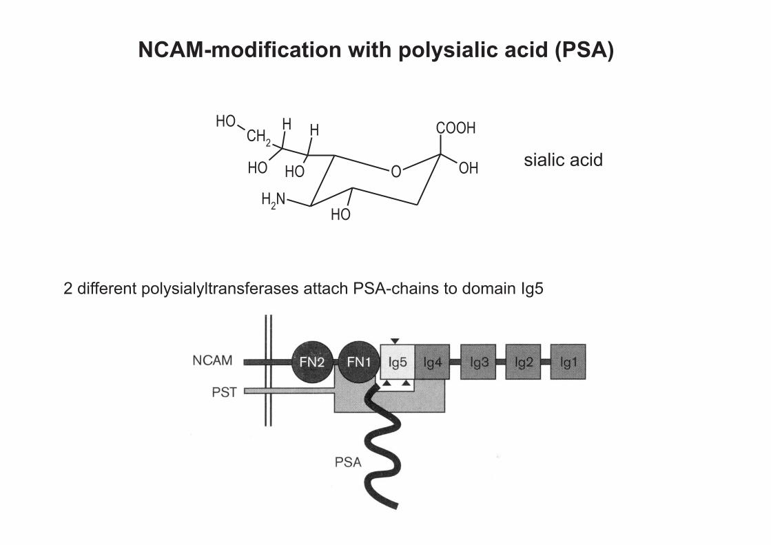
NCAM-modification with polysialic acid (PSA)
sialic acid
2 different polysialyltransferases attach PSA-chains to domain Ig5
O

Biological Functions of Polysialic Acid
modulation of cell adhesion strength
modulation of fasciculation and branching of axons
olfactory bulb
trajectory of motor axons in the embryonic chicken hindlimb
-
-
neuraminidasetreatedcontrol
wild type NCAM-mutant

normal
L1-mutant
Neural Cell Recognition molecule L1
corticospinal tract (CST) at the levelof the pyramids in the medulla
many mutations in the L1 gene have beendescribed in humans leading to a broadspectrum of diseases:
- mental retardation (IQ < 20 - 50)
- hydrocephalus
- corpus callosum hypoplasia
- absence or diminution of thecorticospinal tract
- paralysis of lower extremities


