Embed Size (px)
Citation preview
APPLIED AND ENVIRONMENTAL MICROBIOLOGY,0099-2240/98/$04.0010
Sept. 1998, p. 3473–3479 Vol. 64, No. 9
Copyright © 1998, American Society for Microbiology. All Rights Reserved.
Cellular Events Involved in Survival of Individual ArbuscularMycorrhizal Symbionts Growing in the
Absence of the HostCABLE LOGI,1 CRISTIANA SBRANA,2 AND MANUELA GIOVANNETTI1*
Dipartimento di Chimica e Biotecnologie Agrarie1 e Centro di Studio per la Microbiologiadel Suolo C.N.R.,2 Universita degli Studi di Pisa, 56124 Pisa, Italy
Received 1 December 1997/Accepted 3 June 1998
A survival strategy operating in the absence of the host was shown in obligately biotrophic arbuscular my-corrhizal (AM) symbionts. When no host-derived signals from the surrounding environment were perceived bygerminating spores, fungal hyphae underwent a programmed growth arrest and resource reallocation, allowinglong-term maintenance of viability and host infection capability. The early stages of mycelial growth of AM fun-gi were studied by a combination of time-lapse and video-enhanced light microscopy, image analysis, and im-munodetection, with the aim of acquiring knowledge of cell events leading to the arrest of mycelial growth. Thetime-course of growth arrest was resolved by precisely timing the growth rate and magnitude of the myceliumoriginating from individual spores of Glomus caledonium. Extensive mycelial growth was observed during thefirst 15 days; thereafter, fungal hyphae showed retraction of protoplasm from the tips, with formation of retrac-tion septa separating viable from empty hyphal segments. This active process involved migration of nuclei andcellular organelles and appeared to be functional in the ability of the fungus to survive in the absence of a host.Immunodetection of cytoskeletal proteins, metabolic activity, and the retention of infectivity of germinated sporesconfirmed the developmental data. The highest amounts of tubulins were detected when hyphal growth hadceased but when retraction of protoplasm was most active. This was consistent with the role of the cytoskeletonduring protoplasm retraction. Succinate dehydrogenase activity in hyphae proximal to the mother spore wasstill detectable in 6-month-old mycelium, which remained viable and able to form appressoria and producesymbiotic structures.
Arbuscular mycorrhizal (AM) fungi are obligately biotrophicorganisms, widespread in soil, which live symbiotically with mostplant roots. It has been calculated that they are able to colonizethe roots of 80% of plant species belonging to all phyla of landplants (29).
Fossil records and DNA sequence data have provided evi-dence that arbuscular mycorrhizas were established as early as410 million to 360 million years ago (24, 25, 27). Because ofthis long-lasting coevolution with their host plants, AM fungihave evolved unequivocal mechanisms of host recognitionwhich play a fundamental role in their life cycle. Only in thepresence of signals released by host roots does morphogeneticdifferentiation of infection structures occur, allowing a func-tional symbiosis to be established (8, 11). Despite this, the rec-ognition process does not involve specific regulation of the ger-mination event by host-derived signals. AM fungal spores arecapable of germination and growth from a quiescent-like statein response to different edaphic and environmental conditions,irrespective of the presence of host plants (7, 12, 15, 19, 20, 30).This is contrary to findings described for other biotrophs, suchas some pathogenic fungi, whose resting structures germinateonly as a direct response to the presence of host plants (4, 5).Such behavior by AM fungal spores is apparently inconsistent,given that in the absence of the host these fungi are not capa-ble of extensive independent mycelial growth and consequentlyof completion of the life cycle (8, 13, 19, 21). Thus, many au-thors have observed that germlings of different AM fungal spe-cies ceased growth within 15 days of germination (2, 9, 13, 26).
The fate and behavior of individual AM fungal spores thatgerminate in the absence of appropriate hosts may affect thesurvival of individuals and populations. However, this funda-mental event in the life cycle of AM fungi has so far not beenadequately investigated. In this work we studied the early stagesof mycelial growth of AM fungi, adopting a combination oftime-lapse and video-enhanced light microscopy, image analy-sis, and immunodetection in order to acquire knowledge of cellevents leading to the arrest of mycelial growth in the absenceof host plants.
Our work was aimed at investigating (i) the time course offungal growth in the absence of the host, from spore germina-tion to growth arrest; (ii) cell events involved in developmentalarrest; (iii) metabolic activity of the mycelium after growth ar-rest and its ability to restore growth and to differentiate infec-tion structures; and (iv) differential expression of cytoskeletalproteins during the various growth phases.
MATERIALS AND METHODS
Fungal material. Spores of Glomus caledonium (Nicolson et Gerdemann)Trappe et Gerdemann (Rothamsted isolate) and spores or sporocarps of Glomusmosseae (Nicolson et Gerdemann) Gerdemann et Trappe (Kent isolate) wereobtained from the collection of the Dipartimento di Chimica e BiotecnologieAgrarie, University of Pisa, Italy. Voucher specimens of these isolates weredeposited in the Herbarium of the Department of Botanical Sciences, Universityof Pisa, Herbarium Horti Botanici Pisani (PI), respectively, as PI-HMZ 8 andPI-HMZ 4.
Time course of hyphal growth in the absence of the host plant. Spores ofG. caledonium were surface sterilized with 2% chloramine T supplemented withstreptomycin (400 mg ml21) for 20 min, rinsed five times in sterile water, put on20- by 20-mm cellophane membranes (Hoefer, San Francisco, Calif.), and placedon 1% water agar (WA) in 9-cm-diameter petri dishes. After 4 days’ incubationin the dark at 28°C, spores were checked for germination. Cellophane mem-branes bearing germinated spores were transferred to 5.5-cm-diameter petridishes on a thin layer of 1% WA for growth measurements, which were carried
* Corresponding author. Mailing address: Dipartimento di Chimicae Biotecnologie Agrarie, Via del Borghetto 80, 56124 Pisa, Italy.Phone: 39-50-571561. Fax: 39-50-571562. E-mail: [email protected].
3473
on May 12, 2018 by guest
http://aem.asm
.org/D
ownloaded from
out under a Reichert-Jung Polyvar light microscope. Hyphal length was assessedby using Quantimet 500 image analysis software (Leica, Milan, Italy).
The lengths of both protoplasm-containing and empty mycelium were mea-sured daily on a group of 12 spores, up to the time of growth arrest. The meangrowth rate of the mycelium during the early phase was calculated on eightspores by dividing the mycelial length by the elapsed time (about 6 days). Thelength of total mycelium of a group of 12 spores was measured monthly, up to 6months.
Time-lapse video microscopy. Surface-sterilized spores of G. caledonium andG. mosseae were germinated as previously described. After 1 week of incubation,individual spores were mounted on microscope slides in sterile distilled water(SDW). The coverslips were sealed with 1% WA, which was periodically wettedwith SDW. With this method, individual spores could be held for more than 6 h,allowing the entire process of septum formation to be monitored. Spores wereobserved under a Polyvar light microscope equipped with differential interfer-ence contrast optics and with a 3 CCD color video camera connected to a video-cassette recorder (Hi 8, EV-C2000E; Sony Co., Tokyo, Japan) and to a video-graphic printer (UP-890 CE; Sony Co.).
To observe vacuoles and vacuolar strands in hyphal tips at different stages ofprotoplasm retraction, 1-month-old germinated spores were stained with thefluorescent dye MDY-64 (yeast vacuole membrane marker; Molecular Probes,Inc., Eugene, Oreg.) (10 mM) in dimethyl sulfoxide–10 mM HEPES buffer, pH7.4. Other spores were stained with the cyanine dye 3,39-dihexyloxacarbocyanineiodide (Sigma Chemical Co., St. Louis, Mo.) (0.87 mM) in dimethyl sulfoxide-water. All spores were observed under epifluorescence by using a Polyvar mi-croscope equipped with an HBO 200 mercury lamp, with filter combination B1(BP 450-495, LP 520, DS 510).
To visualize the occurrence and location of nuclei and mitochondria in hyphaeduring the phase of protoplasm retraction, 1-month-old germinated spores weremounted on microscope slides in diamidinophenylindole (DAPI) (5 mg/ml) in a1:1 water-glycerol solution and observed under epifluorescence, with filter com-bination U1 (BP 330-380, LP 418, DS 420).
Immunoblotting of cytoskeletal proteins. To yield comparable amounts oftotal proteins from mycelium of different ages and from ungerminated spores,different numbers of fungal propagules were used: 300 sporocarps of G. mosseaeand 1,000 spores of G. caledonium were incubated for 30 days; 350 sporocarps ofG. mosseae and 2,000 spores of G. caledonium were incubated for 15 days; and400 sporocarps of G. mosseae and 2,000 spores of G. caledonium were utilized asnongerminated controls.
One-dimensional sodium dodecyl sulfate-polyacrylamide gel electrophoresiswas carried out with a discontinuous buffer system (18). Extracts (4 mg ofprotein) were loaded onto linear 12% polyacrylamide gels (10 by 8 by 0.75 mm)and run in a Bio-Rad (Richmond, Calif.) Mini Protean II slab cell system.
Immunodetection of cytoskeletal proteins was carried out as previously de-scribed (1). After electrophoresis, the proteins were transferred to nitrocellulosemembranes by using a Trans-Blot apparatus (Bio-Rad). Electrotransfer wasperformed first overnight at 100 mA, 20 V, and then for 1 h at 350 mA, 100 V,at 0°C. Nitrocellulose sheets were then blocked for 1 h in 2% bovine serumalbumin in phosphate-buffered saline (PBS) (pH 7.3) and incubated for 1 h atroom temperature with mouse monoclonal anti-a- and anti-b-tubulin and anti-actin antibodies, which were diluted 1:500 in PBS containing 0.1% bovine serumalbumin and 0.1% Triton X-100. Bound antibodies were visualized with rabbitanti-mouse immunoglobulin G antibody conjugated to peroxidase and diluted1:3,000 in PBS containing 0.1% Triton X-100. The peroxidase reaction wascarried out with 0.3 mg of 3,39-diaminobenzidine per ml in 1:1 (vol/vol) Tris-HCl(100 mM) (pH 7.6)–water, supplemented with 25 ml of H2O2 immediately priorto use. Color development was stopped by rinsing the membranes with SDW.
Metabolic activity and infectivity of 1- to 6-month-old mycelium. Succinatedehydrogenase (SDH) activity was assessed, as described by Smith and Giani-nazzi-Pearson (28), on 1- to 6-month-old germinated spores of G. caledonium.
Surface-sterilized spores were prepared as described previously, and at leastfive germinated spores were utilized for each assay. The lengths of hyphae showingformazan salt depositions on 6-month-old mycelium and the numbers of stained and
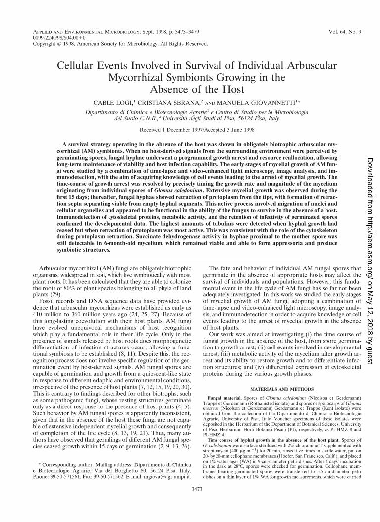
FIG. 1. Growth curves of mycelia developing from eight individual spores ofG. caledonium.
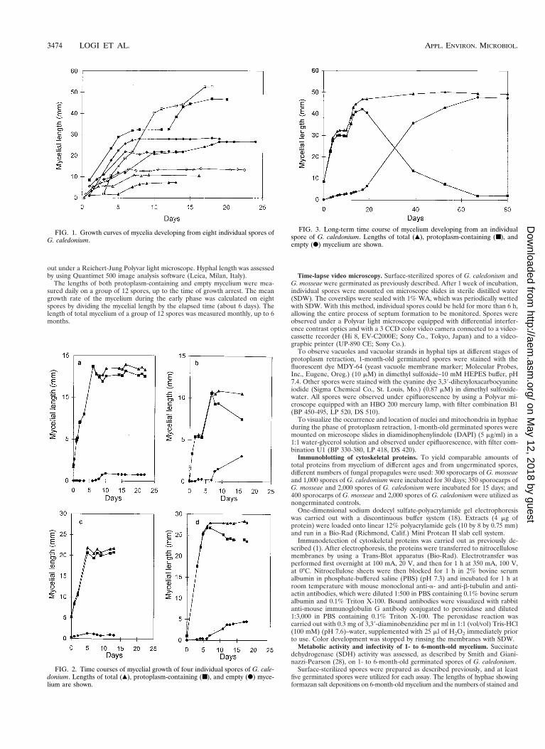
FIG. 2. Time courses of mycelial growth of four individual spores of G. cale-donium. Lengths of total (Œ), protoplasm-containing (■), and empty (F) myce-lium are shown.
FIG. 3. Long-term time course of mycelium developing from an individualspore of G. caledonium. Lengths of total (Œ), protoplasm-containing (■), andempty (F) mycelium are shown.
3474 LOGI ET AL. APPL. ENVIRON. MICROBIOL.
on May 12, 2018 by guest
http://aem.asm
.org/D
ownloaded from
unstained hyphal tips on 1- to 6-month-old mycelium were recorded by using aPolyvar light microscope equipped with Quantimet 500 image analysis software.
To test infectivity by the mycelium, surface-sterilized spores were germinatedas previously described and individually used to inoculate the roots of six 2-week-old Ocimum basilicum L. (basil) plants placed on a 47-mm-diameter Milliporemembrane (0.45-mm-diameter pores). Another Millipore membrane was used tomake up a sandwich system. Ten replicates were set up for each trial, whichconsisted of 1-, 2-, 3-, 4-, 5-, and 6-month-old germinated spores. One monthafter inoculation, sandwiches were carefully opened and roots were stained toreveal fungal structures (23). The number of entry points formed by each ger-minated spore was recorded for each plant root system and for each sandwich.
RESULTS
Time course of hyphal growth in the absence of the hostplant. Observations carried out for 15 to 20 days on spores ofG. caledonium growing in the absence of the host showed thatwithin a few days the elongating germ tubes gave rise to amycelial network whose extension was highly variable betweenindividuals. After a period ranging between 5 and 10 dayspostgermination, total hyphal length reached a plateau and themycelium entered a state of developmental arrest (Fig. 1). Themean growth rate of the mycelium during this early phase was1.97 6 0.39 mm/min (mean 6 standard error). An interesting
FIG. 4. Long-term time course of mycelium developing from an individualspore of G. caledonium. Lengths of total (Œ), protoplasm-containing (■), andempty (F) mycelium are shown.
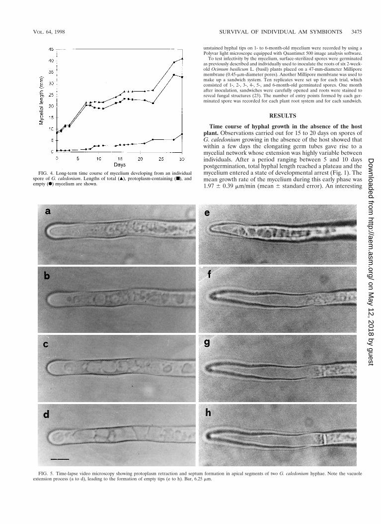
FIG. 5. Time-lapse video microscopy showing protoplasm retraction and septum formation in apical segments of two G. caledonium hyphae. Note the vacuoleextension process (a to d), leading to the formation of empty tips (e to h). Bar, 6.25 mm.
VOL. 64, 1998 SURVIVAL OF INDIVIDUAL AM SYMBIONTS 3475
on May 12, 2018 by guest
http://aem.asm
.org/D
ownloaded from
feature of the mycelial net was the occurrence of many hyphaecompletely devoid of protoplasm. Time course observationsshowed that during the plateau growth phase, corresponding toarrest of hyphal elongation, fundamental modifications oc-curred in the mycelium: protoplasm was retracted from mosthyphae, leading to the formation of cross walls delimiting anempty mycelial network. This phenomenon was further studiedby daily measurements of the extension of protoplasm-contain-ing hyphae and empty hyphae. These showed that when totalhyphal growth reached the plateau, the length of protoplasm-containing mycelium remained constant (Fig. 2a and c) ortended to decrease (Fig. 2b and d). Long-term observations, upto 80 days, showed that this decrease was paralleled by risingvalues of the length of empty hyphae (Fig. 3).
In some spores, a renewal of mycelial elongation followinggrowth arrest was observed. An example is shown in Fig. 4: thespore, 23 days old, resumed growth after 16 days of stasis. Ac-cordingly, assessment of spore growth was carried out monthly,for a period ranging from 1 to 6 months after germination. Theresults showed that total mycelial length increased from the 1stto the 3rd month (from 45.13 6 2.94 to 68.19 6 6.05 mm,respectively), remaining constant up to the 6th month (70.83 67.08 mm).
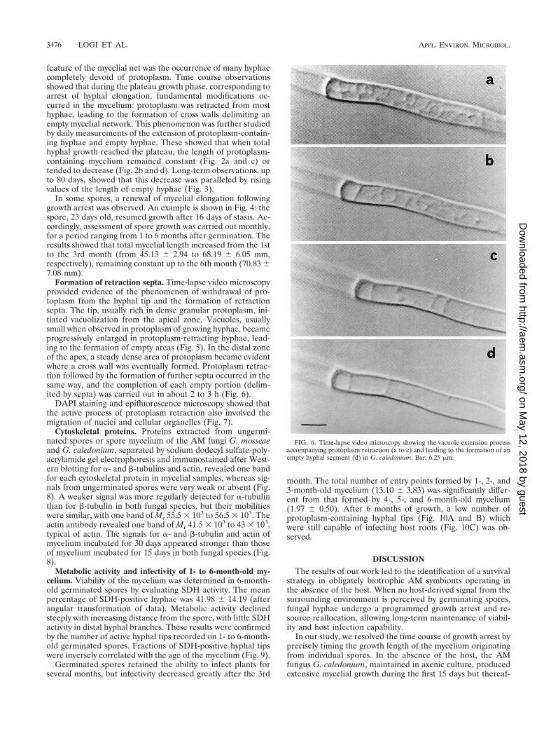
Formation of retraction septa. Time-lapse video microscopyprovided evidence of the phenomenon of withdrawal of pro-toplasm from the hyphal tip and the formation of retractionsepta. The tip, usually rich in dense granular protoplasm, ini-tiated vacuolization from the apical zone. Vacuoles, usuallysmall when observed in protoplasm of growing hyphae, becameprogressively enlarged in protoplasm-retracting hyphae, lead-ing to the formation of empty areas (Fig. 5). In the distal zoneof the apex, a steady dense area of protoplasm became evidentwhere a cross wall was eventually formed. Protoplasm retrac-tion followed by the formation of further septa occurred in thesame way, and the completion of each empty portion (delim-ited by septa) was carried out in about 2 to 3 h (Fig. 6).
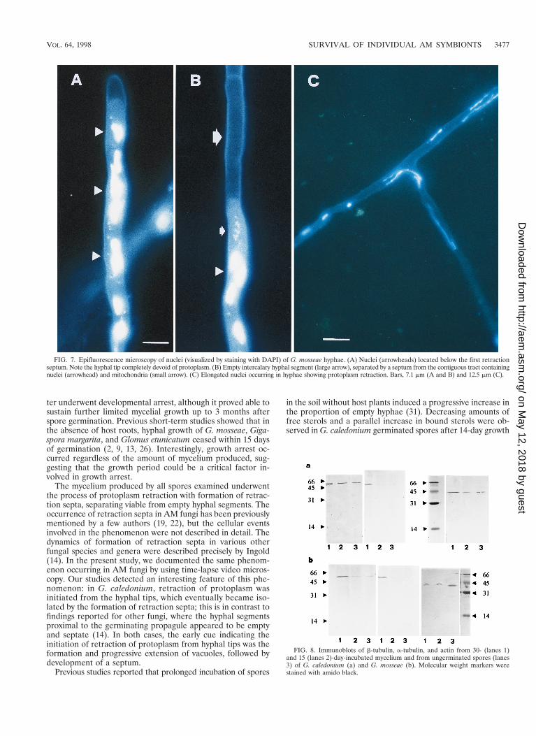
DAPI staining and epifluorescence microscopy showed thatthe active process of protoplasm retraction also involved themigration of nuclei and cellular organelles (Fig. 7).
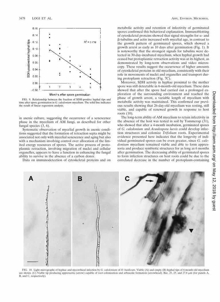
Cytoskeletal proteins. Proteins extracted from ungermi-nated spores or spore mycelium of the AM fungi G. mosseaeand G. caledonium, separated by sodium dodecyl sulfate-poly-acrylamide gel electrophoresis and immunostained after West-ern blotting for a- and b-tubulins and actin, revealed one bandfor each cytoskeletal protein in mycelial samples, whereas sig-nals from ungerminated spores were very weak or absent (Fig.8). A weaker signal was more regularly detected for a-tubulinthan for b-tubulin in both fungal species, but their mobilitieswere similar, with one band of Mr 55.5 3 103 to 56.5 3 103. Theactin antibody revealed one band of Mr 41.5 3 103 to 43 3 103,typical of actin. The signals for a- and b-tubulin and actin ofmycelium incubated for 30 days appeared stronger than thoseof mycelium incubated for 15 days in both fungal species (Fig.8).
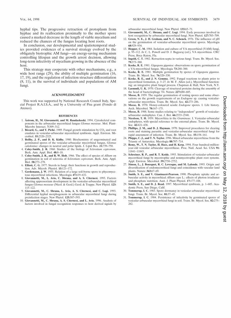
Metabolic activity and infectivity of 1- to 6-month-old my-celium. Viability of the mycelium was determined in 6-month-old germinated spores by evaluating SDH activity. The meanpercentage of SDH-positive hyphae was 41.98 6 14.19 (afterangular transformation of data). Metabolic activity declinedsteeply with increasing distance from the spore, with little SDHactivity in distal hyphal branches. These results were confirmedby the number of active hyphal tips recorded on 1- to 6-month-old germinated spores. Fractions of SDH-positive hyphal tipswere inversely correlated with the age of the mycelium (Fig. 9).
Germinated spores retained the ability to infect plants forseveral months, but infectivity decreased greatly after the 3rd
month. The total number of entry points formed by 1-, 2-, and3-month-old mycelium (13.10 6 3.83) was significantly differ-ent from that formed by 4-, 5-, and 6-month-old mycelium(1.97 6 0.50). After 6 months of growth, a low number ofprotoplasm-containing hyphal tips (Fig. 10A and B) whichwere still capable of infecting host roots (Fig. 10C) was ob-served.
DISCUSSIONThe results of our work led to the identification of a survival
strategy in obligately biotrophic AM symbionts operating inthe absence of the host. When no host-derived signal from thesurrounding environment is perceived by germinating spores,fungal hyphae undergo a programmed growth arrest and re-source reallocation, allowing long-term maintenance of viabil-ity and host infection capability.
In our study, we resolved the time course of growth arrest byprecisely timing the growth length of the mycelium originatingfrom individual spores. In the absence of the host, the AMfungus G. caledonium, maintained in axenic culture, producedextensive mycelial growth during the first 15 days but thereaf-
FIG. 6. Time-lapse video microscopy showing the vacuole extension processaccompanying protoplasm retraction (a to c) and leading to the formation of anempty hyphal segment (d) in G. caledonium. Bar, 6.25 mm.
3476 LOGI ET AL. APPL. ENVIRON. MICROBIOL.
on May 12, 2018 by guest
http://aem.asm
.org/D
ownloaded from
ter underwent developmental arrest, although it proved able tosustain further limited mycelial growth up to 3 months afterspore germination. Previous short-term studies showed that inthe absence of host roots, hyphal growth of G. mosseae, Giga-spora margarita, and Glomus etunicatum ceased within 15 daysof germination (2, 9, 13, 26). Interestingly, growth arrest oc-curred regardless of the amount of mycelium produced, sug-gesting that the growth period could be a critical factor in-volved in growth arrest.
The mycelium produced by all spores examined underwentthe process of protoplasm retraction with formation of retrac-tion septa, separating viable from empty hyphal segments. Theoccurrence of retraction septa in AM fungi has been previouslymentioned by a few authors (19, 22), but the cellular eventsinvolved in the phenomenon were not described in detail. Thedynamics of formation of retraction septa in various otherfungal species and genera were described precisely by Ingold(14). In the present study, we documented the same phenom-enon occurring in AM fungi by using time-lapse video micros-copy. Our studies detected an interesting feature of this phe-nomenon: in G. caledonium, retraction of protoplasm wasinitiated from the hyphal tips, which eventually became iso-lated by the formation of retraction septa; this is in contrast tofindings reported for other fungi, where the hyphal segmentsproximal to the germinating propagule appeared to be emptyand septate (14). In both cases, the early cue indicating theinitiation of retraction of protoplasm from hyphal tips was theformation and progressive extension of vacuoles, followed bydevelopment of a septum.
Previous studies reported that prolonged incubation of spores
in the soil without host plants induced a progressive increase inthe proportion of empty hyphae (31). Decreasing amounts offree sterols and a parallel increase in bound sterols were ob-served in G. caledonium germinated spores after 14-day growth
FIG. 7. Epifluorescence microscopy of nuclei (visualized by staining with DAPI) of G. mosseae hyphae. (A) Nuclei (arrowheads) located below the first retractionseptum. Note the hyphal tip completely devoid of protoplasm. (B) Empty intercalary hyphal segment (large arrow), separated by a septum from the contiguous tract containingnuclei (arrowhead) and mitochondria (small arrow). (C) Elongated nuclei occurring in hyphae showing protoplasm retraction. Bars, 7.1 mm (A and B) and 12.5 mm (C).
FIG. 8. Immunoblots of b-tubulin, a-tubulin, and actin from 30- (lanes 1)and 15 (lanes 2)-day-incubated mycelium and from ungerminated spores (lanes3) of G. caledonium (a) and G. mosseae (b). Molecular weight markers werestained with amido black.
VOL. 64, 1998 SURVIVAL OF INDIVIDUAL AM SYMBIONTS 3477
on May 12, 2018 by guest
http://aem.asm
.org/D
ownloaded from
in axenic culture, suggesting the occurrence of a senescencephase in the mycelium of AM fungi, as described for otherfungal species (3, 6).
Systematic observation of mycelial growth in axenic condi-tions suggested that the formation of retraction septa might beassociated not only with mycelial senescence and aging but alsowith a mechanism involving control over allocation of the lim-ited energy resources of spores. The active process of proto-plasmic retraction, involving migration of nuclei and cellularorganelles, appears to have a function in enhancing the fungalability to survive in the absence of a carbon donor.
Data on immunodetection of cytoskeletal proteins and on
metabolic activity and retention of infectivity of germinatedspores confirmed this behavioral explanation. Immunoblottingof cytoskeletal proteins showed that signal strengths for a- andb-tubulins and actin increased with mycelial age, in contrast tothe growth pattern of germinated spores, which showed agrowth arrest as early as 10 days after germination (Fig. 1). Itis noteworthy that the strongest signals for tubulins were de-tected in 30-day-incubated mycelium, when hyphal growth hadceased but protoplasmic retraction activity was at its highest, asdemonstrated by long-term observations and video micros-copy. These results suggest the occurrence of higher amountsof cytoskeletal proteins in old mycelium, consistently with theirrole in movements of nuclei and organelles and transport dur-ing protoplasm retraction (Fig. 7C).
Moreover, SDH activity in hyphae proximal to the motherspore was still detectable in 6-month-old mycelium. These datashowed that after the spore had carried out a prolonged ex-ploration of the surrounding environment and reached thephase of growth arrest, a variable length of mycelium withmetabolic activity was maintained. This confirmed our previ-ous results showing that 26-day-old mycelium was resting, stillviable, and capable of renewed growth in response to hostroots (10).
The long-term ability of AM mycelium to retain infectivity inthe absence of the host was tested in soil by Tommerup (31),who showed that after a 4-month incubation, germinated sporesof G. caledonium and Acaulospora laevis could develop infec-tion structures and colonize Trifolium roots. Experimentalevidence presented here indicates that the longevity of indi-vidual germinated spores can be even greater, since G. cale-donium mycelium remained viable and able to form appres-soria and produce symbiotic structures for as long as 6 monthsafter germination. The decreasing ability of germinated sporesto form infection structures on host roots could be due to thecorrelated decrease in the number of protoplasm-containing
FIG. 9. Relationship between the fraction of SDH-positive hyphal tips andtime after spore germination in G. caledonium mycelium. The solid line indicatesthe result of linear regression analysis.
FIG. 10. Light micrographs of hyphae and mycorrhizal infection by G. caledonium of O. basilicum. Viable (A) and empty (B) hyphal tips of 6-month-old myceliumare shown. (C) Viable tip producing appressoria (arrow) capable of root colonization and arbuscule formation (arrowhead). Bar, 25, 25, and 27.8 mm (for panels A,B, and C, respectively).
3478 LOGI ET AL. APPL. ENVIRON. MICROBIOL.
on May 12, 2018 by guest
http://aem.asm
.org/D
ownloaded from
hyphal tips. The progressive retraction of protoplasm fromhyphae and its reallocation proximally to the mother sporecaused a marked decrease in the length of viable mycelium andreduced the chances of the fungus locating host roots.
In conclusion, our developmental and spatiotemporal stud-ies provided evidences of a survival strategy evolved by theobligately biotrophic AM fungi—an energy-saving mechanismcontrolling lifespan and the growth arrest decision, allowinglong-term infectivity of mycelium growing in the absence of thehost.
This strategy may cooperate with other mechanisms, e.g., awide host range (29), the ability of multiple germination (16,17, 19), and the regulation of infection structure differentiation(8, 11), in the survival of individuals and populations of AMfungi.
ACKNOWLEDGMENT
This work was supported by National Research Council Italy, Spe-cial Project R.A.I.S.A., and by a University of Pisa grant (Fondo diAteneo).
REFERENCES
1. Astrom, H., M. Giovannetti, and M. Raudaskoski. 1994. Cytoskeletal com-ponents in the arbuscular mycorrhizal fungus Glomus mosseae. Mol. Plant-Microbe Interact. 7:309–312.
2. Becard, G., and Y. Piche. 1989. Fungal growth stimulation by CO2 and rootexudates in vesicular-arbuscular mycorrhizal symbiosis. Appl. Environ. Mi-crobiol. 55:2320–2325.
3. Beilby, J. P., and D. K. Kidby. 1980. Biochemistry of ungerminated andgerminated spores of the vesicular-arbuscular mycorrhizal fungus, Glomuscaledonius: changes in neutral and polar lipids. J. Lipid Res. 21:739–750.
4. Coley-Smith, J. R. 1960. Studies of the biology of Sclerotium cepivorum.Berk. Ann. Appl. Biol. 48:8–18.
5. Coley-Smith, J. R., and R. W. Holt. 1966. The effect of species of Allium ongermination in soil of sclerotia of Sclerotium cepivorum. Berk. Ann. Appl.Biol. 58:273–278.
6. Elliott, C. G. 1977. Sterols in fungi: their functions in growth and reproduc-tion. Adv. Microb. Physiol. 15:121–172.
7. Gerdemann, J. W. 1955. Relation of a large soil-borne spore to phycomyce-tous mycorrhizal infections. Mycologia 47:619–632.
8. Giovannetti, M., L. Avio, C. Sbrana, and A. S. Citernesi. 1993. Factorsaffecting appressorium development in the vesicular-arbuscular mycorrhizalfungus Glomus mosseae (Nicol. & Gerd.) Gerd. & Trappe. New Phytol. 123:115–122.
9. Giovannetti, M., C. Sbrana, L. Avio, A. S. Citernesi, and C. Logi. 1993.Differential hyphal morphogenesis in arbuscular mycorrhizal fungi duringpreinfection stages. New Phytol. 125:587–593.
10. Giovannetti, M., C. Sbrana, A. S. Citernesi, and L. Avio. 1996. Analysis offactors involved in fungal recognition responses to host derived signals by
arbuscular mycorrhizal fungi. New Phytol. 133:65–71.11. Giovannetti, M., C. Sbrana, and C. Logi. 1994. Early processes involved in
host recognition by arbuscular mycorrhizal fungi. New Phytol. 127:703–709.12. Green, N. E., J. H. Graham, and N. C. Schenck. 1976. The influence of pH
on the germination of vesicular-arbuscular mycorrhizal spores. Mycologia68:929–934.
13. Hepper, C. M. 1984. Isolation and culture of VA mycorrhizal (VAM) fungi,p. 95–112. In C. L. Powell and D. J. Bagyaraj (ed.), VA mycorrhizas. CRCPress, Boca Raton, Fla.
14. Ingold, C. T. 1982. Retraction-septa in various fungi. Trans. Br. Mycol. Soc.79:373–380.
15. Koske, R. E. 1981. Gigaspora gigantea: observations on spore germination ofa VA-mycorrhizal fungus. Mycologia 73:288–300.
16. Koske, R. E. 1981. Multiple germination by spores of Gigaspora gigantea.Trans. Br. Mycol. Soc. 76:320–330.
17. Koske, R. E., and J. N. Gemma. 1992. Fungal reactions to plants prior tomycorrhizal formation, p. 3–27. In M. F. Allen (ed.), Mycorrhizal function-ing: an integrative plant fungal process. Chapman & Hall, New York, N.Y.
18. Laemmli, U. K. 1970. Cleavage of structural proteins during the assembly ofthe head of bacteriophage T4. Nature 227:680–685.
19. Mosse, B. 1959. The regular germination of resting spores and some obser-vations on the growth requirements of an Endogone sp. causing vesicular-arbuscular mycorrhiza. Trans. Br. Mycol. Soc. 42:273–286.
20. Mosse, B. 1970. Honey-coloured sessile Endogone spores. I. Life history.Arch. Microbiol. 70:167–175.
21. Mosse, B. 1988. Some studies relating to “independent” growth of vesicular-arbuscular endophytes. Can. J. Bot. 66:2533–2540.
22. Nicolson, T. H. 1959. Mycorrhiza in the Gramineae. I. Vesicular-arbuscularendophytes, with special reference to the external phase. Trans. Br. Mycol.Soc. 42:421–438.
23. Phillips, J. M., and D. J. Hayman. 1970. Improved procedures for clearingroots and staining parasitic and vesicular-arbuscular mycorrhizal fungi forrapid assessment of infection. Trans. Br. Mycol. Soc. 55:158–161.
24. Phipps, C. J., and T. N. Taylor. 1996. Mixed arbuscular mycorrhizae from theTriassic of Antarctica. Mycologia 88:707–714.
25. Remy, W., T. N. Taylor, H. Hass, and H. Kerp. 1994. Four hundred-million-year-old vesicular arbuscular mycorrhizae. Proc. Natl. Acad. Sci. USA 91:11841–11843.
26. Schreiner, R. P., and R. T. Koide. 1993. Stimulation of vesicular-arbuscularmycorrhizal fungi by mycotrophic and nonmycotrophic plant root systems.Appl. Environ. Microbiol. 59:2750–2752.
27. Simon, L., J. Bousquet, R. C. Levesque, and M. Lalonde. 1993. Origin anddiversification of endomycorrhizal fungi and coincidence with vascular landplants. Nature 363:67–69.
28. Smith, S. E., and V. Gianinazzi-Pearson. 1990. Phosphate uptake and ar-buscular activity in mycorrhizal Allium cepa L.: effects of photon irradianceand phosphate nutrition. Aust. J. Plant Physiol. 17:177–188.
29. Smith, S. E., and D. J. Read. 1997. Mycorrhizal symbiosis, p. 1–605. Aca-demic Press, San Diego, Calif.
30. Tommerup, I. C. 1983. Spore dormancy in vesicular-arbuscular mycorrhizalfungi. Trans. Br. Mycol. Soc. 81:37–45.
31. Tommerup, I. C. 1984. Persistence of infectivity by germinated spores ofvesicular-arbuscular mycorrhizal fungi in soil. Trans. Br. Mycol. Soc. 82:275–282.
VOL. 64, 1998 SURVIVAL OF INDIVIDUAL AM SYMBIONTS 3479
on May 12, 2018 by guest
http://aem.asm
.org/D
ownloaded from


![· Web viewBy targeting cellular or viral genes, these miRNAs are involved in the regulation of multiple cellular responses such as host cell proliferation, apoptosis [12-15], and](https://img.pdfslide.net/doc/110x75/5f0b48857e708231d42fc04c/web-view-by-targeting-cellular-or-viral-genes-these-mirnas-are-involved-in-the.jpg)