Embed Size (px)
Citation preview
Cellular/Molecular
Deletion of CPEB3 Enhances Hippocampus-DependentMemory via Increasing Expressions of PSD95 and NMDAReceptors
Hsu-Wen Chao,1* Li-Yun Tsai,1* Yi-Ling Lu,1 Pei-Yi Lin,1 Wen-Hsuan Huang,1 Hsin-Jung Chou,1 Wen-Hsin Lu,1,2
Hsiu-Chen Lin,1 Ping-Tao Lee,1 and Yi-Shuian Huang1,2
1Institute of Biomedical Sciences, Academia Sinica, Taipei 115, Taiwan and 2Taiwan International Graduate Program, National Yang-Ming University,Taipei 112, Taiwan
Long-term memory requires activity-dependent synthesis of plasticity-related proteins (PRPs) to strengthen synaptic efficacy and con-sequently consolidate memory. Cytoplasmic polyadenylation element binding protein (CPEB)3 is a sequence-specific RNA-bindingprotein that regulates translation of several PRP RNAs in neurons. To understand whether CPEB3 plays a part in learning and memory,we generated CPEB3 knock-out (KO) mice and found that the null mice exhibited enhanced hippocampus-dependent, short-term fearmemory in the contextual fear conditioning test and long-term spatial memory in the Morris water maze. The basal synaptic transmissionof Schaffer collateral-CA1 neurons was normal but long-term depression evoked by paired-pulse low-frequency stimulation was mod-estly facilitated in the juvenile KO mice. Molecular and cellular characterizations revealed several molecules in regulating plasticity ofglutamatergic synapses are translationally elevated in the CPEB3 KO neurons, including the scaffolding protein PSD95 and the NMDAreceptors along with the known CPEB3 target, GluA1. Together, CPEB3 functions as a negative regulator to confine the strength ofglutamatergic synapses by downregulating the expression of multiple PRPs and plays a role underlying certain forms of hippocampus-dependent memories.
IntroductionModulation of long-lasting synaptic strength requires activity-induced synthesis of plasticity-related proteins (PRPs) to sustainmorphological and functional changes of synapses that are cru-cial for the establishment and consolidation of long-term mem-ory (LTM). Several RNA-binding proteins control specific PRPsyntheses in neurons via regulating RNA transport, translation,and/ or stability; and when genetically mutated or ablated, resultsin aberrant memory performance in humans and mice (Richterand Klann, 2009; for review, see Costa-Mattioli et al., 2009; Dar-nell and Richter, 2012; Gal-Ben-Ari et al., 2012). Cytoplasmicpolyadenylation element binding protein (CPEB) family of RNA-
binding proteins in vertebrates contains four members,CPEBs1– 4, all of which are expressed in the brain (Theis et al.,2003) and share structure and sequence identity in the RNA-binding domain (RBD; Huang et al., 2006). Disruption of cpeb1gene in mice reduced extinction of hippocampus-dependentLTM (Berger-Sweeney et al., 2006) and altered synaptic electro-physiology in the Schaffer collateral (SC) pathway (Alarcon et al.,2004). In contrast, the contribution of other CPEB members inlearning and memory is not clear. Among them, CPEB3 wasshown to repress the translation of several PRP RNAs, whichencode for epidermal growth factor receptor and the subunits ofAMPA receptor (AMPAR), GluA1, and GluA2 (Huang et al.,2006; Peng et al., 2010; Pavlopoulos et al., 2011). NMDA receptor(NMDAR) signaling triggers calpain 2-mediated cleavage ofCPEB3 and subsequently results in translation of CPEB3-targeted RNAs (Huang et al., 2006; Chen and Huang, 2012; Wangand Huang, 2012). In this model, CPEB3 is a repressor and ame-liorates its repression ability via NMDAR-activated proteolysis.Moreover, monoubiquitination of CPEB3 by neuralized1(Neurl1) can switch CPEB3 from a repressor to an activator toincrease the translation of GluA1 and GluA2 RNAs (Pavlopouloset al., 2011). Thus, CPEB3 bilaterally regulates translation de-pending on its ubiquitin-modified state. Although there are acouple of mechanisms identified to modulate CPEB3-repressedtranslation in neurons, the role of CPEB3 in learning and mem-ory is not clear. Thus, we generated CPEB3 knock-out (KO) mice,which demonstrated normal physical performance and home-cage behaviors, but showed reduced exploratory activity in the
Received July 18, 2013; revised Sept. 1, 2013; accepted Sept. 18, 2013.Author contributions: H.-W.C., L.-Y.T., and Y.-S.H. designed research; H.-W.C., L.-Y.T., Y.-L.L., P.-Y.L., W.-H.H.,
H.-J.C., W.-H.L., H.-C.L., P.-T.L., and Y.-S.H. performed research; H.-W.C., L.-Y.T., Y.-L.L., P.-Y.L., W.-H.H., H.-J.C.,W.-H.L., and Y.-S.H. analyzed data; Y.-S.H. wrote the paper.
This work was supported by National Science Council [NSC 99-2311-B-001-020-MY3] and Academia Sinica[AS-100-TP-B09] in Taiwan. We thank the Taiwan Mouse Clinic, which is funded by the National Research Programfor Biopharmaceuticals from the NSC, for conducting the home-cage experiments and SHIRPA modified assays. Weappreciate Ching-Pang Chang for assistance on the behavior study, the transgenic core facility for embryonic stemcell gene targeting, and blastocyst microinjection and Su-Ping Lee and Huei-Fang Wu in the IMB cores for helpingconfocal and calcium image analyses. We thank Drs. Lih-Chu Chiou, Po-Wu Gean, Kuei-Sen Hsu, and Ming-Yuan Minfor their assistance setting up the field recording system.
*H.-W.C. and L.-Y.T. contributed equally to this work.The authors declare no competing financial interests.Correspondence should be addressed to Yi-Shuian Huang, Institute of Biomedical Sciences, Academia Sinica, 128
Section 2, Academia Road, Taipei 11529, Taiwan. E-mail: [email protected]:10.1523/JNEUROSCI.3043-13.2013
Copyright © 2013 the authors 0270-6474/13/3317008-15$15.00/0
17008 • The Journal of Neuroscience, October 23, 2013 • 33(43):17008 –17022
open field. The learning and memory abilities were assessed usingcontextual fear conditioning and Morris water maze. In the for-mer assay, the KO animals showed elevated short-term fearresponses during acquisition and extinction trainings, but exhib-ited normal long-term fear memory. In the latter task, the nullmice displayed better consolidated spatial memory, which ap-peared to hinder their ability to locate a new platform position inthe reversal probe test. Electro-recordings identified enhancedpaired-pulse low-frequency stimulation (PP-LFS)-induced long-term depression (LTD) at the SC-CA1 synapses of KO mice.Molecular and cellular characterizations revealed that the en-largement of dendritic spines and elevated expression of severalPRPs, including GluA1, NMDARs, and PSD95, in the KO neu-rons. The protein, but not the RNA level of these CPEB3 targets,was elevated, which was accompanied by a shift of the RNA dis-tribution toward polysomes in the KO brains. These results re-vealed the in vivo role of CPEB3 as a negative regulator toconstrain the translation of multiple PRP RNAs and the strengthof consolidated memory.
Materials and MethodsAntibodies and chemicalsAntibodies used in the study were as follows: myc-tag and LRP130 fromSanta Cruz Biotechnology; NR1, NR2A, NR2B, GluA1, PSD95, and syn-aptophysin from Millipore; and GFP, �-actin, and �-tubulin fromSigma-Aldrich. The CPEB3 antibodies have been described previously(Chao et al., 2012; Wang and Huang, 2012). Alexa Fluor-conjugatedsecondary antibodies were obtained from Invitrogen. With the exceptionof the Fura-2 AM calcium indicator (Invitrogen), all of the other chem-icals were purchased from Sigma-Aldrich.
AnimalsAll of the experimental protocols were performed in accordance withthe guidelines of the Institutional Animal Care and Utilization Committee.C57BL/6J mice were housed under a 12 h light/dark cycle in a climate-controlled room with ad libitum access to food and water. All efforts weremade to minimize the number of animals used and their suffering. Thewild-type (WT) and KO male mice used for behavior, electrophysiology, andbiochemical studies were littermates from heterozygous matings.
Construction of the CPEB3 targeting vector and generation ofmutant miceThe genomic BAC clone (RP23-56A17) containing the 5�-portion of theC57BL/6J mouse CPEB3 gene was used to construct the targeting vectorby the recombineering technique according to the manufacturer’s in-structions (Gene Bridges). Briefly, a loxP-Neo-loxP cassette was firstrecombined into the 3�-end of exon 2 and excised with recombinant Cre(New England Biolabs) in vitro to result in a single loxP site. The resultingplasmid was recombineered with the Frt-PGK-Neo-Frt-loxP cassette tothe 5�-end of exon 2. The plasmid was linearized with NruI and electro-porated into C57BL/6J ES cells. Four correct clones of 258 G418-resistantclones were injected into c2J blastocysts. Only one clone derived agermline-transmitted line. The mouse carrying floxed allele was firstcrossed with Frt recombinase driven by the �-actin promoter to removethe Frt-PGK-Neo-Frt cassette. The resulting line was maintained asfCPEB3 mice and then crossed with the protamine-Cre transgenic mouseto derive KO mice.
Genotyping of CPEB3 embryos and miceThe genotypes were determined by PCR using tail biopsies and the KAPAmouse genotyping kit (KAPA Biosystems). Briefly, tail samples werelysed in 20 �l of KAPA extract buffer for 20 min at 75°C and then 5 minat 95�100°C. The DNA sample was then diluted with 60 �l of H2O, and0.5 �l was used for a 10 �l PCR reaction. The sense primer, CP3F15�-TTTGATCCTTCTGCCTCTCCCTC-3� and two antisense primers,CP3R1 5�-TTGGTACACGACCCTCTTTCCCC-3� and CP3R2 5�-TATGGCTCTGAAGGTCGTGTCCT-3� at a 2:1:1 ratio were used to am-plify the WT and KO alleles, respectively.
Primary neuronal cultures, DNA transfection, andlentivirus infectionTo culture WT and KO neurons, the cortices and hippocampi of E18embryos from heterozygous matings were isolated and maintained indi-vidually in HBSS for 2 h on ice. At the same time, the tails were collectedfor genotyping. Once the genotypes were determined, the WT and KOcerebral cortices regardless of sex were pooled and used for neuronalcultures as previously described (Chao et al., 2012). The cell density wasat 3 � 10 5 cells/ well in a 12-well plate, 2 � 10 6 cells/60 mm dish, and 5 �10 6 cells/100 mm dish. Delivery of DNA into the neurons was performedusing calcium phosphate transfection or lentivirus infection as previ-ously reported (Chao et al., 2012).
Immunohistochemistry, immunofluorescence staining, imagingacquisition, and quantificationCoronal sections of WT and KO male brains after 10 min fixation in 4%formaldehyde and 20 min antigen retrieval in 10 mM sodium citratebuffer, pH 6 at 70°C, were washed twice with Tris-buffered saline (TBS)and permeabilized with 0.2% Triton X-100 in TBS. After three washeswith TBS and 1 h blocking in 10% horse serum, the slices were incu-bated with affinity-purified CPEB3 antibody at 4°C overnight. Afterwashes with TBS, the immunobinding signal was developed using theVectastain Elite ABC kit (Vector Laboratories) following the manufac-turer’s protocol. Neurons at 14 different days in vitro (DIV) transfectedwith the enhanced green fluorescent protein (EGFP) plasmid were pro-cessed on 18 DIV for immunofluorescence staining and confocal imag-ing as previously described (Chao et al., 2012). The images werequantified using the MetaMorph software and then exported into Exceland GraphPad Prism for the analyses. Approximately 3000 spines within20 �m dendritic segments 30 �m away from the somas of 30 neuronswere measured in each group (Chao et al., 2008).
Synaptosome, synaptic density, and synaptic cytosol preparationThe cortices and hippocampi rapidly removed from the 3-month-oldmale mouse were homogenized in 2 ml sucrose buffer (10 mM HEPES,pH 7.5, 1.5 mM MgCl2, 320 mM sucrose, 5 mM EDTA, 5 mM dithiothreitol(DTT), 0.1 mM PMSF, 10 �M MG132, and 1X protease and phosphataseinhibitors). Homogenates were centrifuged at 700 � g for 10 min at 4°Cto remove nuclei and cell debris. The supernatant was then centrifugedagain at 9250 � g for 15 min to obtain the pellet containing the crudesynaptosome. The crude synaptosome fraction was resuspended in su-crose buffer with 1% Triton X-100 at 4°C for 60 min, followed by40,000 � g centrifugation for 30 min to obtain the pellet (synaptic den-sity) and supernatant (synaptic cytosol).
RNA immunoprecipitation and quantitative PCRCerebral cortices isolated from 3-month-old male mice were homogenizedin 2 ml of RNA immunoprecipitation (RIP) buffer (DEPC-treated watercontaining 20 mM HEPES, pH 7.4, 150 mM NaCl, 1 mM MgCl2, 0.5% TritonX-100, 0.5% NP-40, 10% glycerol, 0.5 mM DTT, 1X protease inhibitor cock-tail, and 40 U/ml RNase inhibitor), and incubated at 4°C for 60 min on arotator. Insoluble debris was pelleted at 10,000 g for 5 min. The supernatantwas divided and incubated with either control or CPEB3 IgG-bound beadsfor 3 h. The beads were washed with RIP buffer five times, and 1/5 of the beadvolume was used for immunoblotting. The remaining beads were elutedwith 4 M guanidine thiocyanate, followed by phenol/chloroform extractionand ethanol precipitation in the presence of 5 �g of glycogen. The RNAprecipitates were reverse transcribed using oligo-dT and ImPromII ReverseTranscriptase (Promega). Quantitative PCR (qPCR) was conducted usingthe Universal Probe Library and Lightcycler 480 system (Roche). The dataanalysis was performed using the comparative Ct (threshold cycle value)method with the nonCPEB3-targeted RNA, GAPDH mRNA (total RNAand RIP experiments), or synaptophysin mRNA (polysome experiment) asthe reference. The PCR primers used were as follows: PSD95, 5�-GGCGCACAAGTTCATTGAG and 5�-TGAGACATCAAGGATGCAGTG; NR1, 5�-TACAAGCGACACAAGGATGC and 5�-TCAGTGGGATGGTACTGCTG;NR2A,5�-CTGCTCCAGTTTGTTGGTGAand5�-AGATGCCCGTAAGCCACA; NR2B, 5�-GGGTTACAACCGGTGCCTA and 5�-CTTTGCCGATGGTGAAAGAT;GluA1,5�-CGGAAATTGCTTATGGGACAand5�-ACACAGCGATTTTAGACCTCCT; GAPDH, 5�-GCCAAAAGGGTCATCA
Chao, Tsai et al. • CPEB3 Negatively Regulates Memory Consolidation J. Neurosci., October 23, 2013 • 33(43):17008 –17022 • 17009
TCTC-3�and5�-CACACCCATCACAAACATGG-3�;synaptophysin,5�-CAAGGCTACGGCCAACAG-3� and 5�-GTCTTCGTGGGCTTCACTG-3�.
Sucrose density gradient for polysomal profilingTwo plates of WT or KO cortical neuron cultures at 18 DIV (�10 7
neurons) were washed with PBS, lysed in 600 �l buffer (25 mM HEPES,pH 7.5, 100 mM NaCl, 10 mM MgCl2, 100 �g/ml cycloheximide, 0.25 M
sucrose, 40 u/ml RNase inhibitor, and 1X protease inhibitor cocktail),and centrifuged for 5 min at 10,000 g at 4°C. Approximately 500 �l ofsupernatant was layered on top of a linear 15%–50% (w/v) sucrose gra-dient. Centrifugation was performed in a SW41 rotor at 37,000 rpm for2 h. Polysome profiles were monitored by absorbance of light with awavelength of 254 nm (A254).
RNA extraction, Northern blotting, and RT-PCRSucrose gradient fractions were treated with 100 �g/ml Proteinase K and0.2% SDS for 20 min at 37°C, phenol/chloroform extracted and precip-itated with isopropanol to obtain RNAs. Total RNA was extracted usingTrizol reagent (Invitrogen). Thirty micrograms total RNA isolated fromWT, heterozygous, and KO tissues was used for Northern blotting. A 285bp CPEB3 cDNA region, amplified using the sense exon3, 5�-GGTAAACACTACCCTCCC-3�, and the antisense exon6, 5�-CACCTTTCTAGAGTAGCGTTC-3� primers, was used to synthesize the radiolabeledprobe by random primer labeling. The cDNAs reverse transcribedfrom brain and testis RNAs were used for PCR with the sense primerexon2, 5�-ATGGAGGATAACGCTTTCCG-3�, exon3 or exon7, 5�-ACTGCCAGCTTTCGCAGG-3� along with the antisense primerexon11, 5�-TACTGCAGACAGGTGACG-3�.
Plasmid construction, in vitro transcription, and luciferasereporter assayMouse PSD95 3�-UTR was PCR-amplified from brain cDNA usingthe primers 5�-CGGAATTCTTCCTGCCCTGGCTTGGCC-3� and 5�-CCGCTCGAGCAAGTGTCTGTCTCTTCCTTTC-3�. The DNA frag-ment was cloned into the pcDNA3.1 and pcDNA3.1-FLuc plasmid. TheRNAs used for transfection were synthesized using the mMessage mMa-chine T3 and T7 Ultra kits (Invitrogen). Hippocampal neurons at 10 –11DIV were cotransfected with 1.1 �g EGFP, myc-CPEB3, or myc-CPEB3CRNA along with 0.2 �g of firefly luciferase RNA appended to the PSD953�-UTR and 0.05 �g of Renilla luciferase RNA using the TransMessengerTransfection reagent (Qiagen). Six hours after transfection, the neuronswere harvested for the dual luciferase assay (Promega).
Behavior assaysOpen field. Each male mouse was released into a corner of the arena andallowed to explore for 60 min. The recorded moving trace of each mousewas analyzed using the TopScan system (Clever Sys).
Elevated plus maze. The elevated plus maze (EPM) consisted of twoopen arms with 1 cm ledges and two enclosed arms with 15 cm walls. Themaze was elevated to a height of 50 cm above the floor during the task.The mouse behaviors were recorded in a 5 min testing period and ana-lyzed using the TopScan system.
Rotarod. The male mice were placed on rotating drums with graduallyaccelerated speeds from 0 to 40 rpm. The time each mouse was able tomaintain its balance on the rod and the minimum speed at which themice would fall were recorded.
Shock activity. To measure the sensory and pain thresholds in mice, themale mice were given a 1 s shock with increasing intensity from 0.05 mAto 0.4 mA at a 0.05 mA interval until the mice became aware, vocalized, orjumped in response to the shock (Irvine et al., 2011).
Contextual fear conditioning. A chamber with an electrified floor grid anda video camera (Clever Sys FreezeScan) was used to measure the freezingresponse in mice. On the day of fear conditioning, the male mice were placedin the chamber, and a 2 s 0.5 mA foot shock was given every 2 min for fourtimes. The mice were then placed back into their cages 2 min after the finalshock. Extinction trials were followed 24 h later in the same chamber.
Morris water maze. The male mice were trained in a circular pool filledwith milky water, which was maintained at 20°C (Morris, 1984). A cir-cular platform was placed 1 cm beneath the water surface in the center ofone quadrant. Training for the hidden platform version of Morris water
maze (MWZ) consisted of four trials each day for 4 consecutive days. Theprobe trial was administered 24 h after the four training days. During thereversal training, the acquisition training of the new platform positionconsisted of four trials for 1 d, followed by a probe test 24 h later. Thereversal learning was repeated two more times. Last, the escape platform,which was marked by a visible flag, was placed to ensure the swimmingability and visual acuity of the mice. For all of the trials, the maximalswimming duration was 60 s and the intertrial interval (ITI) was 15 min.The trajectories of the mice were recorded and analyzed using the videotracking and measuring system, TrackMot (Singa Technology). All of thebehavioral data were analyzed using STATISTICA software. Student’s ttest, one-way ANOVA, two-way ANOVA, and Fisher’s LSD post hoc testwere used to determine the statistical differences.
Slice preparation and field recordingA male mouse was decapitated and the brain was immediately isolatedand placed in ice-cold artificial CSF (aCSF), containing the following (inmM): 124 NaCl, 4.4 KCl, 1 NaH2PO4, 1.3 MgSO4, 10 D-glucose, 26NaHCO3, 2.5 CaCl2, and 0.5 ascorbic acid, pH 7.4, and oxygenated with95% O2 and 5% CO2. Transverse hippocampal slices (400 �m thick)were prepared using a microslicer and recovered in a submerged holdingchamber perfused with oxygenated aCSF at 28°C for at least 2 h. The slicewas then transferred to an immersion-type chamber perfused with aCSFat a flow rate of 2–3 ml/min and maintained at 30 � 1°C to record thefield EPSP (fEPSP). A concentric bipolar tungsten stimulating electrode(No. 795500; A-M Systems) was placed in the stratum radiatum near theCA2 region and a glass recording microelectrode (No. 615500; A-M Sys-tems) filled with aCSF was placed in the stratum radiatum of the CA1region. The input– output responses were measured using the stimulusintensity from 20 to 110 �A. PPF was measured at interpulse interval(IPI) from 10 to 250 ms. Baseline stimulation (0.017 Hz, 0.1 ms pulseduration, biphasic) was adjusted to evoke 30 – 40% and 50% of the max-imal response for long-term potentiation (LTP) and LTD, respectively.The quantification of synaptic transmission strength was measured usingthe slope of fEPSP (using the minimum slope from 10 to 90% of the risingphase). A stable baseline was acquired 20 –30 min before stimulation.LTD was induced by LFS (1 Hz 15 min) or PP-LFS (50 ms IPI, 1 Hz 15min). Long-term potentiation (LTP) was evoked by high-frequencystimulation (HFS): one train of 100 Hz, two trains of 100 Hz (20 s ITI),four trains of 100 Hz (5 min ITI), one train of theta-burst stimulation(TBS; nine bursts of four pulses at 100 Hz, 200 ms interburst interval) orfour trains of TBS (5 min ITI). The average fEPSP slope measured at theindicated time after stimulation was used for statistical comparisons byStudent’s t test.
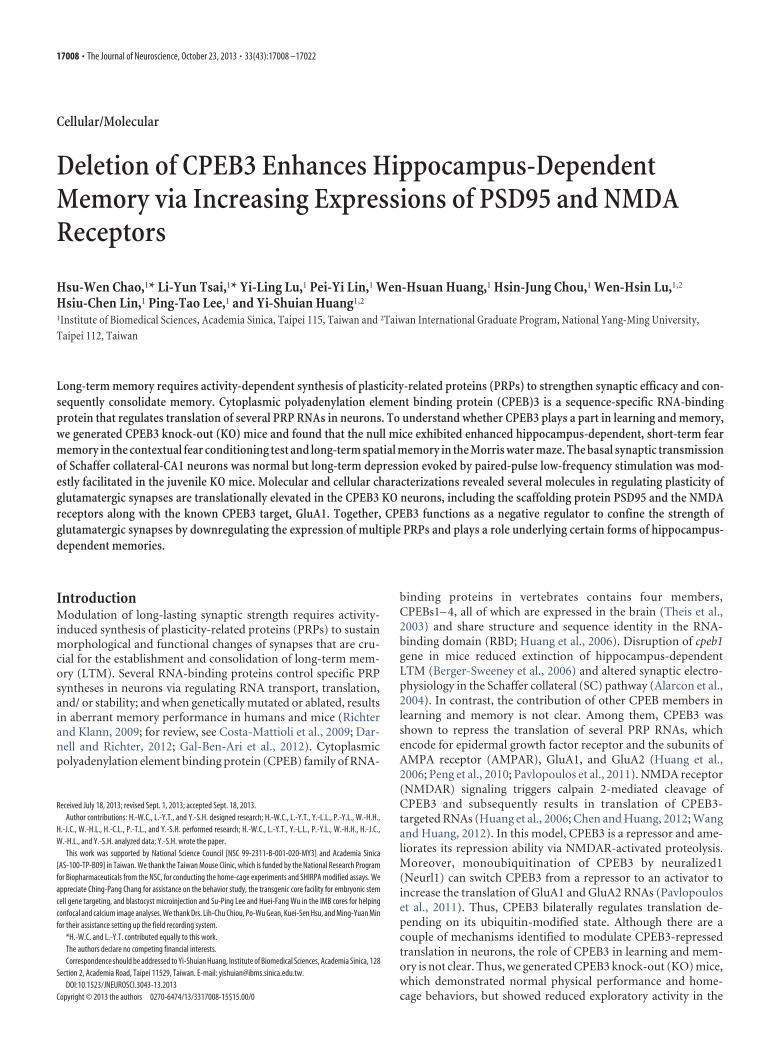
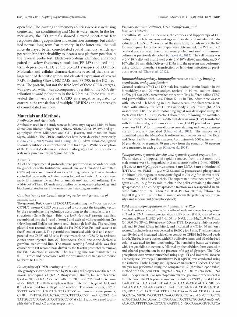
ResultsGeneration of CPEB3-deficient miceTo investigate the role of CPEB3 in learning and memory, weused the cre-loxP strategy to generate CPEB3 KO mice in aC57BL/6 genetic background (Fig. 1A). The targeting vector waselectroporated into C57BL/6 embryonic stem (ES) cells. Fourcorrect targeting ES clones were injected into C57BL/6-Tyrc-2J(c2J) blastocysts for chimera production, where one of the in-jected blastocysts successfully produced germline-transmittedchimeras. After multiple crossings with C57BL/6 mice, C57BL/6mouse lines expressing ubiquitous Flp recombinase or sperm-specific Cre recombinase to obtain CPEB3 heterozygous mice(Fig. 1A), the littermates from these heterozygous matings wereused for all of the experiments in this study. When exon 2 wasdeleted, alternative usage of the first methionine codon in exon 3resulted in premature termination. Using a probe against theexon 3– 6 region, we found the presence of a truncated CPEB3RNA in the heterozygous and KO tissues (Fig. 1B), suggestingthat the premature stop codon in the truncated transcript did notefficiently trigger non-sense-mediated RNA decay (NMD)(Schoenberg and Maquat, 2012). The shorter CPEB3 transcriptin the testis is caused by alternative polyadenylation (Morgan et
17010 • J. Neurosci., October 23, 2013 • 33(43):17008 –17022 Chao, Tsai et al. • CPEB3 Negatively Regulates Memory Consolidation
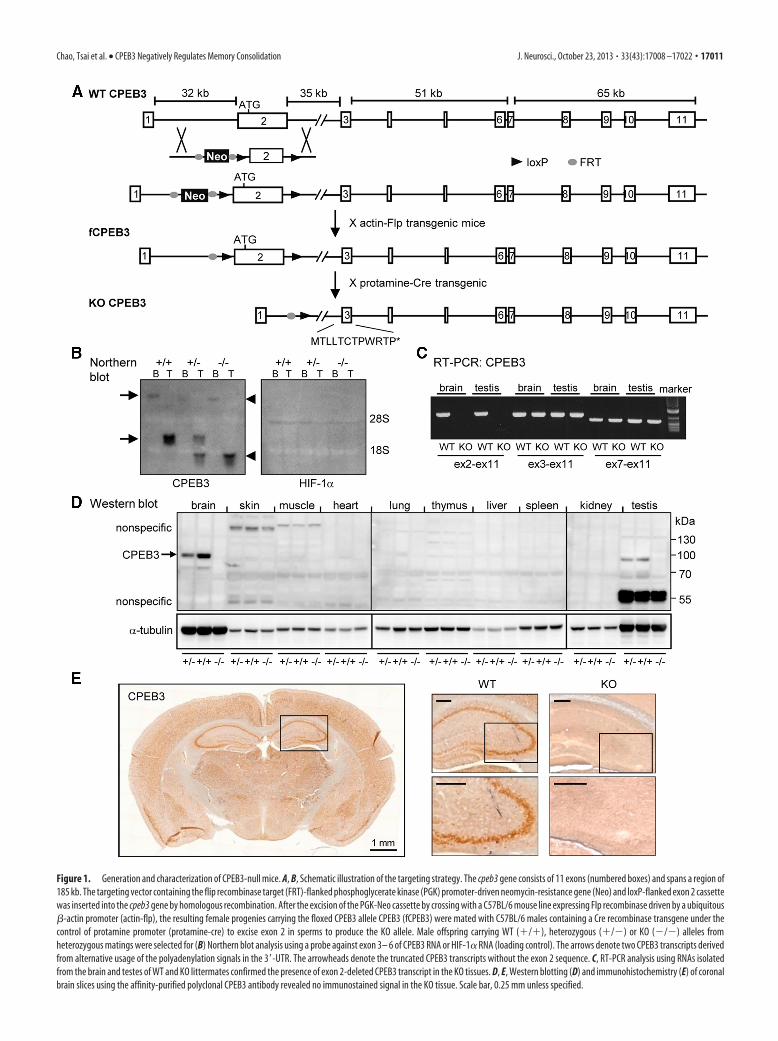
Figure 1. Generation and characterization of CPEB3-null mice. A, B, Schematic illustration of the targeting strategy. The cpeb3 gene consists of 11 exons (numbered boxes) and spans a region of185 kb. The targeting vector containing the flip recombinase target (FRT)-flanked phosphoglycerate kinase (PGK) promoter-driven neomycin-resistance gene (Neo) and loxP-flanked exon 2 cassettewas inserted into the cpeb3 gene by homologous recombination. After the excision of the PGK-Neo cassette by crossing with a C57BL/6 mouse line expressing Flp recombinase driven by a ubiquitous�-actin promoter (actin-flp), the resulting female progenies carrying the floxed CPEB3 allele CPEB3 (fCPEB3) were mated with C57BL/6 males containing a Cre recombinase transgene under thecontrol of protamine promoter (protamine-cre) to excise exon 2 in sperms to produce the KO allele. Male offspring carrying WT (�/�), heterozygous (�/�) or KO (�/�) alleles fromheterozygous matings were selected for (B) Northern blot analysis using a probe against exon 3– 6 of CPEB3 RNA or HIF-1� RNA (loading control). The arrows denote two CPEB3 transcripts derivedfrom alternative usage of the polyadenylation signals in the 3�-UTR. The arrowheads denote the truncated CPEB3 transcripts without the exon 2 sequence. C, RT-PCR analysis using RNAs isolatedfrom the brain and testes of WT and KO littermates confirmed the presence of exon 2-deleted CPEB3 transcript in the KO tissues. D, E, Western blotting (D) and immunohistochemistry (E) of coronalbrain slices using the affinity-purified polyclonal CPEB3 antibody revealed no immunostained signal in the KO tissue. Scale bar, 0.25 mm unless specified.
Chao, Tsai et al. • CPEB3 Negatively Regulates Memory Consolidation J. Neurosci., October 23, 2013 • 33(43):17008 –17022 • 17011
al., 2010). Using the more sensitive assay, RT-PCR, we confirmedthe absence of exon 2 in CPEB3 RNA in the KO tissues (Fig. 1C).Moreover, no CPEB3 protein was detected in the KO tissues us-ing Western blotting (Fig. 1D) and immunohistochemistry assay(Fig. 1E) with the affinity-purified polyclonal CPEB3 antibody.Thus, we produced a mouse line deficient in CPEB3 protein.
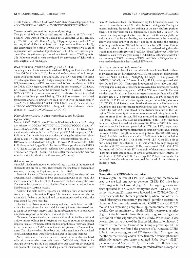
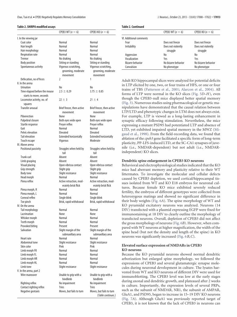
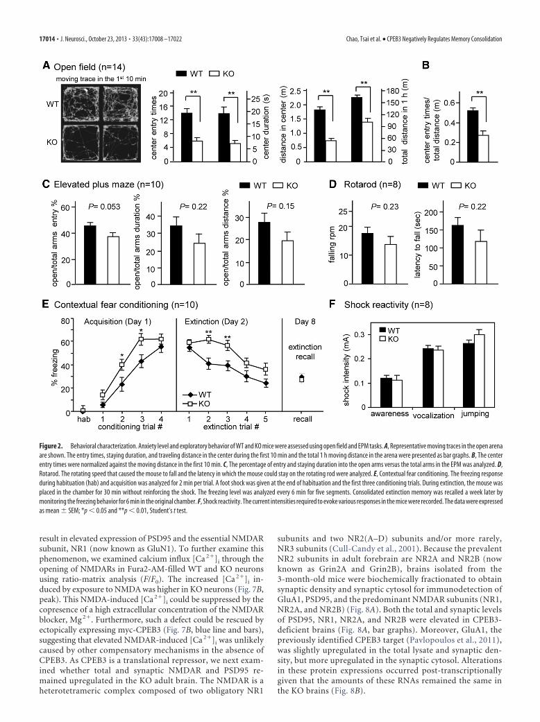
CPEB3 KO mice showed higher anxiety in the open field andelevated short-term fear response in the contextual fearconditioningThe following behavior studies were blindly conducted to thegenotypes using 2- to 3-month-old male WT and KO littermates.The body weight (WT: 22.84 � 0.4 g; KO: 22.58 � 0.5 g, n � 40),home-cage behaviors (Table 1), and physical performance as-sessed using the modified SmithKline/Harwell/Imperial College/Royal Hospital/Phenotype Assessment (SHIRPA) procedures(Masuya et al., 2005, Table 2) were similar between the twogroups of mice. Anxiety-like responses and exploratory behaviorswere studied in the open field and EPM. Normally, mice fear anopen environment and tend to avoid the center of the field or theelevated open arm. The extent of anxiety is determined by count-ing the number of entries and the duration of stay by the testanimal into the center zone in the open field (Fig. 2A,B) or intothe open arm in the EPM (Fig. 2C). The KO mice showed elevatedanxiety in the open field, but not the EPM because they spent lesstime and showed a reduced number of crosses into the centerarena (Fig. 2A). Although the locomotor activity of the null micewas significantly lower (i.e., total distance) in the open field, theKO mice preferred to not enter the center even after taking thisfactor into consideration (Fig. 2B). The reduced locomotor ac-tivity in the open field was not due to motor problems given thatthe KO mice showed normal motor coordination on the rotarod(Fig. 2D) and demonstrated similar locomotor abilities to WTlittermates in the home-cage environment (Table 1).
We next used contextual fear conditioning and MWZ to ex-amine hippocampus-dependent memory. In the former assay,the mice learned to express a fear response (i.e., freezing) whenfaced with a conditioned stimulus (CS, the chamber environ-ment), which was previously paired with a noxious uncondi-tioned stimulus (US, electrical foot shock). In contrast, if themice were exposed only to the CS without US pairing, then pre-viously acquired fear responses would gradually decline. Thisprocess is known as extinction. The freezing levels in WT and KOmice were similar during habituation, increased significantly af-ter CS–US paired trainings (i.e., acquisition, F(4,72) � 115.57, p �0.001), and were significantly reduced during extinction(F(4,72) � 18.998, p � 0.001), indicating that both groups of micecould perform associated learning tasks (Fig. 2E). Importantly,the KO group learned more rapidly during acquisition (F(1,18) �
4.9791, p � 0.05), but slower during extinction (F(1,18) �20.4272, p � 0.001). Post hoc comparisons revealed that the KOmice showed increased freezing in the second and third trialsduring acquisition (p � 0.05) and extinction (p � 0.01). Never-theless, given sufficient training, the consolidated long-term fearmemory and extinction memory, as determined 1 d after acqui-sition (i.e., the freezing response in the first extinction trial) andrecalled 7 d after extinction, respectively, were normal in the KOmice (Fig. 2E). The average current intensities required to triggera specific response, awareness, vocalization, or jumping in WTand KO mice were similar (Fig. 2F), suggesting that the KO ani-mals displayed a normal perception of shock.
CPEB3 KO mice had enhanced consolidated spatial memoryin the MWZDuring spatial acquisition, the mice learned to locate a hiddenplatform using visual cues around the maze. Both WT and KOmice learned where the platform was positioned as evidenced bythe decreasing latencies over the 4 d training period (F(3,42) �68.2152, p � 0.001); however, no difference in spatial learningbetween groups was observed (F(1,14) � 2.744, p � 0.1198; Fig.3A). During the probe trial, the hidden platform was removedand the amount of time that the mice spent in the target quad-rant, where the platform was previously placed, was recorded todetermine their consolidated LTM. Both WT and KO mice spenta significant time in the target quadrant (WT: F(3,28) � 5.1255,p � 0.001; KO: F(3,28) � 50.208, p � 0.001). Moreover, the KOmice showed better consolidated spatial memory because theyspent more time in the target quadrant relative to their WT lit-termates (F(1,7) � 1.797, p � 0.05, Student’s t test; Fig. 3B). Dur-ing the reversal trials (Fig. 3C), the hidden platform was moved toa different quadrant and the mice learned to locate the platformin its new position for 1 d of the four spatial trainings. The probetest was followed 24 h later. Although there was no difference inreversal spatial learning between the two groups of animals (thelatency to escape in seconds, on day 9, WT: 31.68 � 7.98 vs KO:29.19 � 6.71; day 11, WT: 30.50 � 10.76 vs KO: 28.63 � 8.47; day13, WT: 30.69 � 10.42 vs KO: 33.94 � 8.50), the KO mice clearlyspent less time in the newly acquired Q4 quadrant compared withWT littermates in the first reversal probe test (Fig. 3C). Such adifference was likely caused by the stronger consolidated memoryfor the previous Q2 platform position in the KO mice (Fig. 3B).Both of the groups of mice navigated 25% of time in the Q2quadrant in the first reversal probe trial, but soon learned torelocate the platform in the most recently trained quadrant in thesecond and third reversal probe tests (Fig. 3C). Last, using thevisible platform, we ensured that the swimming ability and visualacuity of the KO mice were normal (Fig. 3D).
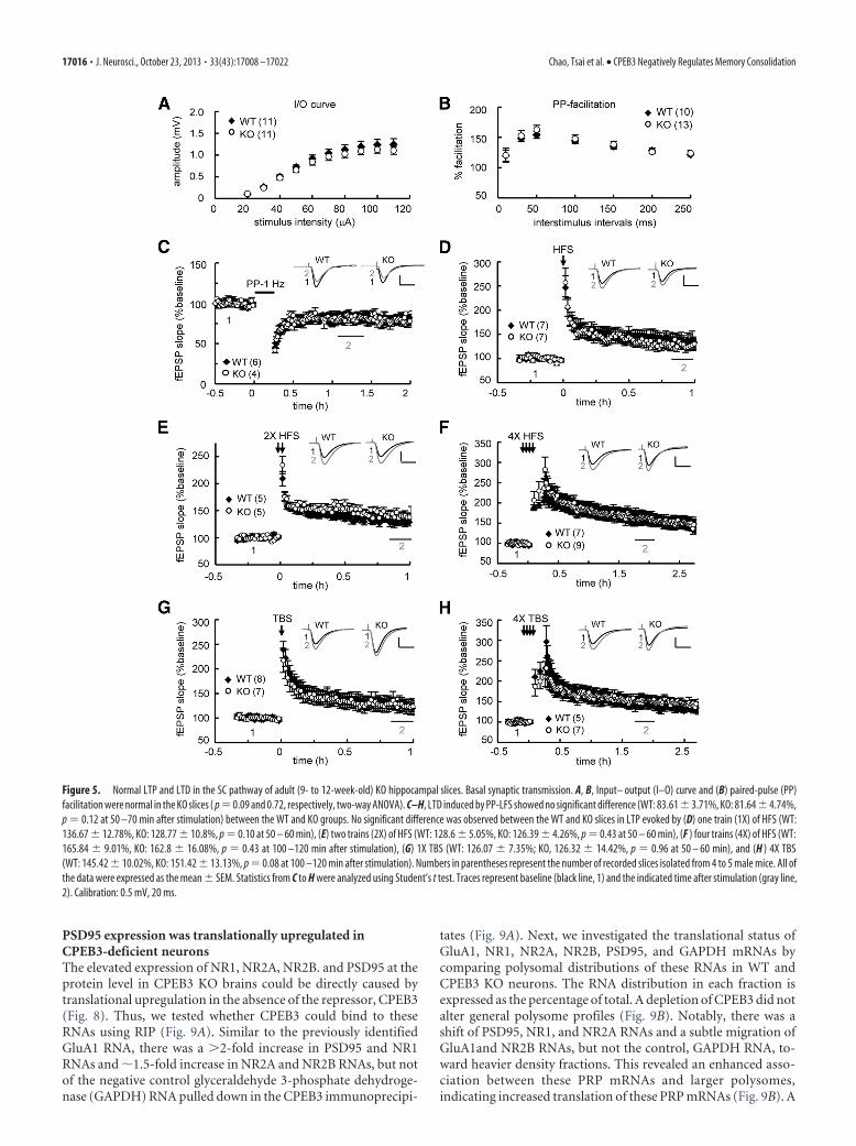
Enhanced PP-LFS-elicited LTD in juvenile CPEB3 KO miceTo examine whether specific forms of synaptic plasticity werealtered in the SC-CA1 pathway of the KO hippocampus, we usedhippocampal slices isolated from juvenile and adult mice for LTDand LTP studies. The basic synaptic responses, including the in-put– output relationship (Figs. 4A, 5A) and PPF (Figs. 4B, 5B),did not differ between the WT and KO groups. LTD induced byLFS was normal (Fig. 4C), but evoked by PP-LFS was enhanced inyoung KO hippocampal slices (Fig. 4D). PP-LFS-evoked LTDrequired the activation of NMDARs in 3- to 4-week-old WT andKO mice (Fig. 4D). PP-LFS is a stronger induction protocol,which can also evoke NMDAR-independent LTD in adult mice(Oliet et al., 1997; Kemp et al., 2000). However, no difference wasfound in this form of LTD if adult slices were used (Fig. 5C).
Table 1. Home-cage monitoring
CPEB3 WT (n � 6) CPEB3 KO (n � 6)
Time percentage Day Night Day Night
Awakening 0.017 � 0.001 0.011 � 0.001 0.016 � 0.002 0.011 � 0.001Drinking 0.025 � 0.018 0.852 � 0.112 0.160 � 0.019 1.180 � 0.078Feeding 1.779 � 0.167 8.324 � 0.498 1.884 � 0.337 7.322 � 0.755Grooming 6.072 � 0.498 11.12 � 0.428 6.292 � 0.581 12.04 � 0.425Hanging 0.468 � 0.139 5.460 � 0.852 0.154 � 0.070 3.803 � 0.724Rearing 0.188 � 0.060 0.848 � 0.149 0.101 � 0.021 0.781 � 0.114Resting 43.380 � 0.778 19.16 � 1.247 44.43 � 1.104 18.72 � 1.609Twitching 0.407 � 0.049 0.211 � 0.020 0.388 � 0.025 0.209 � 0.029Walking 0.418 � 0.061 1.244 � 0.131 0.619 � 0.134 1.886 � 0.222Travel distance (cm) 87.322 � 11.82 369.07 � 46.78 104.87 � 16.65 435.96 � 63.84
17012 • J. Neurosci., October 23, 2013 • 33(43):17008 –17022 Chao, Tsai et al. • CPEB3 Negatively Regulates Memory Consolidation
Adult KO hippocampal slices were analyzed for potential deficitsin LTP elicited by one, two, or four trains of HFS, or one or fourtrains of TBS (Patterson et al., 2001; Alarcon et al., 2004). Allforms of LTP were normal in the KO slices (Fig. 5D–H), eventhough the CPEB3-null mice displayed better spatial memory(Fig. 3). Numerous studies using pharmacological or genetic ma-nipulations have demonstrated that the causal relation betweenLTP/LTD and phenotypic changes in LTM does not always exist.For example, LTP is viewed as a long-lasting enhancement insynaptic efficacy following stimulation. Nevertheless, the miceexpressing a mutant PSD95 had potentiated LTP and absence ofLTD, yet exhibited impaired spatial memory in the MWZ (Mi-gaud et al., 1998). From the field-recording data, we found thatablation of the cpeb3 gene facilitated a specific form of long-termplasticity, PP-LFS-induced LTD, at the SC-CA1 synapses of juve-nile (i.e., NMDAR-dependent) but not adult (i.e., NMDAR-independent) KO slices.
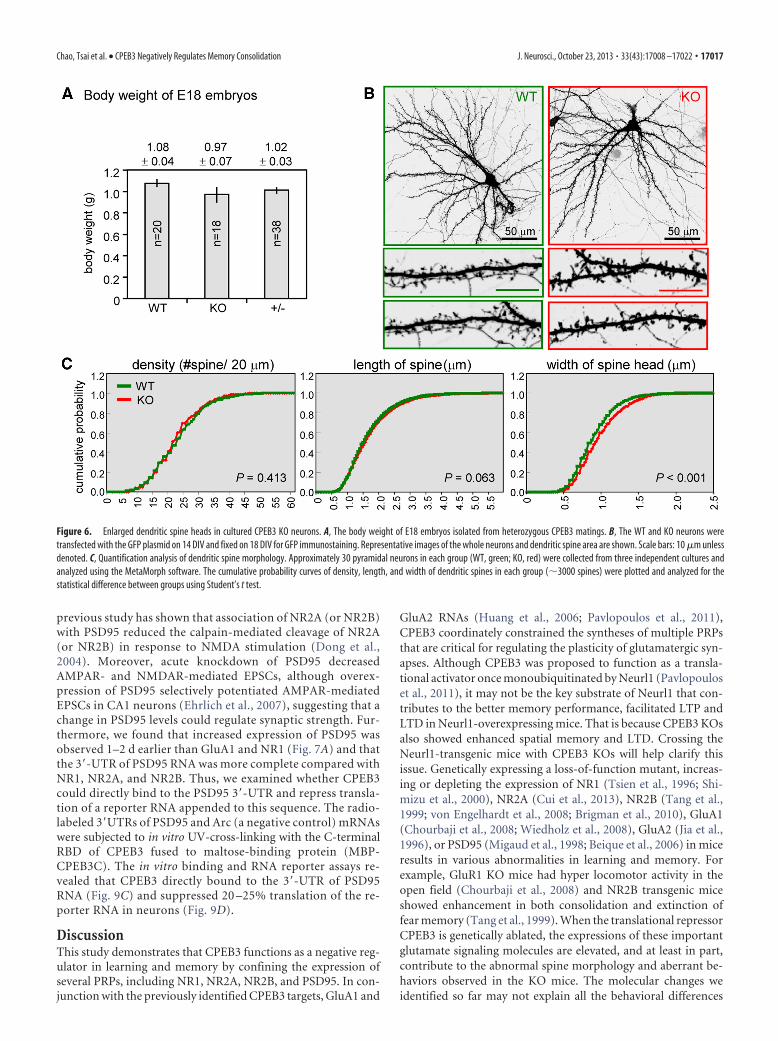
Dendritic spine enlargement in CPEB3 KO neuronsBehavioral and electrophysiological studies indicated that the KOmice had aberrant memory and plasticity relative to their WTlittermates. To investigate the molecular and cellular defectscaused by CPEB3 depletion, we used cortical/hippocampal tis-sues isolated from WT and KO E18 embryos for neuronal cul-tures. Because female KO mice exhibited severely reducedfertility, the embryos of different genotypes were collected fromheterozygous matings and showed no significant difference intheir body weights (Fig. 6A). The spine morphology of WT andKO pyramidal excitatory neurons was analyzed. Neurons (14DIV) transfected with a plasmid expressing EGFP were fixed forimmunostaining at 18 DIV to clearly outline the morphology oftransfected neurons. Overall, depletion of CPEB3 did not affectthe gross morphology of neurons (Fig. 6B). However, when com-pared with WT neurons at higher magnification, the width of thespine head (but not the density and length of the spine) in KOneurons was significantly increased (Fig. 6B,C).
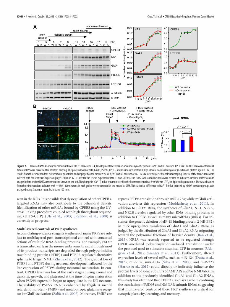
Elevated surface expression of NMDARs in CPEB3KO neuronsBecause the KO pyramidal neurons showed normal dendriticarborization but enlarged spine morphology, we followed theexpressions of CPEB3 and several glutamatergic synapse mole-cules during neuronal development in culture. The lysates har-vested from WT and KO neurons of different DIV were used forimmunoblotting. The CPEB3 level was low at the early stagesduring axonal and dendritic growth, and plateaued after 2 weeksin culture. Importantly, the expression levels of several PRPs,such as the subunit of NMDAR, NR1, the subunit of AMPAR,GluA1, and PSD95, began to increase in 15–19 DIV KO neurons(Fig. 7A). Although GluA1 was previously reported target ofCPEB3, it is not known that the lack of CPEB3 in neurons can
Table 2. SHIRPA modified assays
CPEB3 WT (n � 6) CPEB3 KO (n � 6)
I. In the viewing jarCoat color Normal NormalHair length Normal NormalHair morphology Normal NormalRespiration rate Normal NormalTremor No shaking No shakingBody position Sitting or standing Sitting or standingSpontaneous activity Vigorous scratching,
grooming, moderatemovement
Vigorous scratching,grooming, moderatemovement
Defecation, no of feces 2 1II. In the arena
Urination No NoTime elapsed before the mouse
starts to move, seconds2.5 � 0.29 3.75 � 0.85
Locomotor activity, no. ofsquares
22 � 3 21 � 4
Transfer arousal Brief freeze, then activemovement
Brief freeze, then activemovement
Piloerection None NonePalpebral closure Both eyes wide open Both eyes wide openStartle response Preyer reflex Preyer reflexGait Normal NormalPelvic elevation Elevated ElevatedTail elevation Extended horizontally Extended horizontallyTouch escape Vigorous Moderate
III. Above arenaPositional passivity Struggles when held by
tailStruggles when held by
tailTrunk curl Absent AbsentLimb grasping Absent AbsentVisual placing Upon vibrissa contact Upon vibrissa contactGrip strength Slight grip Slight gripBody tone Slight resistance Slight resistanceHead morph Normal NormalPinna reflex Active retraction, mod-
erately brisk flickActive retraction, mod-
erately brisk flickPinna morph, R Normal NormalPinna morph, L Normal NormalCorneal reflex Single blink Single blinkToe pinch Brisk, rapid withdrawal Brisk, rapid withdrawal
IV. On the arenaTail morphology Normal NormalLacrimation None NoneWhisker morph Normal NormalTeeth morph Normal NormalProvoked biting Present PresentSalivation Slight margin of the
submaxillary areaSlight margin of the
submaxillary areaHeart rate Normal NormalAbdominal tone Slight resistance Slight resistanceSkin color Pink PinkLimb morph FR Normal NormalLimb morph FL Normal NormalLimb morph HR Normal NormalLimb morph HL Normal NormalLimb tone Slight resistance Slight resistance
V. In the arena, part 2Wire maneuver Unable to grip with a
hindlimbUnable to grip with a
hindlimbRighting reflex No impairment No impairmentContact righting reflex Tries TriesNegative geotaxis Moves, but fails to turn Moves, but fails to turn
(Table continues.)
Table 2. Continued
CPEB3 WT (n � 6) CPEB3 KO (n � 6)
VI. Additional commentsFear Does not freeze Does not freezeIrritability Does not violently
struggleDoes not violently
struggleAggression No NoVocalization Yes YesBizarre behavior No bizarre behavior No bizarre behaviorConvulsion No phenotype No phenotype
Chao, Tsai et al. • CPEB3 Negatively Regulates Memory Consolidation J. Neurosci., October 23, 2013 • 33(43):17008 –17022 • 17013
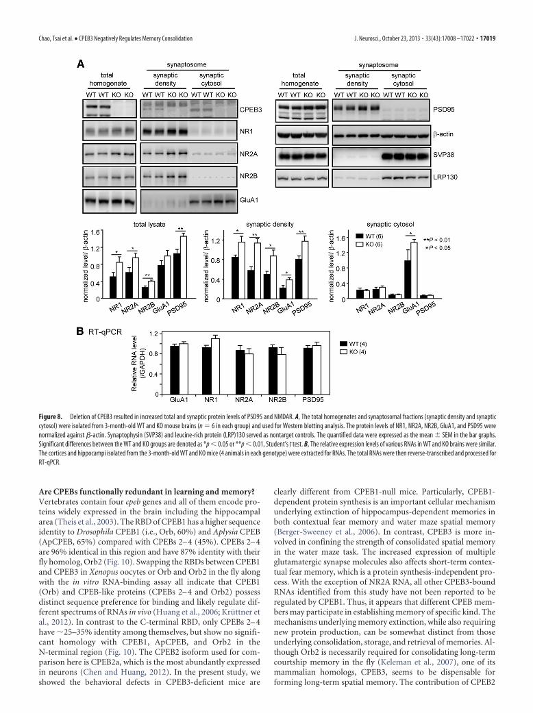
result in elevated expression of PSD95 and the essential NMDARsubunit, NR1 (now known as GluN1). To further examine thisphenomenon, we examined calcium influx [Ca 2�]i through theopening of NMDARs in Fura2-AM-filled WT and KO neuronsusing ratio-matrix analysis (F/F0). The increased [Ca 2�]i in-duced by exposure to NMDA was higher in KO neurons (Fig. 7B,peak). This NMDA-induced [Ca 2�]i could be suppressed by thecopresence of a high extracellular concentration of the NMDARblocker, Mg 2�. Furthermore, such a defect could be rescued byectopically expressing myc-CPEB3 (Fig. 7B, blue line and bars),suggesting that elevated NMDAR-induced [Ca 2�]i was unlikelycaused by other compensatory mechanisms in the absence ofCPEB3. As CPEB3 is a translational repressor, we next exam-ined whether total and synaptic NMDAR and PSD95 re-mained upregulated in the KO adult brain. The NMDAR is aheterotetrameric complex composed of two obligatory NR1
subunits and two NR2(A–D) subunits and/or more rarely,NR3 subunits (Cull-Candy et al., 2001). Because the prevalentNR2 subunits in adult forebrain are NR2A and NR2B (nowknown as Grin2A and Grin2B), brains isolated from the3-month-old mice were biochemically fractionated to obtainsynaptic density and synaptic cytosol for immunodetection ofGluA1, PSD95, and the predominant NMDAR subunits (NR1,NR2A, and NR2B) (Fig. 8A). Both the total and synaptic levelsof PSD95, NR1, NR2A, and NR2B were elevated in CPEB3-deficient brains (Fig. 8A, bar graphs). Moreover, GluA1, thepreviously identified CPEB3 target (Pavlopoulos et al., 2011),was slightly upregulated in the total lysate and synaptic den-sity, but more upregulated in the synaptic cytosol. Alterationsin these protein expressions occurred post-transcriptionallygiven that the amounts of these RNAs remained the same inthe KO brains (Fig. 8B).
Figure 2. Behavioral characterization. Anxiety level and exploratory behavior of WT and KO mice were assessed using open field and EPM tasks. A, Representative moving traces in the open arenaare shown. The entry times, staying duration, and traveling distance in the center during the first 10 min and the total 1 h moving distance in the arena were presented as bar graphs. B, The centerentry times were normalized against the moving distance in the first 10 min. C, The percentage of entry and staying duration into the open arms versus the total arms in the EPM was analyzed. D,Rotarod. The rotating speed that caused the mouse to fall and the latency in which the mouse could stay on the rotating rod were analyzed. E, Contextual fear conditioning. The freezing responseduring habituation (hab) and acquisition was analyzed for 2 min per trial. A foot shock was given at the end of habituation and the first three conditioning trials. During extinction, the mouse wasplaced in the chamber for 30 min without reinforcing the shock. The freezing level was analyzed every 6 min for five segments. Consolidated extinction memory was recalled a week later bymonitoring the freezing behavior for 6 min in the original chamber. F, Shock reactivity. The current intensities required to evoke various responses in the mice were recorded. The data were expressedas mean � SEM; *p � 0.05 and **p � 0.01, Student’s t test.
17014 • J. Neurosci., October 23, 2013 • 33(43):17008 –17022 Chao, Tsai et al. • CPEB3 Negatively Regulates Memory Consolidation
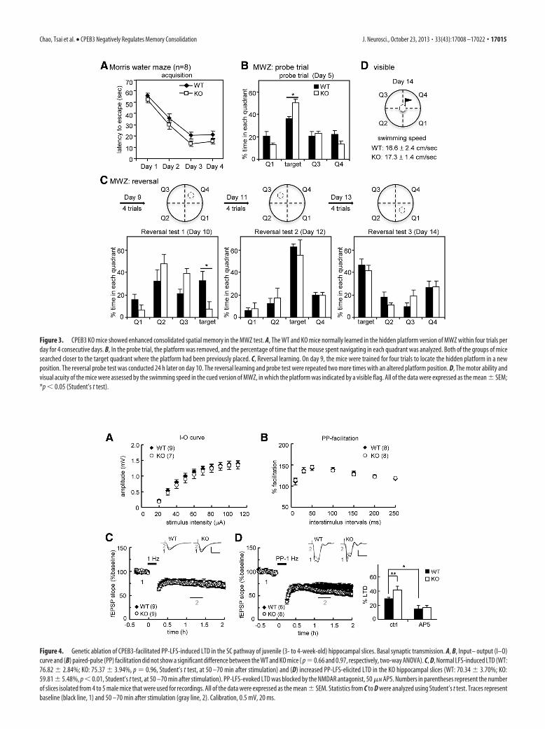
Figure 4. Genetic ablation of CPEB3-facilitated PP-LFS-induced LTD in the SC pathway of juvenile (3- to 4-week-old) hippocampal slices. Basal synaptic transmission. A, B, Input– output (I–O)curve and (B) paired-pulse (PP) facilitation did not show a significant difference between the WT and KO mice ( p � 0.66 and 0.97, respectively, two-way ANOVA). C, D, Normal LFS-induced LTD (WT:76.82 � 2.84%; KO: 75.37 � 3.94%, p � 0.96, Student’s t test, at 50 –70 min after stimulation) and (D) increased PP-LFS-elicited LTD in the KO hippocampal slices (WT: 70.34 � 3.70%; KO:59.81 � 5.48%, p � 0.01, Student’s t test, at 50 –70 min after stimulation). PP-LFS-evoked LTD was blocked by the NMDAR antagonist, 50 �M AP5. Numbers in parentheses represent the numberof slices isolated from 4 to 5 male mice that were used for recordings. All of the data were expressed as the mean � SEM. Statistics from C to D were analyzed using Student’s t test. Traces representbaseline (black line, 1) and 50 –70 min after stimulation (gray line, 2). Calibration, 0.5 mV, 20 ms.
Figure 3. CPEB3 KO mice showed enhanced consolidated spatial memory in the MWZ test. A, The WT and KO mice normally learned in the hidden platform version of MWZ within four trials perday for 4 consecutive days. B, In the probe trial, the platform was removed, and the percentage of time that the mouse spent navigating in each quadrant was analyzed. Both of the groups of micesearched closer to the target quadrant where the platform had been previously placed. C, Reversal learning. On day 9, the mice were trained for four trials to locate the hidden platform in a newposition. The reversal probe test was conducted 24 h later on day 10. The reversal learning and probe test were repeated two more times with an altered platform position. D, The motor ability andvisual acuity of the mice were assessed by the swimming speed in the cued version of MWZ, in which the platform was indicated by a visible flag. All of the data were expressed as the mean � SEM;*p � 0.05 (Student’s t test).
Chao, Tsai et al. • CPEB3 Negatively Regulates Memory Consolidation J. Neurosci., October 23, 2013 • 33(43):17008 –17022 • 17015
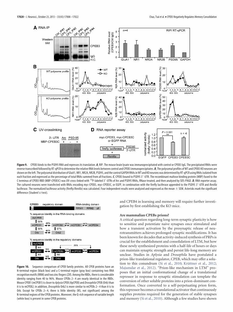
PSD95 expression was translationally upregulated inCPEB3-deficient neuronsThe elevated expression of NR1, NR2A, NR2B. and PSD95 at theprotein level in CPEB3 KO brains could be directly caused bytranslational upregulation in the absence of the repressor, CPEB3(Fig. 8). Thus, we tested whether CPEB3 could bind to theseRNAs using RIP (Fig. 9A). Similar to the previously identifiedGluA1 RNA, there was a 2-fold increase in PSD95 and NR1RNAs and �1.5-fold increase in NR2A and NR2B RNAs, but notof the negative control glyceraldehyde 3-phosphate dehydroge-nase (GAPDH) RNA pulled down in the CPEB3 immunoprecipi-
tates (Fig. 9A). Next, we investigated the translational status ofGluA1, NR1, NR2A, NR2B, PSD95, and GAPDH mRNAs bycomparing polysomal distributions of these RNAs in WT andCPEB3 KO neurons. The RNA distribution in each fraction isexpressed as the percentage of total. A depletion of CPEB3 did notalter general polysome profiles (Fig. 9B). Notably, there was ashift of PSD95, NR1, and NR2A RNAs and a subtle migration ofGluA1and NR2B RNAs, but not the control, GAPDH RNA, to-ward heavier density fractions. This revealed an enhanced asso-ciation between these PRP mRNAs and larger polysomes,indicating increased translation of these PRP mRNAs (Fig. 9B). A
Figure 5. Normal LTP and LTD in the SC pathway of adult (9- to 12-week-old) KO hippocampal slices. Basal synaptic transmission. A, B, Input– output (I–O) curve and (B) paired-pulse (PP)facilitation were normal in the KO slices ( p � 0.09 and 0.72, respectively, two-way ANOVA). C–H, LTD induced by PP-LFS showed no significant difference (WT: 83.61 � 3.71%, KO: 81.64 � 4.74%,p � 0.12 at 50 –70 min after stimulation) between the WT and KO groups. No significant difference was observed between the WT and KO slices in LTP evoked by (D) one train (1X) of HFS (WT:136.67 � 12.78%, KO: 128.77 � 10.8%, p � 0.10 at 50 – 60 min), (E) two trains (2X) of HFS (WT: 128.6 � 5.05%, KO: 126.39 � 4.26%, p � 0.43 at 50 – 60 min), (F ) four trains (4X) of HFS (WT:165.84 � 9.01%, KO: 162.8 � 16.08%, p � 0.43 at 100 –120 min after stimulation), (G) 1X TBS (WT: 126.07 � 7.35%; KO, 126.32 � 14.42%, p � 0.96 at 50 – 60 min), and (H ) 4X TBS(WT: 145.42 � 10.02%, KO: 151.42 � 13.13%, p � 0.08 at 100 –120 min after stimulation). Numbers in parentheses represent the number of recorded slices isolated from 4 to 5 male mice. All ofthe data were expressed as the mean � SEM. Statistics from C to H were analyzed using Student’s t test. Traces represent baseline (black line, 1) and the indicated time after stimulation (gray line,2). Calibration: 0.5 mV, 20 ms.
17016 • J. Neurosci., October 23, 2013 • 33(43):17008 –17022 Chao, Tsai et al. • CPEB3 Negatively Regulates Memory Consolidation
previous study has shown that association of NR2A (or NR2B)with PSD95 reduced the calpain-mediated cleavage of NR2A(or NR2B) in response to NMDA stimulation (Dong et al.,2004). Moreover, acute knockdown of PSD95 decreasedAMPAR- and NMDAR-mediated EPSCs, although overex-pression of PSD95 selectively potentiated AMPAR-mediatedEPSCs in CA1 neurons (Ehrlich et al., 2007), suggesting that achange in PSD95 levels could regulate synaptic strength. Fur-thermore, we found that increased expression of PSD95 wasobserved 1–2 d earlier than GluA1 and NR1 (Fig. 7A) and thatthe 3�-UTR of PSD95 RNA was more complete compared withNR1, NR2A, and NR2B. Thus, we examined whether CPEB3could directly bind to the PSD95 3�-UTR and repress transla-tion of a reporter RNA appended to this sequence. The radio-labeled 3�UTRs of PSD95 and Arc (a negative control) mRNAswere subjected to in vitro UV-cross-linking with the C-terminalRBD of CPEB3 fused to maltose-binding protein (MBP-CPEB3C). The in vitro binding and RNA reporter assays re-vealed that CPEB3 directly bound to the 3�-UTR of PSD95RNA (Fig. 9C) and suppressed 20 –25% translation of the re-porter RNA in neurons (Fig. 9D).
DiscussionThis study demonstrates that CPEB3 functions as a negative reg-ulator in learning and memory by confining the expression ofseveral PRPs, including NR1, NR2A, NR2B, and PSD95. In con-junction with the previously identified CPEB3 targets, GluA1 and
GluA2 RNAs (Huang et al., 2006; Pavlopoulos et al., 2011),CPEB3 coordinately constrained the syntheses of multiple PRPsthat are critical for regulating the plasticity of glutamatergic syn-apses. Although CPEB3 was proposed to function as a transla-tional activator once monoubiquitinated by Neurl1 (Pavlopouloset al., 2011), it may not be the key substrate of Neurl1 that con-tributes to the better memory performance, facilitated LTP andLTD in Neurl1-overexpressing mice. That is because CPEB3 KOsalso showed enhanced spatial memory and LTD. Crossing theNeurl1-transgenic mice with CPEB3 KOs will help clarify thisissue. Genetically expressing a loss-of-function mutant, increas-ing or depleting the expression of NR1 (Tsien et al., 1996; Shi-mizu et al., 2000), NR2A (Cui et al., 2013), NR2B (Tang et al.,1999; von Engelhardt et al., 2008; Brigman et al., 2010), GluA1(Chourbaji et al., 2008; Wiedholz et al., 2008), GluA2 (Jia et al.,1996), or PSD95 (Migaud et al., 1998; Beique et al., 2006) in miceresults in various abnormalities in learning and memory. Forexample, GluR1 KO mice had hyper locomotor activity in theopen field (Chourbaji et al., 2008) and NR2B transgenic miceshowed enhancement in both consolidation and extinction offear memory (Tang et al., 1999). When the translational repressorCPEB3 is genetically ablated, the expressions of these importantglutamate signaling molecules are elevated, and at least in part,contribute to the abnormal spine morphology and aberrant be-haviors observed in the KO mice. The molecular changes weidentified so far may not explain all the behavioral differences
Figure 6. Enlarged dendritic spine heads in cultured CPEB3 KO neurons. A, The body weight of E18 embryos isolated from heterozygous CPEB3 matings. B, The WT and KO neurons weretransfected with the GFP plasmid on 14 DIV and fixed on 18 DIV for GFP immunostaining. Representative images of the whole neurons and dendritic spine area are shown. Scale bars: 10 �m unlessdenoted. C, Quantification analysis of dendritic spine morphology. Approximately 30 pyramidal neurons in each group (WT, green; KO, red) were collected from three independent cultures andanalyzed using the MetaMorph software. The cumulative probability curves of density, length, and width of dendritic spines in each group (�3000 spines) were plotted and analyzed for thestatistical difference between groups using Student’s t test.
Chao, Tsai et al. • CPEB3 Negatively Regulates Memory Consolidation J. Neurosci., October 23, 2013 • 33(43):17008 –17022 • 17017
seen in the KOs. It is possible that dysregulation of other CPEB3-targeted RNAs may also contribute to the behavioral deficits.Identification of other mRNAs bound by CPEB3 using the UV-cross-linking procedure coupled with high throughout sequenc-ing (HITS-CLIP) (Ule et al., 2003; Licatalosi et al., 2008) iscurrently in progress.
Multilayered controls of PRP synthesesAccumulating evidence suggests syntheses of many PRPs are sub-ject to multilayered post-transcriptional control with concertedactions of multiple RNA-binding proteins. For example, PSD95is transcribed early in the mouse embryonic brain, although mostof its product transcripts are degraded through polypyrimidinetract binding protein (PTBP)1 and PTBP2-regulated alternativesplicing to trigger NMD (Zheng et al., 2012). The gradual loss ofPTBP1 and PTBP2 during neuronal development allowed for thelate expression of PSD95 during neuronal maturation. In con-trast, CPEB3 level was low at the early stages during axonal anddendritic growth, and plateaued at the time of spine maturationwhen PSD95 expression began to upregulate in the KO neurons.The stability of PSD95 RNA is enhanced by fragile X mentalretardation protein (FMRP) and metabotropic glutamate recep-tor (mGluR) activation (Zalfa et al., 2007). Moreover, FMRP can
repress PSD95 translation through miR-125a; while mGluR acti-vation alleviates this repression (Muddashetty et al., 2011). Inaddition to PSD95 RNA, the syntheses of GluA1, NR1, NR2A,and NR2B are also regulated by other RNA-binding proteins inaddition to CPEB3 as well as many microRNAs (miRs). For in-stance, the genetic deletion of eIF-4E binding protein 2 (4E-BP2)in mice upregulates translation of GluA1 and GluA2 RNAs asjudged by the distribution of GluA1 and GluA2 RNAs migratingtoward the polysomal fractions of heavier density (Ran et al.,2013). NR2A was recently reported to be regulated throughCPEB1-mediated polyadenylation-induced translation underthe protocol used to stimulate chemical LTP in neurons (Uda-gawa et al., 2012; Swanger et al., 2013). Furthermore, alteredexpression levels of several miRs, such as miR-124 (Dutta et al.,2013), miR-132, miR-181a (Saba et al., 2012), and miR-223(Harraz et al., 2012) could directly or indirectly influence theprotein levels of some subunits of AMPARs and/or NMDARs. Inaddition to the previously identified GluA1 and GluA2 RNAs,this study has identified that CPEB3 also plays a role in confiningthe translation of PSD95 and NMDAR subunit RNAs, suggestingthat multilayered control of these PRP syntheses is critical forsynaptic plasticity, learning, and memory.
Figure 7. Elevated NMDAR-induced calcium influx in CPEB3 KO neurons. A, Developmental expression of various synaptic proteins in WT and KO neurons. CPEB3 WT and KO neurons obtained atdifferent DIV were harvested for Western blotting. The protein levels of NR1, GluA1, PSD95, CPEB3, and leucine-rich protein (LRP)130 were normalized against �-actin and plotted against DIV. Theresults from three independent cultures were quantified and displayed as the mean � SEM. B, WT and KO neurons at 16 –17 DIV were subjected to calcium imaging. Several of the KO neurons wereinfected with the lentivirus expressing myc-CPEB3 on 12–13 DIV for the rescue experiment (KO � myc-CPEB3). The Fura2-AM-loaded neurons were treated as indicated. Representative calciumimages before or after NMDA treatment are shown on the left. The change in [Ca 2�] influx was monitored by the fluorescence ratio at 340/380 nm (F/F0) and plotted against time. The data obtainedfrom three independent cultures with �250 –300 neurons in each group were expressed as the mean � SEM. The statistical difference in [Ca 2�] influx induced by NMDA between groups wasanalyzed using Student’s t test. Scale bars: 100 mm.
17018 • J. Neurosci., October 23, 2013 • 33(43):17008 –17022 Chao, Tsai et al. • CPEB3 Negatively Regulates Memory Consolidation
Are CPEBs functionally redundant in learning and memory?Vertebrates contain four cpeb genes and all of them encode pro-teins widely expressed in the brain including the hippocampalarea (Theis et al., 2003). The RBD of CPEB1 has a higher sequenceidentity to Drosophila CPEB1 (i.e., Orb, 60%) and Aplysia CPEB(ApCPEB, 65%) compared with CPEBs 2– 4 (45%). CPEBs 2– 4are 96% identical in this region and have 87% identity with theirfly homolog, Orb2 (Fig. 10). Swapping the RBDs between CPEB1and CPEB3 in Xenopus oocytes or Orb and Orb2 in the fly alongwith the in vitro RNA-binding assay all indicate that CPEB1(Orb) and CPEB-like proteins (CPEBs 2– 4 and Orb2) possessdistinct sequence preference for binding and likely regulate dif-ferent spectrums of RNAs in vivo (Huang et al., 2006; Kruttner etal., 2012). In contrast to the C-terminal RBD, only CPEBs 2– 4have �25–35% identity among themselves, but show no signifi-cant homology with CPEB1, ApCPEB, and Orb2 in theN-terminal region (Fig. 10). The CPEB2 isoform used for com-parison here is CPEB2a, which is the most abundantly expressedin neurons (Chen and Huang, 2012). In the present study, weshowed the behavioral defects in CPEB3-deficient mice are
clearly different from CPEB1-null mice. Particularly, CPEB1-dependent protein synthesis is an important cellular mechanismunderlying extinction of hippocampus-dependent memories inboth contextual fear memory and water maze spatial memory(Berger-Sweeney et al., 2006). In contrast, CPEB3 is more in-volved in confining the strength of consolidated spatial memoryin the water maze task. The increased expression of multipleglutamatergic synapse molecules also affects short-term contex-tual fear memory, which is a protein synthesis-independent pro-cess. With the exception of NR2A RNA, all other CPEB3-boundRNAs identified from this study have not been reported to beregulated by CPEB1. Thus, it appears that different CPEB mem-bers may participate in establishing memory of specific kind. Themechanisms underlying memory extinction, while also requiringnew protein production, can be somewhat distinct from thoseunderlying consolidation, storage, and retrieval of memories. Al-though Orb2 is necessarily required for consolidating long-termcourtship memory in the fly (Keleman et al., 2007), one of itsmammalian homologs, CPEB3, seems to be dispensable forforming long-term spatial memory. The contribution of CPEB2
Figure 8. Deletion of CPEB3 resulted in increased total and synaptic protein levels of PSD95 and NMDAR. A, The total homogenates and synaptosomal fractions (synaptic density and synapticcytosol) were isolated from 3-month-old WT and KO mouse brains (n � 6 in each group) and used for Western blotting analysis. The protein levels of NR1, NR2A, NR2B, GluA1, and PSD95 werenormalized against �-actin. Synaptophysin (SVP38) and leucine-rich protein (LRP)130 served as nontarget controls. The quantified data were expressed as the mean � SEM in the bar graphs.Significant differences between the WT and KO groups are denoted as *p � 0.05 or **p � 0.01, Student’s t test. B, The relative expression levels of various RNAs in WT and KO brains were similar.The cortices and hippocampi isolated from the 3-month-old WT and KO mice (4 animals in each genotype) were extracted for RNAs. The total RNAs were then reverse-transcribed and processed forRT-qPCR.
Chao, Tsai et al. • CPEB3 Negatively Regulates Memory Consolidation J. Neurosci., October 23, 2013 • 33(43):17008 –17022 • 17019
and CPEB4 in learning and memory will require further investi-gation by first establishing the KO mice.
Are mammalian CPEBs prions?A critical question regarding long-term synaptic plasticity is howto sensitize and potentiate naive synapses once stimulated andhow a transient activation by the presynaptic release of neu-rotransmitters achieves prolonged synaptic modifications. It hasbeen known for decades that activity-induced synthesis of PRPs iscrucial for the establishment and consolidation of LTM, but howthese newly synthesized proteins with a half-life of hours or dayscan maintain synaptic strength and persist life-long memory areunclear. Studies in Aplysia and Drosophila have postulated aprion-like translational regulator, CPEB, which may offer a solu-tion to this conundrum (Si et al., 2010; Kruttner et al., 2012;Majumdar et al., 2012). “Prion-like mechanism in LTM” pro-poses that an initial conformational change of a translationalrepressor in response to synaptic stimulation can template theconversion of other soluble proteins into a prion-dominant con-formation. Once converted to a self-perpetuating prion form,this repressor becomes a translational activator that continuouslysupplies proteins required for the generation of stable synapsesand memory (Si et al., 2010). Although a few studies have shown
Figure 10. Sequence comparison of CPEB family proteins. All CPEB proteins have anN-terminal region (black box) and a C-terminal region (gray box) containing two RNArecognition motifs (RRM) and two zinc fingers (Zif). Among the RBDs, there is considerableidentity ranging from 40 to 96%. Mouse CPEBs 2– 4 are nearly identical in the RBDs.Mouse CPEB1 (mCPEB1) is closer to Aplysia CPEB (ApCPEB) and Drosophila CPEB (Orb) thanit is to mCPEB2; in addition, Drosophila Orb2 is more similar to mCPEBs 2– 4 than it is toOrb. Except for CPEBs 2– 4, there is little identity (NS, not significant) among theN-terminal regions of the CPEB proteins. Moreover, the Q-rich sequence of variable length(white box) is present in some CPEB proteins.
Figure 9. CPEB3 binds to the PSD95 RNA and represses its translation. A, RIP. The mouse brain lysate was immunoprecipitated with control or CPEB3 IgG. The precipitated RNAs werereverse transcribed followed by RT-qPCR to determine the relative RNA levels between control and CPEB3 immunoprecipitates. B, The polysomal profiles of WT and CPEB3 KO neurons areshown on the left. The polysomal distribution of GluA1, NR1, NR2A, NR2B, PSD95, and the control GAPDH RNAs in WT and KO neurons was determined by RT-qPCR using RNAs isolated fromeach fraction and expressed as the percentage of total RNAs summed from all fractions. C, CPEB3 bound to PSD95 3�-UTR. The recombinant maltose binding protein (MBP) fused to theC terminus of CPEB3 RBD (MBP-CPEB3C) was UV-cross-linked with 32P-labeled 3�-UTRs of Arc and PSD95 RNAs, RNase treated, and then analyzed by SDS-PAGE. D, RNA reporter assay.The cultured neurons were transfected with RNAs encoding myc-CPEB3, myc-CPEB3C, or EGFP, in combination with the firefly luciferase appended to the PSD95 3�-UTR and Renillaluciferase. The normalized luciferase activity (firefly/Renilla) was calculated. Four independent results were analyzed and expressed as the mean � SEM. Asterisks mark the significantdifference (Student’s t test).
17020 • J. Neurosci., October 23, 2013 • 33(43):17008 –17022 Chao, Tsai et al. • CPEB3 Negatively Regulates Memory Consolidation
that prion-like oligomerization of Orb2 can be activity inducedand was essential for LTM of the fly courtship behavior (Kruttneret al., 2012; Majumdar et al., 2012), it remains largely unknownhow the physical conversion of Orb2 from a soluble to a prionstructure changes its function from a repressor (Mastushita-Sakai et al., 2010) to an activator. Mammals express four CPEBsin neurons, but only CPEB2 and CPEB3 contain short Q-stretchmotifs in the N terminus. Although the prion domain in Orb2can be substituted with the Q-containing motif from mouseCPEB3 (Kruttner et al., 2012), in this study, CPEB3 KO micedisplayed a potentiated short-term fear response in contextualfear conditioning and enhanced long-term spatial memory in theMWZ, indicating that CPEB3 unlikely employs a prion-likemechanism to persist in all types of long-term memories. Bio-chemical studies also supported a role of CPEB3 as a translationalrepressor (Huang et al., 2006) to constrain the expressions ofAMPAR, PSD95, and NMDAR. Although CPEB3 KO mice havebetter spatial memory in the MWZ, such potentiated memoryappears to jeopardize their ability to rapidly acquire new spatialinformation during reversal learning, suggesting that a slight al-teration in the synaptic proteome composition could shift thebalance of learning and memory toward a better response for onetask and a worse response for the other. Whether CPEB3 mayemploy a prion-like mechanism to influence specific kinds ofmemories or other mammalian homologs of Orb2 (i.e., CPEB2and CPEB4) can employ a prion-like mechanism to positivelyregulate learning and memory, will require further investigation.
ReferencesAlarcon JM, Hodgman R, Theis M, Huang YS, Kandel ER, Richter JD (2004)
Selective modulation of some forms of schaffer collateral-CA1 synapticplasticity in mice with a disruption of the CPEB-1 gene. Learn Mem11:318 –327. CrossRef Medline
Beïque JC, Lin DT, Kang MG, Aizawa H, Takamiya K, Huganir RL (2006)Synapse-specific regulation of AMPA receptor function by PSD-95. ProcNatl Acad Sci U S A 103:19535–19540. CrossRef Medline
Berger-Sweeney J, Zearfoss NR, Richter JD (2006) Reduced extinction ofhippocampal-dependent memories in CPEB knock-out mice. LearnMem 13:4 –7. CrossRef Medline
Brigman JL, Wright T, Talani G, Prasad-Mulcare S, Jinde S, Seabold GK,Mathur P, Davis MI, Bock R, Gustin RM, Colbran RJ, Alvarez VA, Naka-zawa K, Delpire E, Lovinger DM, Holmes A (2010) Loss of GluN2B-containing NMDA receptors in CA1 hippocampus and cortex impairslong-term depression, reduces dendritic spine density, and disrupts learn-ing. J Neurosci 30:4590 – 4600. CrossRef Medline
Chao HW, Hong CJ, Huang TN, Lin YL, Hsueh YP (2008) SUMOylation ofthe MAGUK protein CASK regulates dendritic spinogenesis. J Cell Biol182:141–155. CrossRef Medline
Chao HW, Lai YT, Lu YL, Lin CL, Mai W, Huang YS (2012) NMDAR sig-naling facilitates the IPO5-mediated nuclear import of CPEB3. NucleicAcids Res 40:8484 – 8498. CrossRef Medline
Chen PJ, Huang YS (2012) CPEB2-eEF2 interaction impedes HIF-1alphaRNA translation. EMBO J 31:959 –971. Medline
Chourbaji S, Vogt MA, Fumagalli F, Sohr R, Frasca A, Brandwein C, HortnaglH, Riva MA, Sprengel R, Gass P (2008) AMPA receptor subunit 1(GluR-A) knockout mice model the glutamate hypothesis of depression.FASEB J 22:3129 –3134. CrossRef Medline
Costa-Mattioli M, Sossin WS, Klann E, Sonenberg N (2009) Translationalcontrol of long-lasting synaptic plasticity and memory. Neuron 61:10 –26.CrossRef Medline
Cui Z, Feng R, Jacobs S, Duan Y, Wang H, Cao X, Tsien JZ (2013) IncreasedNR2A:NR2B ratio compresses long-term depression range and constrainslong-term memory. Sci Rep 3:1036. Medline
Cull-Candy S, Brickley S, Farrant M (2001) NMDA receptor subunits: di-versity, development and disease. Curr Opin Neurobiol 11:327–335.CrossRef Medline
Darnell JC, Richter JD (2012) Cytoplasmic RNA-binding proteins and the
control of complex brain function. Cold Spring Harb Perspect Biol4:a012344. CrossRef Medline
Dong YN, Waxman EA, Lynch DR (2004) Interactions of postsynapticdensity-95 and the NMDA receptor 2 subunit control calpain-mediatedcleavage of the NMDA receptor. J Neurosci 24:11035–11045. CrossRefMedline
Dutta R, Chomyk AM, Chang A, Ribaudo MV, Deckard SA, Doud MK,Edberg DD, Bai B, Li M, Baranzini SE, Fox RJ, Staugaitis SM, MacklinWB, Trapp BD (2013) Hippocampal demyelination and memory dys-function are associated with increased levels of the neuronal microRNAmiR-124 and reduced AMPA receptors. Ann Neurol 73:637– 645.CrossRef Medline
Ehrlich I, Klein M, Rumpel S, Malinow R (2007) PSD-95 is required foractivity-driven synapse stabilization. Proc Natl Acad Sci U S A 104:4176 –4181. CrossRef Medline
Gal-Ben-Ari S, Kenney JW, Ounalla-Saad H, Taha E, David O, Levitan D,Gildish I, Panja D, Pai B, Wibrand K, Simpson TI, Proud CG, BramhamCR, Armstrong JD, Rosenblum K (2012) Consolidation and translationregulation. Learn Mem 19:410 – 422. CrossRef Medline
Harraz MM, Eacker SM, Wang X, Dawson TM, Dawson VL (2012)MicroRNA-223 is neuroprotective by targeting glutamate receptors. ProcNatl Acad Sci U S A 109:18962–18967. CrossRef Medline
Huang YS, Kan MC, Lin CL, Richter JD (2006) CPEB3 and CPEB4 in neu-rons: analysis of RNA-binding specificity and translational control ofAMPA receptor GluR2 mRNA. EMBO J 25:4865– 4876. CrossRefMedline
Irvine EE, Drinkwater L, Radwanska K, Al-Qassab H, Smith MA, O’Brien M,Kielar C, Choudhury AI, Krauss S, Cooper JD, Withers DJ, Giese KP(2011) Insulin receptor substrate 2 is a negative regulator of memoryformation. Learn Mem 18:375–383. CrossRef Medline
Jia Z, Agopyan N, Miu P, Xiong Z, Henderson J, Gerlai R, Taverna FA,Velumian A, MacDonald J, Carlen P, Abramow-Newerly W, Roder J(1996) Enhanced LTP in mice deficient in the AMPA receptor GluR2.Neuron 17:945–956. CrossRef Medline
Keleman K, Kruttner S, Alenius M, Dickson BJ (2007) Function of the Dro-sophila CPEB protein Orb2 in long-term courtship memory. Nat Neuro-sci 10:1587–1593. CrossRef Medline
Kemp N, McQueen J, Faulkes S, Bashir ZI (2000) Different forms of LTD inthe CA1 region of the hippocampus: role of age and stimulus protocol.Eur J Neurosci 12:360 –366. CrossRef Medline
Kruttner S, Stepien B, Noordermeer JN, Mommaas MA, Mechtler K, DicksonBJ, Keleman K (2012) Drosophila CPEB Orb2A mediates memory inde-pendent of Its RNA-binding domain. Neuron 76:383–395. CrossRefMedline
Licatalosi DD, Mele A, Fak JJ, Ule J, Kayikci M, Chi SW, Clark TA, SchweitzerAC, Blume JE, Wang X, Darnell JC, Darnell RB (2008) HITS-CLIPyields genome-wide insights into brain alternative RNA processing. Na-ture 456:464 – 469. CrossRef Medline
Majumdar A, Cesario WC, White-Grindley E, Jiang H, Ren F, Khan MR, Li L,Choi EM, Kannan K, Guo F, Unruh J, Slaughter B, Si K (2012) Criticalrole of amyloid-like oligomers of Drosophila Orb2 in the persistence ofmemory. Cell 148:515–529. CrossRef Medline
Mastushita-Sakai T, White-Grindley E, Samuelson J, Seidel C, Si K (2010)Drosophila Orb2 targets genes involved in neuronal growth, synapse for-mation, and protein turnover. Proc Natl Acad Sci U S A 107:11987–11992. CrossRef Medline
Masuya H, Inoue M, Wada Y, Shimizu A, Nagano J, Kawai A, Inoue A,Kagami T, Hirayama T, Yamaga A, Kaneda H, Kobayashi K, Minowa O,Miura I, Gondo Y, Noda T, Wakana S, Shiroishi T (2005) Implementa-tion of the modified-SHIRPA protocol for screening of dominant pheno-types in a large-scale ENU mutagenesis program. Mamm Genome 16:829 – 837. CrossRef Medline
Migaud M, Charlesworth P, Dempster M, Webster LC, Watabe AM, Makh-inson M, He Y, Ramsay MF, Morris RG, Morrison JH, O’Dell TJ, GrantSG (1998) Enhanced long-term potentiation and impaired learning inmice with mutant postsynaptic density-95 protein. Nature 396:433– 439.CrossRef Medline
Morgan M, Iaconcig A, Muro AF (2010) CPEB2, CPEB3 and CPEB4 arecoordinately regulated by miRNAs recognizing conserved binding sites inparalog positions of their 3�-UTRs. Nucleic Acids Res 38:7698 –7710.CrossRef Medline
Chao, Tsai et al. • CPEB3 Negatively Regulates Memory Consolidation J. Neurosci., October 23, 2013 • 33(43):17008 –17022 • 17021
Morris R (1984) Developments of a water-maze procedure for studying spa-tial learning in the rat. J Neurosci Methods 11:47– 60. CrossRef Medline
Muddashetty RS, Nalavadi VC, Gross C, Yao X, Xing L, Laur O, Warren ST,Bassell GJ (2011) Reversible inhibition of PSD-95 mRNA translation bymiR-125a, FMRP phosphorylation, and mGluR signaling. Mol Cell 42:673– 688. CrossRef Medline
Oliet SH, Malenka RC, Nicoll RA (1997) Two distinct forms of long-termdepression coexist in CA1 hippocampal pyramidal cells. Neuron 18:969 –982. CrossRef Medline
Patterson SL, Pittenger C, Morozov A, Martin KC, Scanlin H, Drake C, Kan-del ER (2001) Some forms of cAMP-mediated long-lasting potentiationare associated with release of BDNF and nuclear translocation ofphospho-MAP kinase. Neuron 32:123–140. CrossRef Medline
Pavlopoulos E, Trifilieff P, Chevaleyre V, Fioriti L, Zairis S, Pagano A,Malleret G, Kandel ER (2011) Neuralized1 activates CPEB3: a functionfor nonproteolytic ubiquitin in synaptic plasticity and memory storage.Cell 147:1369 –1383. CrossRef Medline
Peng SC, Lai YT, Huang HY, Huang HD, Huang YS (2010) A novel role ofCPEB3 in regulating EGFR gene transcription via association with Stat5bin neurons. Nucleic Acids Res 38:7446 –7457. CrossRef Medline
Ran I, Gkogkas CG, Vasuta C, Tartas M, Khoutorsky A, Laplante I, Parsyan A,Nevarko T, Sonenberg N, Lacaille JC (2013) Selective regulation ofGluA subunit synthesis and AMPA receptor-mediated synaptic functionand plasticity by the translation repressor 4E-BP2 in hippocampal pyra-midal cells. J Neurosci 33:1872–1886. CrossRef Medline
Richter JD, Klann E (2009) Making synaptic plasticity and memory last:mechanisms of translational regulation. Genes Dev 23:1–11. CrossRefMedline
Saba R, Storchel PH, Aksoy-Aksel A, Kepura F, Lippi G, Plant TD, Schratt GM(2012) Dopamine-regulated microRNA MiR-181a controls GluA2 sur-face expression in hippocampal neurons. Mol Cell Biol 32:619 – 632.CrossRef Medline
Schoenberg DR, Maquat LE (2012) Regulation of cytoplasmic mRNA de-cay. Nat Rev Genet 13:246 –259. CrossRef Medline
Shimizu E, Tang YP, Rampon C, Tsien JZ (2000) NMDA receptor-dependent synaptic reinforcement as a crucial process for memory con-solidation. Science 290:1170 –1174. CrossRef Medline
Si K, Choi YB, White-Grindley E, Majumdar A, Kandel ER (2010) AplysiaCPEB can form prion-like multimers in sensory neurons that contributeto long-term facilitation. Cell 140:421– 435. CrossRef Medline
Swanger SA, He YA, Richter JD, Bassell GJ (2013) Dendritic GluN2A syn-
thesis mediates activity-induced NMDA receptor insertion. J Neurosci33:8898 – 8908. CrossRef Medline
Tang YP, Shimizu E, Dube GR, Rampon C, Kerchner GA, Zhuo M, Liu G,Tsien JZ (1999) Genetic enhancement of learning and memory in mice.Nature 401:63– 69. CrossRef Medline
Theis M, Si K, Kandel ER (2003) Two previously undescribed members ofthe mouse CPEB family of genes and their inducible expression in theprincipal cell layers of the hippocampus. Proc Natl Acad Sci U S A 100:9602–9607. CrossRef Medline
Tsien JZ, Huerta PT, Tonegawa S (1996) The essential role of hippocampalCA1 NMDA receptor-dependent synaptic plasticity in spatial memory.Cell 87:1327–1338. CrossRef Medline
Udagawa T, Swanger SA, Takeuchi K, Kim JH, Nalavadi V, Shin J, Lorenz LJ,Zukin RS, Bassell GJ, Richter JD (2012) Bidirectional control of mRNAtranslation and synaptic plasticity by the cytoplasmic polyadenylationcomplex. Mol Cell 47:253–266. CrossRef Medline
Ule J, Jensen KB, Ruggiu M, Mele A, Ule A, Darnell RB (2003) CLIP identi-fies Nova-regulated RNA networks in the brain. Science 302:1212–1215.CrossRef Medline
von Engelhardt J, Doganci B, Jensen V, Hvalby Ø, Gongrich C, Taylor A, BarkusC, Sanderson DJ, Rawlins JN, Seeburg PH, Bannerman DM, Monyer H(2008) Contribution of hippocampal and extra-hippocampal NR2B-containing NMDA receptors to performance on spatial learning tasks. Neu-ron 60:846–860. CrossRef Medline
Wang CF, Huang YS (2012) Calpain 2 activated through N-methyl-D-aspartic acid receptor signaling cleaves CPEB3 and abrogates CPEB3-repressed translation in neurons. Mol Cell Biol 32:3321–3332. CrossRefMedline
Wiedholz LM, Owens WA, Horton RE, Feyder M, Karlsson RM, Hefner K,Sprengel R, Celikel T, Daws LC, Holmes A (2008) Mice lackingthe AMPA GluR1 receptor exhibit striatal hyperdopaminergia and‘schizophrenia-related’ behaviors. Mol Psychiatry 13:631– 640.CrossRef Medline
Zalfa F, Eleuteri B, Dickson KS, Mercaldo V, De Rubeis S, di Penta A, Tabo-lacci E, Chiurazzi P, Neri G, Grant SG, Bagni C (2007) A new functionfor the fragile X mental retardation protein in regulation of PSD-95mRNA stability. Nat Neurosci 10:578 –587. CrossRef Medline
Zheng S, Gray EE, Chawla G, Porse BT, O’Dell TJ, Black DL (2012) PSD-95is post-transcriptionally repressed during early neural development byPTBP1 and PTBP2. Nat Neurosci 15:381–388:S1. Medline
17022 • J. Neurosci., October 23, 2013 • 33(43):17008 –17022 Chao, Tsai et al. • CPEB3 Negatively Regulates Memory Consolidation