Embed Size (px)
Citation preview

Cellular/Molecular
An Acto-Myosin II Constricting Ring Initiates the Fission ofActivity-Dependent Bulk Endosomes in Neurosecretory Cells
Rachel S. Gormal,* Tam H. Nguyen,* X Sally Martin, Andreas Papadopulos, and Frederic A. MeunierThe Clem Jones Centre for Ageing Dementia Research, Queensland Brain Institute, University of Queensland, Brisbane 4072, Queensland, Australia
Activity-dependent bulk endocytosis allows neurons to internalize large portions of the plasma membrane in response to stimulation.However, whether this critical type of compensatory endocytosis is unique to neurons or also occurs in other excitable cells is currentlyunknown. Here we used fluorescent 70 kDa dextran to demonstrate that secretagogue-induced bulk endocytosis also occurs in bovinechromaffin cells. The relatively large size of the bulk endosomes found in this model allowed us to investigate how the neck of the buddingendosomes constricts to allow efficient recruitment of the fission machinery. Using time-lapse imaging of Lifeact–GFP-transfectedchromaffin cells in combination with fluorescent 70 kDa dextran, we detected acto-myosin II rings surrounding dextran-positive bud-ding endosomes. Importantly, these rings were transient and contracted before disappearing, suggesting that they might be involved inrestricting the size of the budding endosome neck. Based on the complete recovery of dextran fluorescence after photobleaching, wedemonstrated that the actin ring-associated budding endosomes were still connected with the extracellular fluid. In contrast, no suchrecovery was observed following the constriction and disappearance of the actin rings, suggesting that these structures were pinched-offendosomes. Finally, we showed that the rings were initiated by a circular array of phosphatidylinositol(4,5)bisphosphate microdomains,and that their constriction was sensitive to both myosin II and dynamin inhibition. The acto-myosin II rings therefore play a key role inconstricting the neck of budding bulk endosomes before dynamin-dependent fission from the plasma membrane of neurosecretory cells.
Key words: actin; cytoskeleton; dynamin; endocytosis; myosin II; PIP2
IntroductionActivity-dependent bulk endocytosis (ADBE) involves the re-trieval of large amounts of plasma membrane in response to sus-tained stimulation, thereby generating bulk endosomes that cantake up a significant volume of nerve terminals (Miller and Heu-ser, 1984; Richards et al., 2000; Clayton et al., 2008). Contrary tothe well defined molecular understanding of clathrin-mediatedendocytosis (CME), little is known about the mechanism under-pinning ADBE. Whether this important mode of endocytosis islimited to neurons or can also occur in other innervated cell typessuch as neurosecretory cells remains unclear. Furthermore, themechanism leading the neck of such large plasma membraneinvaginations to constrict to a size that allows the fission machin-ery to be recruited and become active is currently unknown. Most
studies have investigated ADBE in neurons, the relatively smallsize of the nerve terminals is a major hindrance to further defin-ing the underpinning mechanism. Although chromaffin cellshave been shown to undergo compensatory endocytosis (Perraiset al., 2004; Ceridono et al., 2011), whether they also undergoADBE is currently unknown. Here, we demonstrate that bovineadrenal chromaffin cells also undergo bulk endocytosis in re-sponse to secretagogue stimulation. Importantly, we discoveredthat budding bulk endosomes are surrounded by an acto-myosinII ring, the contraction of which is key to promoting fission from theplasma membrane. Moreover, phosphatidylinositol(4,5)bisphosphate[PtdIns(4,5)P2], dynamin, and myosin II also contribute to initiat-ing the bulk invagination before fission. Our results provide the firstdemonstration that ADBE is not limited to neurons but also occursin neurosecretory cells. We further reveal how, in these cells, acto-myosin II constricting rings directly constrict the neck of nascentbulk endosome before dynamin-mediated fission.
Materials and MethodsChromaffin cell preparation and transfection. Chromaffin cells were iso-lated from protease digestion of bovine adrenal glands (derived fromeither sex), as described previously (Meunier et al., 2002, 2005) andmaintained in DMEM supplemented with 10% serum supreme, 2.5�g/ml fungizone, 50 �g/ml gentamycin, and 10 mM HEPES on poly-D-lysine-coated culture dishes (MatTek Corporation). Cells were trans-fected by electroporation using Amaxa Rat Neuron Nucleofector Kits(Lonza), plated onto glass-bottom dishes (MatTek Corporation), and cul-tured at 37°C/5% CO2 for 24–72 h before experimentation. Lifeact-GFPand Lifeact-RFP (Riedl et al., 2008) were provided by RolandWedlich Soldner (University of Münster, Germany), glycosyl phosphatidyl
Received Aug. 5, 2014; revised Nov. 19, 2014; accepted Nov. 26, 2014.Author contributions: R.S.G., T.H.N., and F.A.M. designed research; R.S.G., T.H.N., S.M., and A.P. performed
research; R.S.G., T.H.N., and S.M. analyzed data; R.S.G., T.H.N., and F.A.M. wrote the paper.This work was supported by Project Grant DP120104057 from the Australia Research Council (to F.A.M.) and
Australian Research Council LIEF Grant LE130100078. F.A.M. is a National Health and Medical Research CouncilSenior Research Fellow (APP569596). We thank Phil Robinson (Children’s Medical Research Institute, Sydney, NewSouth Wales, Autstralia), Alpha Yap, and Annette Shewan (University of Queensland, Brisbane, Queensland, Aus-tralia) for the gift of plasmids and dynamin inhibitor, and for insightful discussions. We also thank Luke Hammondfor technical support with microscopy experiments, Rowan Tweedale for corrections to this manuscript, and TomasKirchhausen for invaluable comments.
*R.S.G. and T.H.N. contributed equally to this work.Correspondence should be addressed to Frederic A. Meunier, The Clem Jones Centre for Ageing Dementia Re-
search, Queensland Brain Institute, The University of Queensland, Brisbane 4072, Queensland, Australia. E-mail:[email protected].
DOI:10.1523/JNEUROSCI.3228-14.2015Copyright © 2015 the authors 0270-6474/15/351380-10$15.00/0
1380 • The Journal of Neuroscience, January 28, 2015 • 35(4):1380 –1389

inositol (GPI)-anchored GFP (GFP-GPI) was a gift from Kai Simons (MaxPlanck Institute of Molecular Cell Biology and Genetics, Germany), andpleckstrin homology domain of phospholipase C � (PH-PLC�-RFP) was agift from Tamas Balla (NIH, Bethesda, MD) (Varnai et al., 2002).
Confocal microscopy. Transfected cells were visualized with a 510 Metaconfocal inverted microscope (Zeiss). The laser power was set at 1% forthe 488 nm argon laser and �2% for the 561 nm laser, and a 63� oilobjective (numerical aperture, 1.4) was used. Cells were washed with and
Figure 1. Evidence for activity-dependent bulk endocytosis in chromaffin cells. A, Bovine adrenal chromaffin cells were incubated with 70 kDa dextran-Tetramethylrodamine (TMRh) in the presence orabsence of nicotine (100 �M) while being imaged by time-lapse confocal microscopy at the level of the footprint (surface area in contact with the coverslip). Internal dextran-positive structures appeared afterstimulation. B, Bovine chromaffin cells were treated with vehicle (control) or nicotine (100�M) for 8 min, and the cell surface stained with ruthenium red during fixation. Electron microscopy images show the appearance oflargecellsurfaceconnected(redasterisk,electrondensedeposits)anddisconnected(blueasterisk)membrane-boundvacuolesinstimulatedcells.C,Lifeact-GFPtransfectedchromaffincellswerestimulatedwithnicotine(100�M) in the presence of 70 kDa dextran-TMRh and imaged after 15 min of stimulation. Note that some of the internal dextran-positive structures are surrounded by one actin (Lifeact-positive) ring. D, Another example of anicotine-stimulatedchromaffincellshowingadextran-positivestructurenotassociatedwithanactinring(arrowhead)andseveralotherssurroundedbyanactinring(arrow).E,Lifeact-GFP-transfectedchromaffincellswerestimulatedwithnicotine(100�M)inthepresenceof70kDadextran-TMRhandtime-lapseimaged(0.5Hz).Aregionofinteresthighlightingtheappearanceofanindividualdextran-positivestructuresurroundedbyanactinringis illustrated.Thepanelsmatchthetimingonthenormalizedfluorescenceintensitygraphbelow.Notetheappearanceofthedextran-positivestructurepriortotheformationoftheactinringandthechangeinactinringdiameter.F,Dextran-positivestructureswerephotobleachedusing100%laser(20iterations)andtime-lapseimagedat0.5Hz(n�3fromthreecells(ImmediateRecovery),n�3from3cells(Ring,Recovery),n�6from4cells(Noring,norecovery).Dextransurroundedbyanactinringrecoveredfullywitharangeofkineticsfromfast(top,bluetrace)togradual(middle,greentrace).Dextran-positivestructuresnotassociatedwithactinringsfailedtorecover(bottom,redtrace).Thediagramis indicativeofthedextran-refillingmodelproposedforeachcondition.Scalebars,5�munlessotherwisestated.Errorbarsareexpressedasmean�SEM.
Gormal, Nguyen et al. • Bulk Endosomes in Neurosecretory Cells J. Neurosci., January 28, 2015 • 35(4):1380 –1389 • 1381

bathed in Buffer A (145 mM NaCl, 5 mM KCl,1.2 mM Na2HPO4, 10 mM D-glucose, 20 mM
HEPES-NaOH, 2 mM CaCl2, pH 7.4) beforestimulation with either nicotine (100 �M) orbarium chloride (2 mM). For dextran uptakeexperiments, cells were bathed in Buffer A con-taining 70 kDa dextran conjugated to eitherfluorescein or tetramethyl rhodamine (5 �M;Life Technologies). For experiments involvingpharmacological treatment with inhibitors,cells were pretreated with blebbistatin (10 �M),cytochalasin D (10 �M), or Dyngo4a (30 �M)for 10 and 30 min respectively, before stimula-tion with nicotine. Time-lapse images were ac-quired at 1 frame/2 s over the 15–25 minstimulation period.
Structured illumination microscopy. Trans-fected chromaffin cells expressing Lifeact-GFPwere imaged using structured illumination mi-croscopy (SIM; ELYRA PS1, Zeiss) equippedwith a 100� objective (� Plan-Apochromat100�/1.46 oil-immersion) and a PCO scientificCMOS camera. Images were obtained by acquir-ing z-stacks of 10 slices with a spacing of 0.101�m, an exposure time of 100 ms, a SIM gratingsize of 42 �m, and using three rotations. 3Dstructured illumination images were then pro-cessed using Zen software.
Electron microscopy. Bovine chromaffin cellswere incubated in growth medium � 100 �M
nicotine for 8 min before fixation in 2.5%glutaraldehyde in the presence or absence of0.1% ruthenium red (Electron Microscopy Sci-ences). Fixed cells were contrasted with 1% os-mium tetroxide � 0.1% ruthenium red and4% uranyl acetate before dehydration and em-bedding in LX-112 resin (Martin et al., 2013).Sections (50 nm) were cut using an ultrami-crotome (model UC64; Leica) and imaged us-ing a transmission electron microscope (model1011; JEOL) equipped with a cooled charge-coupled device camera (Morada; Olympus).All images were processed using PhotoshopCS5.1 (Adobe), and figures were compiledwith Illustrator CS5.1 (Adobe).
Fluorescence recovery after photobleachinganalysis. Fluorescence recovery after photo-bleaching (FRAP) experiments were per-formed by exposing defined regions of cells to100% argon 488 or 561 nm laser intensity for20 iterations. The fluorescence intensity wasmeasured in the photobleached area and back-ground using Zeiss ZEN software. The back-ground was subtracted from each data point.Fluorescence recovery was determined by ei-ther normalization of values by (1) the percent-age of initial fluorescence after photobleaching (Figs. 1F, 2B) or (2) thepercentage of fluorescence intensity before bleaching (see Figs. 5G, 6B).Mobile fractions were calculated using Zen software from regions ofinterest of photobleached GPI (Fig. 2C; see Fig. 5H ).
Immunocytochemistry. Cells cultured on coverslips were fixed with 4%paraformaldehyde/PBS for 20 min, quenched with NHCl4/PBS (50 mM)for 10 min, permeabilized in 0.1% Triton X100/PBS for 10 min, and thenblocked with blocking buffer (0.2% BSA/0.2% fish skin gelatin in PBS)for 10 min. Coverslips were incubated with primary antibodies for 2 h atroom temperature or 4°C overnight and washed with PBS, followed by 1 hincubation with fluorescently labeled secondary antibodies (Life Technolo-gies). For actin-labeling experiments, phalloidin-488 (13 nM; Life Technol-ogies) was included during the secondary antibody step. Coverslips were
mounted on glass slides with Prolong Gold (Life Technologies) for con-focal imaging on a Zeiss 510 Meta confocal microscope. Myosin IIa andIIb primary antibodies were used as per the manufacturer’s instruction(Sigma-Aldrich).
Statistical analysis. All experiments were repeated at least three times.Data analysis was performed using Student’s t test, and data were con-sidered significant at p � 0.05. Values are expressed as the mean � SEM.
ResultsEvidence for activity-dependent bulk endocytosis inchromaffin cellsDue to its large size, high-molecular-weight dextran (70 kDa) canbe used as a marker for bulk endocytosis (Clayton and Cousin,
Figure 2. Actin rings colocalize with GFP-GPI, a plasma membrane probe. A, Chromaffin cells cotransfected with GFP-GPI andLifeact-RFP were stimulated with nicotine (100 �M) and visualized by time-lapse confocal microscopy. GPI-positive internalizedstructures were subsequently subjected to FRAP (100% argon laser, 20 iterations), and fluorescence recovery (mobile fraction) wasanalyzed. Scale bars: A, 10 �m, inset, 1 �m. B, Plasma membrane area were used as a control for recovery. GPI-positive rings thatwere also positive for Lifeact-RFP actin ring (GPI/Actin Ring Positive: AR�) were shown to recover to a greater extent thanGPI-positive rings with no Lifeact-RFP positive actin ring association (GPI/Actin Ring Negative: AR�). C, GPI-positive rings withoutactin ring association (GPI/AR�) were unable to recover ( p � 0.01; n � 16 cells). Error bars are mean � SEM.
1382 • J. Neurosci., January 28, 2015 • 35(4):1380 –1389 Gormal, Nguyen et al. • Bulk Endosomes in Neurosecretory Cells

Figure 3. PtdIns(4,5)P2 microdomains precede the appearance of actin rings. A, Bovine adrenal chromaffin cells transfected with Lifeact-GFP were imaged at the level of the footprint corticalactin network by time-lapse confocal microscopy before and during nicotine stimulation (100 �M). Scale bar, 5 �m. The three panels are indicative of the change in the cortical actin networkoccurring during stimulation, including a partial depolymerization/remodeling followed by the appearance of actin rings in some areas. B, Time series of a single actin ring from the cell shown in Ahighlighting its formation and constriction (time after stimulation in seconds is indicated in the panels). C–E, Quantitative analysis of actin ring size (C), duration (D), and time of appearancefollowing the onset of stimulation [E; n � 30 rings from six cells (C, D); n � 75 rings from six cells (E)]. F, Bovine chromaffin cells were treated with nicotine (100 �M) for 2 min and were processedfor electron microscopy. Nicotine stimulation triggered the appearance of large membrane-bound endosomes, a subset of which were surrounded by a detectable cytoskeleton (arrowheads). G,Bovine chromaffin cells transfected with Lifeact-GFP were imaged following stimulation with either nicotine (100 �M) in the presence or absence of cytochalasin (Figure legend continues.)
Gormal, Nguyen et al. • Bulk Endosomes in Neurosecretory Cells J. Neurosci., January 28, 2015 • 35(4):1380 –1389 • 1383

2009). Using dextran, we first assessed whether bulk endocytosiscould be induced in response to nicotine stimulation in culturedbovine chromaffin cells. Time-lapse confocal imaging of chro-maffin cells bathed in a solution containing 70 kDa tetramethylrhodamine-conjugated dextran (5 �M) during nicotine stimula-tion (100 �M) showed a clear uptake of the probe into numerouslarge intracellular structures (Fig. 1A), an effect that was absent inunstimulated cells (Fig. 1A). We noted that the intracellulardextran-positive structures were mostly detected in the basal re-gion, an area of the cell known to be actin rich (Trifaro et al.,2008). We next examined the effect of nicotine stimulation byelectron microscopy and found the presence of large endocyticstructures in stimulated cells (Fig. 1B). Some of these endocyticstructures were still connected with the plasma membrane, asindicated by the presence of Ruthenium red staining applied dur-ing fixation (Fig. 1B, bottom). In view of the role of actin in bulkendocytosis (Nguyen et al., 2012), we next visualized actin net-work dynamics by repeating the dextran uptake experiments inchromaffin cells expressing Lifeact-GFP (Riedl et al., 2008), a 17residue peptide fused to GFP that binds actin without alteringneuroexocytosis in neurosecretory cells (Wen et al., 2011). Wenoted that all of the actin rings were dextran positive, but not allof the dextran-positive structures were surrounded by a ring ofactin (Fig. 1C,D). Analysis of the temporal profile revealed thatthe dextran signal appeared slightly earlier than the actin ring(Fig. 1E). Importantly, as the actin rings appear to contract beforedisappearing and we observed dextran-positive structures lack-ing actin rings, we hypothesized that actin rings could contributeto reducing the diameter of the neck of budding endosomes be-fore fission from the plasma membrane. In other words, we spec-ulated that the dextran-positive structures lacking actin ringshave been fully internalized by the cell, whereas the actin ring-associated structures are still in contact with the plasma mem-brane. To test this, we performed FRAP experiments. Thisrevealed that most dextran-positive structures associated withrings exhibited full and fast recovery, consistent with immediatereplacement of the dextran after photobleaching and denotingcontinuity of the dextran structure with the extracellular medium(Fig. 1F, top panels). We also noted that some dextran-positivestructures associated with actin rings exhibited slower, albeit fullfluorescence recovery, suggestive of a restricted access to the ex-tracellular medium, perhaps due to the narrowing of the neck(Fig. 1F, middle panels). In contrast, dextran-positive structureslacking actin rings failed to recover following photobleaching,consistent with an endosome that was no longer in contact with
the extracellular medium (Fig. 1F, bottom panels). We confirmedthe activity-dependent appearance of membrane invaginationsby coexpressing GFP-GPI, a plasma membrane marker, andLifeact-RFP (Fig. 2A). Consistent with the dextran experiments,the presence of actin rings was predictive of continuity betweenthe plasma membrane and the GPI-positive endocytic structures(Fig. 2A–C).
Characterization of the actin ringsWe next quantified the constriction and transient nature of theactin rings by performing time-lapse imaging of chromaffin cellsexpressing Lifeact-GFP alone. Before nicotine stimulation, actinwas present at the basal plasma membrane region as actin stressfilaments (Fig. 3A, left), as previously described (Wen et al.,2011). Following nicotine stimulation, we observed a slightreduction in Lifeact-GFP intensity, consistent with partial depo-lymerization (Fig. 3A, middle). This was followed by the appear-ance of actin ring structures (Fig. 3A, right). Time-lapse analysisof individual actin rings revealed that the diameter of the ringsdecreased over time (Fig. 3A,C), confirming their contractilenature. The average size and duration of the actin rings were0.87 � 0.04 �m and 2.22 � 0.15 min, respectively (Fig. 3D,E),with the majority of rings appearing 7.03 � 0.32 min after theonset of nicotine stimulation. We noted that some cytoskeletalelement surrounded the large endocytic structures as early as 2min following the onset of nicotine stimulation by electron mi-croscopy (Fig. 3F). The number and overall size and shape ofthese rings were similar in barium-stimulated cells (Fig. 3G), an-other highly potent secretagogue (Papadopulos et al., 2013).Consistent with a key role of actin remodeling, pretreatment ofcells with cytochalasin-D (10 �M) for 30 min inhibited the ringformation elicited by nicotine stimulation (Fig. 3G). Addition-ally, 3D high-resolution SIM imaging revealed that actin ringsformed following stimulation with either secretagogue were lo-cated just above the level of the cortical actin network, as indi-cated by the difference in color coding between the stress fibersand abutting rings (Fig. 3H, I).
As PtdIns(4,5)P2 is a major modulator of actin remodeling(Lassing and Lindberg, 1985; Saarikangas et al., 2010; Wen et al.,2011), we tested whether PtdIns(4,5)P2 could initiate the forma-tion of these actin rings. The PtdIns(4,5)P2-selective pleckstrinhomology domain of PH-PLC�-RFP was coexpressed withLifeact-GFP in chromaffin cells. Following nicotine stimulation,PH-PLC� recruitment was detected 9.8 � 2.5 s before actin ringformation (Fig. 3J–L). The appearance of PtdIns(4,5)P2 before thatof actin rings suggests that PtdIns(4,5)P2 acts upstream by promot-ing actin nucleation at the site of nascent endosome formation.
Myosin II is involved in actin ring formationThe contractile nature of the actin rings suggested the involve-ment of molecular motors, such as myosin II, where it can cross-link actin filaments, and exert tension in both muscle andnonmuscle cells (for review, see Berg et al., 2001). Furthermore,myosin II is a key player in other types of contractile rings, such asthose that occur during cell division (Yumura et al., 2008; Ueharaet al., 2010), and stimulated chromaffin cells exhibited numerousphalloidin-positive ring structures that directly colocalized withmyosin IIA and IIB (Fig. 4A,B).
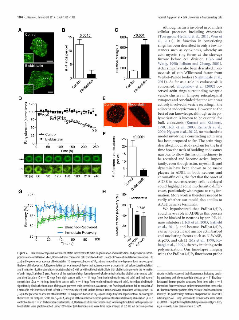
Using the myosin II-specific inhibitor blebbistatin, we found asignificant reduction in the number of Lifeact-positive rings thatfailed to contract during the time course of our experiment (Fig.5A–D). Not surprisingly, the number of dextran-positive struc-tures was also significantly reduced by myosin II inhibition (Fig.
4
(Figure legend continued.) D (10 �M, 30 min preincubation) or with BaCl2 (2 mM), and thenumber of rings per cell was analyzed (n � 6 and n � 8 cells, respectively). H, I, Bovine chromaffincells transfected with Lifeact-GFP were imaged using SIM, and z-stack images were acquired beforeand after stimulation with either nicotine (100�M; H) or BaCl2 (2 mM; I). The color scale indicates thedistance from the plasma membrane to the value indicated in the panels (micrometers inz-stack). Scale bar, 5 �m. J, Chromaffin cells cotransfected with Lifeact-GFP and PH-PLC�-RFPwere time lapse imaged (0.5 Hz) during stimulation with nicotine (100 �M). The panels show aregion of interest highlighting the appearance of highly intense PH-PLC�-positive microdo-mains preceding an individual actin ring. K, 3D surface plot of Lifeact-GFP ring and PH-PLC�-RFP-positive microdomains taken from G 1218 s after stimulation. Note that the intensity of theactin ring appears to be continuous, whereas those of the PtdIns(4,5)P2 microdomains arediscrete. L, Analysis of the time course of Lifeact-GFP and PH-PLC�-RFP fluorescence intensity atthe level of identified actin rings. To measure the latency of actin ring formation, the peakintensity of PH-PLC�-RFP was used as a time reference, and its intensity as normalized. Notethat the PtdIns(4,5)P2 signal precedes actin ring formation (n � 6 rings from three cells). Errorbars are mean � SEM.
1384 • J. Neurosci., January 28, 2015 • 35(4):1380 –1389 Gormal, Nguyen et al. • Bulk Endosomes in Neurosecretory Cells

5E,F). FRAP analysis of the few dextran-positive structures thatwere present resulted in either (1) immediate recovery or (2)slower recovery of fluorescence indicating continuity with theplasma membrane in both cases (Fig. 5G). Consistent with thedextran experiments, FRAP analysis of GFP-GPI was also indic-ative of a continuity between endocytic structures and the plasmamembrane (Fig. 5H).
Our data point to a role of contractile actin rings before fission ofthe nascent bulk endosome. We hypothesize that the contractile ac-tin ring could narrow the neck of the budding endosome, therebycontributing to the initiation of dynamin recruitment and fissionactivity. To test this hypothesis, we used the potent dynamin inhib-itor Dyngo4a (Harper et al., 2011; McCluskey et al., 2013). Dyngo4atreatment reduced the number of dextran-positive structures, whichall remained connected to the plasma membrane, as revealed byFRAP experiments (Fig. 6A–C). Time-lapse imaging of Lifeact-GFP
also revealed a reduction in the number ofactin rings elicited by secretagogue stimula-tion (Fig. 6D,E). Interestingly, the few ringsthat formed under Dyngo4a treatment re-mained stable and failed to contractthroughout the experiment (Fig. 6F,G). Wealso tested the effect of expressingdynamin-2 (K44A) dominant-negative inchromaffin cells and found that the numberof dextran-positive structures were signifi-cantly reduced (Fig. 6H) to a level compara-ble to Dyngo4a-treated cells. These data notonly indicate that dynamin promotes effec-tive fission of bulk endosomes from theplasma membrane, but also suggest an in-volvement of dynamin in the formation andcontractility of the actin rings.
DiscussionIn this study, we demonstrate that, as inneurons, neurosecretory cells undergo bulkendocytosis in response to secretagoguestimulation. This has allowed us to identify amolecular mechanism whereby actin anddynamin actively cooperate to form acto-myosin II rings that are capable of narrow-ing the neck of budding bulk endosomesbefore fission from the plasma membrane(Fig. 6I).
Together with CME and kiss-and-run,ADBE is emerging as a major contributorto synaptic vesicle recycling in nerve ter-minals (Clayton et al., 2007, 2008; Wu andWu, 2007; Nguyen et al., 2012). A numberof key proteins has been shown to be crit-ical for ADBE in neurons. These includesyndapin (Clayton et al., 2009), calcineu-rin (Evans and Cousin, 2007), actin (Ku-romi and Kidokoro, 1998; Holt et al.,2003; Richards et al., 2004; Nguyen et al.,2012), dynamin (Ferguson et al., 2007;Clayton et al., 2009, 2010; Nguyen et al.,2012), and, more recently, myosin II(Flores et al., 2014). Our study demon-strates a clear role for dynamin in the gen-eration of acto-myosin II rings and thefission of budding bulk endosomes. Theseresults are in good agreement with the es-
tablished role for dynamin in ADBE and in actin polymerization(Kessels et al., 2001; Gu et al., 2010).
We used a potent dynamin inhibitor, Dyngo4a (Howes et al.,2010; Harper et al., 2011; McCluskey et al., 2013), which is not iso-form specific and has potential off-target effects (Park et al., 2013).However, our data were corroborated with a dominant-negative ap-proach strongly pointing to a key role for dynamin in this process(Harper et al., 2013). Further studies to identify the dynamin iso-form involved would need to be performed in light of the recentstudy (Gonzalez-Jamett et al., 2013) suggesting that dynamin 2 maybe the predominant isoform in neurosecretory cells despite dynaminI being neuron specific. Further work will also be needed to establishwhether syndapin and calcineurin are required for bulk endocytosisin neurosecretory cells.
Figure 4. Myosin IIa and IIb localize to actin rings. A, B, Resting and nicotine-stimulated (100�M for 8 min) bovine adrenal chromaffincellswerefixedandstainedforeithermyosinIIa(A)ormyosinIIb(B)andphalloidin-AlexaFluor488. Insets illustratethepresenceofmyosinIIa and IIb associating with actin at rest (top panels) and in actin ring structures after stimulation (bottom panels). Scale bars, 5 �m.
Gormal, Nguyen et al. • Bulk Endosomes in Neurosecretory Cells J. Neurosci., January 28, 2015 • 35(4):1380 –1389 • 1385
Although actin is involved in countlesscellular processes including exocytosis(Torregrosa-Hetland et al., 2011; Wen etal., 2011), its function in constrictingrings has been described in only a few in-stances such as cytokinesis, whereby anacto-myosin ring forms at the cleavagefurrow before cell division (Cao andWang, 1990; Pelham and Chang, 2001).Actin rings have also been described in ex-ocytosis of von Willebrand factor fromWeibel–Palade bodies (Nightingale et al.,2011). As far as a role in endocytosis isconcerned, Shupliakov et al. (2002) ob-served actin rings surrounding synapticvesicle clusters in lamprey reticulospinalsynapses and concluded that the actin wasactively involved in vesicle recycling in theadjacent endocytic zones. However, to thebest of our knowledge, although actin po-lymerization is known to be essential forbulk endocytosis (Kuromi and Kidokoro,1998; Holt et al., 2003; Richards et al.,2004; Nguyen et al., 2012), no mechanisticmodel involving a constricting actin ringhas been proposed to far. The actin ringsdescribed in our study explain for the firsttime how the neck of budding endosomesnarrows to allow the fission machinery tobe recruited and become active. Impor-tantly, even though actin, myosin II, anddynamin have been shown to be majorplayers in ADBE in both neurons andchromaffin cells, the fact that the onset ofADBE in neurosecretory cells is delayedcould highlight some mechanistic differ-ences, particularly with regard to ring for-mation. More work is therefore needed toverify whether our model also applies toADBE in nerve terminals.
We hypothesized that PtdIns(4,5)P2
could have a role in ADBE as this processcan be blocked in neurons by pan PI3 ki-nase inhibitors (Holt et al., 2003; Gaffieldet al., 2011), and because PtdIns(4,5)P2
can act to recruit and anchor actin barbedend nucleating factors such as N-WASP,Arp2/3, and cdc42 (Ma et al., 1998; Ro-hatgi et al., 1999), thereby initiating actinpolymerization. Our time-lapse imagingusing the PtdIns(4,5)P2 fluorescent probeFigure 5. Inhibition of myosin II with blebbistatin interferes with actin ring formation and constriction, and prevents dextran-
positive endosomal fission. A–D, Bovine adrenal chromaffin cells transfected with Lifeact-GFP were stimulated with nicotine (100�M) in the presence or absence of blebbistatin (10 min preincubation at 10 �M) and imaged by time-lapse confocal microscopy atthe level of the footprint. A, Representative confocal image of the cortical actin network of a chromaffin cell before (prestimulation)and 8 min after nicotine stimulation (poststimulation) with or without blebbistatin. Note that blebbistatin prevents the formationof actin rings. Scale bar, 5 �m. Analysis of the number of rings formed per cell (B; six control cells; five blebbistatin-treated cells)and their duration (C; n � 52 rings from eight control cells; n � 14 rings from four blebbistatin-treated cells) and their rate ofconstriction (D; n � 10 rings from three control cells; n � 6 rings from two blebbistatin-treated cells). Note that blebbistatinsignificantly blocks the formation of rings and prevents their constriction. As a result, the few rings that form fail to constrict. E,Chromaffin cells transfected with Lifeact-GFP were incubated with 70 kDa dextran-TMRh and were stimulated with nicotine (100�M) in the presence or absence of blebbistatin (10 min preincubation at 10 �M) and imaged by time-lapse confocal microscopy atthe level of the footprint. Scale bar, 5 �m. F, Analysis of the number of dextran-positive structures following stimulation (n � 6control cells and n � 21 blebbistatin-treated cells). G, Dextran-positive structures formed following stimulation in the presence ofblebbistatin were photobleached using 100% laser (20 iterations) and were time lapse imaged at 0.5 Hz. All dextran-positive
4
structures fully recovered their fluorescence, indicating persist-ing continuity with the extracellular dextran (n � 11 Bleached-Recovered dextran-positive structures from three cells; n � 3Immediate Recovery dextran-positive structures from three cells).H, Plasma membrane portions of the cell were used as a control forrecovery. GPI-positive rings that were also positive for Lifeact-RFPactin ring GPI/AR� rings were able to recover to the same extentasGPI/AR� ringsfollowingblebbistatinpretreatment( p�0.05,ns; n � 6 cells). Error bars are mean � SEM.
1386 • J. Neurosci., January 28, 2015 • 35(4):1380 –1389 Gormal, Nguyen et al. • Bulk Endosomes in Neurosecretory Cells

Figure 6. Dynamin inhibition prevents actin ring formation and dextran-positive endosomal fission. A, Bovine adrenal chromaffin cells were incubated with 70 kDa dextran-TMRh in the presenceor absence of Dyngo4a (30 �M) and were imaged by time-lapse confocal microscopy during nicotine stimulation (100 �M). Scale bar, 5 �m. B, Dextran-positive structures formed followingstimulation in the presence of Dyngo4a were photobleached using 100% laser stimulation (20 iterations) and were time lapse imaged at 0.5 Hz. All dextran-positive structures fully recovered theirfluorescence, indicating persisting continuity with the extracellular dextran (n � 7 Bleached-Recovered dextran-positive structures from four cells; n � 5 Immediate Recovery dextran-positivestructures from three cells). C, Analysis of the number of dextran-positive structures appearing following stimulation (n � 6 control cells and n � 29 Dyngo4a-treated cells). D, Chromaffin cellstransfected with Lifeact-GFP were stimulated with nicotine (100 �M) in the presence or absence of Dyngo4a (30 �M) and were imaged by time-lapse confocal microscopy. Scale bar, 5 �m. E–G,Analysis of the number of rings formed per cell (E; n � 12 control cells; n � 35 Dyngo4a-treated cells), their duration (F; n � 52 rings from 8 control cells; n � 20 rings from 4 Dyngo4a-treated cells),and their rate of constriction (G; n � 4 rings from 3 control cells; n � 4 rings from 2 Dyngo4a-treated cells). Note that Dyngo4a significantly blocks the number of (Figure legend continues.)
Gormal, Nguyen et al. • Bulk Endosomes in Neurosecretory Cells J. Neurosci., January 28, 2015 • 35(4):1380 –1389 • 1387
PH-PLC� suggests an involvement of PtdIns(4,5)P2 in generatingthe acto-myosin II rings. Consistent with an initiating roleof PtdIns(4,5)P2, we detect an array of PtdIns(4,5)P2 microdomainsthat precedes the generation of the actin ring. These PtdIns(4,5)P2
hotspots are located at strategic points of the impending actin ring,and it is tempting to speculate that they represent multiple points ofactin nucleation and that myosin II and dynamin may be activelyinvolved in cross-linking these streams of growing filaments, therebystabilizing the actin ring (Fig. 6I). PtdIns(4,5)P2 is also present andrequired for the correct organization of the cleavage furrow, and hasa role in adhesion between the contractile ring and the plasma mem-brane (Field et al., 2005; Liu et al., 2012).
Our data also point to a role of dynamin and myosin II in induc-ing actin ring formation. Indeed, pharmacological inhibition ofdynamin and myosin II drastically reduced the actual number ofacto-myosin II rings (Figs. 5, 6). Further, along with the well estab-lished role of dynamin in mediating membrane fission, a number ofstudies (Kessels et al., 2001; Gu et al., 2010) have shown that dy-namin can directly interact with actin and regulate its polymeriza-tion. Our data are therefore consistent with an additional role ofdynamin in polymerizing specific pools of actin to form rings.
Our results demonstrate that the contractility of the actin ringcan be blocked by pharmacological inhibition of myosin II (Fig. 5).The critical role of myosin II in cell division as a force-generatingfission mechanism has two current hypotheses. In the classic model,bipolar myosin II filaments move along F-actin in a similar mannerto that of the muscle sarcomere (Egelhoff et al., 1993). In the alter-native model, depolymerization of the cross-linked actin networkdoes not drive constriction because many actin filaments of the cy-tokinesis ring are not oriented in the plane of division (Kruse andJulicher, 2003; Reichl et al., 2008). More work will be needed todetermine which model best suits the constriction of activity-dependent actin rings involved in ADBE. On another note, myosinII and dynamin have been implicated in the fusion pore formation(Berberian et al., 2009; Chan et al., 2010; Gonzalez-Jamett et al.,2013), and altering the dynamics of the fusion pore could have sec-ondary effects on the mode of endocytosis elicited.
It is worth noting that dynamin inhibition also affects ringcontractility, suggesting a possible role in acto-myosin II con-striction. In this view dynamin II was shown to coordinate acto-myosin II interaction in epithelial cells (Chua et al., 2009).
Regardless of the molecular underpinning of actin ring con-striction, our study is the first to demonstrate that actin ringconstriction is critical to driving bulk endocytosis to completion.Indeed, we show that inhibition of either myosin II or dynaminfunction leads to a defect in dextran internalization and the fail-ure of fission from the plasma membrane (Figs. 5, 6).
It should be noted that our interpretation of the FRAP data isbased on the principle that a fully “detached” nascent bulk endo-
some is unable to recover fluorescence following photobleachingas it is spatially separated from any other source of dextran. Wecannot rule out the possibility that bulk endosomes could fuse withother dextran-containing organelles. However, high-molecular-weight dextran is only internalized in bulk endosomes that arenot being re-released upon secondary stimulation in neurons,ruling out the potential contribution of the CME pathway (Clay-ton and Cousin, 2009). Bulk endosome homotypic fusion couldalso theoretically contribute to the fluorescence recovery. How-ever, we did not observe any noticeable morphological change inthe size or shape of the dextran-positive endosomes, and theassociated actin rings were always relatively immobile, stronglysuggesting that they were in close contact with the plasma mem-brane as they appear to originate from the plane of remodeledcortical actin network after stimulation (Fig. 3H, I).
In conclusion, we demonstrate that constricting acto-myosinII rings appearing following stimulation are critical for the fissionof bulk endosomes from the plasma membrane in neurosecretorycells. In this context, PtdIns(4,5)P2, actin, myosin II, and dy-namin provide a molecular framework on which to build a morecomprehensive picture of the mechanism underpinning bulk en-docytosis. Revealing bulk endocytosis in neurosecretory cells maylead to further understanding of the molecular mechanism driv-ing ADBE in other cellular systems.
ReferencesBerberian K, Torres AJ, Fang Q, Kisler K, Lindau M (2009) F-actin and
myosin II accelerate catecholamine release from chromaffin granules.J Neurosci 29:863– 870. CrossRef Medline
Berg JS, Powell BC, Cheney RE (2001) A millennial myosin census. Mol BiolCell 12:780 –794. CrossRef Medline
Cao LG, Wang YL (1990) Mechanism of the formation of contractile ring individing cultured animal cells. II. Cortical movement of microinjectedactin filaments. J Cell Biol 111:1905–1911. CrossRef Medline
Ceridono M, Ory S, Momboisse F, Chasserot-Golaz S, Houy S, Calco V,Haeberle AM, Demais V, Bailly Y, Bader MF, Gasman S (2011) Selectiverecapture of secretory granule components after full collapse exocytosis inneuroendocrine chromaffin cells. Traffic 12:72– 88. CrossRef Medline
Chan SA, Doreian B, Smith C (2010) Dynamin and myosin regulate differ-ential exocytosis from mouse adrenal chromaffin cells. Cell Mol Neuro-biol 30:1351–1357. CrossRef Medline
Chua J, Rikhy R, Lippincott-Schwartz J (2009) Dynamin 2 orchestrates theglobal actomyosin cytoskeleton for epithelial maintenance and apical con-striction. Proc Natl Acad Sci USA 106:20770–20775. CrossRef Medline
Clayton EL, Cousin MA (2009) Quantitative monitoring of activity-dependent bulk endocytosis of synaptic vesicle membrane by fluorescentdextran imaging. J Neurosci Methods 185:76 – 81. CrossRef Medline
Clayton EL, Evans GJ, Cousin MA (2007) Activity-dependent control ofbulk endocytosis by protein dephosphorylation in central nerve termi-nals. J Physiol 585:687– 691. CrossRef Medline
Clayton EL, Evans GJ, Cousin MA (2008) Bulk synaptic vesicle endocytosisis rapidly triggered during strong stimulation. J Neurosci 28:6627– 6632.CrossRef Medline
Clayton EL, Anggono V, Smillie KJ, Chau N, Robinson PJ, Cousin MA(2009) The phospho-dependent dynamin-syndapin interaction triggersactivity-dependent bulk endocytosis of synaptic vesicles. J Neurosci 29:7706 –7717. CrossRef Medline
Clayton EL, Sue N, Smillie KJ, O’Leary T, Bache N, Cheung G, Cole AR,Wyllie DJ, Sutherland C, Robinson PJ, Cousin MA (2010) Dynamin Iphosphorylation by GSK3 controls activity-dependent bulk endocytosisof synaptic vesicles. Nat Neurosci 13:845– 851. CrossRef Medline
Egelhoff TT, Lee RJ, Spudich JA (1993) Dictyostelium myosin heavy chainphosphorylation sites regulate myosin filament assembly and localizationin vivo. Cell 75:363–371. CrossRef Medline
Evans GJ, Cousin MA (2007) Activity-dependent control of slow synapticvesicle endocytosis by cyclin-dependent kinase 5. J Neurosci 27:401– 411.CrossRef Medline
Ferguson SM, Brasnjo G, Hayashi M, Wolfel M, Collesi C, Giovedi S, Rai-mondi A, Gong LW, Ariel P, Paradise S, O’toole E, Flavell R, Cremona O,
4
(Figure legend continued.) ring forming and prevent their constriction. H, Chromaffin cellstransfected with dynamin II (K44A) dominant-negative mutant were incubated with 70 kDadextran-TMRh and incubated with or without nicotine (100 �M) while being imaged by time-lapse confocal microscopy at the level of the footprint (surface area in contact with the cover-slip). Numerous internal dextran-positive structures appeared after stimulation in control butare significantly reduced in cells expressing Dynamin mutant. Scale bars, 5 �m. I, Left, Modelhighlighting the initiating role of PtdIns(4,5)P2 in generating actin filaments from regularlyspaced hotspots in the plasma membrane delineating the future actin ring. In this model,myosin II and dynamin stabilize the actin ring by cross-linking the intersecting fibers (inset).Right, Model highlighting the role of actin rings in generating bulk endosomes. Myosin II anddynamin play critical roles in generating and stabilizing the actin ring in addition to the role ofdynamin in pinching off the nascent bulk endosomes from the plasma membrane.
1388 • J. Neurosci., January 28, 2015 • 35(4):1380 –1389 Gormal, Nguyen et al. • Bulk Endosomes in Neurosecretory Cells

Miesenbock G, Ryan TA, De Camilli P (2007) A selective activity-dependent requirement for dynamin 1 in synaptic vesicle endocytosis.Science 316:570 –574. CrossRef Medline
Field SJ, Madson N, Kerr ML, Galbraith KA, Kennedy CE, Tahiliani M,Wilkins A, Cantley LC (2005) PtdIns(4,5)P2 functions at the cleavagefurrow during cytokinesis. Curr Biol 15:1407–1412. CrossRef Medline
Flores JA, Balseiro-Gomez S, Cabeza JM, Acosta J, Ramirez-Ponce P, Ales E(2014) A new role for myosin II in vesicle fission. PLoS One 9:e100757.CrossRef Medline
Gaffield MA, Romberg CF, Betz WJ (2011) Live imaging of bulk endocytosisin frog motor nerve terminals using FM dyes. J Neurophysiol 106:599 –607. CrossRef Medline
Gonzalez-Jamett AM, Momboisse F, Guerra MJ, Ory S, Baez-Matus X,Barraza N, Calco V, Houy S, Couve E, Neely A, Martínez AD, Gasman S,Cardenas AM (2013) Dynamin-2 regulates fusion pore expansion andquantal release through a mechanism that involves actin dynamics inneuroendocrine chromaffin cells. PLoS One 8:e70638. CrossRef Medline
Gu C, Yaddanapudi S, Weins A, Osborn T, Reiser J, Pollak M, Hartwig J, SeverS (2010) Direct dynamin-actin interactions regulate the actin cytoskel-eton. EMBO J 29:3593–3606. CrossRef Medline
Harper CB, Martin S, Nguyen TH, Daniels SJ, Lavidis NA, Popoff MR, Hadzic G,Mariana A, Chau N, McCluskey A, Robinson PJ, Meunier FA (2011) Dy-namin inhibition blocks botulinum neurotoxin type-A endocytosis in neu-rons and delays botulism. J Biol Chem 286:35966–35976. CrossRef Medline
Harper CB, Popoff MR, McCluskey A, Robinson PJ, Meunier FA (2013)Targeting membrane trafficking in infection prophylaxis: dynamin inhib-itors. Trends Cell Biol 23:90 –101. CrossRef Medline
Holt M, Cooke A, Wu MM, Lagnado L (2003) Bulk membrane retrieval inthe synaptic terminal of retinal bipolar cells. J Neurosci 23:1329 –1339.Medline
Howes MT, Mayor S, Parton RG (2010) Molecules, mechanisms, and cellu-lar roles of clathrin-independent endocytosis. Curr Opin Cell Biol 22:519 –527. CrossRef Medline
Kessels MM, Engqvist-Goldstein AE, Drubin DG, Qualmann B (2001)Mammalian Abp1, a signal-responsive F-actin-binding protein, links theactin cytoskeleton to endocytosis via the GTPase dynamin. J Cell Biol153:351–366. CrossRef Medline
Kruse K, Julicher F (2003) Self-organization and mechanical properties ofactive filament bundles. Phys Rev E Stat Nonlin Soft Matter Phys 67:051913. CrossRef Medline
Kuromi H, Kidokoro Y (1998) Two distinct pools of synaptic vesicles insingle presynaptic boutons in a temperature-sensitive Drosophila mu-tant, shibire. Neuron 20:917–925. CrossRef Medline
Lassing I, Lindberg U (1985) Specific interaction between phosphatidylino-sitol 4,5-bisphosphate and profilactin. Nature 314:472– 474. CrossRefMedline
Liu J, Fairn GD, Ceccarelli DF, Sicheri F, Wilde A (2012) Cleavage furroworganization requires PIP(2)-mediated recruitment of anillin. Curr Biol22:64 – 69. CrossRef Medline
Ma L, Rohatgi R, Kirschner MW (1998) The Arp2/3 complex mediates actinpolymerization induced by the small GTP-binding protein Cdc42. ProcNatl Acad Sci USA 95:15362–15367. CrossRef Medline
Martin S, Tomatis VM, Papadopulos A, Christie MP, Malintan NT, Gormal RS,Sugita S, Martin JL, Collins BM, Meunier FA (2013) The Munc18-1 domain3a loop is essential for neuroexocytosis but not for syntaxin-1A transport tothe plasma membrane. J Cell Sci 1:2353–2360. CrossRef Medline
McCluskey A, Daniel JA, Hadzic G, Chau N, Clayton EL, Mariana A, WhitingA, Gorgani NN, Lloyd J, Quan A, Moshkanbaryans L, Krishnan S, PereraS, Chircop M, von Kleist L, McGeachie AB, Howes MT, Parton RG,Campbell M, Sakoff JA, et al. (2013) Building a better dynasore: thedyngo compounds potently inhibit dynamin and endocytosis. Traffic 14:1272–1289. CrossRef Medline
Meunier FA, Feng ZP, Molgo J, Zamponi GW, Schiavo G (2002) Glycero-toxin from Glycera convoluta stimulates neurosecretion by up-regulatingN-type Ca2� channel activity. EMBO J 21:6733– 6743. CrossRef Medline
Meunier FA, Osborne SL, Hammond GR, Cooke FT, Parker PJ, Domin J,Schiavo G (2005) Phosphatidylinositol 3-kinase C2alpha is essential forATP-dependent priming of neurosecretory granule exocytosis. Mol BiolCell 16:4841– 4851. CrossRef Medline
Miller TM, Heuser JE (1984) Endocytosis of synaptic vesicle membrane at the frogneuromuscular junction. J Cell Biol 98:685–698. CrossRef Medline
Nguyen TH, Maucort G, Sullivan RK, Schenning M, Lavidis NA, McCluskey
A, Robinson PJ, Meunier FA (2012) Actin- and dynamin-dependentmaturation of bulk endocytosis restores neurotransmission followingsynaptic depletion. PLoS One 7:e36913. CrossRef Medline
Nightingale TD, White IJ, Doyle EL, Turmaine M, Harrison-Lavoie KJ, WebbKF, Cramer LP, Cutler DF (2011) Actomyosin II contractility expels vonWillebrand factor from Weibel-Palade bodies during exocytosis. J CellBiol 194:613– 629. CrossRef Medline
Papadopulos A, Martin S, Tomatis VM, Gormal RS, Meunier FA (2013)Secretagogue stimulation of neurosecretory cells elicits filopodial exten-sions uncovering new functional release sites. J Neurosci 33:19143–19153.CrossRef Medline
Park Rj, Shen H, Liu L, Liu X, Ferguson SM, De Camilli P (2013) Dynamintriple knockout cells reveal off target effects of commonly used dynamininhibitors. J Cell Sci 126:5305–5312. CrossRef Medline
Pelham RJ Jr, Chang F (2001) Role of actin polymerization and actin cablesin actin-patch movement in Schizosaccharomyces pombe. Nat Cell Biol3:235–244. CrossRef Medline
Perrais D, Kleppe IC, Taraska JW, Almers W (2004) Recapture after exocyto-sis causes differential retention of protein in granules of bovine chromaf-fin cells. J Physiol 15:413– 428. Medline
Reichl EM, Ren Y, Morphew MK, Delannoy M, Effler JC, Girard KD, Divi S,Iglesias PA, Kuo SC, Robinson DN (2008) Interactions between myosinand actin crosslinkers control cytokinesis contractility dynamics and me-chanics. Curr Biol 18:471– 480. CrossRef Medline
Richards DA, Guatimosim C, Betz WJ (2000) Two endocytic recyclingroutes selectively fill two vesicle pools in frog motor nerve terminals.Neuron 27:551–559. CrossRef Medline
Richards DA, Rizzoli SO, Betz WJ (2004) Effects of wortmannin and latrun-culin A on slow endocytosis at the frog neuromuscular junction. J Physiol557:77–91. CrossRef Medline
Riedl J, Crevenna AH, Kessenbrock K, Yu JH, Neukirchen D, Bista M, BradkeF, Jenne D, Holak TA, Werb Z, Sixt M, Wedlich-Soldner R (2008) Life-act: a versatile marker to visualize F-actin. Nat Methods 5:605– 607.CrossRef Medline
Rohatgi R, Ma L, Miki H, Lopez M, Kirchhausen T, Takenawa T, KirschnerMW (1999) The interaction between N-WASP and the Arp2/3 complexlinks Cdc42-dependent signals to actin assembly. Cell 97:221–231.CrossRef Medline
Saarikangas J, Zhao H, Lappalainen P (2010) Regulation of the actincytoskeleton-plasma membrane interplay by phosphoinositides. PhysiolRev 90:259 –289. CrossRef Medline
Shupliakov O, Bloom O, Gustafsson JS, Kjaerulff O, Low P, Tomilin N, Pieri-bone VA, Greengard P, Brodin L (2002) Impaired recycling of synapticvesicles after acute perturbation of the presynaptic actin cytoskeleton.Proc Natl Acad Sci USA 99:14476 –14481. CrossRef Medline
Torregrosa-Hetland CJ, Villanueva J, Giner D, Lopez-Font I, Nadal A, Que-sada I, Viniegra S, Exposito-Romero G, Gil A, Gonzalez-Velez V, Segura J,Gutierrez LM (2011) The F-actin cortical network is a major factor influ-encing the organization of the secretory machinery in chromaffin cells.J Cell Sci 1:727–734. CrossRef Medline
Trifaro JM, Gasman S, Gutierrez LM (2008) Cytoskeletal control of vesicletransport and exocytosis in chromaffin cells. Acta Physiol (Oxf) 192:165–172. CrossRef Medline
Uehara R, Goshima G, Mabuchi I, Vale RD, Spudich JA, Griffis ER (2010)Determinants of myosin II cortical localization during cytokinesis. CurrBiol 20:1080 –1085. CrossRef Medline
Varnai P, Lin X, Lee SB, Tuymetova G, Bondeva T, Spat A, Rhee SG, Hajnoc-zky G, Balla T (2002) Inositol lipid binding and membrane localizationof isolated pleckstrin homology (PH) domains. Studies on the PH do-mains of phospholipase C delta 1 and p130. J Biol Chem 277:27412–27422. CrossRef Medline
Wen PJ, Osborne SL, Zanin M, Low PC, Wang HT, Schoenwaelder SM,Jackson SP, Wedlich-Soldner R, Vanhaesebroeck B, Keating DJ, MeunierFA (2011) Phosphatidylinositol(4,5)bisphosphate coordinates actin-mediatedmobilizationandtranslocationofsecretoryvesiclestotheplasmamem-brane of chromaffin cells. Nat Commun 2:491. CrossRef Medline
Wu W, Wu LG (2007) Rapid bulk endocytosis and its kinetics of fission poreclosure at a central synapse. Proc Natl Acad Sci USA 104:10234 –10239.CrossRef Medline
Yumura S, Ueda M, Sako Y, Kitanishi-Yumura T, Yanagida T (2008) Mul-tiple mechanisms for accumulation of myosin II filaments at the equatorduring cytokinesis. Traffic 9:2089 –2099. CrossRef Medline
Gormal, Nguyen et al. • Bulk Endosomes in Neurosecretory Cells J. Neurosci., January 28, 2015 • 35(4):1380 –1389 • 1389