Embed Size (px)
Citation preview
Cellular/Molecular
Preferential Targeting of Lateral Entorhinal Inputs ontoNewly Integrated Granule Cells
X Nicholas I. Woods,* X Christopher E. Vaaga,* Christina Chatzi, X Jaimie D. Adelson, X Matthew F. Collie,Julia V. Perederiy, Kenneth R. Tovar, and X Gary L. WestbrookVollum Institute, Oregon Health and Science University, Portland, Oregon 97239
Mature dentate granule cells in the hippocampus receive input from the entorhinal cortex via the perforant path in precisely arrangedlamina, with medial entorhinal axons innervating the middle molecular layer and lateral entorhinal cortex axons innervating the outermolecular layer. Although vastly outnumbered by mature granule cells, adult-generated newborn granule cells play a unique role inhippocampal function, which has largely been attributed to their enhanced excitability and plasticity (Schmidt-Hieber et al., 2004; Ge etal., 2007). Inputs from the medial and lateral entorhinal cortex carry different informational content. Thus, the distribution of inputs ontonewly integrated granule cells will affect their function in the circuit. Using retroviral labeling in combination with selective optogeneticactivation of medial or lateral entorhinal inputs, we examined the functional innervation and synaptic maturation of newly generateddentate granule cells in the mouse hippocampus. Our results indicate that lateral entorhinal inputs provide the majority of functional innerva-tion of newly integrated granule cells at 21 d postmitosis. Despite preferential functional targeting, the dendritic spine density of immaturegranule cells was similar in the outer and middle molecular layers, which we speculate could reflect an unequal distribution of shaft synapses.However, chronic blockade of neurotransmitter release of medial entorhinal axons with tetanus toxin disrupted normal synapse development ofboth medial and lateral entorhinal inputs. Our results support a role for preferential lateral perforant path input onto newly generated neuronsin mediating pattern separation, but also indicate that medial perforant path input is necessary for normal synaptic development.
Key words: dentate gyrus; neurogenesis; pattern separation; retroviral labeling; synapse formation; tetanus toxin
IntroductionAs the entry point to the trisynaptic hippocampal circuit, the dentategyrus has several interesting features, including a “sparse” networkdesign (Boss et al., 1985; Rolls et al., 1998), laminated inputscarrying distinct informational content (Witter, 2007; Knierim et
al., 2014), and the participation of mature granule cells alongsidethe continuous integration of newly generated neurons (Overstreet-Wadiche and Westbrook, 2006; Ming and Song, 2011). Hippocam-pal granule cells receive highly laminar inputs from the entorhinalcortex within the molecular layer of the dentate gyrus. Input fromthe medial entorhinal cortex, conveying spatial cues, is restrictedto the middle molecular layer (MML; Ferbinteanu et al., 1999;Hafting et al., 2005; Hargreaves et al., 2005; Yasuda and Mayford,2006; Witter, 2007; Van Cauter et al., 2013), whereas input fromthe lateral entorhinal cortex conveying contextual information isrestricted to the outer molecular layer (OML; Hargreaves et al.,2005; Hunsaker et al., 2007; Witter, 2007; Deshmukh and Knierim,2011; Yoganarasimha et al., 2011; Tsao et al., 2013). Despite beingsubstantially outnumbered by mature granule cells, newly inte-grated granule cells are thought to uniquely contribute to patternseparation (Clelland et al., 2009; Sahay et al., 2011; Nakashiba etal., 2012; Tronel et al., 2012). That is, they foster the ability todistinguish between subtly different contexts, one of the primaryfunctions of the dentate gyrus (Deng et al., 2010; Aimone et al.,
Received June 21, 2017; revised April 26, 2018; accepted May 17, 2018.Author contributions: N.I.W. and G.L.W. designed research; N.I.W., C.E.V., C.C., J.D.A., M.F.C., J.V.P., and K.R.T.
performed research; N.I.W., C.E.V., C.C., J.D.A., M.F.C., J.V.P., and K.R.T. analyzed data; N.I.W., C.E.V., and G.L.W.wrote the paper.
This work was supported by National Institutes of Health Grants R01 NS080979 and P30 NS061800 and by theEllison Medical Foundation (G.L.W.). We thank Stefanie Kaech Petrie for help with imaging, Eric Schnell for guidancewith electrophysiology, and Sue Aicher for assistance with electron microscopy.
*N.I.W and C.E.V. contributed equally to this work.The authors declare no competing financial interests.Correspondence should be addressed to Gary L. Westbrook, Vollum Institute, L474, Oregon Health and Science
University, 3181 SW Sam Jackson Park Road, Portland OR, 97008. E-mail: [email protected]. Woods’ present address: Medical Scientist Training Program, University of California San Francisco, San
Francisco, CA 94143.DOI:10.1523/JNEUROSCI.1737-17.2018
Copyright © 2018 the authors 0270-6474/18/385843-11$15.00/0
Significance Statement
The formation of episodic memories involves the integration of contextual and spatial information. Newly integrated neurons inthe dentate gyrus of the hippocampus play a critical role in this process, despite constituting only a minor fraction of the totalnumber of granule cells. Here we demonstrate that these neurons preferentially receive information thought to convey the contextof an experience. Each newly integrated granule cell plays this unique role for �1 month before reaching maturity.
The Journal of Neuroscience, June 27, 2018 • 38(26):5843–5853 • 5843
2011). This unique function must occur during a narrow timewindow between initial integration into the perforant path circuit(�3 weeks postmitosis) and the complete maturation of synapses(�8 weeks postmitosis; van Praag et al., 2002; Ambrogini et al.,2004; Overstreet et al., 2004; Laplagne et al., 2006; Overstreet-Wadiche et al., 2006; Zhao et al., 2006; Kumamoto et al., 2012;Brunner et al., 2014).
The search for unique functions of newly integrated neuronshas largely focused on intrinsic properties, such as enhanced ex-citability and plasticity (Schmidt-Hieber et al., 2004; Abrous etal., 2005; Ge et al., 2007; Marín-Burgin et al., 2012; Dieni et al.,2013). However, newborn neurons also undergo rapid changes inconnectivity in the first few weeks after mitosis. This process likelydiffers from synapse remodeling in early development (Goodmanand Shatz, 1993; Katz and Shatz, 1996; Walsh and Lichtman,2003), as newborn neurons integrate into an already establishedcircuit (Toni et al., 2007, 2008) and compete for synaptic innervationwith pre-existing axons of the perforant path. Rabies-based circuitmapping also suggests that inputs to newly integrated neurons maydiffer from mature granule cells (Vivar et al., 2012).
Here, using retroviral labeling of newborn neurons and laminar-specific optogenetic stimulation of entorhinal inputs, we directlyassayed functional synaptic integration of newborn granule cellsover the course of excitatory synapse development. Our resultsindicate that newly integrated granule cells preferentially receivefunctional synaptic input from the lateral entorhinal cortex,whereas mature granule cells receive balanced input from themedial and lateral entorhinal cortex. Although medial perforantpath (MPP) input was weak in newly integrated granule cells,chronic silencing of this pathway using tetanus toxin impairedthe functional and morphological development of lateral per-forant path (LPP) inputs.
Materials and MethodsAnimals. We used male and female C57BL/6 mice. Animal procedureswere performed in accordance with the Oregon Health and Science Uni-versity Institutional Animal Care and Use Committee, Biosafety Com-mittee protocols, and National Institutes of Health guidelines for the safehandling of animals.
Viral constructs. To selectively transfect and visualize adult-born hip-pocampal granule cells, we used a replication-deficient Moloney murineleukemia virus-based retroviral vector that requires cell mitosis fortransduction (Luikart et al., 2011). The retrovirus contained an inter-nal ubiquitin 6 promoter that drives expression of GFP (viral titer 10 5) asdescribed previously (pRubi-GFP; Luikart et al., 2011). To express thelight-activated ion channel channelrhodopsin-2 (ChR2) selectively inentorhinal cortex axons projecting to the MML or OML, we stereotaxi-cally injected an AAV9-CAG-ChR2-eGFP viral construct (University ofNorth Carolina Viral Core) into the medial or lateral entorhinal cortex.Laminar-specific labeling was confirmed by examination of GFP fluores-cence in the entorhinal cortex and in the dentate molecular layer. Injectionsaffecting both the lateral and medial entorhinal cortex were excluded fromthe analysis. To selectively silence axons, we made a custom tetanus neu-rotoxin (TeNT) virus (AAV9-CAG-TeNT-mCherry, viral titer 10 13) bycloning a 2kb fragment encoding the light chain of tetanus toxin fusedwith mCherry into an AAV backbone using InFusion cloning. AAV vec-tors were serotyped with AAV9 coat proteins and packaged at the Uni-versity of North Carolina Vector Core.
Stereotaxic injections. Stereotaxic viral injections into 6 – 8-week-oldmale and female C57BL/6 mice were performed using a Model 1900Stereotaxic Alignment System (David Kopf Instruments). Mice wereanesthetized with 2% isoflurane, and a small incision was created overthe skull following application of artificial tears to the eyes and antibiotic/iodine around the incision site. A Model 1911 Stereotaxic Drill (DavidKopf Instruments) was used to create burr holes over the injection site.pRubi-GFP retrovirus to label mitotically active granule cells was injected
into the dentate gyrus (coordinates, from bregma: anteroposterior, �1.9mm; lateromedial: �1.1 mm; dorsoventral, �2.5 mm, �2.3 mm). Onemicroliter of nondiluted virus was injected at 250 nl/min with a 10 �lHamilton syringe fitted with a 30 gauge needle using a Quintessentialstereotaxic injector (Stoetling). After each injection, the needle was left inplace for 1 min to allow for diffusion of the virus and prevent backflow.Injections of AAV9-CAG-CHR2-GFP into the lateral entorhinal cortexwere made at the following coordinates: anteroposterior (from bregma),�3.4 mm; lateromedial (from bregma), �4.0 mm; dorsoventral (frombrain surface), �2.4 mm. Injections of AAV9-CAG-ChR2-GFP or AAV9-CAG-TeNT-mCherry into the medial entorhinal cortex were made at thefollowing coordinates: anteroposterior (from bregma), �4.5 mm; latero-medial (from bregma), �3.0 mm; dorsoventral (from brain surface),�3.2 mm. To control for differences in the degree of innervation fromthe medial and lateral entorhinal cortex (Dolorfo and Amaral, 1998;Ohara et al., 2013), injections in the dentate gyrus were targeted to theintermediate region across the septotemporal axis (van Groen et al.,2003; Strange et al., 2014), which receives approximately equal innerva-tion from the medial and lateral entorhinal cortex. Where appropriate,pRubi and ChR2 were injected during the same surgery. Following injec-tion, mice received topical lidocaine and drinking water with cherry-flavored Tylenol, and were monitored every 24 h for 3 d.
Immunohistochemistry and spine counts. Transcardiac perfusion ofdeeply anesthetized mice [2% 2,2,2-tribromoethanol (0.7– 0.8 ml)] wasperformed with ice-cold 4% sucrose in 0.1 M PBS followed by fixativecontaining 4% sucrose and 4% paraformaldehyde (PFA) in PBS. Brainswere removed and postfixed (4% PFA, 1� PBS) overnight at 4°C. Thehippocampus was sectioned (coronal, 100 �m) using a Leica VT 1000Svibratome. Sections were permeabilized with 0.4% Triton X-100 in PBS(PBS-T), blocked with filtered 10% horse serum in PBS-T and incubatedin primary antibody overnight in 1.5% horse serum in PBS-T. Primaryantibodies included the following: 1:300 Alexa Fluor 488-conjugatedrabbit anti-GFP (catalog #A21311, Invitrogen), 1:500 anti-VGlut2 (cat-alog #135 404, Synaptic Systems), and 1:500 anti-glial-associated fibril-lary protein (GFAP; catalog #Z-0334, DAKO). Sections were then rinsedwith PBS-T and incubated for 2–3 h at room temperature in PBS-T withthe following secondary antibodies: 1:300 Alexa Fluor 488-conjugatedanti-GFP (A21311, Invitrogen), 1:200 goat anti-guinea pig Alexa Fluor568 (A11075, Invitrogen), and 1:200 goat anti-rabbit Alexa Fluor 488(A11034, Invitrogen). The mCherry-TeNT signal was extremely bright andwas visualized without use of secondary enhancement with antibodies. Thetissue was counterstained with DAPI using Fluoromount G with DAPI(SouthernBiotech). All antibodies have been well characterized in priorstudies in our laboratory, and staining was not observed when the primaryantibody was omitted.
Images were acquired using either a Zeiss LSM 770 or LSM 780 laser-scanning confocal microscope on a motorized AxioObserver Z1 invertedscope (Carl Zeiss MicroImaging). Dendrites for spine analysis were im-aged using a 63� objective (1.4 numerical aperture, oil, 2� zoom). Foreach imaged cell, dendritic segments in the MML and the OML wereimaged. The MML and OML were distinguished based on VGluT2 im-munofluorescence pattern, which begins at the border between the innermolecular layer and the MML. MML dendritic segments were thereforeimaged at the beginning of the VGluT2 staining nearest the granule cellbody layer, whereas OML dendritic segments were imaged at the distaltip of the molecular layer. Spine density analysis was performed blindedto experimental condition. The Cell Counter plugin in FIJI (NationalInstitutes of Health) was used to count and categorize spines (Harris etal., 1992), and Simple Neurite Tracer (FIJI) was used to measure den-dritic segment length. Spine density and spine type between the MMLand OML was compared across conditions (control and TeNT overex-pression) and developmental time points (3–12 weeks after viral injec-tion). Spine morphology was classified into two groups: mushroomspines, containing a spine head (2� shaft diameter) or filopodia-like forprotrusions lacking a spine head.
Electrophysiology. Electrophysiological recordings were made 21 d af-ter viral injection into the entorhinal cortex to allow for construct expres-sion. Acute brain slices were prepared as described previously (Perederiyet al., 2013). Briefly, animals were anesthetized with an intraperitoneal
5844 • J. Neurosci., June 27, 2018 • 38(26):5843–5853 Woods, Vaaga et al. • Preferential Targeting of Newly Integrated Neurons
injection of 2% 2,2,2-tribromoethanol (0.7– 0.8 ml), and perfused withan ice-cold, oxygenated modified ACSF, which contained the following(in mM): 110 choline-Cl, 7 MgCl2, 2.5 KCl, 1.25 NaH2PO4, 0.5 CaCl, 1.3Na-ascorbate, and 25 NaHCO3. Hippocampi were resected and cut at300 �m transverse to the longitudinal axis of the hippocampus on a Leica1200S vibratome. Slices from the intermediate region (Strange et al., 2014)were used for these studies. Slices were allowed to incubate for 1 h in 37°Cnormal ACSF, which contained the following (in mM): 125 NaCl, 2.5 KCl,2.0 CaCl, 1.0 MgCl2, 1.25 NaH2PO4, 25 NaHCO3, and 25 glucose.
For extracellular field recording, we used glass pipettes (2–3 M�) filledwith normal ACSF placed into the lamina of interest. Presynaptic fiberswere stimulated using either a bipolar electrode (3–7 V, 0.5 V steps) oroptogenetic stimulation (1 ms pulses of 470 nm blue light). Whole-cellvoltage-clamp recordings were made using glass pipettes (5– 8 M�).Mature granule cells were selected based on input resistance �750 M�(495 � 37 M�) and soma position in the outer one-third of the granulecell layer (Ambrogini et al., 2004; Overstreet-Wadiche and Westbrook,2006). The whole-cell recording solution contained the following (inmM): 100 gluconic acid, 0.2 EGTA, 5 HEPES, 2 Mg-ATP, 0.3 Li-GTP, pH7.2, 295 mOsm, adjusted with 50% CsOH such that final concentrationof Cs-gluconate was 100–120 mM. The liquid junction potential (�7 mV)was not corrected. Series resistance of the cell was continually monitoredwith a 10 mV hyperpolarizing step, and cells with input resistance �25 M�at any point were excluded from analysis. Data were acquired at 10 kHz andBessel filtered at 4 kHz on a Multiclamp 700B (Molecular Devices) andrecorded using AxoGraphX acquisition software (www.axograph.com).
Optogenetic stimulation was provided by a 470 nm LED (ThorLabs).Light pulses (1 ms) were delivered through the microscope objective,which was centered over the appropriate lamina. Optical stimulation wasdelivered over a range of intensities until a maximal response was elicited,which was then used for the remainder of the experiment. Opticallyevoked EPSCs were collected from mature and newly integrated cells inthe same cohort of animals. Peak EPSC amplitudes were measured usinga built-in routine in AxographX. Miniature EPSCs (mEPSCs) were recordedin the presence of SR95531 (10 �M) and TTX (1 �M) to isolate miniatureexcitatory events. Quantal events were detected using a sliding window tem-plate consisting of a single exponential (�10 pA, 1 ms rise time, 6 ms decaytime constant). Individual events were then manually inspected. mEPSCanalysis was performed with the experimenter blinded to condition.
Cell culture. Mouse hippocampal neurons were cultured on glial mi-croislands as described previously (Tovar et al., 2009). Briefly, neonatal(postnatal day 0 –1) male mice were decapitated, and the hippocampiwere dissected. Microislands were generated by plating at 125,000cells/35 mm dish. After 7 d in vitro, cultures were treated with 200 �M
glutamate for 30 min to kill any neurons. Neurons were then plated onthe remaining glial feeder layer at 25,000 cells/35 mm dish and main-tained in a tissue culture incubator (37°C, 5% CO2) until use. The culturemedium consisted of minimum essential media with 2 mM glutaMAX(Invitrogen), 5% heat-inactivated fetal calf serum (Lonza), and 1 ml/LMITO Serum Extender (BD Biosciences). The culture medium wassupplemented with glucose to a final concentration of 21 mM. For validationof tetanus toxin constructs, cultured neurons were transduced at 1 d in vitroby replacing 50% of the culture medium with virus-containing medium (1�l of virus in 500 �l of medium). After 24 h, the virus-containing mediumwas removed and replaced with fresh complete medium.
Whole-cell voltage-clamp recordings were made from cultured neu-rons 3–16 d in vitro. The extracellular recording solution consisted of thefollowing (in mM): 158 NaCl, 2.4 KCl, 1.3 CaCl2, 1 MgCl2, 10 HEPES, and10 D-glucose, pH 7.4, 320 mOsmol. Glass pipettes (2– 6 M�) were filledwith a solution which contained the following (in mM): 140 K-gluconate,4 CaCl2, 8 NaCl, 2 MgCl2, 10 EGTA, 10 HEPES, 4 Na2ATP, and 0.2Na2GTP, pH 7.4, 319 mOsmol. Autaptic EPSCs were elicited from neu-rons in isolation on a glial microisland with a brief voltage command(30 mV, 0.5 ms) to elicit an unclamped action potential. Recordingswere made in the presence of 10 �M SR95531 and 10 �M (R)-CPP [(R)-3-(2-carboxypiperazin-4-yl)propyl-1phosphonic acid] to block GABAA
and NMDA receptors, respectively. Data were acquired using an Axo-patch 1C amplifier and AxographX (www.axograph.com) acquisitionsoftware. In all recordings, the series resistance was �10 M� and was
continuously monitored with a �10 mV step. Data were low-pass Besselfiltered at 4 kHz and sampled at 10 kHz.
Electron microscopy. Ultrastructural analysis of control and TeNT-expressing MPP axons were conducted 21 d after viral injection, as inPerederiy et al., 2013. Two control (pRubi-expressing) and two pRubi-expressing and TeNT-expressing animals were perfused with PBS fol-lowed by a 3.75% acrolein and 2% PFA fixative. The brains were thenextracted and stored in 2% PFA for �1 h before sectioning at 40 �m inthe coronal plane using a Leica VT 1000S vibratome (Leica Microsys-tems). Sections including the dorsal hippocampus were incubated in 1%sodium borohydride for 30 min to reduce nonspecific binding, followedby incubation in 10% Triton-X for 45 min to increase antibody penetra-tion. Next, sections were blocked with 0.5% bovine serum albumin for 1 hfollowed by primary antibody incubation directed against pRubi-GFP (rab-bit �-GFP, 1:500; Millipore, catalog #AB3080) or TeNT-mCherry (Mouse�-mCherry, 1–500; Living Colors, catalog #632543) overnight at 4°C. Fol-lowing primary antibody incubation, the tissue was thoroughly washedwith 0.4% Triton-X. To visualize GFP, tissue was incubated in biotinyl-ated goat �-rabbit secondary antibody (1:200; Vector Laboratories, cat-alog #BA-1000) for 2 h at room temperature followed by avidin-bindingcomplex (Vector Laboratories) for 30 min then reacted with DAB-H2O2
solution for 5.5 min. To visualize mCherry, the tissue was incubated ingoat �-mouse gold-conjugated IgG (1:50; Aurion, catalog #800.422) for2 h at room temperature. Tissue was then washed with citrate buffer andsilver-enhanced for 6.5 min.
Following DAB and/or immunogold reactions, the tissue was fixed in1% osmium tetroxide for 15 min in 0.1 M phosphate buffer. Tissue wasthen washed and dehydrated through an ethanol series before being in-cubated in propylene oxide (10 min) and propylene oxide:EMBed (1:1solution) overnight. Finally, the tissue was embedded in Aclar resin andplaced in an oven at 60°C for 24 h. Coronal sections measuring 700 nmwere made using a Leica EM UC6 vibratome (Leica Microsystems). Somesections were mounted on glass slides and stained with toluidine blue in0.5% sodium tetraborate to assist in region selection. Tissue from thesuprapyramidal blade of the dentate gyrus was sectioned at 70 nm usingan ultramicrotome (Leica Microsystems). Sections were placed on 200square mesh copper/rhodium grids and counterstained with 5% uranylacetate and Reynold’s lead citrate. The MML was imaged at 11,000� onan FEI Technai G 2 12 BioTWIN microscope (Thermo Fisher Scientific)at 80 kV. At least 10 representative images were selected in both controland TeNT-expressing conditions.
Experimental design and statistical analysis. Male and female mice wereused for all experiments except developmental spine analysis, in whichonly males were used to provide more consistent results across animals.All data are reported as mean � SEM unless otherwise noted. Forstatistical analysis of electrophysiological datasets, the number of cellsis reported in the text and was collected from 3– 6 animals for eachexperiment. Statistical analysis was performed in Prism6 (GraphPadSoftware). Data were assumed to be normally distributed, in accordancewith previous datasets in this circuit. Spine density data were analyzedusing a two-way repeated-measures ANOVA (repeated measures: dayspostmitosis, lamina). Pooled data were analyzed using an ANOVA withHolm–Sidak post hoc analysis. Electrophysiology data were analyzed us-ing two-tailed unpaired Student’s t tests. Linear regressions were per-formed using an extra-sum-of-squares F test. Sample sizes were chosento detect an effect size of 20%, based on previous experiments, with apower of 0.8. Statistical significance was defined as � � 0.05, and wasadjusted for post hoc comparisons (Holm–Sidak), as appropriate.
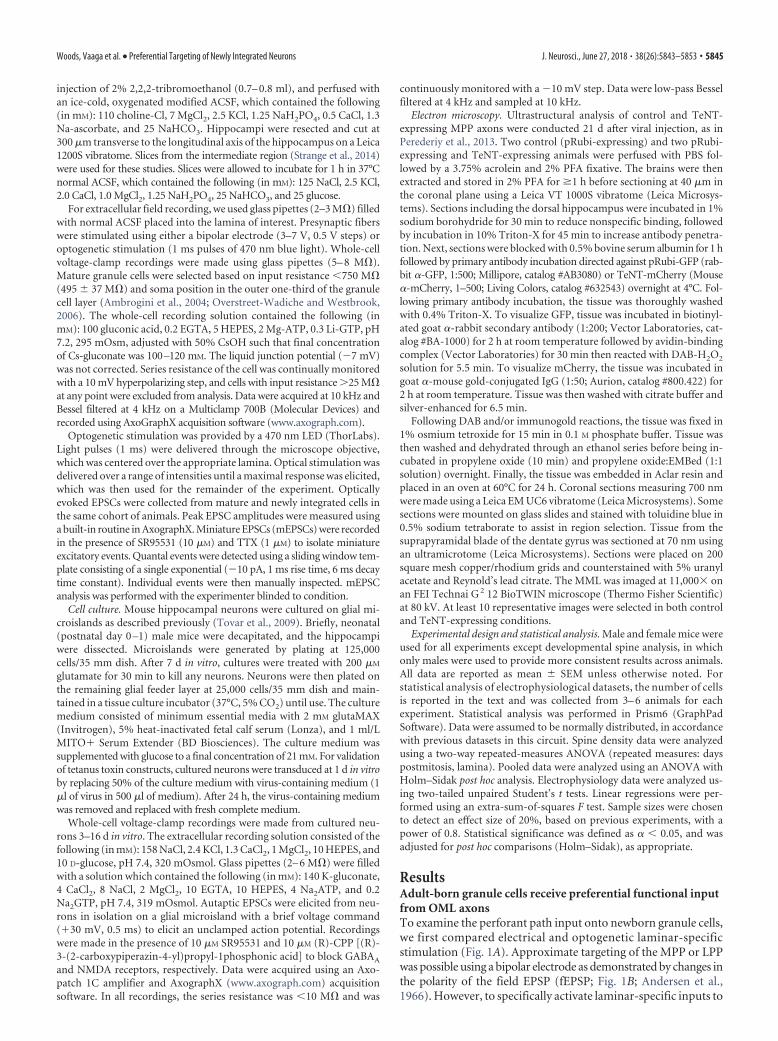
ResultsAdult-born granule cells receive preferential functional inputfrom OML axonsTo examine the perforant path input onto newborn granule cells,we first compared electrical and optogenetic laminar-specificstimulation (Fig. 1A). Approximate targeting of the MPP or LPPwas possible using a bipolar electrode as demonstrated by changes inthe polarity of the field EPSP (fEPSP; Fig. 1B; Andersen et al.,1966). However, to specifically activate laminar-specific inputs to
Woods, Vaaga et al. • Preferential Targeting of Newly Integrated Neurons J. Neurosci., June 27, 2018 • 38(26):5843–5853 • 5845
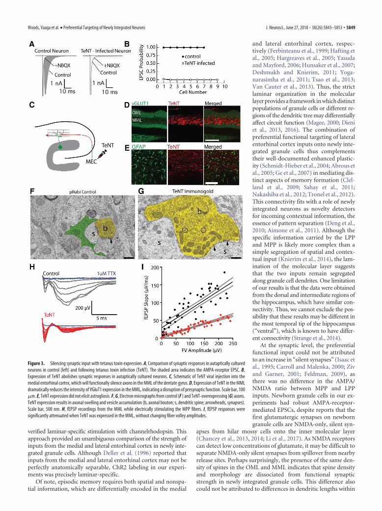
mature and newly integrating granule cells, we optogeneticallylabeled axons by injecting AAV9-CAG-ChR2-eGFP into eitherthe medial or lateral entorhinal cortex, which provided preciselabeling of either the MPP or LPP fibers in the molecular layer(Fig. 1C). To ensure that ChR2 expression was comparable acrossanimals and slices, we used a range of stimulus intensities to deter-mine a maximal evoked EPSC in mature cells from the MPP andLPP. Consistent with effective and laminar-specific ChR2 expres-sion, the maximal EPSC amplitudes were the same for optical andelectrical stimulation in the MPP (electrical stimulation: 105.0 �17.4 pA, n 7 cells; optical stimulation: 123.0 � 24.7 pA, n 19cells; unpaired t test: 0.68) and the LPP (electrical stimulation: 97.1�20.6 pA, n 6 cells; optical stimulation: 105.9 � 31.3 pA; n 16cells; unpaired t test: 0.87) in mature granule cells.
As expected for mature cells, including mature granule cellsgenerated from adult-born granule cells (Laplagne et al., 2006),the similar maximal amplitude of light-evoked EPSCs from theMPP or LPP indicates that mature cells receive robust and balancedexcitatory input from both layers. In newly generated granule cells,inputs from both the medial and lateral entorhinal cortex can bedetected at �14 d postretroviral labeling (Kumamoto et al.,2012). However, surprisingly, in newly integrated neurons (iden-tified by retroviral labeling at 21 d postmitosis), we found that thestrength of LPP inputs was nearly 10-fold larger than inputs fromthe MPP (MPP: 7.8 � 3.1 pA, n 14 cells; LPP: 72.2 � 15.2 pA,n 18 cells; Student’s unpaired t test: t(30) 3.68, p 0.0009;Fig. 1D,E). Despite the larger optically evoked EPSCs resultingfrom optical stimulation of the LPP, the band of fluorescent in-tensity of labeled axons was similar across the MML and the OML(Fig. 1C). Furthermore, because optically evoked responses in ma-ture neurons were similar across laminae, it is unlikely that differ-ences in innervation contribute to these results. Additionally, ourslices were taken from the intermediate region of the longitudinalaxis of the hippocampus, which has similar innervation patterns asthe most dorsal regions (for review, see Strange et al., 2014). Record-ings from mature and newly integrated neurons were obtained fromthe same animals, supporting the preferential functional innervationby LPP to newly integrated neurons (Fig. 1D,E).
The relatively reduced strength of MPP inputs in newly inte-grated granule cells could not be attributed to NMDA-only or“silent” synapses, as NMDA-evoked responses showed similaramplitude differences (MPP: 3.27 � 1.55 pA, n 13 cells; LPP:37.68 � 10.38 pA, n 13 cells; Student’s unpaired t test: t(24) 3.28;p0.0032). There also was no difference in the AMPA/NMDA ratiobetween lamina (MPP: 5.2 � 1.3, n 13 cells; LPP: 3.66 � 0.58, n 13 cells; Student’s unpaired t test: t(24) 1.04; p 0.31; Fig. 1F).These results indicate that newly integrated neurons receive prefer-ential, but not exclusive, functional input from the LPP.
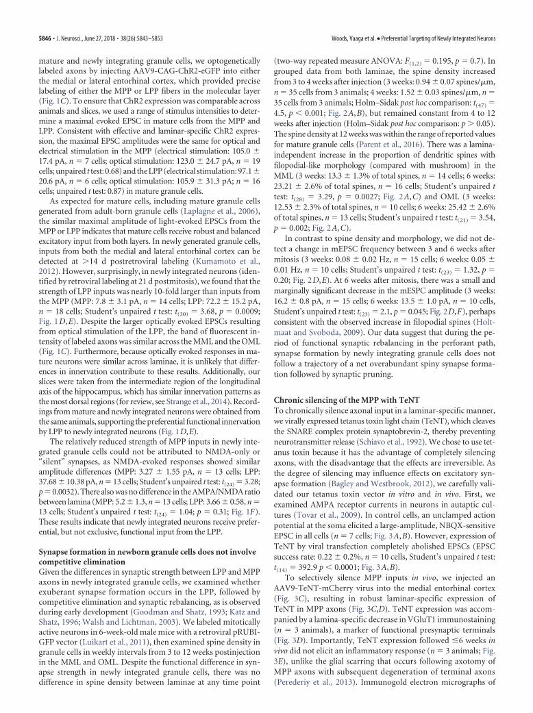
Synapse formation in newborn granule cells does not involvecompetitive eliminationGiven the differences in synaptic strength between LPP and MPPaxons in newly integrated granule cells, we examined whetherexuberant synapse formation occurs in the LPP, followed bycompetitive elimination and synaptic rebalancing, as is observedduring early development (Goodman and Shatz, 1993; Katz andShatz, 1996; Walsh and Lichtman, 2003). We labeled mitoticallyactive neurons in 6-week-old male mice with a retroviral pRUBI-GFP vector (Luikart et al., 2011), then examined spine density ingranule cells in weekly intervals from 3 to 12 weeks postinjectionin the MML and OML. Despite the functional difference in syn-apse strength in newly integrated granule cells, there was nodifference in spine density between laminae at any time point
(two-way repeated measure ANOVA: F(1,2) 0.195, p 0.7). Ingrouped data from both laminae, the spine density increasedfrom 3 to 4 weeks after injection (3 weeks: 0.94 � 0.07 spines/�m,n 35 cells from 3 animals; 4 weeks: 1.52 � 0.03 spines/�m, n 35 cells from 3 animals; Holm–Sidak post hoc comparison: t(47) 4.5, p � 0.001; Fig. 2A,B), but remained constant from 4 to 12weeks after injection (Holm–Sidak post hoc comparison: p � 0.05).The spine density at 12 weeks was within the range of reported valuesfor mature granule cells (Parent et al., 2016). There was a lamina-independent increase in the proportion of dendritic spines withfilopodial-like morphology (compared with mushroom) in theMML (3 weeks: 13.3 � 1.3% of total spines, n 14 cells; 6 weeks:23.21 � 2.6% of total spines, n 16 cells; Student’s unpaired ttest: t(28) 3.29, p 0.0027; Fig. 2A,C) and OML (3 weeks:12.53 � 2.3% of total spines, n 10 cells; 6 weeks: 25.42 � 2.6%of total spines, n 13 cells; Student’s unpaired t test: t(21) 3.54,p 0.002; Fig. 2A,C).
In contrast to spine density and morphology, we did not de-tect a change in mEPSC frequency between 3 and 6 weeks aftermitosis (3 weeks: 0.08 � 0.02 Hz, n 15 cells; 6 weeks: 0.05 �0.01 Hz, n 10 cells; Student’s unpaired t test: t(23) 1.32, p 0.20; Fig. 2D,E). At 6 weeks after mitosis, there was a small andmarginally significant decrease in the mESPC amplitude (3 weeks:16.2 � 0.8 pA, n 15 cells; 6 weeks: 13.5 � 1.0 pA, n 10 cells,Student’s unpaired t test: t(23) 2.1, p 0.045; Fig. 2D,F), perhapsconsistent with the observed increase in filopodial spines (Holt-maat and Svoboda, 2009). Our data suggest that during the pe-riod of functional synaptic rebalancing in the perforant path,synapse formation by newly integrating granule cells does notfollow a trajectory of a net overabundant spiny synapse forma-tion followed by synaptic pruning.
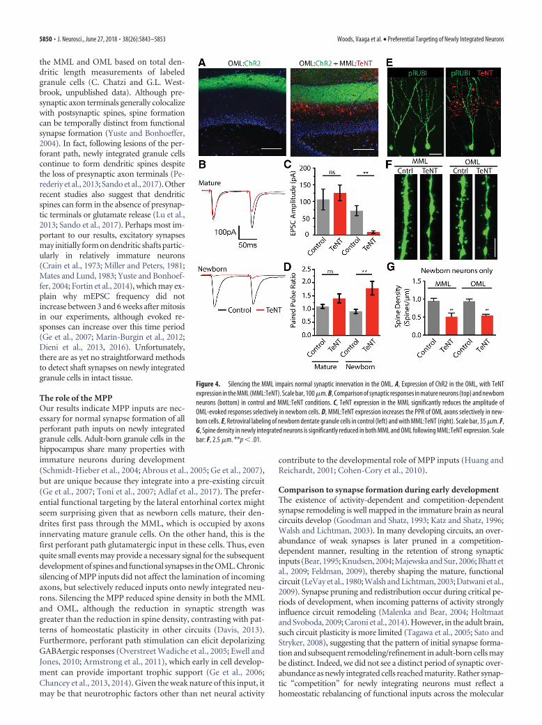
Chronic silencing of the MPP with TeNTTo chronically silence axonal input in a laminar-specific manner,we virally expressed tetanus toxin light chain (TeNT), which cleavesthe SNARE complex protein synaptobrevin-2, thereby preventingneurotransmitter release (Schiavo et al., 1992). We chose to use tet-anus toxin because it has the advantage of completely silencingaxons, with the disadvantage that the effects are irreversible. Asthe degree of silencing may influence effects on excitatory syn-apse formation (Bagley and Westbrook, 2012), we carefully vali-dated our tetanus toxin vector in vitro and in vivo. First, weexamined AMPA receptor currents in neurons in autaptic cul-tures (Tovar et al., 2009). In control cells, an unclamped actionpotential at the soma elicited a large-amplitude, NBQX-sensitiveEPSC in all cells (n 7 cells; Fig. 3A,B). However, expression ofTeNT by viral transfection completely abolished EPSCs (EPSCsuccess rate: 0.22 � 0.2%, n 10 cells, Student’s unpaired t test:t(14) 392.9 p � 0.0001; Fig. 3A,B).
To selectively silence MPP inputs in vivo, we injected anAAV9-TeNT-mCherry virus into the medial entorhinal cortex(Fig. 3C), resulting in robust laminar-specific expression ofTeNT in MPP axons (Fig. 3C,D). TeNT expression was accom-panied by a lamina-specific decrease in VGluT1 immunostaining(n 3 animals), a marker of functional presynaptic terminals(Fig. 3D). Importantly, TeNT expression followed �6 weeks invivo did not elicit an inflammatory response (n 3 animals; Fig.3E), unlike the glial scarring that occurs following axotomy ofMPP axons with subsequent degeneration of terminal axons(Perederiy et al., 2013). Immunogold electron micrographs of
5846 • J. Neurosci., June 27, 2018 • 38(26):5843–5853 Woods, Vaaga et al. • Preferential Targeting of Newly Integrated Neurons
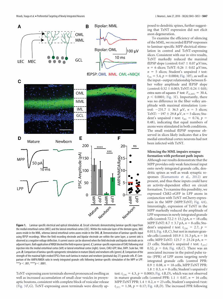
TeNT-expressing axon terminals showed pronounced swelling aswell as increased accumulation of small clear vesicles in presyn-aptic boutons, consistent with complete block of vesicular release(Fig. 3F,G). TeNT-expressing axon terminals were directly ap-
posed to dendritic spines, further suggest-ing that TeNT expression did not elicitaxon degeneration.
To examine the efficiency of silencingof the MML, we recorded fEPSP responsesto laminar-specific MPP electrical stimu-lation in control and TeNT-expressingslices. Consistent with our in vitro results,TeNT markedly reduced the maximalfEPSP slope (control: 0.67 � 0.07 �V/ms,n 6 slices; TeNT: 0.26 � 0.02 �V/ms,n 5 slices; Student’s unpaired t test:t(9) 5.4, p 0.0004; Fig. 3H), as well asthe input– output relationship between fi-ber volley amplitude and fEPSP slope(control: 0.52 � 0.003; TeNT: 0.24 � 0.01;extra-sum-of-squares F test: F(1,105) 30.4,p � 0.0001; Fig. 3I). Importantly, therewas no difference in the fiber volley am-plitude with maximal stimulation (con-trol: �231.7 � 36.5 �V, n 5 slices;TeNT: �197 � 29.8 �V, n 5 slices; Stu-dent’s unpaired t test: t(8) 0.74, p 0.48), indicating that equal numbers ofaxons were stimulated in both conditions.The small residual fEPSP response ob-served in slices likely indicates that a fewmedial entorhinal cortex neurons had notbeen infected with TeNT.
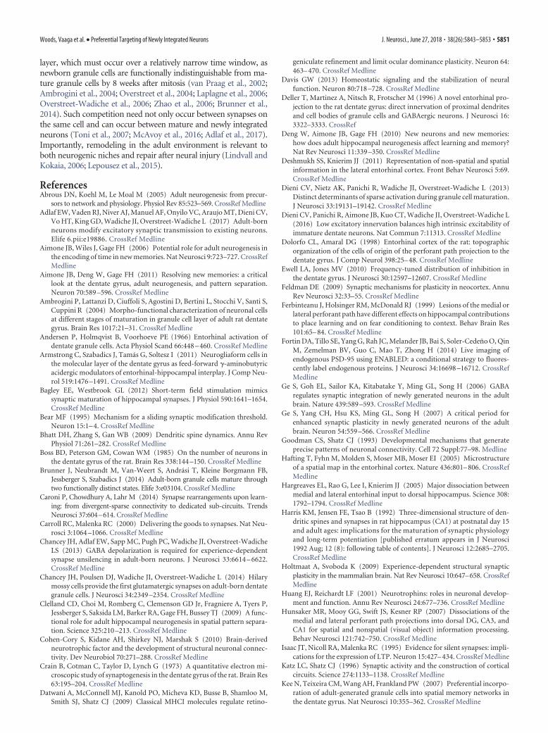
Silencing the MML impairs synapseformation with perforant path inputsAlthough our results demonstrate that theMPP provides only weak functional inputonto newly integrated granule cells, den-dritic spines as well as weak synaptic re-sponses (Kumamoto et al., 2012) arepresent, and thus these inputs could havean activity-dependent effect on circuitformation. To examine this possibility, weexpressed ChR2-eGFP in LPP axons inconjunction with TeNT-mCherry expres-sion in the MPP (MPP:TeNT; Fig. 4A).Interestingly, expression of TeNT in theMPP markedly reduced the amplitude ofLPP responses in newly integrated granulecells (control: 72.2 � 15.2 pA, n 18 cells;MPP:TeNT: 8.7 � 3.7 pA, n 8 cells; Stu-dent’s unpaired t test: t(24) 2.7, p 0.011; Fig. 4B,C), but not in mature gran-ule cells (control: 105.9 � 31.3 pA, n 16cells; MPP:TeNT: 125.7 � 23.24 pA, n 23 cells; Student’s unpaired t test: t(37):0.52, p 0.6; Fig. 4B,C). There was anassociated increase in the paired pulse ra-tio (PPR) of LPP axons targeting newlyintegrated granule cells (control PPR:0.9 � 0.08, n 18 cells, MPP:TeNT PPR:1.8 � 0.3, n 8 cells; Student’s unpaired t
test: t(24) 4.3, p 0.0003; Fig. 4B,D), which was not observedin mature granule cells (control PPR: 1.1 � 0.07, n 16 cells;MPP:TeNT PPR: 1.4 � 0.2, n 23 cells, Student’s unpaired t test:t(37) 1.38, p 0.17; Fig. 4B,D). The increased PPR following
Figure 1. Laminar-specific electrical and optical stimulation. A, Circuit schematic demonstrating laminar-specific input fromthe medial entorhinal cortex (MEC) and the lateral entorhinal cortex (LEC). Within the molecular layer of the dentate gyrus, MECaxons reside in the MML, whereas lateral entorhinal cortex axons reside in the OML. B, Demonstration of laminar-specific inputusing fEPSP recordings. When the field recording electrode and bipolar electrode are within the same layer, a current sink isobserved as a negative voltage deflection. A current source can be observed when the field electrode and bipolar electrode are inadjacent layers. Bath application of NBQX blocked the field response (green). C, Laminar-specific expression of ChR2 following viralinjection into the medial entorhinal cortex (left) or lateral entorhinal cortex (right). Green, ChR2-GFP; blue, DAPI. Scale bar, 100�m. D, Comparison of lamina-specific optogenetic stimulation in mature (black) and newborn cells (green). E, Comparison of thestrength of the maximal light-evoked EPSCs from each lamina in mature and newborn (postnatal day 21) granule cells. F, Com-parison of the AMPA/NMDA ratio in newly integrated granule cells following laminar-specific stimulation of the MPP or LPP.***p � .001, ****p � .0001.
Woods, Vaaga et al. • Preferential Targeting of Newly Integrated Neurons J. Neurosci., June 27, 2018 • 38(26):5843–5853 • 5847
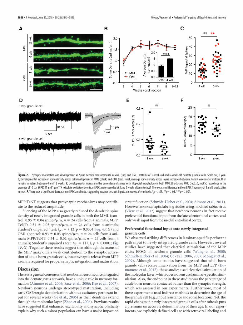
MPP:TeNT suggests that presynaptic mechanisms may contrib-ute to the reduced amplitude.
Silencing of the MPP also greatly reduced the dendritic spinedensity of newly integrated granule cells in both the MML (con-trol: 0.95 � 0.04 spines/�m, n 24 cells from 4 animals; MPP:TeNT: 0.51 � 0.05 spines/�m, n 24 cells from 4 animals;Student’s unpaired t test: t(6) 7.12, p 0.0004; Fig. 4F,G) andOML (control: 0.93 � 0.03 spines/�m, n 24 cells from 4 ani-mals; MPP:TeNT: 0.54 � 0.02 spines/�m, n 24 cells from 4animals; Student’s unpaired t test: t(6) 11.01, p � 0.0001; Fig.4F,G). Together these results suggest that although the axons ofthe MPP make only a weak contribution to the synaptic activa-tion of adult-born granule cells, intact synaptic release from MPPaxons is required for proper synaptic integration and maturation.
DiscussionThere is a general consensus that newborn neurons, once integratedinto the dentate gyrus network, have a unique role in memory for-mation (Aimone et al., 2006; Saxe et al., 2006; Kee et al., 2007).Newborn neurons undergo stereotyped maturation, includingearly GABAergic depolarization without excitatory perforant in-put for several weeks (Ge et al., 2006) as their dendrites extendthrough the molecular layer (Zhao et al., 2006). Previous resultshave suggested that enhanced excitability and synaptic plasticityexplain why such a minor population can have a major impact on
circuit function (Schmidt-Hieber et al., 2004; Aimone et al., 2011).However, monosynaptic labeling studies using modified rabies virus(Vivar et al., 2012) suggest that newborn neurons in fact receivepreferential functional input from the lateral entorhinal cortex, andonly weak input from the medial entorhinal cortex.
Preferential functional input onto newly integratedgranule cellsWe observed striking differences in laminar-specific perforantpath input to newly integrated granule cells. However, severalstudies have suggested that electrical stimulation of the MPPelicits EPSCs in newborn granule cells (Wang et al., 2000;Schmidt-Hieber et al., 2004; Ge et al., 2006, 2007; Mongiat et al.,2009). Although some studies have suggested that adult-borngranule cells receive innervation from the MPP and LPP (Ku-mamoto et al., 2012), these studies used electrical stimulation ofthe molecular layer, which does not ensure laminar-specific stim-ulation. Also, the endpoint in these studies was the percentage ofadult-born neurons contacted rather than the synaptic strength,which was assessed in our experiments. Furthermore, most ofthese experiments used indirect methods to determine the age ofthe granule cell (e.g., input resistance and soma location). Yet, therapid changes in newly integrated granule cells after mitosis putsa premium on accurate determination of their age. In our exper-iments, we explicitly defined cell age with retroviral labeling and
Figure 2. Synaptic maturation and development. A, Spine density measurements in MML (top) and OML (bottom) of 3-week-old and 6-week-old dentate granule cells. Scale bar, 5 �m.B, Developmental increase in spine density across cell development in MML (black) and OML (red). Inset, Average spine density across layers increases between 3 and 4 weeks after mitosis, thenremains constant between 4 and 12 weeks. C, Developmental increase in the percentage of spines with filopodial morphology in both MML (black) and OML (red). D, mEPSC recordings in thepresence of 10 �M SR95531 and 1 �M TTX to isolate excitatory events. mEPSCs were recorded at 3 and 6 weeks after mitosis. E, There was no difference in the mEPSC frequency at 3 and 6 weeks aftermitosis. F, There was a significant decrease in mEPSC amplitude, suggesting weaker synaptic inputs at 6 weeks after mitosis. *p � .05, **p � .01, ***p � .001.
5848 • J. Neurosci., June 27, 2018 • 38(26):5843–5853 Woods, Vaaga et al. • Preferential Targeting of Newly Integrated Neurons
verified laminar-specific stimulation with channelrhodopsin. Thisapproach provided an unambiguous comparison of the strength ofinputs from the medial and lateral entorhinal cortex in newly inte-grated granule cells. Although Deller et al. (1996) reported thatinputs from the medial and lateral entorhinal cortex may not beperfectly anatomically separable, ChR2 labeling in our experi-ments was precisely laminar-specific.
Of note, episodic memory requires both spatial and nonspa-tial information, which are differentially encoded in the medial
and lateral entorhinal cortex, respec-tively (Ferbinteanu et al., 1999; Hafting etal., 2005; Hargreaves et al., 2005; Yasudaand Mayford, 2006; Hunsaker et al., 2007;Deshmukh and Knierim, 2011; Yoga-narasimha et al., 2011; Tsao et al., 2013;Van Cauter et al., 2013). Thus, the strictlaminar organization in the molecularlayer provides a framework in which distinctpopulations of granule cells or different re-gions of the dendritic tree may differentiallyaffect circuit function (Magee, 2000; Dieniet al., 2013, 2016). The combination ofpreferential functional targeting of lateralentorhinal cortex inputs onto newly inte-grated granule cells thus complementstheir well-documented enhanced plastic-ity (Schmidt-Hieber et al., 2004; Abrous etal., 2005; Ge et al., 2007) in mediating dis-tinct aspects of memory formation (Clel-land et al., 2009; Sahay et al., 2011;Nakashiba et al., 2012; Tronel et al., 2012).This connectivity fits with a role of newlyintegrated neurons as novelty detectorsfor incoming contextual information, theessence of pattern separation (Deng et al.,2010; Aimone et al., 2011). Although thespecific information carried by the LPPand MPP is likely more complex than asimple segregation of spatial and contex-tual input (Knierim et al., 2014), the lam-ination of the molecular layer suggeststhat the two inputs remain segregatedalong granule cell dendrites. One limitationof our results is that the data were obtainedfrom the dorsal and intermediate regions ofthe hippocampus, which have similar con-nectivity. Thus, we cannot exclude the pos-sibility that these results may be different inthe most temporal tip of the hippocampus(“ventral”), which is known to have differ-ent connectivity (Strange et al., 2014).
At the synaptic level, the preferentialfunctional input could not be attributedto an increase in “silent synapses” (Isaac etal., 1995; Carroll and Malenka, 2000; Zivand Garner, 2001; Feldman, 2009), asthere was no difference in the AMPA/NMDA ratio between MPP and LPPinputs. Newborn granule cells in our ex-periments had robust AMPA-receptor-mediated EPSCs, despite reports that thefirst glutamatergic synapses on newborngranule cells are NMDA-only, silent syn-
apses from hilar mossy cells onto the inner molecular layer(Chancey et al., 2013, 2014; Li et al., 2017). As NMDA receptorscan detect low concentrations of glutamate, it may be difficult toseparate NMDA-only silent synapses from spillover from nearbyrelease sites. Perhaps surprisingly, the presence of the same den-sity of spines in the OML and MML indicates that spine densityand morphology are dissociated from functional synapticstrength in newly integrated granule cells. This difference alsocould not be attributed to differences in dendritic lengths within
Figure 3. Silencing synaptic input with tetanus toxin expression. A, Comparison of synaptic responses in autaptically culturedneurons in control (left) and following tetanus toxin infection (TeNT). The shaded area indicates the AMPA-receptor EPSC. B,Expression of TeNT abolishes synaptic responses in autaptically cultured neurons. C, Schematic of TeNT viral injection into themedial entorhinal cortex, which will functionally silence axons in the MML of the dentate gyrus. D, Expression of TeNT in the MMLdramatically reduces the intensity of VGluT1 expression in the MML, indicating a disruption of presynaptic function. Scale bar, 100�m. E, TeNT expression did not elicit astrogliosis. F, G, Electron micrographs from control (F ) and TeNT-overexpressing (G) axons.TeNT expression results in axonal swelling and vesicle accumulation (b, axonal bouton; s, dendritic spine; arrowheads, synapses).Scale bar, 500 nm. H, fEPSP recordings from the MML while electrically stimulating the MPP fibers. I, fEPSP responses weresignificantly attenuated when TeNT was expressed in the MML, without changing fiber volley amplitudes.
Woods, Vaaga et al. • Preferential Targeting of Newly Integrated Neurons J. Neurosci., June 27, 2018 • 38(26):5843–5853 • 5849
the MML and OML based on total den-dritic length measurements of labeledgranule cells (C. Chatzi and G.L. West-brook, unpublished data). Although pre-synaptic axon terminals generally colocalizewith postsynaptic spines, spine formationcan be temporally distinct from functionalsynapse formation (Yuste and Bonhoeffer,2004). In fact, following lesions of the per-forant path, newly integrated granule cellscontinue to form dendritic spines despitethe loss of presynaptic axon terminals (Pe-rederiy et al., 2013; Sando et al., 2017). Otherrecent studies also suggest that dendriticspines can form in the absence of presynap-tic terminals or glutamate release (Lu et al.,2013; Sando et al., 2017). Perhaps most im-portant to our results, excitatory synapsesmay initially form on dendritic shafts partic-ularly in relatively immature neurons(Crain et al., 1973; Miller and Peters, 1981;Mates and Lund, 1983; Yuste and Bonhoef-fer, 2004; Fortin et al., 2014), which may ex-plain why mEPSC frequency did notincrease between 3 and 6 weeks after mitosisin our experiments, although evoked re-sponses can increase over this time period(Ge et al., 2007; Marín-Burgin et al., 2012;Dieni et al., 2013, 2016). Unfortunately,there are as yet no straightforward methodsto detect shaft synapses on newly integratedgranule cells in intact tissue.
The role of the MPPOur results indicate MPP inputs are nec-essary for normal synapse formation of allperforant path inputs on newly integratedgranule cells. Adult-born granule cells in thehippocampus share many properties withimmature neurons during development(Schmidt-Hieber et al., 2004; Abrous et al., 2005; Ge et al., 2007),but are unique because they integrate into a pre-existing circuit(Ge et al., 2007; Toni et al., 2007; Adlaf et al., 2017). The prefer-ential functional targeting by the lateral entorhinal cortex mightseem surprising given that as newborn cells mature, their den-drites first pass through the MML, which is occupied by axonsinnervating mature granule cells. On the other hand, this is thefirst perforant path glutamatergic input in these cells. Thus, evenquite small events may provide a necessary signal for the subsequentdevelopment of spines and functional synapses in the OML. Chronicsilencing of MPP inputs did not affect the lamination of incomingaxons, but selectively reduced inputs onto newly integrated neu-rons. Silencing the MPP reduced spine density in both the MMLand OML, although the reduction in synaptic strength wasgreater than the reduction in spine density, contrasting with pat-terns of homeostatic plasticity in other circuits (Davis, 2013).Furthermore, perforant path stimulation can elicit depolarizingGABAergic responses (Overstreet Wadiche et al., 2005; Ewell andJones, 2010; Armstrong et al., 2011), which early in cell develop-ment can provide important trophic support (Ge et al., 2006;Chancey et al., 2013, 2014). Given the weak nature of this input, itmay be that neurotrophic factors other than net neural activity
contribute to the developmental role of MPP inputs (Huang andReichardt, 2001; Cohen-Cory et al., 2010).
Comparison to synapse formation during early developmentThe existence of activity-dependent and competition-dependentsynapse remodeling is well mapped in the immature brain as neuralcircuits develop (Goodman and Shatz, 1993; Katz and Shatz, 1996;Walsh and Lichtman, 2003). In many developing circuits, an over-abundance of weak synapses is later pruned in a competition-dependent manner, resulting in the retention of strong synapticinputs (Bear, 1995; Knudsen, 2004; Majewska and Sur, 2006; Bhatt etal., 2009; Feldman, 2009), thereby shaping the mature, functionalcircuit (LeVay et al., 1980; Walsh and Lichtman, 2003; Datwani et al.,2009). Synapse pruning and redistribution occur during critical pe-riods of development, when incoming patterns of activity stronglyinfluence circuit remodeling (Malenka and Bear, 2004; Holtmaatand Svoboda, 2009; Caroni et al., 2014). However, in the adult brain,such circuit plasticity is more limited (Tagawa et al., 2005; Sato andStryker, 2008), suggesting that the pattern of initial synapse forma-tion and subsequent remodeling/refinement in adult-born cells maybe distinct. Indeed, we did not see a distinct period of synaptic over-abundance as newly integrated cells reached maturity. Rather synap-tic “competition” for newly integrating neurons must reflect ahomeostatic rebalancing of functional inputs across the molecular
Figure 4. Silencing the MML impairs normal synaptic innervation in the OML. A, Expression of ChR2 in the OML, with TeNTexpression in the MML (MML:TeNT). Scale bar, 100 �m. B, Comparison of synaptic responses in mature neurons (top) and newbornneurons (bottom) in control and MML:TeNT conditions. C, TeNT expression in the MML significantly reduces the amplitude ofOML-evoked responses selectively in newborn cells. D, MML:TeNT expression increases the PPR of OML axons selectively in new-born cells. E, Retroviral labeling of newborn dentate granule cells in control (left) and with MML:TeNT (right). Scale bar, 35 �m. F,G, Spine density in newly integrated neurons is significantly reduced in both MML and OML following MML:TeNT expression. Scalebar: F, 2.5 �m. **p � .01.
5850 • J. Neurosci., June 27, 2018 • 38(26):5843–5853 Woods, Vaaga et al. • Preferential Targeting of Newly Integrated Neurons
layer, which must occur over a relatively narrow time window, asnewborn granule cells are functionally indistinguishable from ma-ture granule cells by 8 weeks after mitosis (van Praag et al., 2002;Ambrogini et al., 2004; Overstreet et al., 2004; Laplagne et al., 2006;Overstreet-Wadiche et al., 2006; Zhao et al., 2006; Brunner et al.,2014). Such competition need not only occur between synapses onthe same cell and can occur between mature and newly integratedneurons (Toni et al., 2007; McAvoy et al., 2016; Adlaf et al., 2017).Importantly, remodeling in the adult environment is relevant toboth neurogenic niches and repair after neural injury (Lindvall andKokaia, 2006; Lepousez et al., 2015).
ReferencesAbrous DN, Koehl M, Le Moal M (2005) Adult neurogenesis: from precur-
sors to network and physiology. Physiol Rev 85:523–569. CrossRef MedlineAdlaf EW, Vaden RJ, Niver AJ, Manuel AF, Onyilo VC, Araujo MT, Dieni CV,
Vo HT, King GD, Wadiche JI, Overstreet-Wadiche L (2017) Adult-bornneurons modify excitatory synaptic transmission to existing neurons.Elife 6.pii:e19886. CrossRef Medline
Aimone JB, Wiles J, Gage FH (2006) Potential role for adult neurogenesis inthe encoding of time in new memories. Nat Neurosci 9:723–727. CrossRefMedline
Aimone JB, Deng W, Gage FH (2011) Resolving new memories: a criticallook at the dentate gyrus, adult neurogenesis, and pattern separation.Neuron 70:589 –596. CrossRef Medline
Ambrogini P, Lattanzi D, Ciuffoli S, Agostini D, Bertini L, Stocchi V, Santi S,Cuppini R (2004) Morpho-functional characterization of neuronal cellsat different stages of maturation in granule cell layer of adult rat dentategyrus. Brain Res 1017:21–31. CrossRef Medline
Andersen P, Holmqvist B, Voorhoeve PE (1966) Entorhinal activation ofdentate granule cells. Acta Physiol Scand 66:448 – 460. CrossRef Medline
Armstrong C, Szabadics J, Tamas G, Soltesz I (2011) Neurogliaform cells inthe molecular layer of the dentate gyrus as feed-forward �-aminobutyricacidergic modulators of entorhinal-hippocampal interplay. J Comp Neu-rol 519:1476 –1491. CrossRef Medline
Bagley EE, Westbrook GL (2012) Short-term field stimulation mimicssynaptic maturation of hippocampal synapses. J Physiol 590:1641–1654.CrossRef Medline
Bear MF (1995) Mechanism for a sliding synaptic modification threshold.Neuron 15:1– 4. CrossRef Medline
Bhatt DH, Zhang S, Gan WB (2009) Dendritic spine dynamics. Annu RevPhysiol 71:261–282. CrossRef Medline
Boss BD, Peterson GM, Cowan WM (1985) On the number of neurons inthe dentate gyrus of the rat. Brain Res 338:144 –150. CrossRef Medline
Brunner J, Neubrandt M, Van-Weert S, Andrasi T, Kleine Borgmann FB,Jessberger S, Szabadics J (2014) Adult-born granule cells mature throughtwo functionally distinct states. Elife 3:e03104. CrossRef Medline
Caroni P, Chowdhury A, Lahr M (2014) Synapse rearrangements upon learn-ing: from divergent-sparse connectivity to dedicated sub-circuits. TrendsNeurosci 37:604–614. CrossRef Medline
Carroll RC, Malenka RC (2000) Delivering the goods to synapses. Nat Neu-rosci 3:1064 –1066. CrossRef Medline
Chancey JH, Adlaf EW, Sapp MC, Pugh PC, Wadiche JI, Overstreet-WadicheLS (2013) GABA depolarization is required for experience-dependentsynapse unsilencing in adult-born neurons. J Neurosci 33:6614 – 6622.CrossRef Medline
Chancey JH, Poulsen DJ, Wadiche JI, Overstreet-Wadiche L (2014) Hilarymossy cells provide the first glutamatergic synapses on adult-born dentategranule cells. J Neurosci 34:2349 –2354. CrossRef Medline
Clelland CD, Choi M, Romberg C, Clemenson GD Jr, Fragniere A, Tyers P,Jessberger S, Saksida LM, Barker RA, Gage FH, Bussey TJ (2009) A func-tional role for adult hippocampal neurogenesis in spatial pattern separa-tion. Science 325:210 –213. CrossRef Medline
Cohen-Cory S, Kidane AH, Shirkey NJ, Marshak S (2010) Brain-derivedneurotrophic factor and the development of structural neuronal connec-tivity. Dev Neurobiol 70:271–288. CrossRef Medline
Crain B, Cotman C, Taylor D, Lynch G (1973) A quantitative electron mi-croscopic study of synaptogenesis in the dentate gyrus of the rat. Brain Res63:195–204. CrossRef Medline
Datwani A, McConnell MJ, Kanold PO, Micheva KD, Busse B, Shamloo M,Smith SJ, Shatz CJ (2009) Classical MHCI molecules regulate retino-
geniculate refinement and limit ocular dominance plasticity. Neuron 64:463– 470. CrossRef Medline
Davis GW (2013) Homeostatic signaling and the stabilization of neuralfunction. Neuron 80:718 –728. CrossRef Medline
Deller T, Martinez A, Nitsch R, Frotscher M (1996) A novel entorhinal pro-jection to the rat dentate gyrus: direct innervation of proximal dendritesand cell bodies of granule cells and GABAergic neurons. J Neurosci 16:3322–3333. CrossRef
Deng W, Aimone JB, Gage FH (2010) New neurons and new memories:how does adult hippocampal neurogenesis affect learning and memory?Nat Rev Neurosci 11:339 –350. CrossRef Medline
Deshmukh SS, Knierim JJ (2011) Representation of non-spatial and spatialinformation in the lateral entorhinal cortex. Front Behav Neurosci 5:69.CrossRef Medline
Dieni CV, Nietz AK, Panichi R, Wadiche JI, Overstreet-Wadiche L (2013)Distinct determinants of sparse activation during granule cell maturation.J Neurosci 33:19131–19142. CrossRef Medline
Dieni CV, Panichi R, Aimone JB, Kuo CT, Wadiche JI, Overstreet-Wadiche L(2016) Low excitatory innervation balances high intrinsic excitability ofimmature dentate neurons. Nat Commun 7:11313. CrossRef Medline
Dolorfo CL, Amaral DG (1998) Entorhinal cortex of the rat: topographicorganization of the cells of origin of the perforant path projection to thedentate gyrus. J Comp Neurol 398:25– 48. CrossRef Medline
Ewell LA, Jones MV (2010) Frequency-tuned distribution of inhibition inthe dentate gyrus. J Neurosci 30:12597–12607. CrossRef Medline
Feldman DE (2009) Synaptic mechanisms for plasticity in neocortex. AnnuRev Neurosci 32:33–55. CrossRef Medline
Ferbinteanu J, Holsinger RM, McDonald RJ (1999) Lesions of the medial orlateral perforant path have different effects on hippocampal contributionsto place learning and on fear conditioning to context. Behav Brain Res101:65– 84. CrossRef Medline
Fortin DA, Tillo SE, Yang G, Rah JC, Melander JB, Bai S, Soler-Cedeno O, QinM, Zemelman BV, Guo C, Mao T, Zhong H (2014) Live imaging ofendogenous PSD-95 using ENABLED: a conditional strategy to fluores-cently label endogenous proteins. J Neurosci 34:16698 –16712. CrossRefMedline
Ge S, Goh EL, Sailor KA, Kitabatake Y, Ming GL, Song H (2006) GABAregulates synaptic integration of newly generated neurons in the adultbrain. Nature 439:589 –593. CrossRef Medline
Ge S, Yang CH, Hsu KS, Ming GL, Song H (2007) A critical period forenhanced synaptic plasticity in newly generated neurons of the adultbrain. Neuron 54:559 –566. CrossRef Medline
Goodman CS, Shatz CJ (1993) Developmental mechanisms that generateprecise patterns of neuronal connectivity. Cell 72 Suppl:77–98. Medline
Hafting T, Fyhn M, Molden S, Moser MB, Moser EI (2005) Microstructureof a spatial map in the entorhinal cortex. Nature 436:801– 806. CrossRefMedline
Hargreaves EL, Rao G, Lee I, Knierim JJ (2005) Major dissociation betweenmedial and lateral entorhinal input to dorsal hippocampus. Science 308:1792–1794. CrossRef Medline
Harris KM, Jensen FE, Tsao B (1992) Three-dimensional structure of den-dritic spines and synapses in rat hippocampus (CA1) at postnatal day 15and adult ages: implications for the maturation of synaptic physiologyand long-term potentiation [published erratum appears in J Neurosci1992 Aug; 12 (8): following table of contents]. J Neurosci 12:2685–2705.CrossRef Medline
Holtmaat A, Svoboda K (2009) Experience-dependent structural synapticplasticity in the mammalian brain. Nat Rev Neurosci 10:647–658. CrossRefMedline
Huang EJ, Reichardt LF (2001) Neurotrophins: roles in neuronal develop-ment and function. Annu Rev Neurosci 24:677–736. CrossRef Medline
Hunsaker MR, Mooy GG, Swift JS, Kesner RP (2007) Dissociations of themedial and lateral perforant path projections into dorsal DG, CA3, andCA1 for spatial and nonspatial (visual object) information processing.Behav Neurosci 121:742–750. CrossRef Medline
Isaac JT, Nicoll RA, Malenka RC (1995) Evidence for silent synapses: impli-cations for the expression of LTP. Neuron 15:427– 434. CrossRef Medline
Katz LC, Shatz CJ (1996) Synaptic activity and the construction of corticalcircuits. Science 274:1133–1138. CrossRef Medline
Kee N, Teixeira CM, Wang AH, Frankland PW (2007) Preferential incorpo-ration of adult-generated granule cells into spatial memory networks inthe dentate gyrus. Nat Neurosci 10:355–362. CrossRef Medline
Woods, Vaaga et al. • Preferential Targeting of Newly Integrated Neurons J. Neurosci., June 27, 2018 • 38(26):5843–5853 • 5851
Knierim JJ, Neunuebel JP, Deshmukh SS (2014) Functional correlates of thelateral and medial entorhinal cortex: objects, path integration and local-global reference frames. Philos Trans R Soc Lond B Biol Sci 369:20130369.CrossRef Medline
Knudsen EI (2004) Sensitive periods in the development of the brain andbehavior. J Cogn Neurosci 16:1412–1425. CrossRef Medline
Kumamoto N, Gu Y, Wang J, Janoschka S, Takemaru K, Levine J, Ge S (2012)A role for primary cilia in glutamatergic synaptic integration of adult-born neurons. Nat Neurosci 15:399 – 405, S1. CrossRef Medline
Laplagne DA, Esposito MS, Piatti VC, Morgenstern NA, Zhao C, van Praag H,Gage FH, Schinder AF (2006) Functional convergence of neurons gen-erated in the developing and adult hippocampus. PLoS Biol 4:e409.CrossRef Medline
Lepousez G, Nissant A, Lledo PM (2015) Adult neurogenesis and the futureof the rejuvenating brain circuits. Neuron 86:387– 401. CrossRef Medline
LeVay S, Wiesel TN, Hubel DH (1980) The development of ocular domi-nance columns in normal and visually deprived monkeys. J Comp Neurol191:1–51. CrossRef Medline
Li L, Sultan S, Heigele S, Schmidt-Salzmann C, Toni N, Bischofberger J (2017)Silent synapses generate sparse and orthogonal action potential firing inadult-born hippocampal granule cells. eLife 6.pii::e23612. CrossRef Medline
Lindvall O, Kokaia Z (2006) Stem cells for the treatment of neurologicaldisorders. Nature 441:1094 –1096. CrossRef Medline
Lu W, Bushong EA, Shih TP, Ellisman MH, Nicoll RA (2013) The cell-autonomous role of excitatory synaptic transmission in the regulation ofneuronal structure and function. Neuron 78:433– 439. CrossRef Medline
Luikart BW, Schnell E, Washburn EK, Bensen AL, Tovar KR, Westbrook GL(2011) Pten knockdown in vivo increases excitatory drive onto dentategranule cells. J Neurosci 31:4345– 4354. CrossRef Medline
Magee JC (2000) Dendritic integration of excitatory synaptic input. Nat RevNeurosci 1:181–190. CrossRef Medline
Majewska AK, Sur M (2006) Plasticity and specificity of cortical processingnetworks. Trends Neurosci 29:323–329. CrossRef Medline
Malenka RC, Bear MF (2004) LTP and LTD: an embarrassment of riches.Neuron 44:5–21. CrossRef Medline
Marín-Burgin A, Mongiat LA, Pardi MB, Schinder AF (2012) Unique pro-cessing during a period of high excitation/inhibition balance in adult-born neurons. Science 335:1238 –1242. CrossRef Medline
Mates SL, Lund JS (1983) Spine formation and maturation of type 1 syn-apses on spiny stellate neurons in primate visual cortex. J Comp Neurol221:91–97. CrossRef Medline
McAvoy KM, Scobie KN, Berger S, Russo C, Guo N, Decharatanachart P,Vega-Ramirez H, Miake-Lye S, Whalen M, Nelson M, Bergami M,Bartsch D, Hen R, Berninger B, Sahay A (2016) Modulating neuronalcompetition dynamics in the dentate gyrus to rejuvenate aging memorycircuits. Neuron 91:1356 –1373. CrossRef Medline
Miller M, Peters A (1981) Maturation of rat visual cortex. II. A combinedGolgi-electron microscope study of pyramidal neurons. J Comp Neurol203:555–573. CrossRef Medline
Ming GL, Song H (2011) Adult neurogenesis in the mammalian brain: sig-nificant answers and significant questions. Neuron 70:687–702. CrossRefMedline
Mongiat LA, Esposito MS, Lombardi G, Schinder AF (2009) Reliable activa-tion of immature neurons in the adult hippocampus. PLoS One 4:e5320.CrossRef Medline
Nakashiba T, Cushman JD, Pelkey KA, Renaudineau S, Buhl DL, McHugh TJ,Rodriguez Barrera V, Chittajallu R, Iwamoto KS, McBain CJ, FanselowMS, Tonegawa S (2012) Young dentate granule cells mediate patternseparation, whereas old granule cells facilitate pattern completion. Cell149:188 –201. CrossRef Medline
Ohara S, Sato S, Tsutsui K, Witter MP, Iijima T (2013) Organization ofmultisynaptic inputs to the dorsal and ventral dentate gyrus: retrogradetrans-synaptic tracing with rabies virus vector in the rat. PLoS One 8:e78928.CrossRef Medline
Overstreet LS, Hentges ST, Bumaschny VF, de Souza FS, Smart JL, SantangeloAM, Low MJ, Westbrook GL, Rubinstein M (2004) A transgenic markerfor newly born granule cells in dentate gyrus. J Neurosci 24:3251–3259.CrossRef Medline
Overstreet Wadiche L, Bromberg DA, Bensen AL, Westbrook GL (2005)GABAergic signaling to newborn neurons in dentate gyrus. J Neurophysiol94:4528–4532. CrossRef Medline
Overstreet-Wadiche LS, Westbrook GL (2006) Functional maturation of adult-generated granule cells. Hippocampus 16:208–215. CrossRef Medline
Overstreet-Wadiche LS, Bensen AL, Westbrook GL (2006) Delayed devel-opment of adult-generated granule cells in dentate gyrus. J Neurosci 26:2326 –2334. CrossRef Medline
Parent AS, Pinson A, Woods N, Chatzi C, Vaaga CE, Bensen A, Gerard A,Thome JP, Bourguignon JP, Westbrook GL (2016) Early exposure toaroclor 1254 in vivo disrupts the functional synaptic development of new-born hippocampal granule cells. Eur J Neurosci 44:3001–3010. CrossRefMedline
Perederiy JV, Luikart BW, Washburn EK, Schnell E, Westbrook GL (2013)Neural injury alters proliferation and integration of adult-generated neu-rons in the dentate gyrus. J Neurosci 33:4754 – 4767. CrossRef Medline
Rolls ET, Treves A, Rolls ET (1998) Neural networks and brain function.Oxford: Oxford UP.
Sahay A, Scobie KN, Hill AS, O’Carroll CM, Kheirbek MA, Burghardt NS,Fenton AA, Dranovsky A, Hen R (2011) Increasing adult hippocampalneurogenesis is sufficient to improve pattern separation. Nature 472:466 –470. CrossRef Medline
Sando R, Bushong E, Zhu Y, Huang M, Considine C, Phan S, Ju S, Uytiepo M,Ellisman M, Maximov A (2017) Assembly of excitatory synapses in the ab-sence of glutamatergic neurotransmission. Neuron 94:312–321.e3. CrossRefMedline
Sato M, Stryker MP (2008) Distinctive features of adult ocular dominanceplasticity. J Neurosci 28:10278 –10286. CrossRef Medline
Saxe MD, Battaglia F, Wang JW, Malleret G, David DJ, Monckton JE, GarciaAD, Sofroniew MV, Kandel ER, Santarelli L, Hen R, Drew MR (2006)Ablation of hippocampal neurogenesis impairs contextual fear condi-tioning and synaptic plasticity in the dentate gyrus. Proc Natl Acad SciU S A 103:17501–17506. CrossRef Medline
Schiavo G, Benfenati F, Poulain B, Rossetto O, Polverino de Laureto P,DasGupta BR, Montecucco C (1992) Tetanus and botulinum-B neuro-toxins block neurotransmitter release by proteolytic cleavage of synapto-brevin. Nature 359:832–835. CrossRef Medline
Schmidt-Hieber C, Jonas P, Bischofberger J (2004) Enhanced synaptic plas-ticity in newly generated granule cells of the adult hippocampus. Nature429:184 –187. CrossRef Medline
Strange BA, Witter MP, Lein ES, Moser EI (2014) Functional organizationof the hippocampal longitudinal axis. Nat Rev Neurosci 15:655–669. CrossRefMedline
Tagawa Y, Kanold PO, Majdan M, Shatz CJ (2005) Multiple periods of func-tional ocular dominance plasticity in mouse visual cortex. Nat Neurosci8:380 –388. CrossRef Medline
Toni N, Teng EM, Bushong EA, Aimone JB, Zhao C, Consiglio A, van PraagH, Martone ME, Ellisman MH, Gage FH (2007) Synapse formation onneurons born in the adult hippocampus. Nat Neurosci 10:727–734. CrossRefMedline
Toni N, Laplagne DA, Zhao C, Lombardi G, Ribak CE, Gage FH, Schinder AF(2008) Neurons born in the adult dentate gyrus form functional synapseswith target cells. Nat Neurosci 11:901–907. CrossRef Medline
Tovar KR, Maher BJ, Westbrook GL (2009) Direct actions of carbenoxoloneon synaptic transmission and neuronal membrane properties. J Neuro-physiol 102:974 –978. CrossRef Medline
Tronel S, Belnoue L, Grosjean N, Revest JM, Piazza PV, Koehl M, Abrous DN(2012) Adult-born neurons are necessary for extended contextual dis-crimination. Hippocampus 22:292–298. CrossRef Medline
Tsao A, Moser MB, Moser EI (2013) Traces of experience in the lateralentorhinal cortex. Curr Biol 23:399 – 405. CrossRef Medline
Van Cauter T, Camon J, Alvernhe A, Elduayen C, Sargolini F, Save E (2013)Distinct roles of medial and lateral entorhinal cortex in spatial cognition.Cereb Cortex 23:451– 459. CrossRef Medline
van Groen T, Miettinen P, Kadish I (2003) The entorhinal cortex of themouse: organization of the projection to the hippocampal formation.Hippocampus 13:133–149. CrossRef Medline
van Praag H, Schinder AF, Christie BR, Toni N, Palmer TD, Gage FH (2002)Functional neurogenesis in the adult hippocampus. Nature 415:1030 –1034. CrossRef Medline
Vivar C, Potter MC, Choi J, Lee JY, Stringer TP, Callaway EM, Gage FH, SuhH, van Praag H (2012) Monosynaptic inputs to new neurons in thedentate gyrus. Nat Commun 3:1107. CrossRef Medline
5852 • J. Neurosci., June 27, 2018 • 38(26):5843–5853 Woods, Vaaga et al. • Preferential Targeting of Newly Integrated Neurons

Walsh MK, Lichtman JW (2003) In vivo time-lapse imaging of synaptictakeover associated with naturally occurring synapse elimination. Neu-ron 37:67–73. CrossRef Medline
Wang S, Scott BW, Wojtowicz JM (2000) Heterogenous properties of den-tate granule neurons in the adult rat. J Neurobiol 42:248 –257. CrossRefMedline
Witter MP (2007) The perforant path: projections from the entorhinalcortex to the dentate gyrus. Prog Brain Res 163:43– 61. CrossRefMedline
Yasuda M, Mayford MR (2006) CaMKII activation in the entorhinal cortexdisrupts previously encoded spatial memory. Neuron 50:309–318. CrossRefMedline
Yoganarasimha D, Rao G, Knierim JJ (2011) Lateral entorhinal neurons arenot spatially selective in cue-rich environments. Hippocampus 21:1363–1374. CrossRef Medline
Yuste R, Bonhoeffer T (2004) Genesis of dendritic spines: insights from ul-trastructural and imaging studies. Nat Rev Neurosci 5:24 –34. CrossRefMedline
Zhao C, Teng EM, Summers RG Jr, Ming GL, Gage FH (2006) Distinctmorphological stages of dentate granule neuron maturation in the adultmouse hippocampus. J Neurosci 26:3–11. CrossRef Medline
Ziv NE, Garner CC (2001) Principles of glutamatergic synapse formation:seeing the forest for the trees. Curr Opin Neurobiol 11:536 –543. CrossRefMedline
Woods, Vaaga et al. • Preferential Targeting of Newly Integrated Neurons J. Neurosci., June 27, 2018 • 38(26):5843–5853 • 5853