Embed Size (px)
Citation preview

Cervical Spine Trauma
2016 Nordic Trauma Society
Stuart E. Mirvis. M.D., FACR
Department of Radiology and Maryland
Shock-Trauma Center
University of Maryland School of Medicine

Topics to Review
Definition of stability
Craniocervical distraction
Hyperflexion “sprain”
MRI Applications

2-column method
Supraspinous ligament to PLL
ALL column to PLL
Complete column injury produces instability

Defining Stability
Mechanical - spine able to resist non-
physiologic movement under physiologic load
Neurologic – injury will not cause or worsen a
neurologic injury under physiologic load
Most mechanically unstable injuries are
neurologically unstable. (Exceptions… lo-grade
Hangman, Jefferson)
To be considered stable an injury must not led
to chronic pain, deformity, neurologic damage
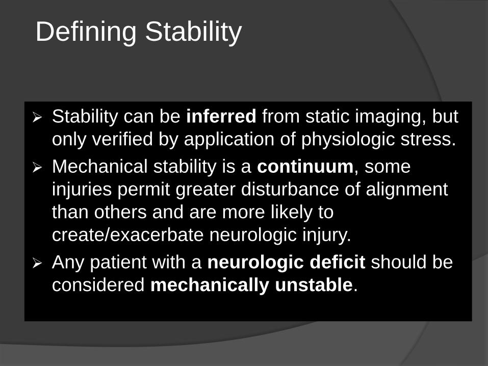
Defining Stability
Stability can be inferred from static imaging, but
only verified by application of physiologic stress.
Mechanical stability is a continuum, some
injuries permit greater disturbance of alignment
than others and are more likely to
create/exacerbate neurologic injury.
Any patient with a neurologic deficit should be
considered mechanically unstable.

Methods of Diagnosis
Most unstable injuries can be diagnosed from
radiographs – but sometimes misleading
More (almost all) unstable injuries diagnosed
by MDCT
MRI can show ligament injuries, but does not
correlate well with surgical findings (tears,
partial tears, stripping, stretching)
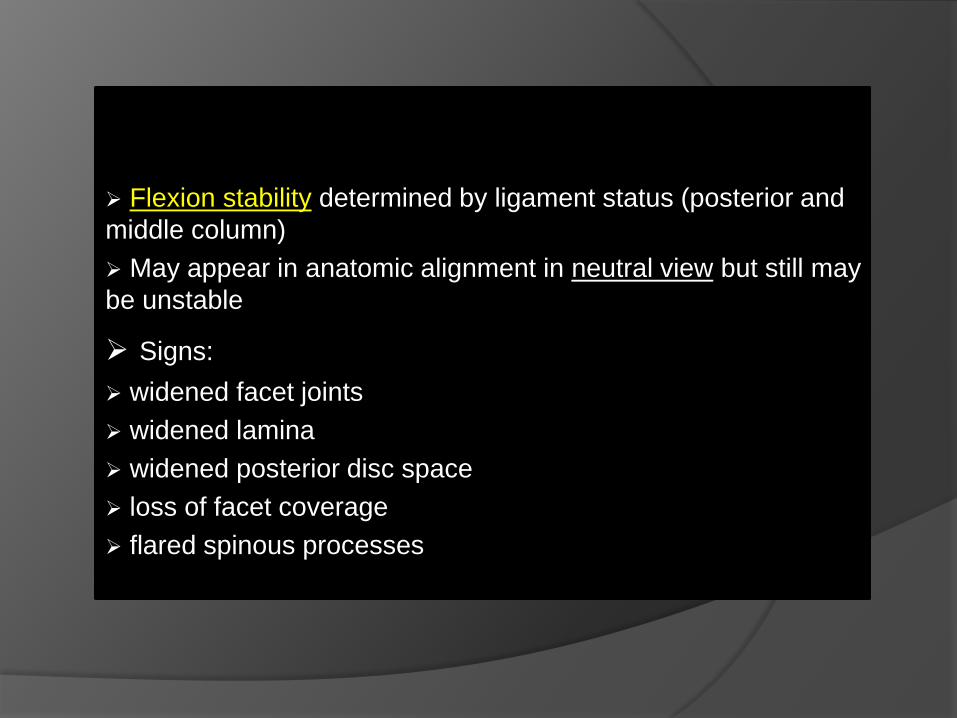
Flexion stability determined by ligament status (posterior and
middle column)
May appear in anatomic alignment in neutral view but still may
be unstable
Signs:
widened facet joints
widened lamina
widened posterior disc space
loss of facet coverage
flared spinous processes

C4-5 Hyperflexion Sprain – Ligament
STABLE?

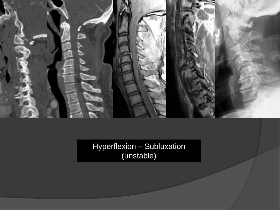
Hyperflexion – Subluxation
(unstable)
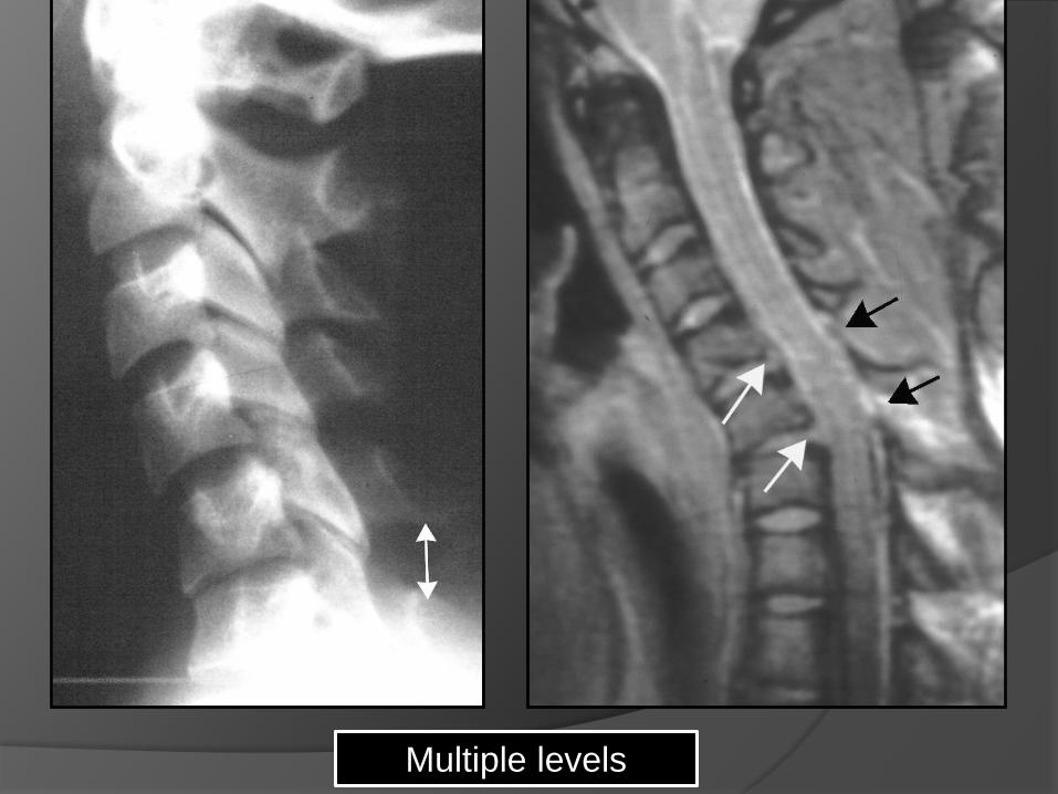
Multiple levels
Hyperflexion – Subluxation
(unstable)
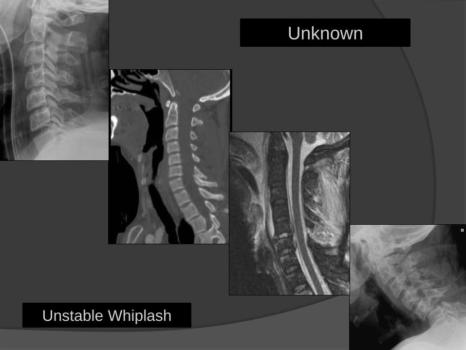
Unknown
Unstable Whiplash


Multi-level distraction injuries

Spinal stenosis
Disc hernation
Anterior distraction
(hyperextension)
Posterior axial
loading
Cord contusion
Unstable

Type 1 Type 2 Type 3
Occipital Condyle Fractures

AOD FINDINGS
Non-congruent arcs Increased basion-dental
distance Increased C1-C2
posterior distance

Basion-Dens
CT > 9.5 mm
Radiograph >12 mm
V-sign
Vertical
atlanto-
dens line
Normal Abnormal
PAL > 5.5 mm
Radiograph >12mm
Occ-C1
C1-C2 > 7.8mm
Normal Abnormal
Condylar sum
> 4.3 mm
Harris JH, Jr., Carson GC, Wagner LK, Kerr N. Radiologic diagnosis of
traumatic occipitovertebral dissociation: 2. Comparison of three methods of
detecting occipitovertebral relationships on lateral radiographs of supine
subjects. AJR Am J Roentgenol 1994;162:887-892.
Chang W, Alexander MT, Mirvis SE. Diagnostic determinants of
craniocervical distraction injury in adults. AJR.2009;192:52-58.

CT Determinants of Craniocervical
Distraction Injury
C1-C2 spinolaminar line > 7.8 mm (p=0.02)
Basion to posterior axial line > 5.5 mm (p=0.0007)
Basion-dental interval > 9.5 mm (p < 0.0001)
Summed condyle to C1 > 4.3 mm (p=0.001)
“V” –shaped atlanto-dental space - trended
Chang W, et al. AJR. 2009;192(1):52-59.

Multi-level Injuries
AOD; C1-C2; tectorial membrane; post
atlanto-occipital lig; atlanto-dental interval;
C5-C6 ligamentum flavum
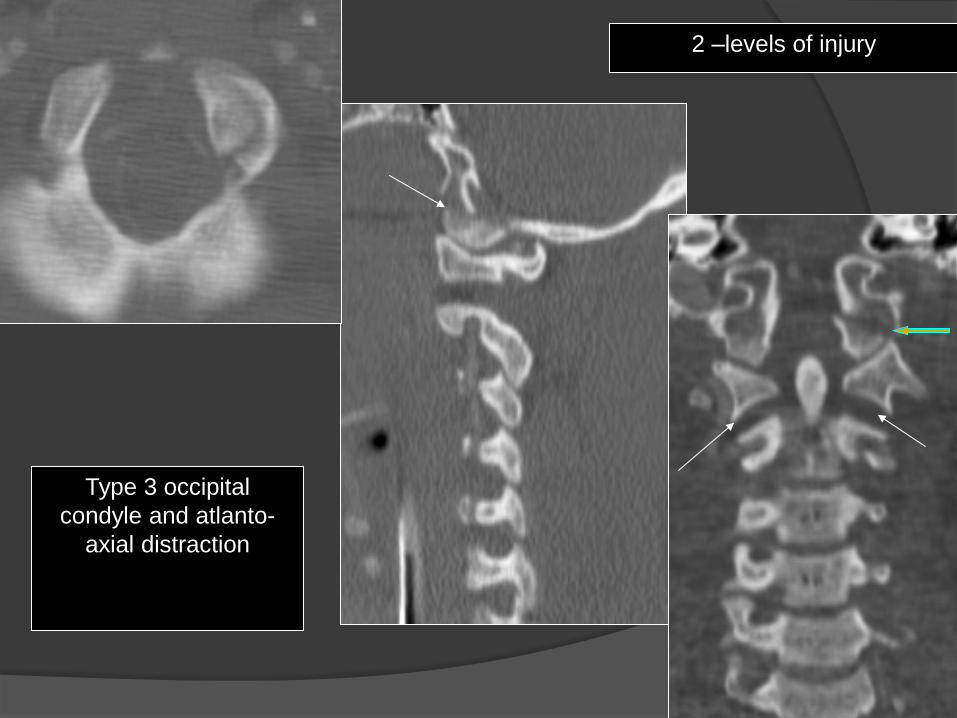
Type 3 occipital
condyle and atlanto-
axial distraction
2 –levels of injury
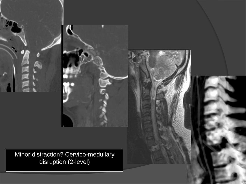
Minor distraction? Cervico-medullary
disruption (2-level)
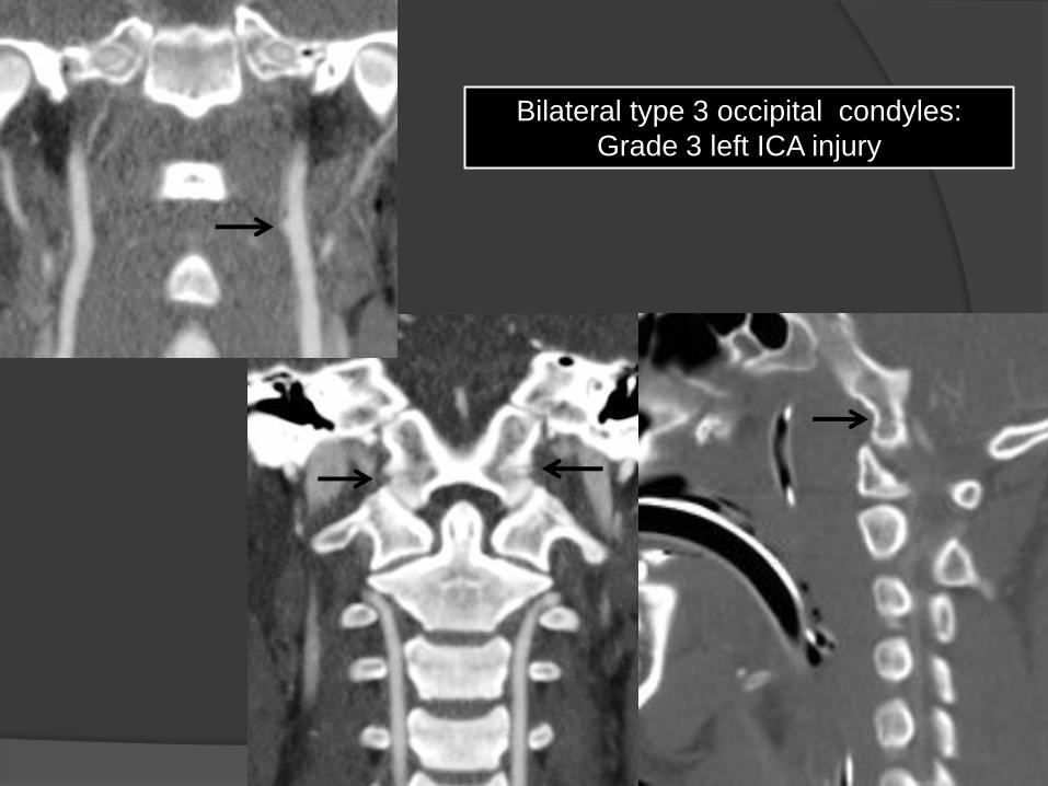
Bilateral type 3 occipital condyles:
Grade 3 left ICA injury

MR Verification
> 7.8 mm
> 9.5 mm
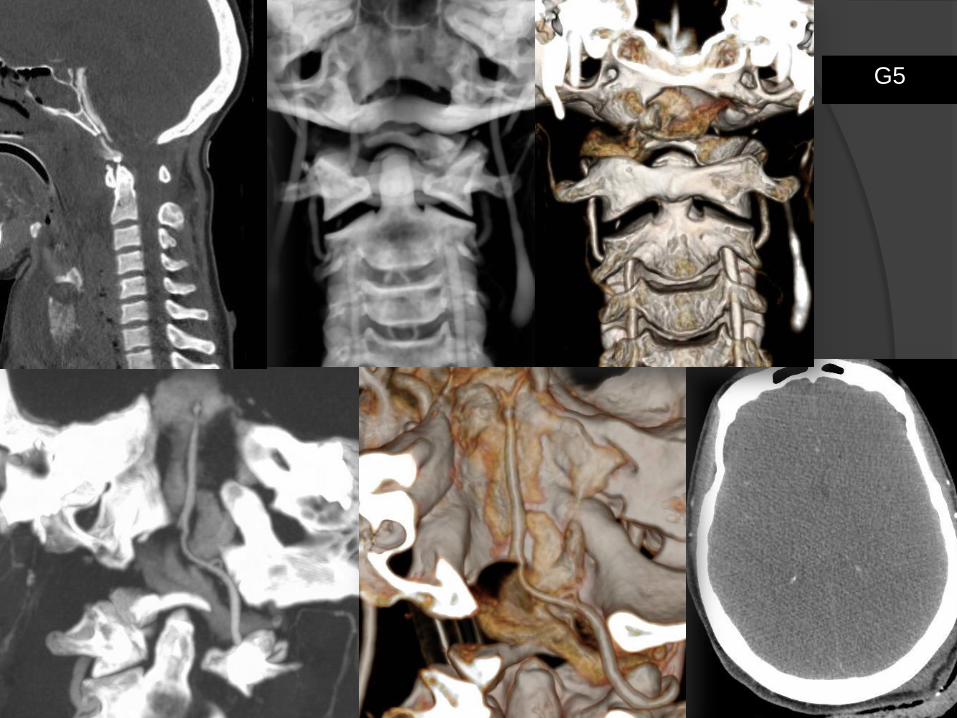
G5
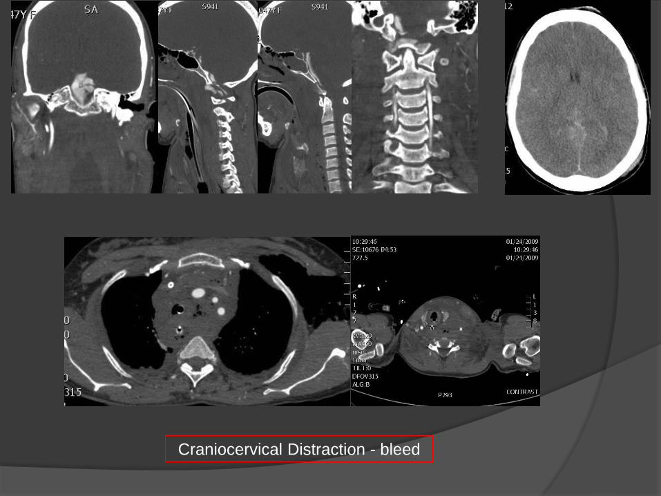
Craniocervical Distraction - bleed

Pre-surgical planning
Assess stability for management (MRI overcalls some ligament injuries)
Neurologic deficit
?? Neck pain with normal exam and high image quality CT
Normal appearing CT with neuro deficit
-herniated disc,
-epidural hematoma,
-traumatic syrinx
Prognostic assistance ? (blood, degree of stenosis, compression?, injury
extent, injury progression)
Detect less apparent concurrent injuries
Assess vessels – especially with selected C-spine injuries (vs. CTA)
?? Obtunded – unexaminable patient with mechanism with normal quality
CT
Trauma MRI Indications
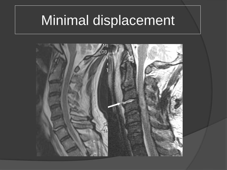
Minimal displacement
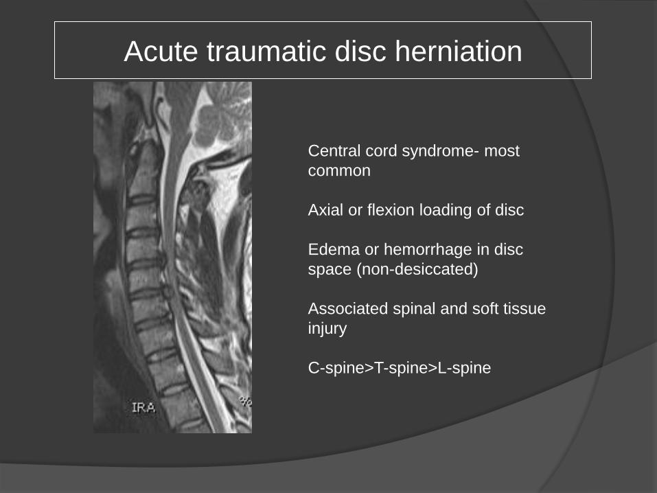
Acute traumatic disc herniation
Central cord syndrome- most
common
Axial or flexion loading of disc
Edema or hemorrhage in disc
space (non-desiccated)
Associated spinal and soft tissue
injury
C-spine>T-spine>L-spine
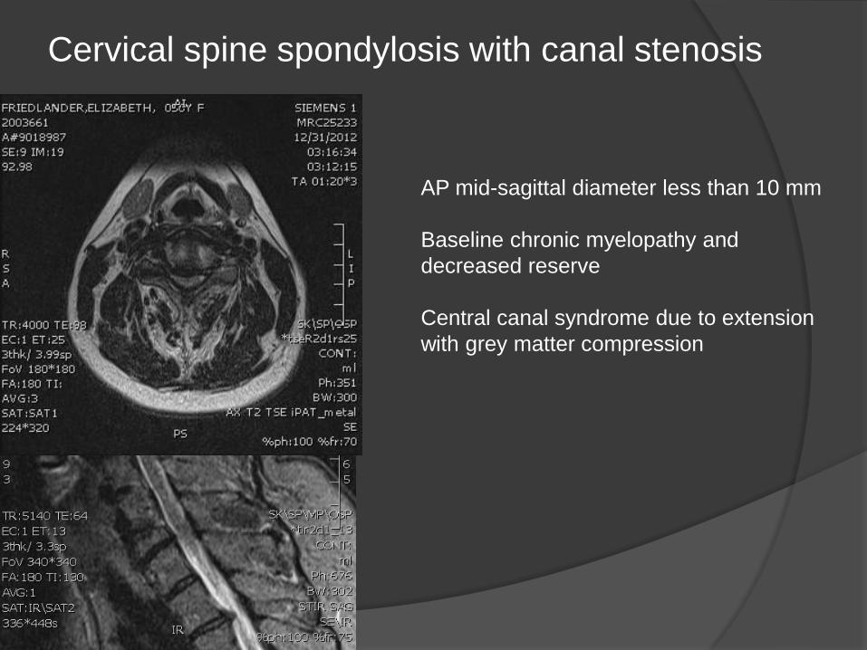
Cervical spine spondylosis with canal stenosis
AP mid-sagittal diameter less than 10 mm
Baseline chronic myelopathy and
decreased reserve
Central canal syndrome due to extension
with grey matter compression
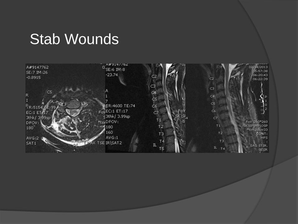
Stab Wounds
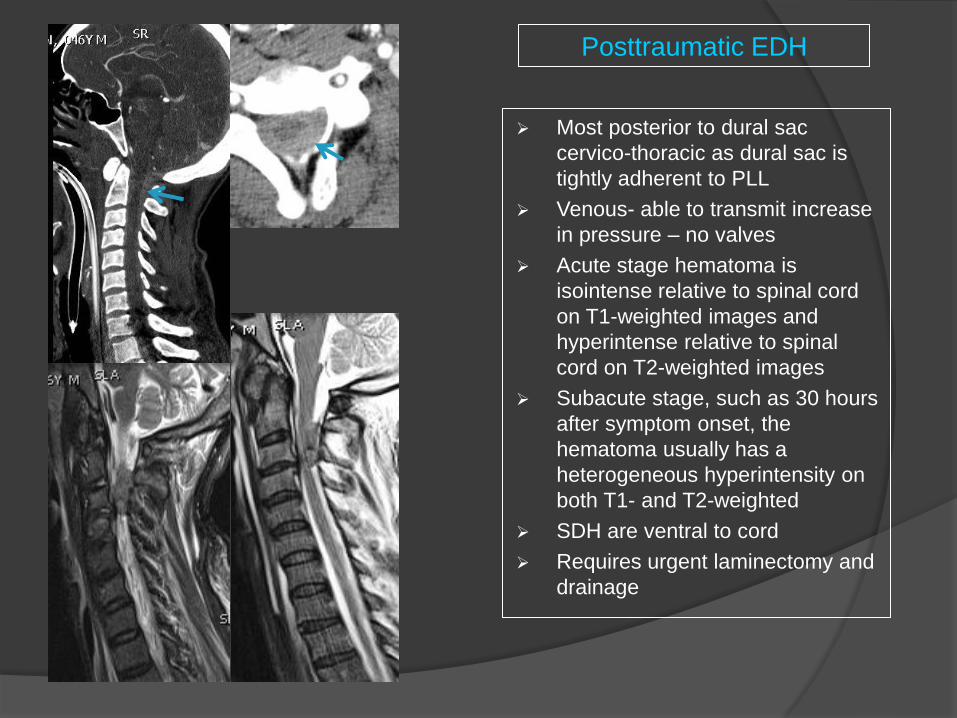
Most posterior to dural sac
cervico-thoracic as dural sac is
tightly adherent to PLL
Venous- able to transmit increase
in pressure – no valves
Acute stage hematoma is
isointense relative to spinal cord
on T1-weighted images and
hyperintense relative to spinal
cord on T2-weighted images
Subacute stage, such as 30 hours
after symptom onset, the
hematoma usually has a
heterogeneous hyperintensity on
both T1- and T2-weighted
SDH are ventral to cord
Requires urgent laminectomy and
drainage
Posttraumatic EDH

Epidural hematomas

Cervical spine trauma and vertebral artery injury

Bullet fragments to neck and cervical spine MRI
17 pts.(3 fragments in canal)
CT before and after MRI to assess for movement
Neurologic exam before and after MRI
No migration or neurologic decline at median 8 weeks
F/U
MRI can be effective to assess neck GSWs without
complications related to metal fragments
Slavin J, et al. Magnetic Resonance Imaging to Evaluate Cervical Cord
Injury from GSWs from Handguns. World Neurosurg. 2015. Epub
ahead of print.
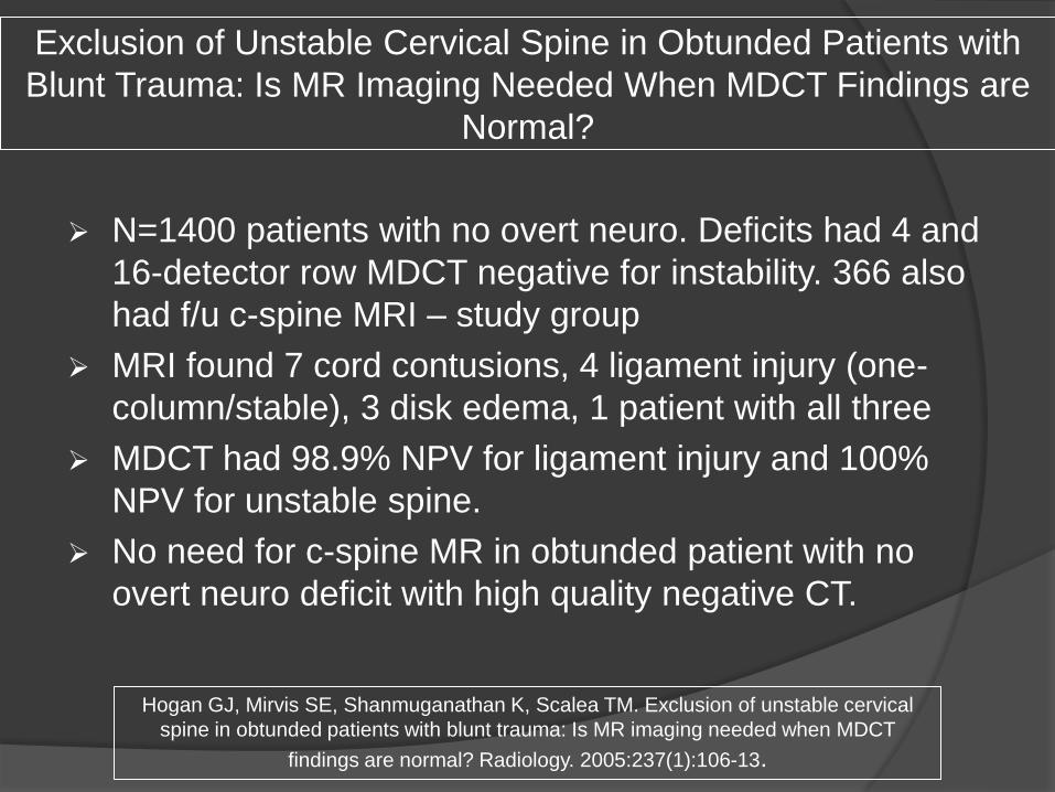
Exclusion of Unstable Cervical Spine in Obtunded Patients with
Blunt Trauma: Is MR Imaging Needed When MDCT Findings are
Normal?
N=1400 patients with no overt neuro. Deficits had 4 and
16-detector row MDCT negative for instability. 366 also
had f/u c-spine MRI – study group
MRI found 7 cord contusions, 4 ligament injury (one-
column/stable), 3 disk edema, 1 patient with all three
MDCT had 98.9% NPV for ligament injury and 100%
NPV for unstable spine.
No need for c-spine MR in obtunded patient with no
overt neuro deficit with high quality negative CT.
Hogan GJ, Mirvis SE, Shanmuganathan K, Scalea TM. Exclusion of unstable cervical
spine in obtunded patients with blunt trauma: Is MR imaging needed when MDCT
findings are normal? Radiology. 2005:237(1):106-13.
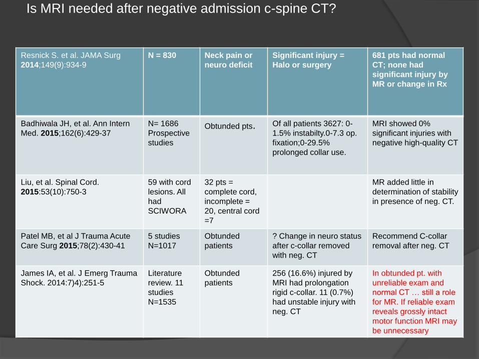
Is MRI needed after negative admission c-spine CT?
Resnick S. et al. JAMA Surg
2014;149(9):934-9
N = 830 Neck pain or
neuro deficit
Significant injury =
Halo or surgery
681 pts had normal
CT; none had
significant injury by
MR or change in Rx
Badhiwala JH, et al. Ann Intern
Med. 2015;162(6):429-37
N= 1686
Prospective
studies
Obtunded pts. Of all patients 3627: 0-
1.5% instabilty.0-7.3 op.
fixation;0-29.5%
prolonged collar use.
MRI showed 0%
significant injuries with
negative high-quality CT
Liu, et al. Spinal Cord.
2015:53(10):750-3
59 with cord
lesions. All
had
SCIWORA
32 pts =
complete cord,
incomplete =
20, central cord
=7
MR added little in
determination of stability
in presence of neg. CT.
Patel MB, et al J Trauma Acute
Care Surg 2015;78(2):430-41
5 studies
N=1017
Obtunded
patients
? Change in neuro status
after c-collar removed
with neg. CT
Recommend C-collar
removal after neg. CT
James IA, et al. J Emerg Trauma
Shock. 2014:7)4):251-5
Literature
review. 11
studies
N=1535
Obtunded
patients
256 (16.6%) injured by
MRI had prolongation
rigid c-collar. 11 (0.7%)
had unstable injury with
neg. CT
In obtunded pt. with
unreliable exam and
normal CT … still a role
for MR. If reliable exam
reveals grossly intact
motor function MRI may
be unnecessary

Is MRI needed after high quality negative admission CT?
Raza, M, et al. Injury
2013;44(11):1589-95 N=1850
(meta-
analysis)
Altered
sensorium
(clear C-spine
Prolonged follow-up
or MRI
NPV (CT) =99.7%
PPV 93.7%; CT
rules out clinically
significant injury.
Perform MR on
case-to-case basis
Mavos MN, et al. World J
Surg. 2015;39(11):2685-90 N=383 -GCS=15
-C-spine
tenderness
without neuro
signs
-Negative CT
Follow-up physical
exam, MRI (36) and or
Flex-ext. films (19)
No neuro.signs after
collar removal. Can
withdraw CT
precautions in pts.
with neck pain but
neg. high quality CT
Pancyzkowski DM, et al. J
Neurosurg.
2011;115(3):541-9
N=14,327
(meta-
analysis) in
17 studies
Positive is injury
requiring
orthotic or
surgical
stabilization
Imaging or clinical f/u
for unstable C-spine
Negative likelihood
of unstable c-spine
with neg. CT
<.0001. NPV of
normal CT= 100%
Tan LA, et al. Clin Neurol
Neurosurg 2014;120:23-6
N=83 GCS 14 or less
= obtunded.
Not high impact
CT and MR to clear c-
spine
CT & MRI + 34%
4 – CT with + MR
(all stable had
decompression)
CT & MRI – in 61%
all collars safely
removed

Intramedullary Lesion Expansion
on MRI with Complete Cervical Cord Deficit
42 patients with ASCI had 2 consecutive MR scans (1st MR ave. 7 hr, 2nd ave. 55 hr)
Rostrocaudal length of cord lesion went from on ave. from 60mm on first to 89mm on second MRI.
Intramedullary length was assoc. with time from injury to 1st MRI (p=0.05) and time to decompression (p=0.03)
Factors influencing expansion rate (0.9 +/- .8 mm/hr) were the maximum amount of cord compression (p=0.03) and mechanism of injury (p=0.05).
Aarabi B, et al. Intramedullary lesion expansion on MRI in
patients with motor complete cervical spinal cord injury. J
Neurosurg Spine. 2012;17(3): 243-50.
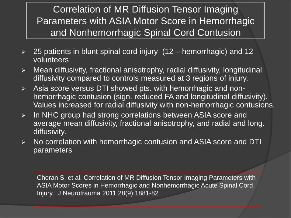
Correlation of MR Diffusion Tensor Imaging
Parameters with ASIA Motor Score in Hemorrhagic
and Nonhemorrhagic Spinal Cord Contusion
25 patients in blunt spinal cord injury (12 – hemorrhagic) and 12 volunteers
Mean diffusivity, fractional anisotrophy, radial diffusivity, longitudinal diffusivity compared to controls measured at 3 regions of injury.
Asia score versus DTI showed pts. with hemorrhagic and non-hemorrhagic contusion (sign. reduced FA and longitudinal diffusivity). Values increased for radial diffusivity with non-hemorrhagic contusions.
In NHC group had strong correlations between ASIA score and average mean diffusivity, fractional anisotrophy, and radial and long. diffusivity.
No correlation with hemorrhagic contusion and ASIA score and DTI parameters
Cheran S, et al. Correlation of MR Diffusion Tensor Imaging Parameters with
ASIA Motor Scores in Hemorrhagic and Nonhemorrhagic Acute Spinal Cord
Injury. J Neurotrauma 2011:28(9):1881-82

Thank you for your attention !