Embed Size (px)
Citation preview

20160820 www.medscape.com/viewarticle/555355_print
http://www.medscape.com/viewarticle/555355_print 1/17
www.medscape.com
Abstract and Introduction
Study Design: Literature review.Objectives: The Spine Trauma Study Group compiled a collection of clinically useful imaging methods used in uppercervical spine trauma and standardized how these measurements are documented.Summary of Background Data: Imaging of the upper cervical spine is crucial for injury detection, description, andtreatment decision making. However, a standard set of imaging measurement techniques for this region does not exist.While most clinicians have developed their own methods of describing radiographic pathology, this variability often leads toconfusion in developing an agreed on classification system and in proposing universal treatment recommendations.Methods: The available literature concerning measurement of injury characteristics after upper cervical trauma wasreviewed. Consensus of the most clinically applicable measurement methods among the surgeon members of the SpineTrauma Study Group was achieved.Results: The techniques include: the basiondens and basionposterior axial line intervals (C0C2); fracture gap andfracture length apposition (a reflection of fragment size) for occipital condyle injuries; lateral articular overhang for C1 ringfractures; the atlantodens and posterior atlantodens intervals for sagittal C1C2 instability; odontoid fracture angulationand displacement; and C2C3 angulation and translation for traumatic spondylolisthesis of the axis.Conclusions: Only through prospective study using a standardized and uniform set of measurement techniques can theclinical significance of these imaging characteristics be fully appreciated.
Imaging examination of the upper cervical spine following trauma is crucial for injury detection and description, especially inlight of the potentially dire neurologic consequences of a missed bone or discoligamentous injury.[14] Because of therelatively low incidence of occipital cervical trauma, clinicians are often not well versed in the normal spatial relationshipsbetween the occiput, atlas, and axis. While surgical treatment for survivors of occipitocervical dissociation is almostuniversally accepted,[58] the optimal radiologic method of detecting this often subtle injury is not clear.[813] Furtherinconsistencies and paradoxes are illustrated by 2 common fractures, in which treatment is dependant on accurateradiographic measurements. The classification of traumatic C2 spondylolisthesis relies on the detection of angulation andtranslation, with accepted methods of measurement of these parameters described.[14] In contrast, measurement methodsof displacement and angulation of odontoid fractures have not been universally established.[15]
Possibly because of familiarity with the techniques, many clinicians have applied commonly recommended measurementtools described for instability in patients with rheumatoid arthritis to the setting of upper cervical spine trauma. Thesemeasurements, although accurate in defining relationships between anatomic structures, are often difficult to use in thetraumatic setting because of their complexity and difficulty with visualizing pertinent anatomic landmarks.
It is the purpose of this paper to present a systematic review of various radiographic measurements in order to developuniversal standards for the assessment (measurement) of traumatic upper cervical spine injuries. Furthermore, it is hopedthat standardized radiographic methods will lead to more universal acceptance and use among clinicians, leading to betterpatient care and improved storage, retrieval, and analysis of relevant data, thus facilitating multicenter studies and/ormetaanalysis.
Materials and Methods
The available literature concerning measurement of injury characteristics after upper cervical trauma was reviewed. Thisincluded computerbased searches using PubMed[16] as well as available textbooks. Consensus of the most clinicallyapplicable measurement methods among the surgeon members of the Spine Trauma Study Group (STSG) was achievedthrough discussion, debate, and resolution.
A taskoriented subcommittee that was comprised of the authors of this paper developed a narrative with a pictorial,describing in detail how the measurements should be performed, for each chosen method. These were again presented to
Measurement Techniques for Upper Cervical SpineInjuries: Consensus Statement of the Spine TraumaStudy GroupChristopher M. Bono, MD; Alexander R. Vaccaro, MD; Michael Fehlings, MD; Charles Fisher, MD;Marcel Dvorak, MD; Steven Ludwig, MD; James Harrop, MDSpine. 2007;32(5):593600.

20160820 www.medscape.com/viewarticle/555355_print
http://www.medscape.com/viewarticle/555355_print 2/17
all of the members of the STSG for final discussion, modification, and approval.
The STSG performed the current study. The group was formed in early 2002 and had its first official meeting in October2002. Its purpose of formation was to plan multicenter studies concerning spinal trauma. The international membershiprepresents many of the busiest spine trauma centers in the world, including those in the United States, Canada, Mexico,Sweden, France, Italy, India, Belgium, and Turkey. All of the nearly 50 members are either an orthopedic surgeon orneurosurgeon that specializes in spine surgery. Participants were selected based on their extensive clinical experienceand/or established reputations as spine trauma researchers.
The STSG meets as a whole twice a year. The taskoriented committee in charge of the current study had met on 2separate occasions. The time line of these meetings was:
1. An initial STSG meeting at which time the study goal was outlined, and 1 of the authors (C.M.B.) was assigned tobe principal investigator.
2. A subcommittee meeting that focused on the detailed review of the literature and available measurementtechniques. From these techniques, the subcommittee members selected those techniques that had the greatestsupporting evidence or perceived clinical relevance.
3. One of the authors (C.M.B.) then prepared a slide presentation of narratives and pictorials of each technique beforethe next STSG meeting and distributed it to all of the STSG members.
4. At the next STSG meeting, the group as a whole presented, reviewed, and critiqued each measurement technique.During this meeting, individual members had the opportunity to suggest modifications/deletions/additions to theselected measurement techniques. One of the authors (C.M.B.) acted as a mediator for this discussion. A show ofhands assessed consensus.
5. The members of the subcommittee prepared and reviewed a manuscript with illustrations at a subsequent meeting.With its approval, the manuscript was forwarded to all members of the STSG.
6. At the latest STSG meeting, slides of the narratives and pictorials of the selected measurement techniques werepresented. One of the authors (C.M.B.) arbitrated final suggestions of modifications to these techniques. A show ofhands assessed consensus. These modifications were then incorporated into a final manuscript text and figures.
Results
There are several methods to describe the spatial relationship between the occiput, atlas, and axis.[11] The most popular inthe setting of trauma are the basiondental interval (BDI) and the basionposterior axial line interval (BAI). Originallydescribed by Harris et al,[9,10] the BDI and BAI have been carefully examined (Figure 1). In the first of 2 companionstudies, Harris et al[10] measured the BAI and BDI on lateral radiographs of 400 normal adults. The BAI and BDI did notexceed 12 mm in 98% and 95% of adults, respectively. The BDI and BAI have come to be known as Harris Measurements,and more descriptively as the Rule of Twelve. Deliganis et al[12] recommended use of the BDI and the BAI to help detectoccipitocervical dissociations. Likewise, Fisher et al[8] found these to be the most useful radiographic parameters for theseinjuries.

20160820 www.medscape.com/viewarticle/555355_print
http://www.medscape.com/viewarticle/555355_print 3/17
Measurement technique for the BDI and BAIs.
In their second study, the investigators retrospectively reassessed lateral radiographs of 37 patients in whomoccipitoatlantal dissociation had been previously diagnosed at the time of admission.[9] They found the BDI and BAI to begreater than 12 mm in 23 patients (group 1) with frank occipitoatlantal dislocation and 8 (group 2) with occipitoatlantalsubluxation/dissociation. Measurements were less than 12 mm for 6 patients who initially had suspected instability but didnot have supportive clinical findings. In the same patients, the Power ratio (Figure 2) and Lee X line method (Figure 3)could not be measured in 17 of 37 cases because either the opisthion could not be seen or the posterior C1 arch had adevelopmental anomaly (i.e., not fused). In the remaining patients, these methods enabled detection of only 60% and 20%of injuries, respectively. While these data strongly suggest that BDI/BAI are superior to the Power ratio and Lee X lines,the interobserver and intraobserver reproducibility of these measurements has not been assessed to the authors'knowledge.

20160820 www.medscape.com/viewarticle/555355_print
http://www.medscape.com/viewarticle/555355_print 4/17
Measurement technique for the Power ratio.

20160820 www.medscape.com/viewarticle/555355_print
http://www.medscape.com/viewarticle/555355_print 5/17
Measurement of Lee's lines.
Other lines and measurements have also been described for the craniocervical junction. The Power ratio (Figure 2)describes the relationship between the occiput and C1 through a ratio of the distance between the basion and C1 posteriorarch divided by the distance between the opisthion and anterior C1 arch. Ahuja et al[11] found lower Power ratios in 5surviving patients compared to a higher ratio seen in 1 fatal case of occipitoatlantal dislocation.
Most other descriptive measurements of the occipitocervical junction were originally developed to assess rheumatoidpatients with basilar invagination (cranial settling). Thus, the critical pathologic values are usually seen in the lower, ratherthan upper limits of normal due to the process of settling rather than distraction. These measurements, although possiblyuseful for some injuries, were not intended to detect widening or translation of the occipitocervical junction. They include:(1) the Chamberlain line, drawn from the hard palate to the tip of the opisthion (Figure 4); and (2) McCrae's line, drawnfrom the basion to the opisthion, representing the anteroposterior width of the foramen magnum (Figure 5).
The Chamberlain line.
20160820 www.medscape.com/viewarticle/555355_print
http://www.medscape.com/viewarticle/555355_print 6/17
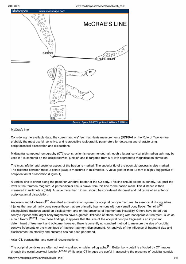
McCrae's line.
Considering the available data, the current authors' feel that Harris measurements (BDI/BAI or the Rule of Twelve) areprobably the most useful, sensitive, and reproducible radiographic parameters for detecting and characterizingoccipitocervical dissociation and dislocations.
Midsagittal computed tomography (CT) reconstruction is recommended, although a lateral cervical plain radiograph may beused if it is centered on the occipitocervical junction and is targeted from 6 ft with appropriate magnification correction.
The most inferior and posterior aspect of the basion is marked. The superior tip of the odontoid process is also marked.The distance between these 2 points (BDI) is measured in millimeters. A value greater than 12 mm is highly suggestive ofoccipitoatlantal dissociation (Figure 1).
A vertical line is drawn along the posterior vertebral border of the C2 body. This line should extend superiorly, just past thelevel of the foramen magnum. A perpendicular line is drawn from this line to the basion mark. This distance is thenmeasured in millimeters (BAI). A value more than 12 mm should be considered abnormal and indicative of an anterioroccipitoatlantal dissociation.
Anderson and Montesano[17] described a classification system for occipital condyle fractures. In essence, it distinguishesinjuries that are primarily bony versus those that are primarily ligamentous with only small bony flecks. Tuli et al[18]
distinguished fractures based on displacement and on the presence of ligamentous instability. Others have noted thatcondyle injuries with larger bony fragments have a greater likelihood of stable healing with nonoperative treatment, such asa halo fixator.[19,20] From these findings, it appears that the size of the occipital condyle fragment is an importantdeterminant of treatment and outcome; however, there is currently no standard method to measure the size of occipitalcondyle fragments or the magnitude of fracture fragment displacement. An analysis of the influence of fragment size anddisplacement on stability and outcome has not been performed.
Axial CT, parasagittal, and coronal reconstructions.
The occipital condyles are often not well visualized on plain radiographs.[21] Better bony detail is afforded by CT imagesthrough the occipitocervical junction.[20,22] While axial CT images are useful in assessing the presence of occipital condyle

20160820 www.medscape.com/viewarticle/555355_print
http://www.medscape.com/viewarticle/555355_print 7/17
fractures, they do not usually enable visualization of the entire condyle in 1 image. For these reasons, the authors suggestthat a parasagittal and coronal CT reconstruction through the approximate midaspect of the condyle be used. As principlesof fracture healing dictate, the area of apposed fracture surfaces can influence the potential for fracture healing. Thus, asimilar quantification of the apposed contacting surfaces of occipital condyle fracture fragments is proposed. In regards todisplacement, greater amounts imply greater concomitant soft tissue disruption and instability. The ability to reduce thefractured fragments would also influence healing potential.
Because actual detailed quantitative measurements would be exceedingly difficult, the length of the fracture line ismeasured in millimeters on the midsagittal and coronal condyle images (Figure 6). The coronal and sagittal measurementsare added to derive a total amount of bony apposition for each condyle. For shelltype avulsion fractures that leave a rim ofsubarticular cortical bone juxtaposed to the upper C1 articular process, the length of apposed fracture surface would beessentially complete. This would suggest that the fracture would have a high likelihood of healing. The same would be truefor a fracture through the junction of the condyle and occiput, with minimal displacement. In contrast, a primarilyligamentous injury with a small bony fleck would have a short measured distance of apposed bone and a presumed lowerrate of healing. Future prospective evaluations using this measurement technique and its relationship to stability andhealing rates need to be performed to support or disqualify the current belief that fragment size is a prognosticallyimportant factor.
Technique for measuring displacement (gap) and apposition of occipital condyle fracture fragments. It is suggested thatinjuries with small fracture fragments (flecks) are primarily ligamentous that may have less healing potential than broadfracture surfaces that are well aligned and minimally gapped.
On coronal and parasagittal reconstructed images, the maximal gap between the fractured fragments are measured inmillimeters (Figure 6).
Lee et al[23] used an openmouth view to measure displacement of C1 lateral mass fractures. Most authors seem to agreeon this method of assessment. Spence determined that 6.9 mm is the critical amount of total displacement necessary todisrupt the transverse ligament. This measurement was derived by direct cadaveric measurements, however. Heller et al[24]
warned of overestimating the amount of displacement based on plain films. They determined that the transverse ligamentshould be considered intact in patients with less than 8.1mm total displacement as measured on a plain openmouthradiograph. This consideration may be obviated using calibrated axial and coronally reconstructed CT images.
Coronal CTreformatted images through the center of the lateral masses of the atlas.
Vertical lines are drawn along the most lateral aspect of the bone of the C1 and C2 articular processes (Figure 7). Thetransverse distance between them is then measured in millimeters. These are then added to calculate the total lateral mass

20160820 www.medscape.com/viewarticle/555355_print
http://www.medscape.com/viewarticle/555355_print 8/17
displacement.
Measurement technique for lateral mass overhang.
While the atlantodens interval (ADI) and posterior atlantodens interval (PADI) are widely used to detect traumatic cervicalinstability, the authors could find no article assessing their reliability in the setting of acute injury. Studying the flexionextension radiographs of 72 patients with Down syndrome, Wellborn et al[13] found ADI to have statistically significantintraobserver agreement in 2 of 3 observers. Although the interobserver agreement was considered fair, it was statisticallybetter than measurements using the Power ratio. Supportively, Cremers et al[25] found the ADI to be reliable in detectinginstability in 279 children with Down syndrome. Intraobserver and interobserver agreement was better using flexion versusthe neutral radiograph. The PADI represents the anteroposterior diameter of the spinal canal at this level. It has beenshown to be a more useful prognosticator in rheumatoid arthritis patients than the ADI. However, it has not been validatedin the same manner for traumatic atlantoaxial instability. With traumatic atlantoaxial instability being relatively uncommonand the aforementioned measurements having undergone significant epidemiological rigor for other pathology, the authorspropose the use of the ADI and PADI as described below.
Lateral cervical radiograph or midsagittal CT reconstruction.
The craniocaudal midpoint of the anterior ring of C1 is marked. A line parallel to the ring of C1 is drawn from this pointtoward the odontoid process. The distance between the C1 mark and intersection with the anterior aspect of the odontoidprocess is measured in millimeters (Figure 8).

20160820 www.medscape.com/viewarticle/555355_print
http://www.medscape.com/viewarticle/555355_print 9/17
The ADI and PADI.
Rotation between the C1 and C2 rings can occur by itself or in combination with a sagittal translational deformity. Mostreports of atlantoaxial rotational instability are in children, commonly associated with nontraumatic upper pharyngealinfections. Most recommendations concerning optimal measurement of atlantoaxial rotation are derived from this body ofliterature.[2628] Most authors agree that CT is the imaging modality of choice for detection and quantification of rotationaldeformity,[26,27,29] though its reliability and accuracy for detecting dynamically unstable joints has been questioned.[30] Inthe authors' experience, dynamic CT is rarely indicated (and can be dangerous) in the adult patient with traumaticinstability.
Transaxial CT images through mid body of C2 and mid body of C1.
For optimal measuring, the CT gantry angle should be aligned along the transverse plane of the upper cervical vertebrae.An axial CT slice at the level of the C2 body and C1 ring are then obtained. An anteroposterior line is drawn from themidpoint of the C2 body to the center of the spinous process. A perpendicular to this line is drawn along the posterior C2vertebral body. On the best axial slice of C1, the midpoints of the transverse (vertebral artery) foramens are marked, and aline is drawn between them. The angulation between these 2 lines represents the degree of static atlantoaxial rotatorydeformity (subluxation) (Figure 9). By convention, the side (right or left) toward which the atlas (head) points is consideredthe side of the rotation.

20160820 www.medscape.com/viewarticle/555355_print
http://www.medscape.com/viewarticle/555355_print 10/17
Measurement of right (R) (A) and left (L) (B) atlantoaxial rotation.

20160820 www.medscape.com/viewarticle/555355_print
http://www.medscape.com/viewarticle/555355_print 11/17
Odontoid fracture displacement and angulation are known to be important prognostic factors of fracture healing. However,in the authors' review of the literature, there are no assessments of the optimal method for measuring these parameters.Carlson et al[15] measured displacement by drawing lines along the anterior aspect of the dens fragment and the intactcaudal body of C2. The angle subtended by these lines would be the degree of fracture angulation. The location of theapex of fracture angulation would be described as anterior or posterior.
Lateral cervical radiograph or midsagittal CT reconstruction.
A tangent line is drawn along the anterior aspect of the odontoid fragment and the anterior aspect of the C2 body. At thelevel of the fracture, a transverse line is drawn connecting these 2 lines. This distance is measured in millimeters andrepresents sagittal fracture displacement (Figure 10).
Odontoid fracture displacement.
A tangent line is drawn along the posterior aspect of the odontoid fragment and the posterior aspect of the C2 body. Theangle subtended by these lines would be the degree of fracture angulation. The location of the fracture apex angulationwould be used for the descriptor anterior or posterior (Figure 11).

20160820 www.medscape.com/viewarticle/555355_print
http://www.medscape.com/viewarticle/555355_print 12/17
Odontoid fracture angulation.
Levine and Edwards[14] described a method of measuring both angulation and translation of C2 with traumaticspondylolisthesis (Hangman's fractures).
Lateral cervical radiograph or midsagittal CT reconstruction.
Endplate method. As depicted in Figure 12, lines are drawn perpendicular to the inferior endplate of C2 and C3 to measureangulation.

20160820 www.medscape.com/viewarticle/555355_print
http://www.medscape.com/viewarticle/555355_print 13/17
C2C3 angulation measurement using the endplate method.
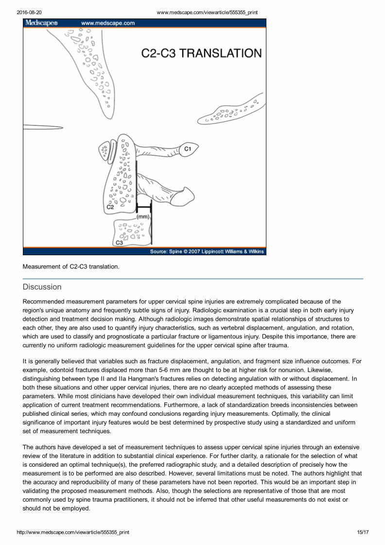
Similarly, posterior vertebral body lines are drawn along the posterior aspect of C2 and C2 to enable measurement ofangulation (Figure 13) and displacement (Figure 14). Both of these parameters are important in classification and treatmentdecision making for this injury.

20160820 www.medscape.com/viewarticle/555355_print
http://www.medscape.com/viewarticle/555355_print 14/17
C2C3 angulation measurement using the posterior vertebral body line method.
20160820 www.medscape.com/viewarticle/555355_print
http://www.medscape.com/viewarticle/555355_print 15/17
Measurement of C2C3 translation.
Discussion
Recommended measurement parameters for upper cervical spine injuries are extremely complicated because of theregion's unique anatomy and frequently subtle signs of injury. Radiologic examination is a crucial step in both early injurydetection and treatment decision making. Although radiologic images demonstrate spatial relationships of structures toeach other, they are also used to quantify injury characteristics, such as vertebral displacement, angulation, and rotation,which are used to classify and prognosticate a particular fracture or ligamentous injury. Despite this importance, there arecurrently no uniform radiologic measurement guidelines for the upper cervical spine after trauma.
It is generally believed that variables such as fracture displacement, angulation, and fragment size influence outcomes. Forexample, odontoid fractures displaced more than 56 mm are thought to be at higher risk for nonunion. Likewise,distinguishing between type II and IIa Hangman's fractures relies on detecting angulation with or without displacement. Inboth these situations and other upper cervical injuries, there are no clearly accepted methods of assessing theseparameters. While most clinicians have developed their own individual measurement techniques, this variability can limitapplication of current treatment recommendations. Furthermore, a lack of standardization breeds inconsistencies betweenpublished clinical series, which may confound conclusions regarding injury measurements. Optimally, the clinicalsignificance of important injury features would be best determined by prospective study using a standardized and uniformset of measurement techniques.
The authors have developed a set of measurement techniques to assess upper cervical spine injuries through an extensivereview of the literature in addition to substantial clinical experience. For further clarity, a rationale for the selection of whatis considered an optimal technique(s), the preferred radiographic study, and a detailed description of precisely how themeasurement is to be performed are also described. However, several limitations must be noted. The authors highlight thatthe accuracy and reproducibility of many of these parameters have not been reported. This would be an important step invalidating the proposed measurement methods. Also, though the selections are representative of those that are mostcommonly used by spine trauma practitioners, it should not be inferred that other useful measurements do not exist orshould not be employed.

20160820 www.medscape.com/viewarticle/555355_print
http://www.medscape.com/viewarticle/555355_print 16/17
Sidebar: Key Points
Measurements of upper cervical spine injury characteristics are often used to determine optimal treatment.
A comprehensive set of radiographic measurement techniques for upper cervical spine injuries has been presentedin the hope of standardizing the methods by which injuries are described and evaluated.
Only through prospective study using a standardized and uniform set of measurement techniques can the clinicalsignificance of these imaging characteristics be fully appreciated.
References
1. Harrison DE, Harrison DD, Cailliet R, et al. Cobb method or Harrison posterior tangent method: Which to choose forlateral cervical radiographic analysis. Spine 2000;25:20728.
2. Troyanovich SJ, Stroink AR, Kattner KA, et al. Does anterior plating maintain cervical lordosis versus conventionalfusion techniques? A retrospective analysis of patients receiving singlelevel fusions. J Spinal Disord Tech2002;15:6974.
3. Loder RT. The sagittal profile of the cervical and lumbosacral spine in Scheuermann thoracic kyphosis. J SpinalDisord 2001;14:22631.
4. Hilibrand AS, Tannenbaum DA, Graziano GP, et al. The sagittal alignment of the cervical spine in adolescentidiopathic scoliosis. J Pediatr Orthop 1995;15:62732.
5. Montane I, Eismont FJ, Green BA. Traumatic occipitoatlantal dislocation. Spine 1991;16:1126.
6. Dickman CA, Papadopoulos SM, Sonntag VK, et al. Traumatic occipitoatlantal dislocation. J Spinal Disord1993;6:30013.
7. Saehueng S, Phuenpathom N. Traumatic occipitoatlantal dislocation. Surg Neurol 2001;55:3540.
8. Fisher CG, Sun JC, Dvorak M. Recognition and management of atlantooccipital dislocation: Improving survival froman often fatal condition. Can J Surg 2001;44:41220.
9. Harris JH, Carson GC, Wagner LK, et al. Radiologic diagnosis of traumatic occipitovertebral dissociation: 2.Comparison of three methods of detecting occipitovertebral relationships on lateral radiographs of supine subjects.AJR Am J Roentgenol 1994;162:88792.
10. Harris JH, Carson GC, Wagner LK. Radiologic diagnosis of traumatic occipitovertebral dissociation: 1. Normaloccipitovertebral relationship on lateral radiographs of supine subjects. AJR Am J Roentgenol 1994;162:8816.
11. Ahuja A, Glasauer FE, Alker CJ, et al. Radiology in survivors of traumatic atlantooccipital dislocation. Surg Neurol1994;41:1128.
12. Deliganis AV, Baxter AB, Hanson JA, et al. Radiologic spectrum of craniocervical distraction injuries. Radiographics2000;20:S23750.
13. Wellborn CC, Sturm PF, Hatch RS, et al. Intraobserver reproducibility and interobserver reliability of cervical spinemeasurements. J Pediatr Orthop 2000;20:6670.
14. Levine AM, Edwards C. The management of traumatic spondylolisthesis of the axis. J Bone Joint Surg Am1985;67:21726.
15. Carlson GD, Heller JG, Abitbol JJ, et al. Odontoid fractures. In: Levine AM, Eismont FJ, Garfin SR, et al., eds.Spine Trauma. Philadelphia, PA: Saunders; 1998:22748.
16. PubMed. Available at: http://www.ncbi.nlm.nih.gov/PubMed. Accessed January 4, 2007.

20160820 www.medscape.com/viewarticle/555355_print
http://www.medscape.com/viewarticle/555355_print 17/17
Funding information
Corporate/Industry funds were received in support of this work. No benifits in any form have been or will be received from acommercial party related directly or indirectly to the subject of this manuscript.
Reprint Address
Christopher M. Bono, MD, Harvard Medical School, Brigham and Women's Hospital, 75 Francis Street, Department ofOrthopedic Surgery, Boston, MA 02115; Email: [email protected]
Spine. 2007;32(5):593600. © 2007 Lippincott Williams & Wilkins
The manuscript submitted does not contain information about medical device(s)/drug(s).
This website uses cookies to deliver its services as described in our Cookie Policy. By using this website, you agree to the use ofcookies.close
17. Anderson PA, Montesano PX. Morphology and treatment of occipital condyle fractures. Spine 1988;13:7316.
18. Tuli S, Tator CH, Fehlings MG, et al. Occipital condyle fractures. Neurosurgery 1997;41:3767.
19. Hanson JA, Deliganis AV, Baxter AB, et al. Radiologic and clinical spectrum of occipital condyle fractures:Retrospective review of 107 consecutive fractures in 95 patients. AJR Am J Roentgenol 2002;178:12618.
20. Young WF, Rosenwasser RH, Getch C, et al. Diagnosis and management of occipital condyle fractures.Neurosurgery 1994;34:25760.
21. Emery E, Saillant G, Ismail M, et al. Fracture of the occipital condyle: Case report and review of the literature. EurSpine J 1995;4:1913.
22. Savoilane ER, Ebraheim NA, Jackson WT, et al. Threedimensional computed tomography in evaluation of occipitalcondyle fracture. J Orthop Trauma 1989;3:715.
23. Lee TT, Green BA, Petrin DR. Treatment of stable burst fracture of the atlas (Jefferson fracture) with rigid cervicalcollar. Spine 1998;23:19637.
24. Heller JG, Viroslav S, Hudson T. Jefferson fractures: The role of magnification artifact in assessing transverseligament integrity. J Spinal Disord 1993;6:3926.
25. Cremers MJ, Ramos L, Bol E, et al. Radiological assessment of the atlantoaxial distance in Down's syndrome. ArchDis Child 1993;69:34750.
26. Hicazi A, Acaroglu E, Alanay A, et al. Atlantoaxial rotatory fixationsubluxation revisited: A computed tomographicanalysis of acute torticollis in pediatric patients. Spine 2002;27:27715.
27. McGuire KJ, Silber J, Flynn JM, et al. Torticollis in children: Can dynamic computed tomography help determineseverity and treatment. J Pediatr Orthop 2002;22:76670.
28. Lee SC, Lui TN, Lee ST. Atlantoaxial rotatory subluxation in skeletally immature patients. Br J Neurosurg2002;16:1547.
29. Ellis GL. Imaging of the atlas (C1) and axial (C2). Emerg Med Clin North Am 1991;9:71932.
30. Alanay A, Hicazi A, Acaroglu E, et al. Reliability and necessity of dynamic computerized tomography in diagnosis ofatlantoaxial rotatory subluxation. J Pediatr Orthop 2002;22:7635.