Embed Size (px)
Citation preview

Dr. Abhay Kulkarni et al. World Journal of Pharmaceutical Research
www.wjpr.net │ Vol 9, Issue 13, 2020. │ ISO 9001:2015 Certified Journal │ 155
CHAIRSIDE INVESTIGATIONS
Dr. Abhay Kulkarni1, Dr. Aditee Karkade
2*, Dr. Nitin Kole
3, Dr. Supriya Sankpal
4,
Dr. Shruti Wadne5
and Dr. Rashmi Rokade6
1Reader, Department of OMR, PDU Dental College, Solapur.
2,4,5,6PG Student, Department of OMR, PDU Dental College, Solapur.
3Professor in Department of General Pathology, P D U Dental College, Solapur.
INTRODUCTION
Investigations are the extensions of physical examination in which
tissue, blood and other specimens are obtained from patients which are
subjected to microscopic, microbiological or immunologic
examinations.[1]
This step is next to examination of the complaints
elicited by the patients and carried after clinical inspection, palpation
methods. The purpose of investigation is to get the information about
the underlying pathology and understand the nature of the disease
which is invisible to naked eyes.
Chairside Investigations are the specialized chairside procedures which
can be done as a chairside exercises. Chair side investigations can be carried out conveniently
in a clinic or dental office set up. They require only simple and relatively inexpensive
equipment and can usually be performed with an acceptable degree of accuracy without
extensive specialized training.[1]
The results of most of these investigations can be known
almost immediately which may facilitate in quick and at least initial management of the
patient’s disease or may give direction to carry out further proper investigations. Chair side
investigations are those chair side procedures after performing which the results are available
immediately to arrive at particular conclusion.
Apart from soft tissue lesions or disorders, investigations of hard tissues are also required to
detect carious lesions, fractures and any other anomalies of the teeth. Sometimes it is not
possible to give the diagnosis based only on examination. Visual examination may aid in
giving a working diagnosis but may not contribute to the final diagnosis. Some conditions
like inter proximal caries, fracture of the teeth, taste and smell abnormalities and for drug
Article Received on
25 August 2020,
Revised on 14 Sept. 2020,
Accepted on 05 October 2020
DOI: 10.20959/wjpr202013-18896
*Corresponding Author
Dr. Aditee Karkade
PG Student, Department of
OMR, PDU Dental College,
Solapur.
World Journal of Pharmaceutical Research SJIF Impact Factor 8.084
Volume 9, Issue 13, 155-167. Review Article ISSN 2277– 7105
Dr. Abhay Kulkarni et al. World Journal of Pharmaceutical Research
www.wjpr.net │ Vol 9, Issue 13, 2020. │ ISO 9001:2015 Certified Journal │ 156
allergies the various chair side investigations can aid in the diagnosis and treatment for the
benefit of the patient.[1]
Chair side investigations give result immediately most of the times. The clinician may get a
brief idea about the underline pathology. These are the simple procedures to be done with
simple instruments or solutions.[1]
In these tests specimens (saliva, fluid) are collected from
the patient’s oral cavity and working diagnosis can be achieved.
The chairside investigations are exclusive for providing the additional information about the
disease.
The advantages of chairside investigations are
1) Non-invasive methods
2) Comfortable to patients
3) Inexpensive
4) These don’t require specialized training and equipment.
The disadvantages of chairside investigations are
1) Subject to observer bias
2) The information obtained by some of investigations may not be completely reliable.
3) The information may not be reliable and conclusive.
The Chairside Investigations can be briefly classified as
1. Chairside investigations giving an immediate impression of disease.
2. Chairside investigations give guide for further investigations to carry out.
1. Chairside investigations giving an immediate impression of disease: These are the some
chairside procedures which are in a position to give some nature of the underlying
pathology and some definitive conclusion of the disease process.
These include
Tooth Mobility test and percussion tests.
Pulp vitality tests
Caries detection test and caries indicator dyes
Tests to detect cracked tooth
Selective anesthesia
Dr. Abhay Kulkarni et al. World Journal of Pharmaceutical Research
www.wjpr.net │ Vol 9, Issue 13, 2020. │ ISO 9001:2015 Certified Journal │ 157
Diagnostic nerve block
Plaque disclosing agents
Mirror test
Tongue Blade sign
Whole mouth test/sip and spit method
Diascopy
Intraoral patch test
Endfeel test
Test for Trauma From Occlusion
3. Chairside investigations give guide for further investigations to carry out
These are chairside investigations which don’t give immediate conclusion of the disease.
These are chairside procedures which give further guidance to carry out the proper
investigations to arrive at a definitive conclusion.
Toluidine blue staining
Lugol’s iodine
Acriding Binding Method
Methylene blue
Rose Bengal
FNAC
Pathergy test
Capillary fragility test
Intraoral patch test
1. Chairside investigations giving immediate impression of disease
Tooth mobility test and percussion test
Mobility is graded clinically by holding the tooth firmly between the handles of two metallic
instruments or with one metallic instrument and one finger. An effort then is made to move it
in all directions. Abnormal mobility most often occurs faciolingually. Mobility is graded
according to the ease and extent of tooth movement. Previously one finger and a blunt end of
instrument was used to check the mobility, But due to finger’s fat it may give false positive
result. This test offers the diagnosis of Perodontitis.

Dr. Abhay Kulkarni et al. World Journal of Pharmaceutical Research
www.wjpr.net │ Vol 9, Issue 13, 2020. │ ISO 9001:2015 Certified Journal │ 158
Percussion test is used to determine whether the inflammatory process has extended into the
periapical tissues or surrounding periodontal ligament. The dentist performs the percussion
test by gently tapping on the incisal or occlusal of the tooth in question with the end of the
mouth mirror handle, which is held parallel to the long axis of the tooth. The idea is pressing
the tooth in the surroundings if it is inflamed the patient complains of pain. It can be carried
out in oblique fashion too. The vertical pain on percussion indicates apical area is inflamed
and confirms the diagnosis of apical periodontitis while oblique or lateral inicates the
surrounding periodontal area is inflamed.[2]
Pulp vitality test
Pulp vitality test is crucial in monitoring the state of health of dental pulp, especially after
traumatic injuries. In thermal testing, Heat test, Cold test, Electric pulp test (EPT), Test cavity
are more commonly used in day to day practice. It indicates prior to operative procedures
where pulp health may be in question, diagnosis of pain, post trauma assessment and
assessment of teeth that have been pulp capped or required deep restoration, and to check the
vitality of the tooth associated with the lesions like tumor or cysts in jaws. The test confirms
the diagnosis of vital or non vital teeth.[3]
Caries detection test and Caries indicator dyes
The most common method of caries detection is visual tactile method with light, mirror, and
gentle probing. Other non-invasive technniques for detection of early caries have been
developed, caries indicator dyes complex with carious tooth structure which is later diclosed
with the help of fluorescence. They aids in both quantitative and qualitative analysis of the
lesion. Various caries indicator dyes are used like, Procion, Calcein, Zyglo ZL-22, Brilliant
blue.The test confirms the diagnosis of caries especially the incipient caries.[4]
Tests to detect cracked tooth (Greenstick fracture)
Most common tests which are used to detect cracked tooth syndrome are, The Fractfinder or
tooth slooth, tactile examination, exploratory excavation, percussion test, dye test,
transillumination, bite tests. The Fractfinder or tooth slooth can be used on each individual
cusp and the patient is asked to bite, thus allowing the placement of selective pressure on one
cusp. If there is pain on biting or release of biting pressure, it is indicative that the cusp is
cracked. Vitality tests are usually positive.[5]

Dr. Abhay Kulkarni et al. World Journal of Pharmaceutical Research
www.wjpr.net │ Vol 9, Issue 13, 2020. │ ISO 9001:2015 Certified Journal │ 159
Selective anesthesia
Selective anesthesia refers to administration of a local anesthetic to facilitate identification of
the tooth causing a painful episode. It may help to identify the possible source of pain. A
block can localize pain to one arch. It has ability to anesthetize a single tooth has been
questioned. This test is restricted to patients who are in pain at the time of the test when the
usual tests have failed to identify the tooth. The objective is to anesthetize one tooth at a time
until the pain disappears and is localized to a specific tooth.[6]
Diagnostic nerve blocking
A diagnostic nerve block can be helpful in establishing a diagnosis, particurly when used to
distinguish peripheral disease, such as dental disorder, from more centrally acting
neuropathic pain. If pain does not resolve after a nerve block, then the neuropathic changes
are likely to be central in origin. Trigeminal nerve block provides hemifacial anesthesia and
is used predominantly in the diagnosis and treatment of neuralgia.[7]
Plaque disclosing agents
Dental plaques are relatively invisible. Certain agents (dyes) may be used to make the
supragingival plaques visible and such agents are called disclosing agents.
Erythrosin: These tablets are dissolved into a solution or chewed to dissolve in the mouth. It
stains the plaque area red but also may stain soft tissues. It is the most widely used disclosing
agent.[8]
Mirror test (Fog test)
Mirror test is used to check the habit of mouth breathing. A double-sided mirror is held
between the nose and the mouth. Fogging on the nasal side of the mirror indicates nasal
breathing while fogging on the oral side indicates mouth breathing.[9]
Dr. Abhay Kulkarni et al. World Journal of Pharmaceutical Research
www.wjpr.net │ Vol 9, Issue 13, 2020. │ ISO 9001:2015 Certified Journal │ 160
Tongue Blade sign test ( Mouth Mirror Sign)
Saliva normally wets mucosa and aids in cleansing the teeth. Tongue blade sign test is carried
out by gently pressing the tongue blade against mucosa. If it adheres the tissue, the tongue
blade sign is positive. The positive tongue blade sign indicates hyposalivation.[10]
Whole mouth test / Sip and spit method
A whole-mouth test is used to assess the patient’s ability to detect, identify and evaluate the
intensity of different concentrations of sweet, sour, salty and bitter taste solutions. In this
method, a solution of a predetermined concentration of a sweet, salty, bitter, or sour
substance is gargled and then spit out. The patient is asked to identify the taste and the
concentration can be varied to determine threshold sensitivity.[11]
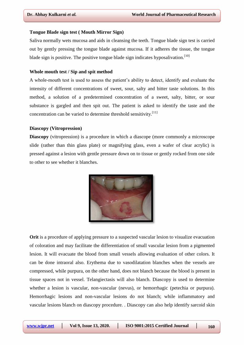
Diascopy (Vitropression)
Diascopy (vitropression) is a procedure in which a diascope (more commonly a microscope
slide (rather than thin glass plate) or magnifying glass, even a wafer of clear acrylic) is
pressed against a lesion with gentle pressure down on to tissue or gently rocked from one side
to other to see whether it blanches.
Orit is a procedure of applying pressure to a suspected vascular lesion to visualize evacuation
of coloration and may facilitate the differentiation of small vascular lesion from a pigmented
lesion. It will evacuate the blood from small vessels allowing evaluation of other colors. It
can be done intraoral also. Erythema due to vasodilatation blanches when the vessels are
compressed, while purpura, on the other hand, does not blanch because the blood is present in
tissue spaces not in vessel. Telangiectasis will also blanch. Diascopy is used to determine
whether a lesion is vascular, non-vascular (nevus), or hemorrhagic (petechia or purpura).
Hemorrhagic lesions and non-vascular lesions do not blanch; while inflammatory and
vascular lesions blanch on diascopy procedure. . Diascopy can also help identify sarcoid skin

Dr. Abhay Kulkarni et al. World Journal of Pharmaceutical Research
www.wjpr.net │ Vol 9, Issue 13, 2020. │ ISO 9001:2015 Certified Journal │ 161
lesions, when tested, turn an apple jelly color. This is detected in lupus vulgaris. It is detected
to differentiate from localized area of vasoconstriction from a hypopigmented and
depigmented skin patch, i.e. vitiligo. In the former the diascopy will blanch the lesion and in
later the patch is still detectable.[12]
End feel test
If mouth opening is restricted, it is helpful to test the “end feel”. End feel describes the
characteristics of restriction. End feel can be evaluated by placing the fingers between
patient’s upper and lower teeth and applying gentle-but-steady force in an attempt to
passively increase the interincisal distance. Muscle restrictions are associated with soft end
feel and results in increase of >5mm above the active opening (wide opening with pain). Joint
disorders such as acute non reducing disc displacement have hard end feel and
characteristically limit assisted opening to <5mm.[13]
Test for trauma from occlusion (Fremitus test/Fuctional Mobility).
Trauma from occlusion
It is a term used to describe pathologic alterations or adaptive changes which develop in the
periodontium as a result of undue force produced by the masticatory muscles. Fremitus is a
measurement of the vibratory pattern of the teeth when the teeth are placed in contacting
position and movements. This test can be assessed by placing the dampened index finger over
the buccal mucosa and palpating the buccal aspect of teeth while asking the patient to close
and move in excursive movements or tap their teeth together. Fremitus is caused by trauma
from occlusion and may be due to periodontal disease destruction. Each tooth is recorded in
the periodontal chart for being positive or negative for fremitus.[14]
2. Chairside investigations give guide for further investigations to carry out
These are chairside investigations which don’t give immediate conclusion of the disease but
may give further guidelines to carry out most proper investigations to reach some conclusion.
It includes
Toluidine blue
It is useful for surgeons to demarcate the extent orofacial lesion prior to excision. It indicates
area of dysplasia and further need of biopsy. Only the dark staining lesions are taken as
positive whereas equivocal stains (faint stains) and the lesions which do not retain at all are

Dr. Abhay Kulkarni et al. World Journal of Pharmaceutical Research
www.wjpr.net │ Vol 9, Issue 13, 2020. │ ISO 9001:2015 Certified Journal │ 162
taken as negative. This was followed by an oral rinse with 1% acetic acid solution which was
given to the patient to hold in the mouth for 20 seconds before expectorating. Toluidine blue
(1% W/W) was applied as an oral rinse for 20 seconds and then 1% acetic acid was used for
20 seconds to eliminate mechanically retained stain. Only the dark staining lesions were
taken as positive whereas equivocal stains (faint stains) and the lesions which did not retain
the stain at all were taken as negative but all the lesions were subjected to histopathological
examination.[15]
Figure 1 Figure 2
Lugol’s Iodine
Lugol’s Iodine solution consists of 5g iodine and 10g Potassium iodide mixed with 85 ml
distilled water to make a brown solution. Lugol’s iodine is used in early screening and also
can be used to identify the lesion margin and extension in precancerous lesions like
homogenous leukoplakia, non-homogenous leukoplakia, erythroplakia, reticular lichen
planus, erosive lichen planus.[16]
Acridine Binding Method
In this method, the uptake of acriflavine by desquamated buccal cells is measured. Since the
DNA content of the dysplastic cells are more, they will stain more intensely than normal
cells.[17]
Dr. Abhay Kulkarni et al. World Journal of Pharmaceutical Research
www.wjpr.net │ Vol 9, Issue 13, 2020. │ ISO 9001:2015 Certified Journal │ 163
Methylene blue
Methylene blue staining is a useful diagnostic adjunct in a large, community-based oral
cancer screening program for high-risk individuals. It indicates area of dysplasia and gives
guidance for exact site of biopsy.[18]
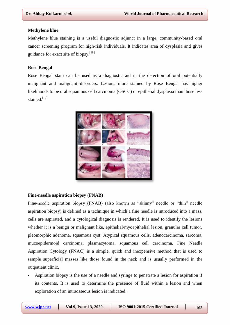
Rose Bengal
Rose Bengal stain can be used as a diagnostic aid in the detection of oral potentially
malignant and malignant disorders. Lesions more stained by Rose Bengal has higher
likelihoods to be oral squamous cell carcinoma (OSCC) or epithelial dysplasia than those less
stained.[19]
Fine-needle aspiration biopsy (FNAB)
Fine-needle aspiration biopsy (FNAB) (also known as “skinny” needle or “thin” needle
aspiration biopsy) is defined as a technique in which a fine needle is introduced into a mass,
cells are aspirated, and a cytological diagnosis is rendered. It is used to identify the lesions
whether it is a benign or malignant like, epithelial/myoepithelial lesion, granular cell tumor,
pleomorphic adenoma, squamous cyst, Atypical squamous cells, adenocarcinoma, sarcoma,
mucoepidermoid carcinoma, plasmacytoma, squamous cell carcinoma. Fine Needle
Aspiration Cytology (FNAC) is a simple, quick and inexpensive method that is used to
sample superficial masses like those found in the neck and is usually performed in the
outpatient clinic.
- Aspiration biopsy is the use of a needle and syringe to penetrate a lesion for aspiration if
its contents. It is used to determine the presence of fluid within a lesion and when
exploration of an intraosseous lesion is indicated.
Dr. Abhay Kulkarni et al. World Journal of Pharmaceutical Research
www.wjpr.net │ Vol 9, Issue 13, 2020. │ ISO 9001:2015 Certified Journal │ 164
Procedure
- An 18 gauge needle on a 5 or 10 ml syringe is inserted into the area under investigsation
after anesthesia is obtained to find the fluid cavity.
- The syringe is aspirated and the needle redirected if necessary.[20]
Pathergy test
Pathergy phenomenon is defined as a state of altered tissue reactivity that occurs in response
to minor trauma. Pathergy test (PT) is an easy to perform skin test to look for the pathergy
phenomenon.
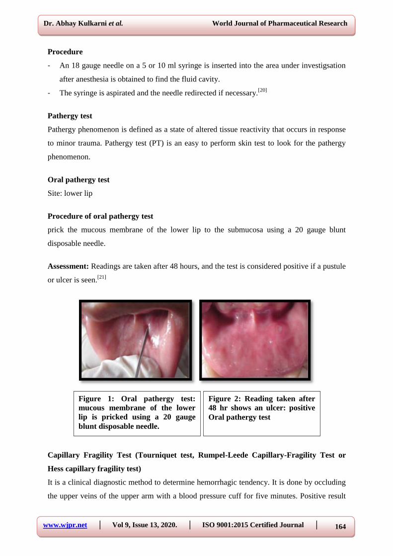
Oral pathergy test
Site: lower lip
Procedure of oral pathergy test
prick the mucous membrane of the lower lip to the submucosa using a 20 gauge blunt
disposable needle.
Assessment: Readings are taken after 48 hours, and the test is considered positive if a pustule
or ulcer is seen.[21]
Capillary Fragility Test (Tourniquet test, Rumpel-Leede Capillary-Fragility Test or
Hess capillary fragility test)
It is a clinical diagnostic method to determine hemorrhagic tendency. It is done by occluding
the upper veins of the upper arm with a blood pressure cuff for five minutes. Positive result
Figure 2: Reading taken after
48 hr shows an ulcer: positive
Oral pathergy test
Figure 1: Oral pathergy test:
mucous membrane of the lower
lip is pricked using a 20 gauge
blunt disposable needle.

Dr. Abhay Kulkarni et al. World Journal of Pharmaceutical Research
www.wjpr.net │ Vol 9, Issue 13, 2020. │ ISO 9001:2015 Certified Journal │ 165
will show unequivocal petechiae distal to cuff and negative result will show 1 or 2 petechiae
distal to cuff. It is used in diagnosis of Dengue fever. It is used to identify thrombocytopenia,
thrombocytopathia.[22]
Intraoral patch test
Patch-testing is a diagnostic procedure that is most commonly used for identifying the
possible causes of contact dermatitis (type IV hypersensitivity reaction). This can be done
type I hypersensitivity reactions and in DRESS (drug reaction with eosinophilia and systemic
symptoms) syndrome (type IVb hypersensitivity reaction). In this procedure mixing of
allergen with white petrolatum (i.e. the vehicle) is done and then applied in close proximity
with the skin. The first reading is taken after 30 minutes to look for type 1 hypersensitivity
reaction, while a second reading is taken after 48 hours to investigate for type 4 delayed
hypersensitivity reaction. Between the 2 readings, the patient should be instructed not to wet,
rub or scratch the testing area, avoid exercise and sweating. The readings are evaluated using
the International Contact Dermatitis Research Group (ICDRG) grading system: minus (–)
stands for negative, plus (+) stands for a weak (non-vesicular) positive reaction, double plus
(++) stands for a strong (vesicular) positive reaction and triple plus (+++) stands for extreme
(bullous) positive reaction.
A pectin, gelatin, sodium carboxymethylcellulose, and plasticized hydrocarbon gel base
(orabase) may be used as a vehicle to test for contact allergy within the oral cavity. The
suspected chemical is incorporated in the vehicle and applied to the oral mucosa. Fenretinide
mucoadhesive patches were attached (q.d. 30 min for 10 consecutive days) to the right buccal
mucosa (blank patches on left buccal mucosa) immediately posterior to the intraoral
commissure of the upper and lower lips. Intraorally patch testing can be done by keeping the
antigen in the maxillary denture base and holding it in the mouth.[23]
REFERENCES
1. Augustin Prabha Xavier, Sarat Gummadapu, et. Al Classification of chair side
investigations in dentistry. International Journal of Applied Dental Sciences, 2019; 5(3):
05-07.
2. Michael G. Newman, Henry Takei, Perry R. Klokkevold, Carranza's Clinical
Periodontology, Second South Asia Edition.
3. Suresh Chandra , Grossman’s Endodontic Practice, 13th
Edition.
Dr. Abhay Kulkarni et al. World Journal of Pharmaceutical Research
www.wjpr.net │ Vol 9, Issue 13, 2020. │ ISO 9001:2015 Certified Journal │ 166
4. Kadandale Sadasiva, Kumarappan Senthil Kumar, et al, Evaluation of the Efficacy of
Visual, Tactile Method, Caries Detector Dye, and Laser Fluorescence in Removal of
Dental Caries and Confirmation by Culture and Polymerase Chain Reaction: An In
Vivo Study, Journal of pharmacy & bioallied sciences, 2019; 11(2): S146-S150.
5. Nisha Garg, Amit Garg , Textbook of Endodontics, 4th
Edition
6. Sebeena Mathew, Boopathi Thangavel, et al, Diagnosis of cracked tooth syndrome. J
Pharm Bioallied Sci., 2012; 4(l2): S242-S244.
7. Jeffrey P. Okeson. Management of Temporomandibular Disorders and Occlusion - E-
Book. 7th Edition
8. Michael G. Newman, Henry Takei, Perry R. Klokkevold , Carranza's Clinical
Periodontology, Second South Asia Edition.
9. Nilufer Nadaf, Krishnapriya .V, et. al. Mouth Breathing-A Harmful Habit in a Young
Child. ARC Journal of Forensic Science, 2018; 3(2): 25-29.
10. Abhay S. Kulkarni, Rajendra S. Birangane, et. al. Clinical and Radiological Signs of
Importance for the Oral Physician and Oral Surgeon. Journal of Indian Academy of Oral
Medicine & Radiology ¦ Volume 31 ¦ Issue 3 ¦ July-September 2019.
11. Vijay Kumar Ambaldhage, Jaishankar Homberhalli Puttabuddi, et. al. Taste disorders: A
review. Journal of Indian Academy of Oral Medicine & Radiology, Jan-Mar, 2014; 26(1).
12. Abhay Suresh Kulkarni. Oral Medicine And Radiology Question-Answer Format for
Review and Exam Preparation. 2019; 1(1): 103.
13. Jeffrey P. Okeson. Management of Temporomandibular Disorders and Occlusion - E-
Book. 7th Edition.
14. Michael G. Newman, Henry Takei, Perry R. Klokkevold, Carranza's Clinical
Periodontology, Second South Asia Edition
15. Nahum Puebla-Osorio, Seri N.E. Sarchio, et. al. Detection of Infiltrating Mast Cells
Using a Modified Toluidine Blue Staining. Fibrosis: Methods and Protocols, Methods in
Molecular Biology, vol. 1627.
16. Shereen Fatima, Rajarshi Basu, et. al. Lugol's iodine identifies dysplastic tissue in
precancerous lesions: A clinical trial. Ann Maxillofac Surg, 2016; 6(2): 172-174.
17. Anil Govindrao Ghom. Textbook of ORAL MEDICINE. Second Edition
18. Akhtar Riaz, Balasundari Shreedhar, et. al. Methylene blue as an early diagnostic marker
for oral precancer and cancer. Riaz et al. SpringerPlus, 2013; 2: 95.

Dr. Abhay Kulkarni et al. World Journal of Pharmaceutical Research
www.wjpr.net │ Vol 9, Issue 13, 2020. │ ISO 9001:2015 Certified Journal │ 167
19. Ge-fei Du, Cheng-zhang Li, et. al. Rose bengal staining in detection of oral precancerous
and malignant lesions with colorimetric evaluation: A pilot study. Int. J. Cancer, 2007;
120: 1958–1963.
20. Ronald G. Amedee, Nina R. Dhurandhar. Fine-Needle Aspiration Biopsy. Laryngoscope,
2001; 111: 1551–1557.
21. Fiona F Sequeira, Deepak Daryani. The oral and skin pathergy test. Indian Journal of
Dermatology, Venereology, Leprology. 2011; 77(4): 526-30.
22. S. Das. A Manual On Clinical Surgery. 9th
Edition.
23. Abhay Suresh Kulkarni. Oral Medicine And Radiology Question-Answer Format for
Review and Exam Preparation. 2019; 1(1): 103.