Embed Size (px)
Citation preview
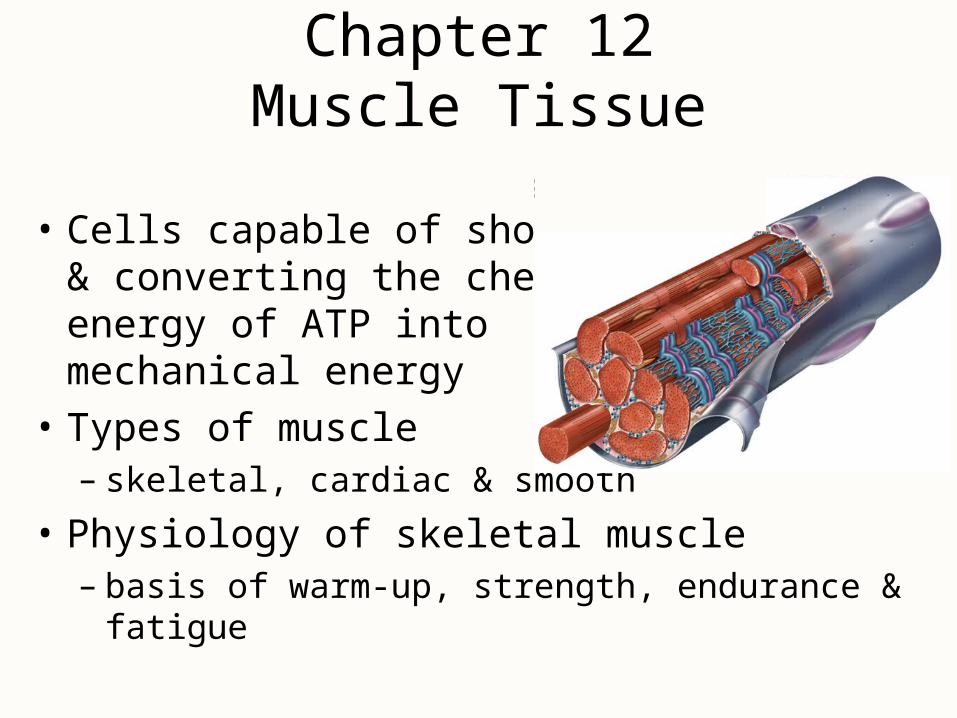
Chapter 12Muscle Tissue
• Cells capable of shortening & converting the chemical energy of ATP into mechanical energy
• Types of muscle– skeletal, cardiac & smooth
• Physiology of skeletal muscle– basis of warm-up, strength, endurance & fatigue
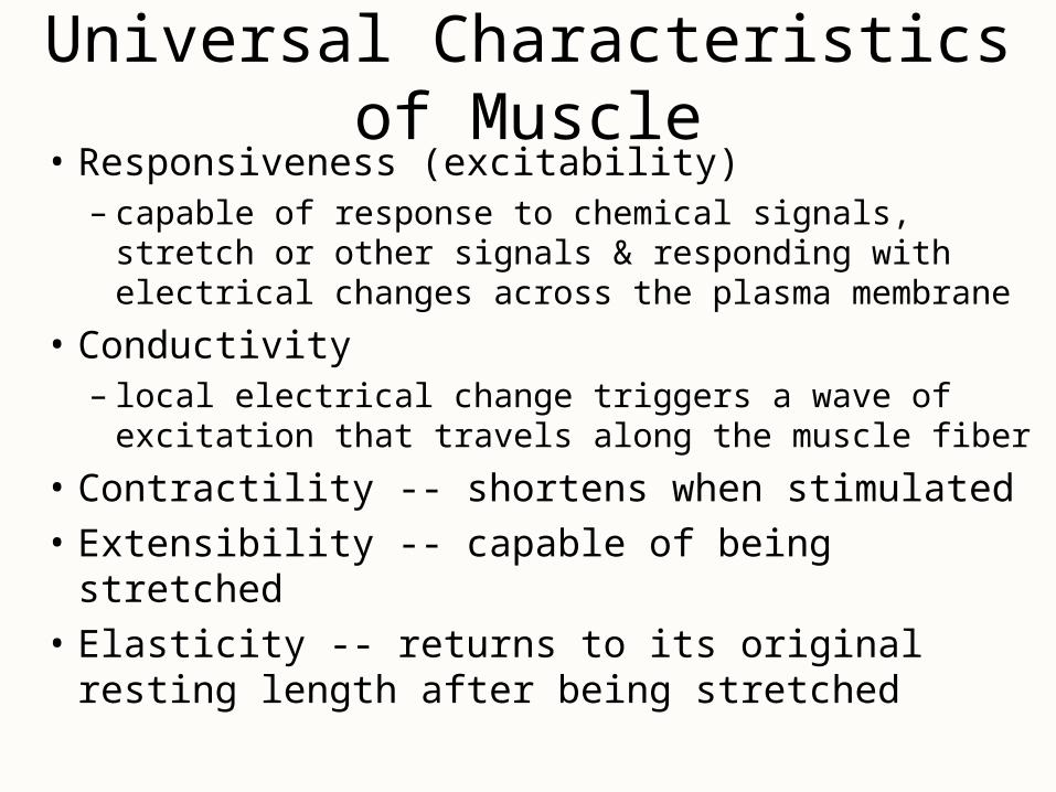
Universal Characteristics of Muscle• Responsiveness (excitability)
– capable of response to chemical signals, stretch or other signals & responding with electrical changes across the plasma membrane
• Conductivity– local electrical change triggers a wave of excitation that
travels along the muscle fiber
• Contractility -- shortens when stimulated
• Extensibility -- capable of being stretched
• Elasticity -- returns to its original resting length after being stretched
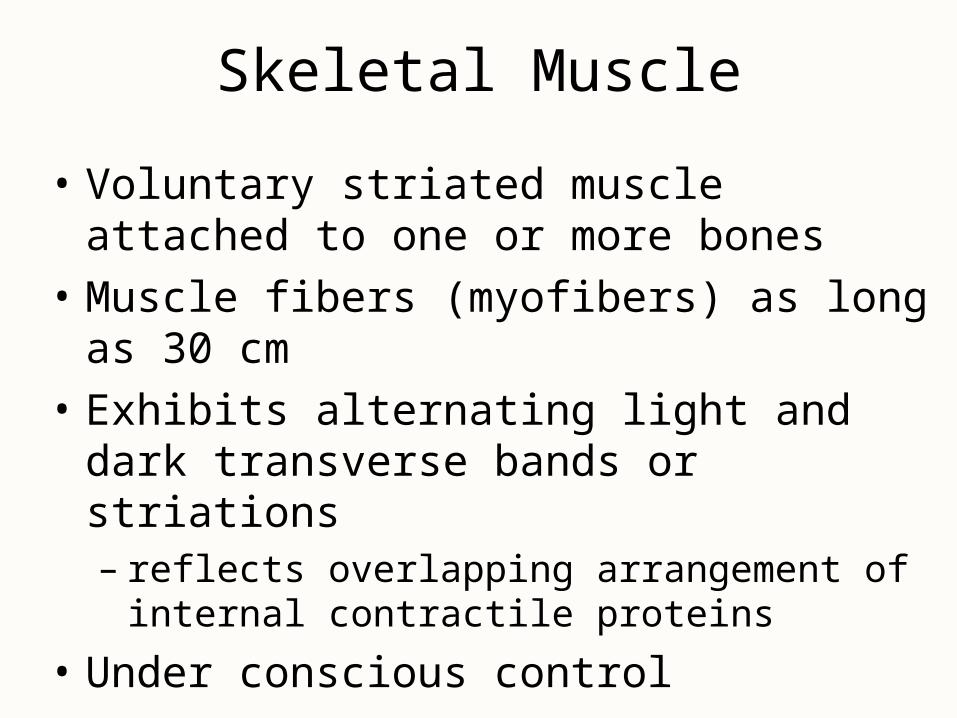
Skeletal Muscle
• Voluntary striated muscle attached to one or more bones
• Muscle fibers (myofibers) as long as 30 cm
• Exhibits alternating light and dark transverse bands or striations– reflects overlapping arrangement of internal
contractile proteins
• Under conscious control
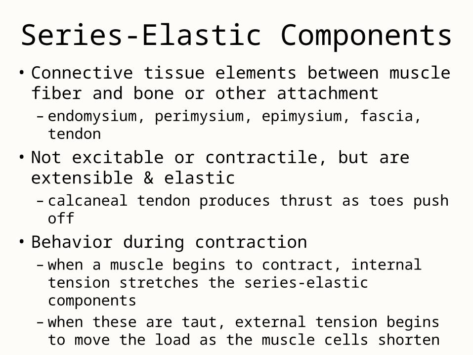
Series-Elastic Components• Connective tissue elements between muscle fiber
and bone or other attachment– endomysium, perimysium, epimysium, fascia, tendon
• Not excitable or contractile, but are extensible & elastic– calcaneal tendon produces thrust as toes push off
• Behavior during contraction– when a muscle begins to contract, internal tension
stretches the series-elastic components– when these are taut, external tension begins to move
the load as the muscle cells shorten
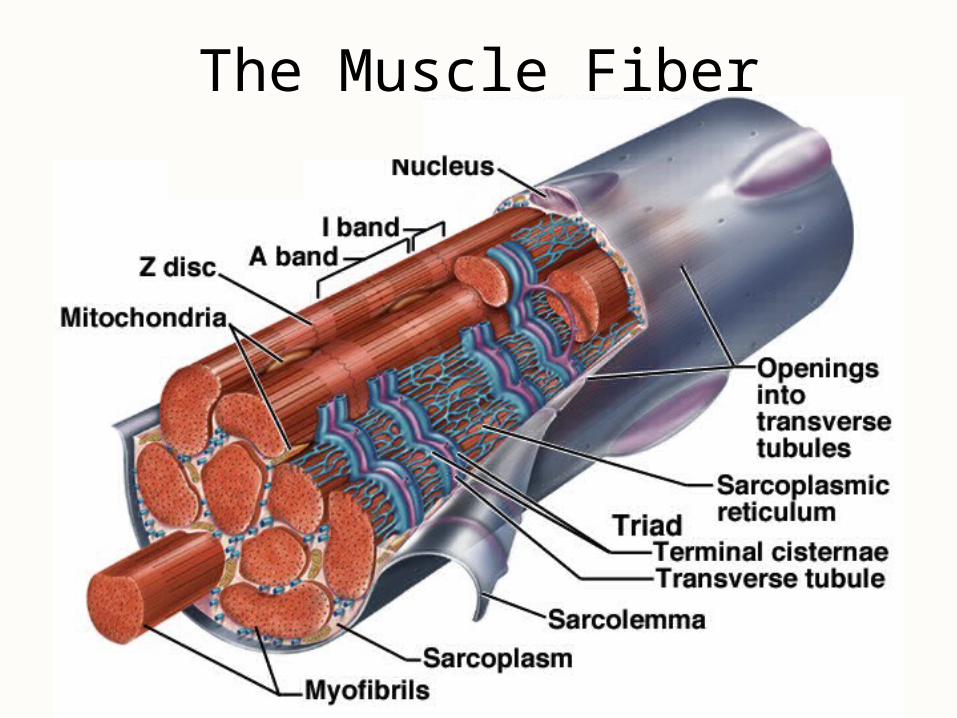
The Muscle Fiber
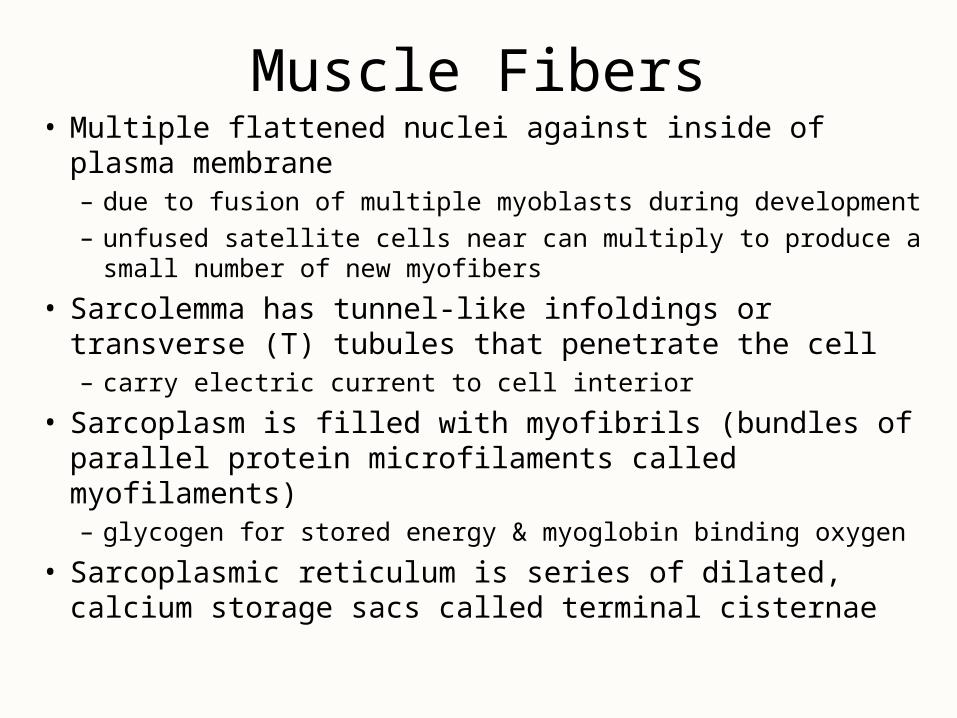
Muscle Fibers• Multiple flattened nuclei against inside of plasma membrane
– due to fusion of multiple myoblasts during development
– unfused satellite cells near can multiply to produce a small number of new myofibers
• Sarcolemma has tunnel-like infoldings or transverse (T) tubules that penetrate the cell– carry electric current to cell interior
• Sarcoplasm is filled with myofibrils (bundles of parallel protein microfilaments called myofilaments)– glycogen for stored energy & myoglobin binding oxygen
• Sarcoplasmic reticulum is series of dilated, calcium storage sacs called terminal cisternae
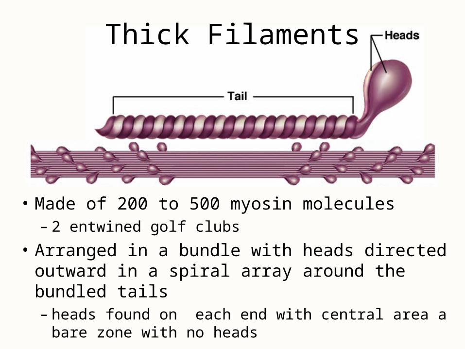
Thick Filaments
• Made of 200 to 500 myosin molecules– 2 entwined golf clubs
• Arranged in a bundle with heads directed outward in a spiral array around the bundled tails– heads found on each end with central area a bare
zone with no heads
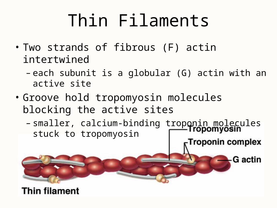
Thin Filaments
• Two strands of fibrous (F) actin intertwined– each subunit is a globular (G) actin with an active site
• Groove hold tropomyosin molecules blocking the active sites– smaller, calcium-binding troponin molecules stuck to
tropomyosin
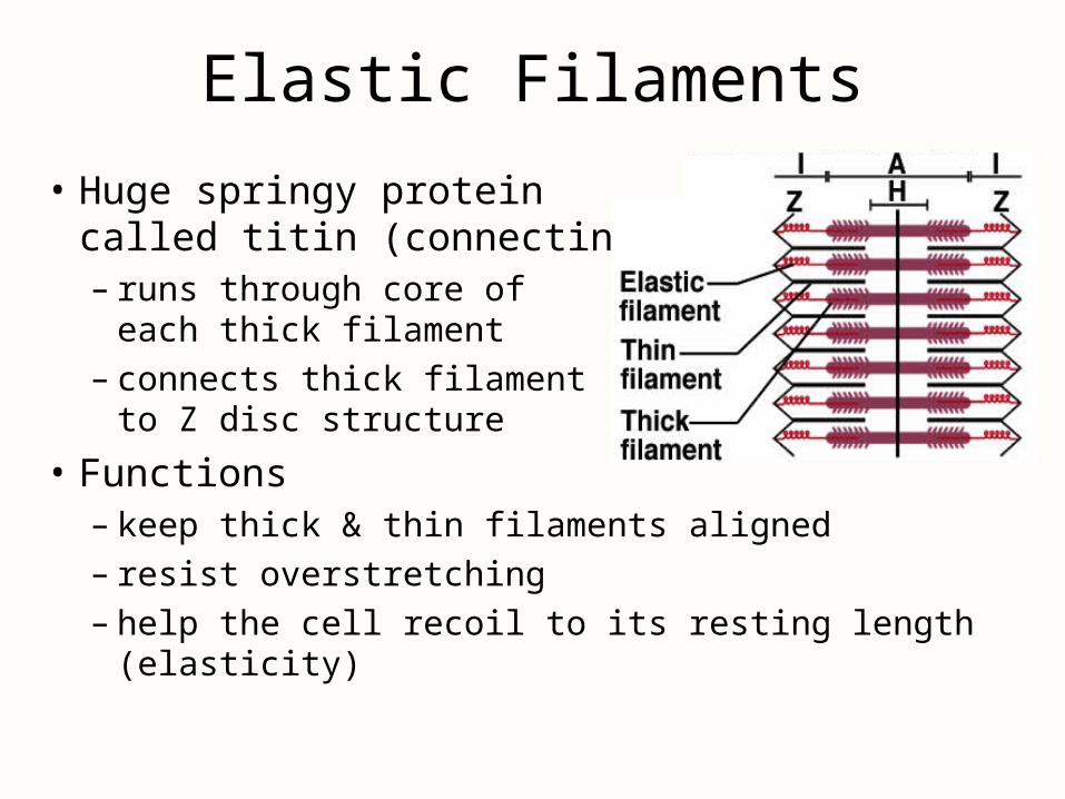
Elastic Filaments
• Huge springy protein called titin (connectin)– runs through core of
each thick filament– connects thick filament
to Z disc structure
• Functions– keep thick & thin filaments aligned– resist overstretching– help the cell recoil to its resting length (elasticity)
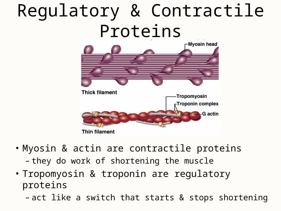
Regulatory & Contractile Proteins
• Myosin & actin are contractile proteins– they do work of shortening the muscle
• Tropomyosin & troponin are regulatory proteins– act like a switch that starts & stops shortening
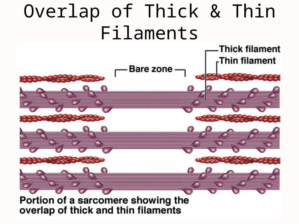
Overlap of Thick & Thin Filaments
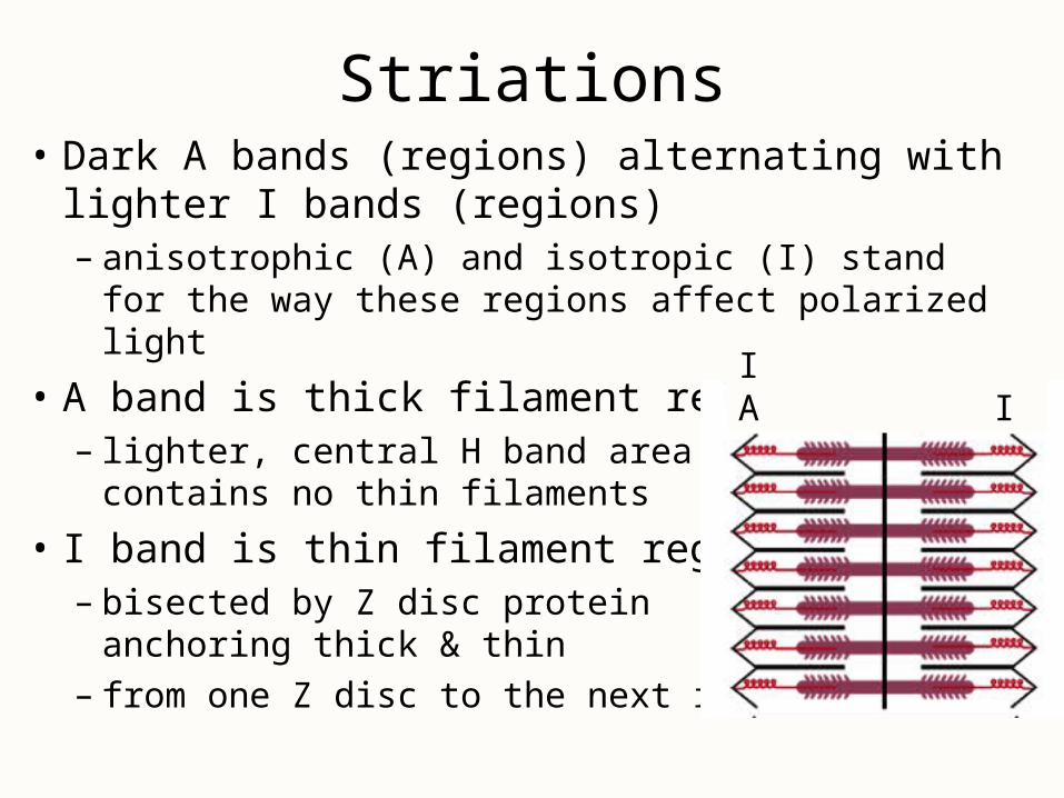
Striations• Dark A bands (regions) alternating with lighter I
bands (regions)– anisotrophic (A) and isotropic (I) stand for the way
these regions affect polarized light
• A band is thick filament region– lighter, central H band area
contains no thin filaments
• I band is thin filament region– bisected by Z disc protein
anchoring thick & thin– from one Z disc to the next is a sarcomere
I A I
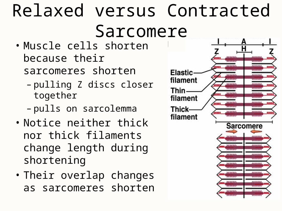
Relaxed versus Contracted Sarcomere• Muscle cells shorten
because their sarcomeres shorten – pulling Z discs closer
together– pulls on sarcolemma
• Notice neither thick nor thick filaments change length during shortening
• Their overlap changes as sarcomeres shorten

Motor Neurons
• Skeletal muscle must be stimulated by a nerve or it will not contract (paralyzed)
• Cell bodies of somatic motor neurons are in brainstem or spinal cord
• Axons of somatic motor neurons are called somatic motor fibers– each branches, on average, into 200 terminal
branches that supply one muscle fiber each
• Each motor neuron and all the muscle fibers it innervates are called a motor unit

Motor Units• A motor neuron & the muscle fibers it innervates
– dispersed throughout the muscle
– when contract together causes weak contraction over wide area
– provides ability to sustain long-term contraction as motor units take turns resting (postural control)
• Fine control– small motor units contain as few as
20 muscle fibers per nerve fiber
– eye muscles
• Strength control– gastrocnemius muscle has 1000
fibers per nerve fiber
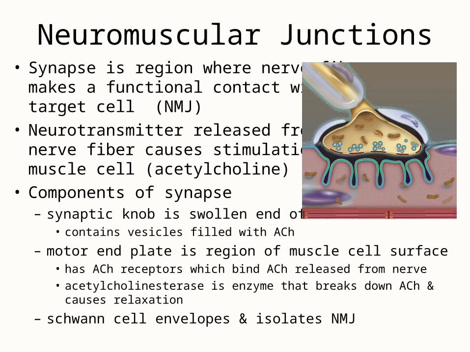
Neuromuscular Junctions• Synapse is region where nerve fiber
makes a functional contact with its target cell (NMJ)
• Neurotransmitter released from nerve fiber causes stimulation of muscle cell (acetylcholine)
• Components of synapse– synaptic knob is swollen end of nerve fiber
• contains vesicles filled with ACh
– motor end plate is region of muscle cell surface• has ACh receptors which bind ACh released from nerve
• acetylcholinesterase is enzyme that breaks down ACh & causes relaxation
– schwann cell envelopes & isolates NMJ

The Neuromuscular Junction
Neuromuscular Toxins & Paralysis
• Pesticides contain cholinesterase inhibitors that bind to acetylcholinesterase & prevent it from degrading ACh– spastic paralysis & possible suffocation
• Tetanus or lockjaw is spastic paralysis caused by toxin of Clostridium bacteria– blocks glycine release in the spinal cord & causes
overstimulation of the muscles
• Flaccid paralysis with limp muscles unable to contract caused by curare that competes with ACh
Electrically Excitable Cells (muscle & nerve)
• Plasma membrane is polarized or charged – resting membrane potential is due to Na+ outside of cell
and K+ & other anions inside of cell– difference in charge across the membrane is potential
• inside is slightly more negative (-90 mV)
• Plasma membranes exhibit voltage changes in response to stimulation– ion gates open allowing Na+ to rush into cell and then
K+ to rush out of cell (quick up-and-down voltage shift is called action potential)
– spreads over cell surface as nerve signal or impulse
Muscle Contraction & Relaxation
• Four phases involved in this process– excitation where action potentials in the nerve lead to
formation of action potentials in muscle fiber– excitation-contraction coupling refers to action
potentials on the sarcolemma activate myofilaments– contraction is shortening of muscle fiber or at least
formation of tension– relaxation is return of fiber to its resting length
• Images will be used to demonstrate the steps of each of these phases
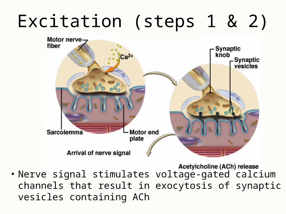
Excitation (steps 1 & 2)
• Nerve signal stimulates voltage-gated calcium channels that result in exocytosis of synaptic vesicles containing ACh
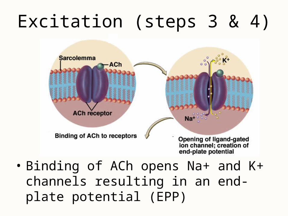
Excitation (steps 3 & 4)
• Binding of ACh opens Na+ and K+ channels resulting in an end-plate potential (EPP)
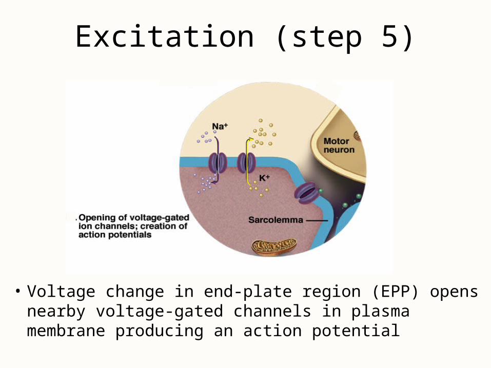
Excitation (step 5)
• Voltage change in end-plate region (EPP) opens nearby voltage-gated channels in plasma membrane producing an action potential
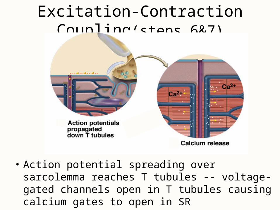
Excitation-Contraction Coupling(steps 6&7)
• Action potential spreading over sarcolemma reaches T tubules -- voltage-gated channels open in T tubules causing calcium gates to open in SR
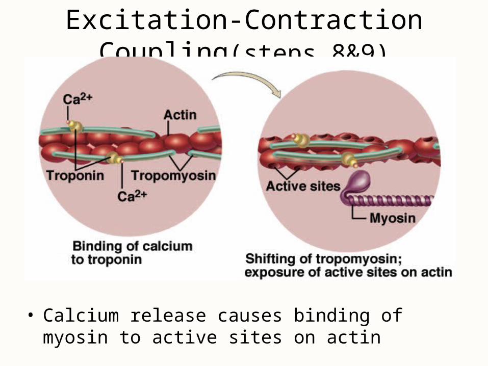
Excitation-Contraction Coupling(steps 8&9)
• Calcium release causes binding of myosin to active sites on actin
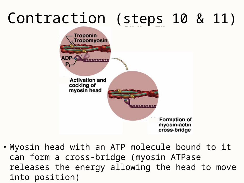
Contraction (steps 10 & 11)
• Myosin head with an ATP molecule bound to it can form a cross-bridge (myosin ATPase releases the energy allowing the head to move into position)
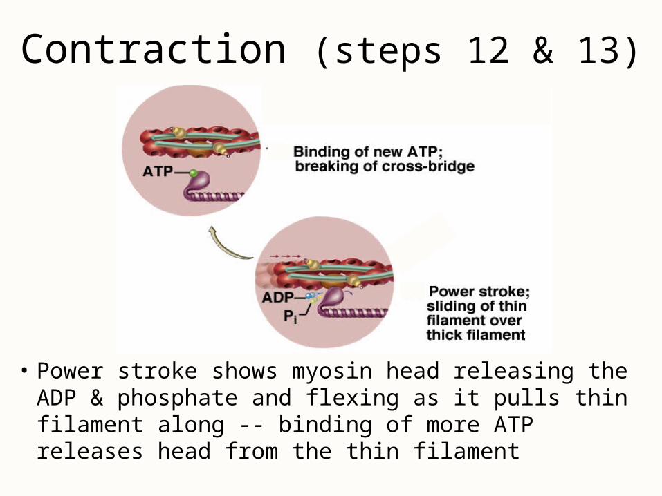
Contraction (steps 12 & 13)
• Power stroke shows myosin head releasing the ADP & phosphate and flexing as it pulls thin filament along -- binding of more ATP releases head from the thin filament
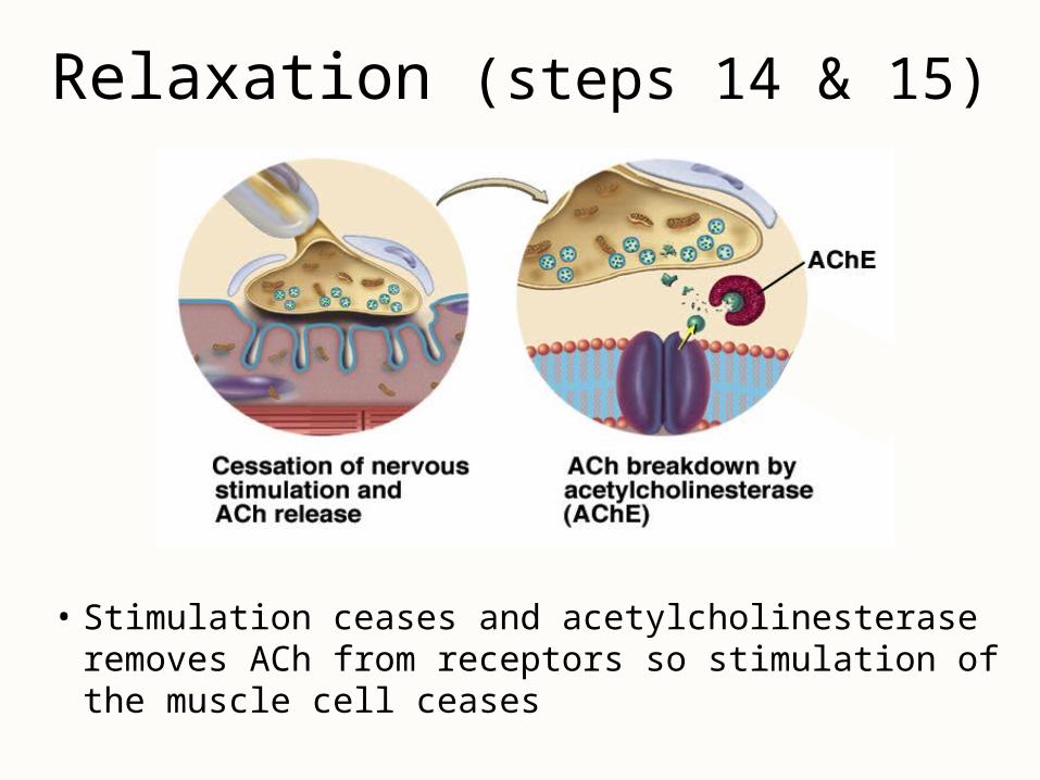
Relaxation (steps 14 & 15)
• Stimulation ceases and acetylcholinesterase removes ACh from receptors so stimulation of the muscle cell ceases
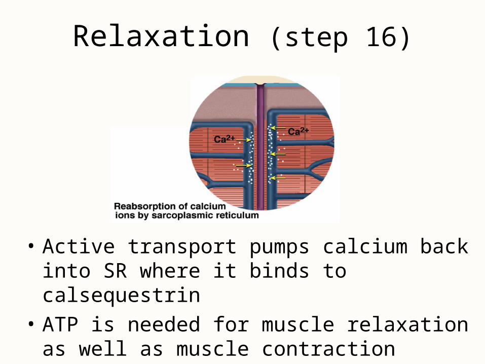
Relaxation (step 16)
• Active transport pumps calcium back into SR where it binds to calsequestrin
• ATP is needed for muscle relaxation as well as muscle contraction
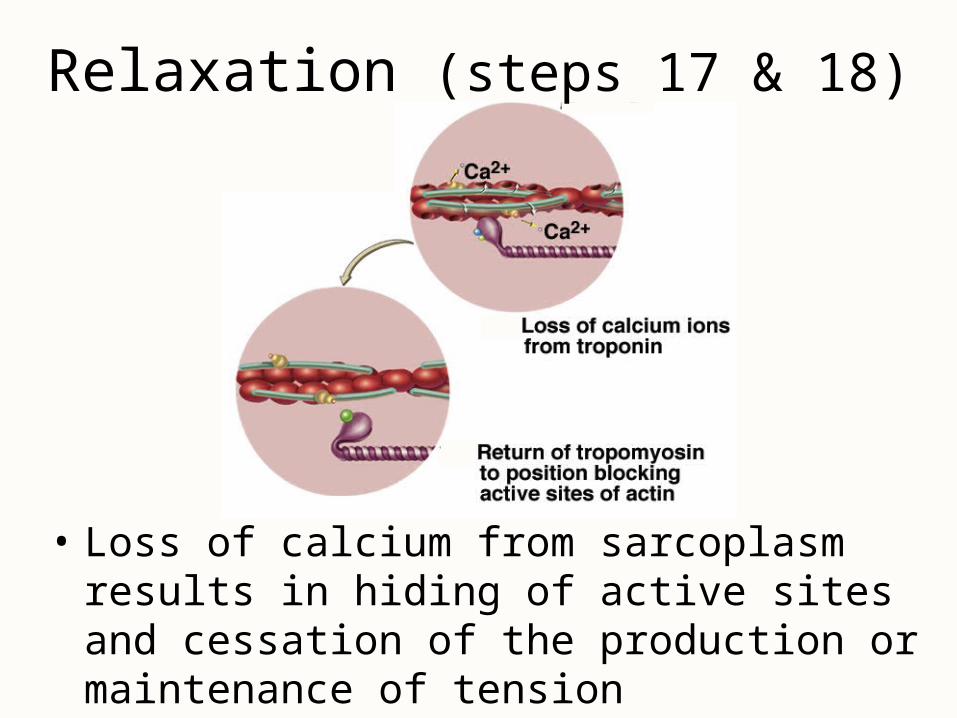
Relaxation (steps 17 & 18)
• Loss of calcium from sarcoplasm results in hiding of active sites and cessation of the production or maintenance of tension

Rigor Mortis• Stiffening of the body beginning 3 to 4 hours after
death -- peaks at 12 hours after death & diminishes over next 48 hours
• Deteriorating sarcoplasmic reticulum releases calcium
• Activates myosin-actin cross bridging & muscle contraction
• Muscle relaxation requires ATP & ATP production is no longer produced after death
• Fibers remain contracted until myofilaments decay
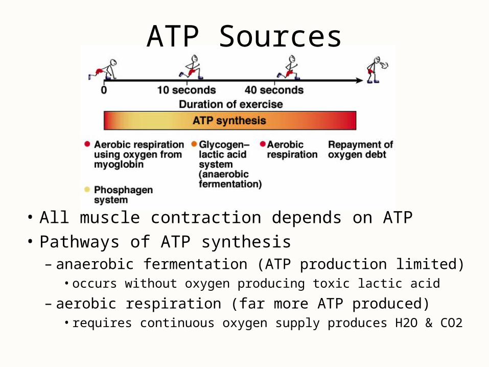
ATP Sources
• All muscle contraction depends on ATP
• Pathways of ATP synthesis– anaerobic fermentation (ATP production limited)
• occurs without oxygen producing toxic lactic acid
– aerobic respiration (far more ATP produced)• requires continuous oxygen supply produces H2O & CO2
Cardiac Muscle
• In comparison to skeletal muscle, the cells are shorter, thicker, branched and linked to each other at intercalated discs– electrical gap junctions allow cells to stimulate their
neighbors & mechanical junctions keep the cells from pulling apart
– sarcoplasmic reticulum is less developed but T tubules are larger to admit Ca+2 from extracellular fluid
• Autorhythmic due to pacemaker cells
• Uses aerobic respiration almost exclusively– large mitochondria make it resistant to fatigue
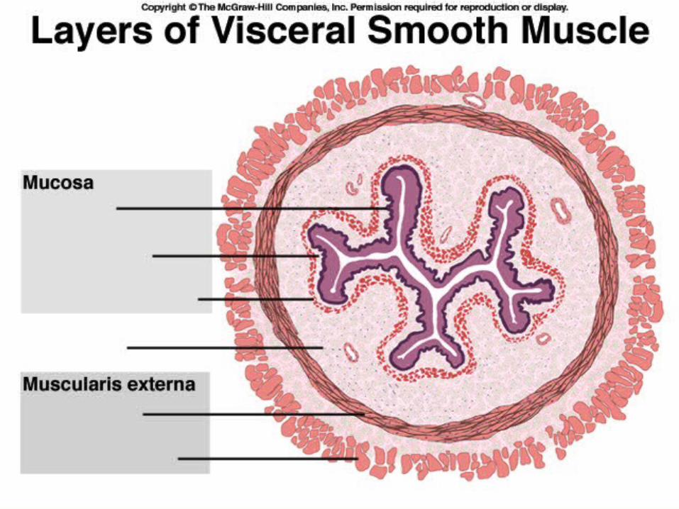
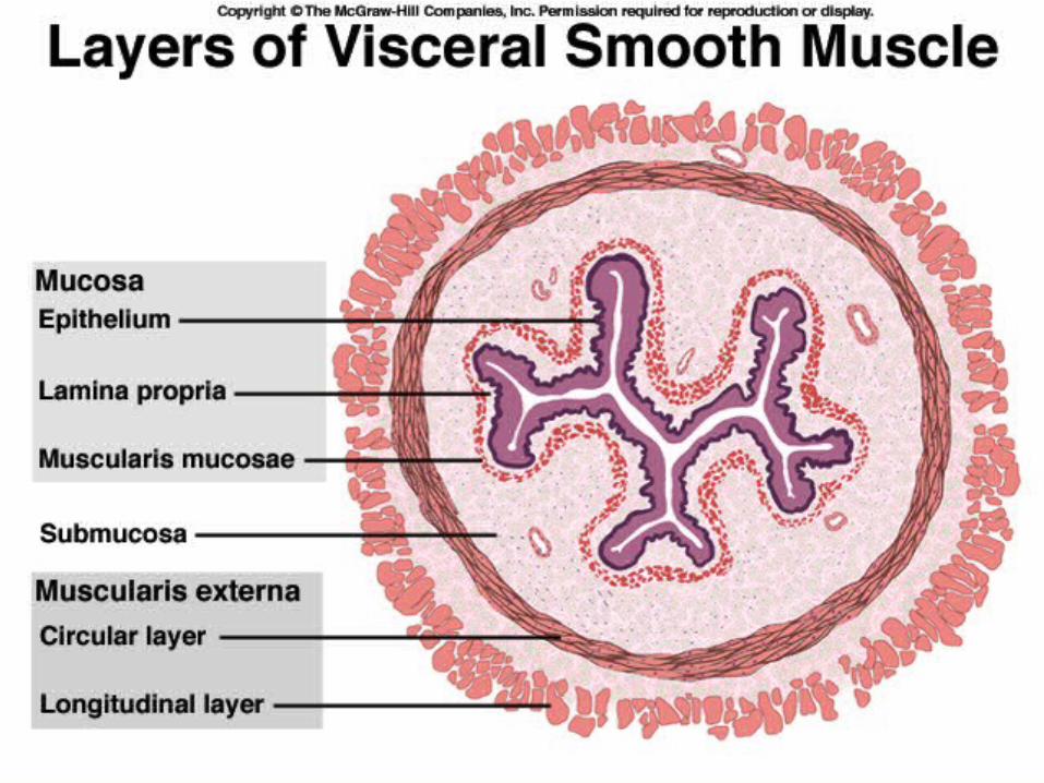
Smooth Muscle
• Fusiform cells with one nucleus– 30 to 200 microns long & 5 to 10 microns wide– no visible striations, sarcomeres or Z discs– thin filaments attach to dense bodies scattered
throughout sarcoplasm & on sarcolemma– SR is scanty & has no T tubules
• calcium for contraction comes from extracellular fluid
• Nerve supply is autonomic, if any is present

Types of Smooth Muscle• Multiunit smooth muscle
– in largest arteries, iris, pulmonary air passages, arrector pili muscles
– terminal nerve branches synapse on individual myocytes in a motor unit
– independent contraction
• Single-unit smooth muscle– in most blood vessels & viscera as
circular & longitudinal muscle layers– electrically coupled by gap junctions– large number of cells contract as a unit
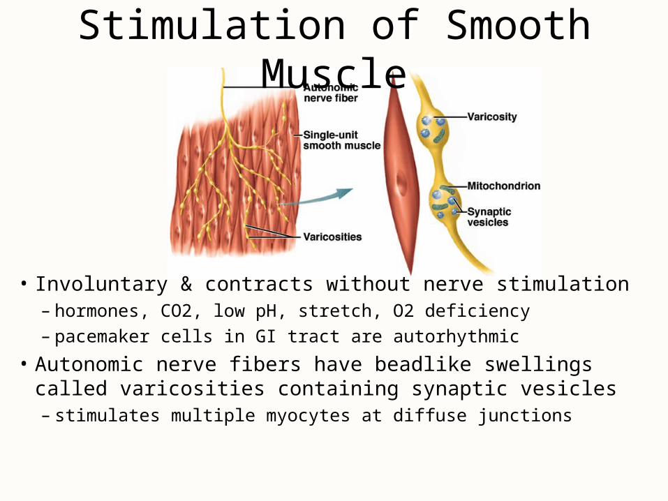
Stimulation of Smooth Muscle
• Involuntary & contracts without nerve stimulation– hormones, CO2, low pH, stretch, O2 deficiency– pacemaker cells in GI tract are autorhythmic
• Autonomic nerve fibers have beadlike swellings called varicosities containing synaptic vesicles– stimulates multiple myocytes at diffuse junctions

Features of Contraction and Relaxation• Calcium triggering contraction is extracellular
– enters cell through channels triggered by voltage, hormones, neurotransmitters or stretching of the cell
• calcium ion binds to calmodulin -- activates myosin light-chain kinase which activates the myosin head with ATP to bind actin -- power stroke occurs when hydrolyzes 2nd ATP
• Thin filaments pull on intermediate filaments attached to dense bodies on the plasma membrane– shortens the entire cell in a twisting fashion
• Contraction & relaxation very slow in comparison– slow myosin ATPase enzyme & slow pumps that remove Ca+2
• Uses l0-300 times less ATP to maintain the same tension– latch-bridge mechanism maintains tetanus (muscle tone)
• keeps arteries in state of partial contraction (vasomotor tone)
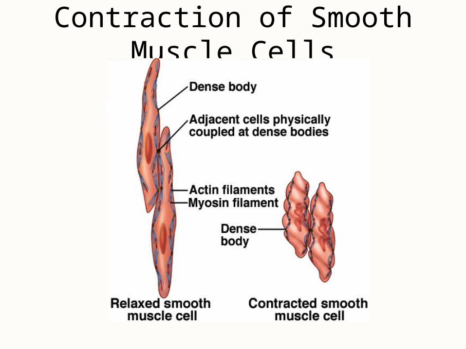
Contraction of Smooth Muscle Cells

Responses to Stretch• Stretch opens mechanically-gated calcium channels
causing muscle response– food entering the esophagus brings on peristalsis
• Stress-relaxation response necessary for hollow organs that gradually fill (urinary bladder)– when stretched, tissue briefly contracts then relaxes
• Must contract forcefully when greatly stretched– thick filaments have heads along their entire length– no orderly filament arrangement -- no Z discs
• Plasticity is ability to adjust tension to degree of stretch such as empty bladder is not flabby
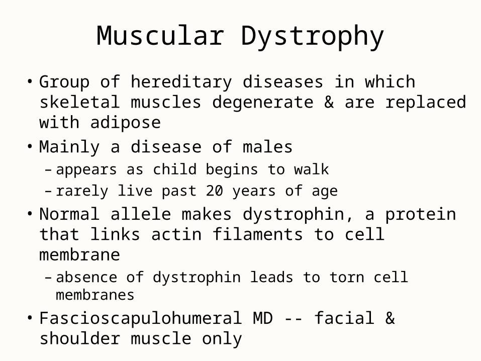
Muscular Dystrophy
• Group of hereditary diseases in which skeletal muscles degenerate & are replaced with adipose
• Mainly a disease of males– appears as child begins to walk– rarely live past 20 years of age
• Normal allele makes dystrophin, a protein that links actin filaments to cell membrane– absence of dystrophin leads to torn cell membranes
• Fascioscapulohumeral MD -- facial & shoulder muscle only
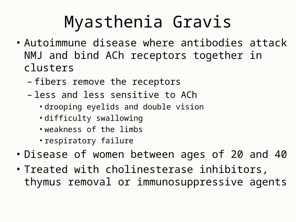
Myasthenia Gravis• Autoimmune disease where antibodies attack
NMJ and bind ACh receptors together in clusters– fibers remove the receptors– less and less sensitive to ACh
• drooping eyelids and double vision
• difficulty swallowing
• weakness of the limbs
• respiratory failure
• Disease of women between ages of 20 and 40
• Treated with cholinesterase inhibitors, thymus removal or immunosuppressive agents