Embed Size (px)
Citation preview
1
Chapter 21:
Respiratory System
Chapter 21: Respiratory System
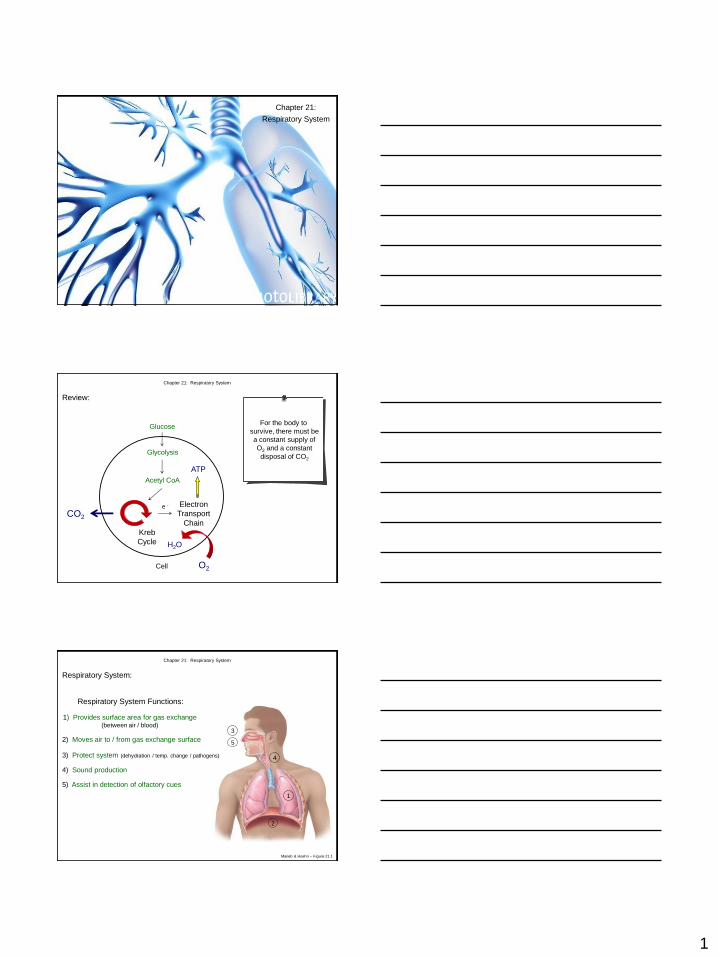
For the body to
survive, there must be
a constant supply of
O2 and a constant
disposal of CO2
Glucose
Glycolysis
Acetyl CoA
Kreb
Cycle
e - Electron
Transport
Chain CO2
O2
H2O
ATP
Review:
Cell
Chapter 21: Respiratory System
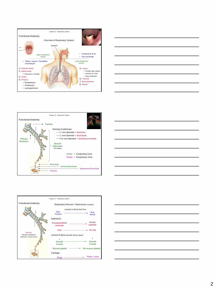
Respiratory System:
4) Sound production
Respiratory System Functions:
1) Provides surface area for gas exchange (between air / blood)
1
2) Moves air to / from gas exchange surface
2
3) Protect system (dehydration / temp. change / pathogens)
3
5) Assist in detection of olfactory cues
4
5
Marieb & Hoehn – Figure 21.1
2
Functional Anatomy:
Upper Respiratory
System
Lower Respiratory
System • Filters / warms / humidifies
incoming air
• Conduction of air
• Gas exchange
1) External nares
2) Nasal cavity
• Resonance chamber
3) Uvula
5) Larynx
• Provide open airway
• channel air / food
Overview of Respiratory System:
Epiglottis
4) Pharynx
• Nasopharynx
• Oropharynx
• Laryngopharynx
7) Bronchial tree
• voice production
6) Trachea
Chapter 21: Respiratory System
8) Alveoli
Trachea
Primary
Bronchus Bronchi
bifurcation (23 orders)
Naming of pathways:
• > 1 mm diameter = bronchus
• < 1 mm diameter = bronchiole
• < 0.5 mm diameter = terminal bronchiole
Bronchiole Terminal Bronchiole
Respiratory Bronchiole Alveolus
Green = Conducting Zone
Purple = Respiratory Zone
Functional Anatomy:
Chapter 21: Respiratory System
Respiratory Mucosa / Submucosa (Variable):
Location in Bronchial Tree
Near
Trachea Near
Alveoli
Epithelium:
Pseudostratified
columnar
Simple
cuboidal
Lamina Propria (areolar tissue layer) :
Cilia No cilia
Smooth
muscle
Smooth
muscle
Cartilage:
Rings Plates / none
Mucosa:
Mucous membrane
(epithelium / areolar tissue)
Functional Anatomy:
Mucous glands No mucous glands
Chapter 21: Respiratory System
3
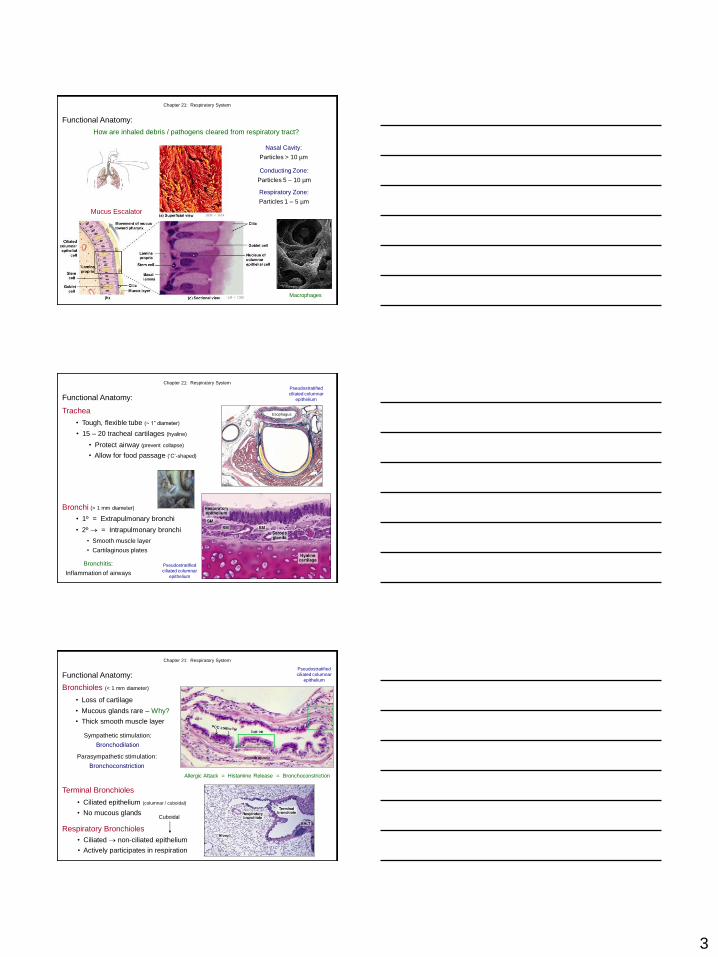
Functional Anatomy:
How are inhaled debris / pathogens cleared from respiratory tract?
Macrophages
Nasal Cavity:
Particles > 10 µm
Conducting Zone:
Particles 5 – 10 µm
Respiratory Zone:
Particles 1 – 5 µm
Mucus Escalator
Chapter 21: Respiratory System
• Tough, flexible tube (~ 1” diameter)
Trachea
• 15 – 20 tracheal cartilages (hyaline)
Functional Anatomy:
Pseudostratified
ciliated columnar
epithelium
Esophagus
Bronchi (> 1 mm diameter)
Pseudostratified
ciliated columnar
epithelium
• 1º = Extrapulmonary bronchi
• 2º = Intrapulmonary bronchi
• Smooth muscle layer
• Cartilaginous plates
Bronchitis:
Inflammation of airways
• Protect airway (prevent collapse)
• Allow for food passage (‘C’-shaped)
Chapter 21: Respiratory System
• Loss of cartilage
Bronchioles (< 1 mm diameter)
• Mucous glands rare – Why?
• Thick smooth muscle layer
Pseudostratified
ciliated columnar
epithelium
Sympathetic stimulation:
Bronchodilation
Parasympathetic stimulation:
Bronchoconstriction
Allergic Attack = Histamine Release = Bronchoconstriction
Terminal Bronchioles
Respiratory Bronchioles
Cuboidal
• Actively participates in respiration
• Ciliated epithelium (columnar / cuboidal)
• No mucous glands
• Ciliated non-ciliated epithelium
Chapter 21: Respiratory System
Functional Anatomy:
4
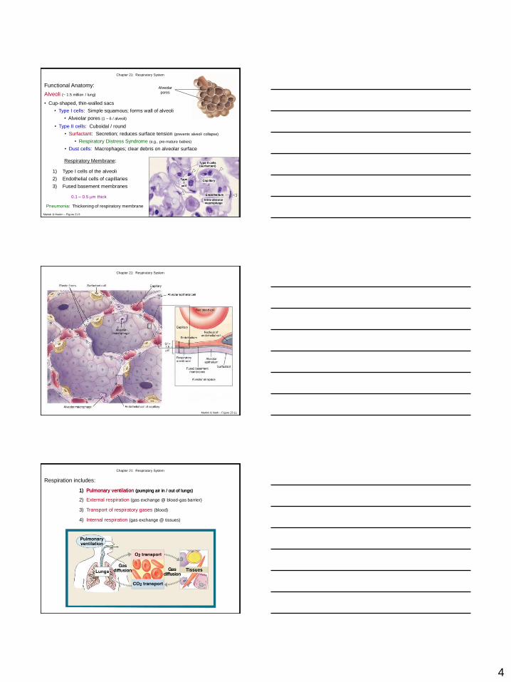
• Cup-shaped, thin-walled sacs
• Type I cells: Simple squamous; forms wall of alveoli
• Alveolar pores (1 – 6 / alveoli)
Alveoli (~ 1.5 million / lung)
Respiratory Membrane:
1) Type I cells of the alveoli
2) Endothelial cells of capillaries
3) Fused basement membranes
0.1 – 0.5 m thick
Pneumonia: Thickening of respiratory membrane
Chapter 21: Respiratory System
Functional Anatomy: Alveolar
pores
Marieb & Hoehn – Figure 21.8
• Type II cells: Cuboidal / round
• Surfactant: Secretion; reduces surface tension (prevents alveoli collapse)
• Respiratory Distress Syndrome (e.g., pre-mature babies)
• Dust cells: Macrophages; clear debris on alveolar surface
Chapter 21: Respiratory System
Martini & Nath – Figure 23.11
Respiration includes:
1) Pulmonary ventilation (pumping air in / out of lungs)
Chapter 21: Respiratory System
2) External respiration (gas exchange @ blood-gas barrier)
3) Transport of respiratory gases (blood)
4) Internal respiration (gas exchange @ tissues)
1) Pulmonary ventilation (pumping air in / out of lungs)
5
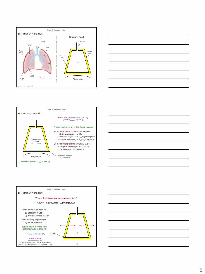
1) Pulmonary Ventilation:
Chapter 21: Respiratory System
Trachea
Thoracic
wall
Visceral
pleura
Parietal
pleura
Pleural
cavity
Lung
Marieb & Hoehn – Figure 21.12
Diaphragm Diaphragm
Simplified Model:
Trachea
Lung
Pleural
cavity
Thoracic
wall
Pressure relationships in the thoracic cavity:
Atmospheric pressure = ~ 760 mm Hg
(Consider Patmospheric = 0 mm Hg)
1) Intrapulmonary Pressure (w/in the alveoli):
• Static conditions = 0 mm Hg
2) Intrapleural pressure (w/in pleural cavity):
• Always relatively negative (~ - 4 mm Hg)
atmospheric pressure = Patm = 0 mm Hg
Intrapleural pressure (Pip = - 4 mm Hg)
1) Pulmonary Ventilation:
Chapter 21: Respiratory System
Diaphragm
Intrapulmonary
pressure (Ppul = 0 mm Hg)
• Inhalation (inspiration) = Ppul slightly negative
• Exhalation (expiration) = Ppul slightly positive
• Prevents lungs from collapsing
Why is the intrapleural pressure negative?
Answer: Interaction of opposing forces
Forces acting to collapse lung:
1) Elasticity of lungs
2) Alveolar surface tension
Force resisting lung collapse:
1) Rigid chest wall
Surface tension of serous fluids
keep lungs “stuck” to chest wall
Forces equilibrate at Pip = - 4 mm Hg
Pneumothorax: (“sucking chest wound”)
Puncture of chest wall – results in inability to
generate negative pressure and expand the lungs
Chapter 21: Respiratory System
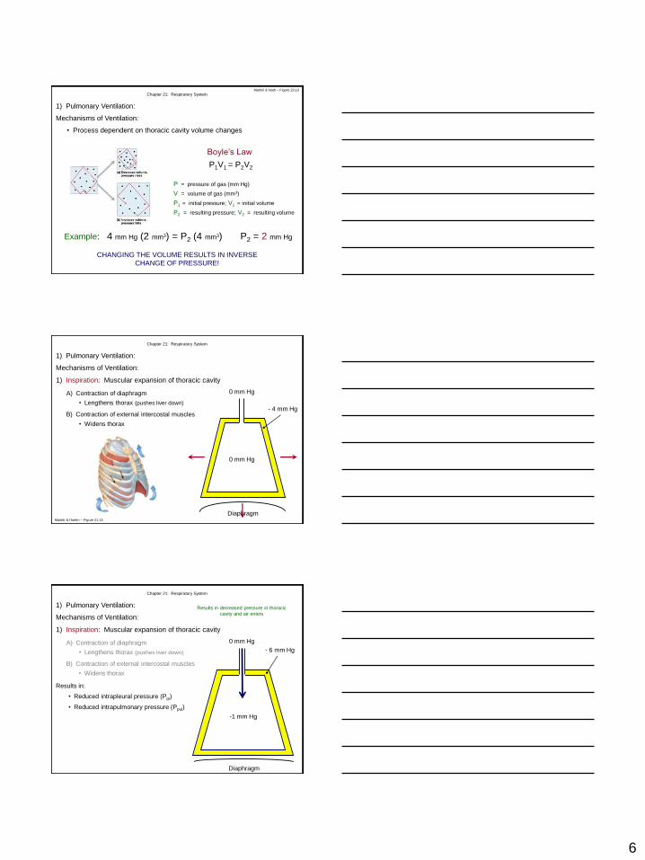
1) Pulmonary Ventilation:
6
Mechanisms of Ventilation:
• Process dependent on thoracic cavity volume changes
CHANGING THE VOLUME RESULTS IN INVERSE
CHANGE OF PRESSURE!
P = pressure of gas (mm Hg)
V = volume of gas (mm3)
P1 = initial pressure; V1 = initial volume
P2 = resulting pressure; V2 = resulting volume
Boyle’s Law
P1V1 = P2V2
Example: 4 mm Hg (2 mm3) = P2 (4 mm3) P2 = 2 mm Hg
Chapter 21: Respiratory System
1) Pulmonary Ventilation:
Martini & Nath – Figure 23.13
1) Inspiration: Muscular expansion of thoracic cavity
Diaphragm
0 mm Hg
0 mm Hg
- 4 mm Hg
A) Contraction of diaphragm
• Lengthens thorax (pushes liver down)
B) Contraction of external intercostal muscles
Mechanisms of Ventilation:
Chapter 21: Respiratory System
1) Pulmonary Ventilation:
Marieb & Hoehn – Figure 21.13
• Widens thorax
1) Inspiration: Muscular expansion of thoracic cavity
A) Contraction of diaphragm
• Lengthens thorax (pushes liver down)
B) Contraction of external intercostal muscles
• Widens thorax
Mechanisms of Ventilation:
Chapter 21: Respiratory System
1) Pulmonary Ventilation:
- 6 mm Hg
-1 mm Hg
Diaphragm
0 mm Hg
Results in:
• Reduced intrapleural pressure (Pip)
• Reduced intrapulmonary pressure (Ppul)
Results in decreased pressure in thoracic
cavity and air enters
7
0 mm Hg
Diaphragm
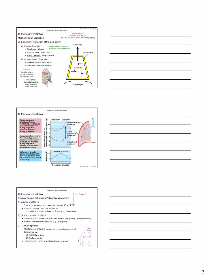
Mechanisms of Ventilation:
2) Expiration: Retraction of thoracic cavity
A) Passive Expiration
• Diaphragm relaxes
- 4 mm Hg
1 mm Hg
Results in increased pressure
in thoracic cavity and air exits
B) Active (“Forced”) Expiration
• Abdominal muscles contract
• Internal intercostals contract
Internal pressure
can reach +100 mm Hg
(e.g., why you should exhale when lifting weights)
Eupnea:
Quiet breathing
(active inspiration;
(passive expiration)
Chapter 21: Respiratory System
1) Pulmonary Ventilation:
Marieb & Hoehn – Figure 21.13
• External intercostals relax
• Elastic rebound (lungs rebound)
Hyperpnea:
Forced breathing
(active inspiration;
(active expiration)
Chapter 21: Respiratory System
1) Pulmonary Ventilation:
Marieb & Hoehn – Figure 21.14
B) Surface tension in alveoli
Physical Factors Influencing Pulmonary Ventilation:
A) Airway resistance
C) Lung compliance Barrel
Chest
R = 1 / radius4
Asthma - allergic response to irritants
• Constriction of bronchioles = radius = resistance
Chapter 21: Respiratory System
1) Pulmonary Ventilation:
• Flow of air = change in pressure / resistance (F = P / R)
• Moist alveolar surfaces attract to one another (H2O polarity – collapses alveoli)
• Alveolar cells secrete surfactant (e.g., detergents)
• “Stretchiness” of lung ( compliance = easier to expand lung)
• Determined by:
a) Elasticity of lung
b) Surface tension
Emphysema: Lungs lose elasticity (too compliant)
8
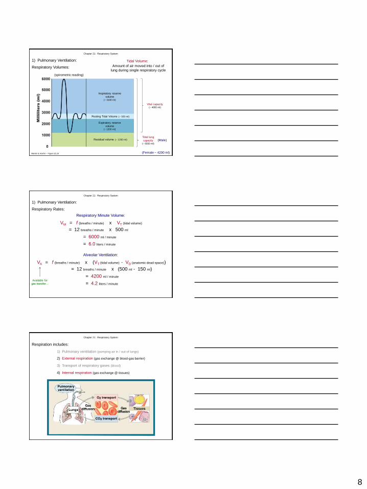
Respiratory Volumes:
Tidal Volume:
Amount of air moved into / out of
lung during single respiratory cycle
Chapter 21: Respiratory System
1) Pulmonary Ventilation:
(spirometric reading)
Resting Tidal Volume (~ 500 ml)
Inspiratory reserve
volume (~ 3100 ml)
Expiratory reserve
volume (~ 1200 ml)
Residual volume (~ 1200 ml)
Vital capacity (~ 4800 ml)
Total lung
capacity (~ 6000 ml)
(Male)
(Female ~ 4200 ml) Marieb & Hoehn – Figure 21.16
VM = f (breaths / minute) x VT (tidal volume)
= 12 breaths / minute x 500 ml
= 6000 ml / minute
Respiratory Minute Volume:
VA = f (breaths / minute) x (VT (tidal volume) - VD (anatomic dead space))
= 12 breaths / minute x (500 ml - 150 ml)
= 4200 ml / minute
Alveolar Ventilation:
Available for
gas transfer…
Chapter 21: Respiratory System
Respiratory Rates:
1) Pulmonary Ventilation:
= 6.0 liters / minute
= 4.2 liters / minute
Respiration includes:
1) Pulmonary ventilation (pumping air in / out of lungs)
Chapter 21: Respiratory System
2) External respiration (gas exchange @ blood-gas barrier)
3) Transport of respiratory gases (blood)
4) Internal respiration (gas exchange @ tissues)
9
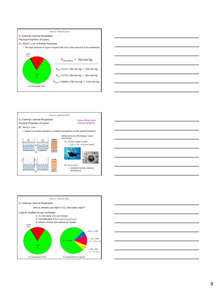
A) Dalton’s Law of Partial Pressures:
Patmosphere = 760 mm Hg
PO2 = 0.21 x 760 mm Hg = 159 mm Hg
PN2 = 0.79 x 760 mm Hg = 601 mm Hg
PCO2 = 0.0004 x 760 mm Hg = 0.30 mm Hg
• The total pressure of a gas is equal to the sum of the pressure of its constituents
Physical Properties of Gases:
% Composition of Air
21%
O2
79%
N2
0.04%
CO2
Chapter 21: Respiratory System
2) External / Internal Respiration:
B) Henry’s Law:
Gases diffuse down
pressure gradients
Additional factors affecting gas / liquid
interchange:
A) Solubility of gas in water
• CO2 >> O2 >> N2 (the “bends”)
B) Temperature
• Solubility inversely related to
temperature
Chapter 21: Respiratory System
Physical Properties of Gases:
• Gases in a mixture dissolve in a liquid in proportion to their partial pressures
2) External / Internal Respiration:
N2 = 74.5%
H2O = 6.2%
O2 = 13%
CO2 = 6%
% Composition of Lung Air
Why is alveolar gas high in CO2 and water vapor?
Lung air modified by gas exchange:
1) O2 into blood; CO2 out of blood
2) Humidification of air (conducting pathways)
3) Mixture of fresh and residual air / breath
% Composition of Air
21%
O2
79%
N2
0.04%
CO2
PO2 ~ 100 mm Hg
PCO2 ~ 40 mm Hg
Chapter 21: Respiratory System
2) External / Internal Respiration:
10
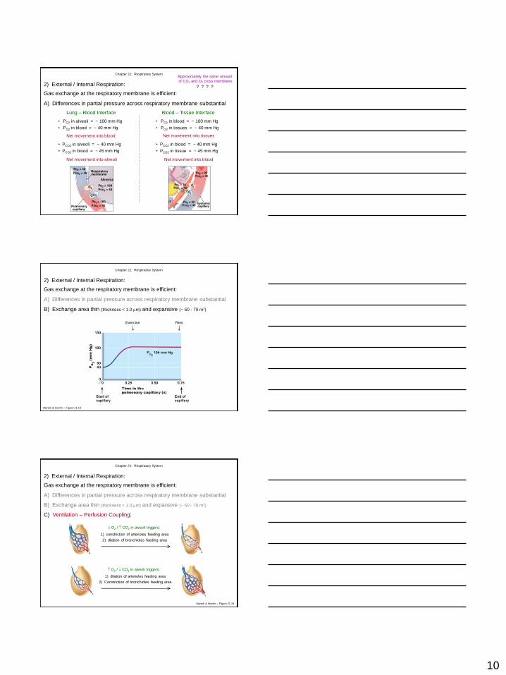
Gas exchange at the respiratory membrane is efficient:
Net movement into blood
A) Differences in partial pressure across respiratory membrane substantial
• PCO2 in alveoli = ~ 40 mm Hg
• PCO2 in blood = ~ 45 mm Hg
Net movement into alveoli
Chapter 21: Respiratory System
2) External / Internal Respiration:
• PO2 in alveoli = ~ 100 mm Hg
• PO2 in blood = ~ 40 mm Hg
Lung – Blood Interface
Approximately the same amount
of CO2 and O2 cross membrane
? ? ? ?
Blood – Tissue Interface
• PO2 in blood = ~ 100 mm Hg
• PO2 in tissues = ~ 40 mm Hg
Net movement into tissues
• PCO2 in blood = ~ 40 mm Hg
• PCO2 in tissue = ~ 45 mm Hg
Net movement into blood
Gas exchange at the respiratory membrane is efficient:
B) Exchange area thin (thickness < 1.0 m) and expansive (~ 50 - 70 m2)
Rest Exercise
Chapter 21: Respiratory System
Marieb & Hoehn – Figure 21.18
2) External / Internal Respiration:
A) Differences in partial pressure across respiratory membrane substantial
Gas exchange at the respiratory membrane is efficient:
B) Exchange area thin (thickness < 1.0 m) and expansive (~ 50 - 70 m2)
Chapter 21: Respiratory System
Marieb & Hoehn – Figure 21.19
2) External / Internal Respiration:
A) Differences in partial pressure across respiratory membrane substantial
C) Ventilation – Perfusion Coupling:
O2 / CO2 in alveoli triggers:
1) constriction of arterioles feeding area
2) dilation of bronchioles feeding area
O2 / CO2 in alveoli triggers:
1) dilation of arterioles feeding area
2) Constriction of bronchioles feeding area
11
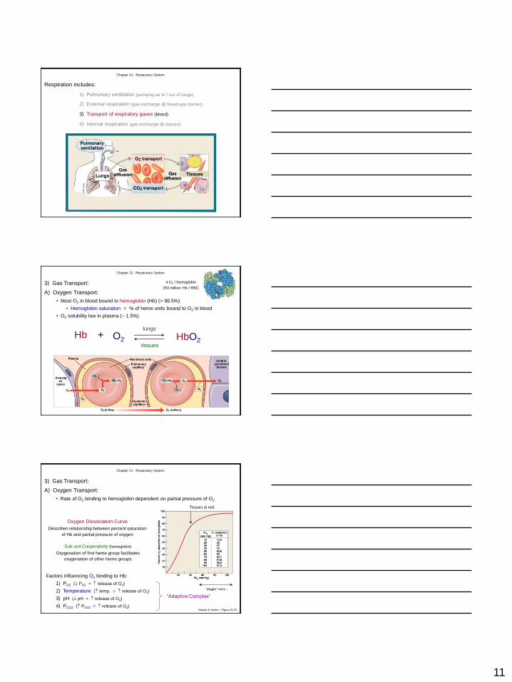
Respiration includes:
1) Pulmonary ventilation (pumping air in / out of lungs)
Chapter 21: Respiratory System
2) External respiration (gas exchange @ blood-gas barrier)
3) Transport of respiratory gases (blood)
4) Internal respiration (gas exchange @ tissues)
A) Oxygen Transport:
• Most O2 in blood bound to hemoglobin (Hb) (> 98.5%)
• Hemoglobin saturation = % of heme units bound to O2 in blood
Hb + O2 HbO2 lungs
tissues
4 O2 / hemoglobin
250 million Hb / RBC
• O2 solubility low in plasma (~ 1.5%)
Chapter 21: Respiratory System
3) Gas Transport:
• Rate of O2 binding to hemoglobin dependent on partial pressure of O2
Oxygen Dissociation Curve
Describes relationship between percent saturation
of Hb and partial pressure of oxygen
Sub-unit Cooperativity (hemoglobin)
Oxygenation of first heme group facilitates
oxygenation of other heme groups
“wiggle” room…
Tissues at rest
Factors influencing O2 binding to Hb:
1) PO2 ( PO2 = release of O2)
2) Temperature ( temp. = release of O2)
3) pH ( pH = release of O2)
4) PCO2 ( PCO2 = release of O2)
“Adaptive Complex”
Chapter 21: Respiratory System
Marieb & Hoehn – Figure 21.20
A) Oxygen Transport:
3) Gas Transport:
12
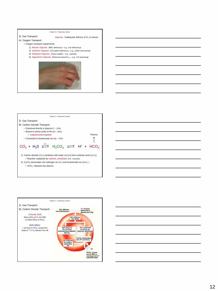
• Oxygen transport impairments:
Hypoxia: Inadequate delivery of O2 to tissues
1) Anemic Hypoxia (RBC deficiency – e.g., iron deficiency)
2) Ischemic Hypoxia (Circulation deficiency – e.g., sickle-cell anemia)
3) Histotoxic Hypoxia (Tissue uptake – e.g., cyanide)
4) Hypoxemic Hypoxia (Reduced arterial PO2 – e.g., CO poisoning)
Chapter 21: Respiratory System
A) Oxygen Transport:
3) Gas Transport:
• Dissolved directly in plasma (7 – 10%)
• Bound to amino acids of Hb (20 – 30%)
• Carbaminohemoglobin
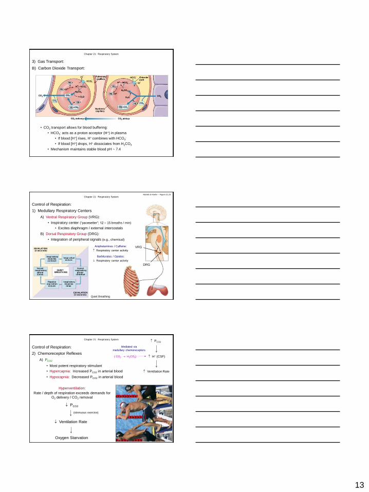
• Converted to bicarbonate ion (60 – 70%)
+ CO2 H20 H2CO3 H+ + HCO3-
1) Carbon dioxide (CO2) combines with water (H2O) to form carbonic acid (H2CO3)
• Reaction catalyzed by carbonic anhydrase (CA - enzyme)
CA
2) H2CO3 dissociates into hydrogen ion (H+) and bicarbonate ion (HCO3- )
• HCO3- released into plasma
Plasma
Chapter 21: Respiratory System
B) Carbon Dioxide Transport:
3) Gas Transport:
Chloride Shift:
Mass influx of Cl- into RBC
to offset efflux of HCO3-
Bohr Effect:
pH due to HCO3- production
leads to in O2 release from Hb
Chapter 21: Respiratory System
B) Carbon Dioxide Transport:
3) Gas Transport:
13
• CO2 transport allows for blood buffering:
• HCO3- acts as a proton acceptor (H+) in plasma
• If blood [H+] rises, H+ combines with HCO3-
• If blood [H+] drops, H+ dissociates from H2CO3
• Mechanism maintains stable blood pH ~ 7.4
Chapter 21: Respiratory System
B) Carbon Dioxide Transport:
3) Gas Transport:
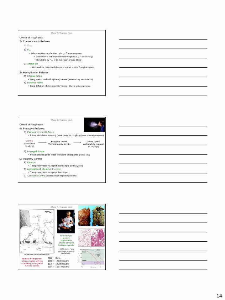
Control of Respiration:
1) Medullary Respiratory Centers
A) Ventral Respiratory Group (VRG):
Chapter 21: Respiratory System
VRG
DRG
Quiet Breathing
Ventral Ventral
Amphetamines / Caffeine:
Respiratory center activity
Barbiturates / Opiates:
Respiratory center activity
Marieb & Hoehn – Figure 21.23
B) Dorsal Respiratory Group (DRG):
• Inspiratory center (“pacesetter”; 12 – 15 breaths / min)
• Excites diaphragm / external intercostals
• Integration of peripheral signals (e.g., chemical)
A) PCO2
PCO2
H+ (CSF)
Ventilation Rate
( CO2 H2CO3)
Mediated via
medullary chemoreceptors
• Hypocapnia: Decreased PCO2 in arterial blood
• Most potent respiratory stimulant
• Hypercapnia: Increased PCO2 in arterial blood
Chapter 21: Respiratory System
Control of Respiration:
2) Chemoreceptor Reflexes
Hyperventilation:
Rate / depth of respiration exceeds demands for
O2 delivery / CO2 removal
PCO2
Ventilation Rate
Oxygen Starvation
(strenuous exercise)
14
A) PCO2
Chapter 21: Respiratory System
Control of Respiration:
2) Chemoreceptor Reflexes
B) PO2
• Minor respiratory stimulant ( O2 = respiratory rate)
• Mediated via peripheral chemoreceptors (e.g., carotid artery)
• Stimulated by PO2 < 60 mm Hg in arterial blood
C) Arterial pH
• Mediated via peripheral chemoreceptors ( pH = respiratory rate)
3) Hering-Breuer Reflexes
A) Inflation Reflex
• Lung stretch inhibits inspiratory center (prevents lung over-inflation)
B) Deflation Reflex
• Lung deflation inhibits expiratory center (during active expiration)
4) Protective Reflexes:
B) Laryngeal Spasm
• Irritant around glottis leads to closure of epiglottis (protect lung)
Apnea
(cessation of
breathing)
Epiglottis closes;
Thoracic cavity shrinks
5) Voluntary Control:
Glottis opens;
Air forcefully released (~ 100 mph)
Chapter 21: Respiratory System
Control of Respiration:
A) Pulmonary Irritant Reflexes:
• Irritant stimulates sneezing (nasal cavity) or coughing (lower conduction system)
A) Emotion:
• respiratory rate via hypothalamic input (limbic system)
B) Anticipation of Strenuous Exercise:
• respiratory rate via sympathetic input
C) Conscious Control (bypass / block respiratory centers)
1900 = Rare…
1956 = 29,000 deaths
1978 = 105,000 deaths
2004 = 160,440 deaths
Increase in lung cancer
rates correlated with rise
in smoking among adult
men and women
formaldehyde
benzene
vinyl chloride
arsenic ammonia
hydrogen cyanide
~ 3,400 deaths / year
contributed to second-
hand smoke…
Chapter 21: Respiratory System