Embed Size (px)
Citation preview
The Respiratory
System
2
Respiration Includes Pulmonary ventilation
Air moves in and out of lungs Continuous replacement of gases in alveoli (air sacs)
External respiration Gas exchange between blood and air at alveoli O2 (oxygen) in air diffuses into blood CO2 (carbon dioxide) in blood diffuses into air
Transport of respiratory gases Between the lungs and the cells of the body Performed by the cardiovascular system Blood is the transporting fluid
Internal respiration Gas exchange in capillaries between blood and tissue cells O2 in blood diffuses into tissues CO2 waste in tissues diffuses into blood
3
Cellular Respiration
Oxygen (O2) is used by the cells O2 needed in conversion of glucose to
cellular energy (ATP) All body cells Carbon dioxide (CO2) is produced as a
waste product The body’s cells die if either the
respiratory or cardiovascular system fails
4
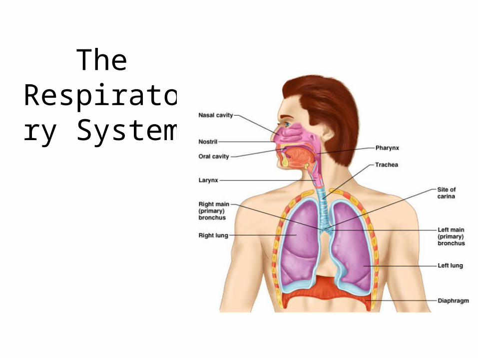
The Respiratory Organs
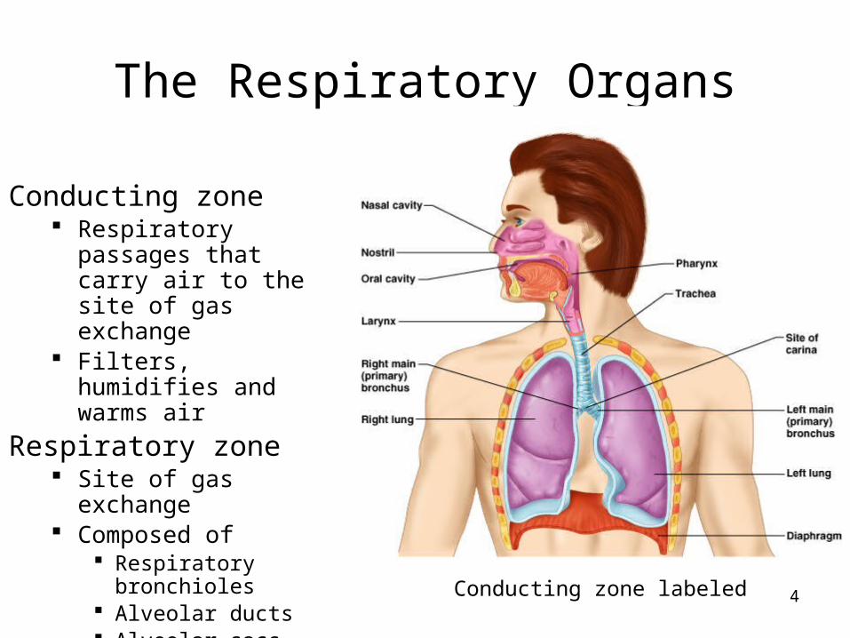
Conducting zone Respiratory passages
that carry air to the site of gas exchange
Filters, humidifies and warms air
Respiratory zone Site of gas exchange Composed of
Respiratory bronchioles Alveolar ducts Alveolar sacs
Conducting zone labeled
5
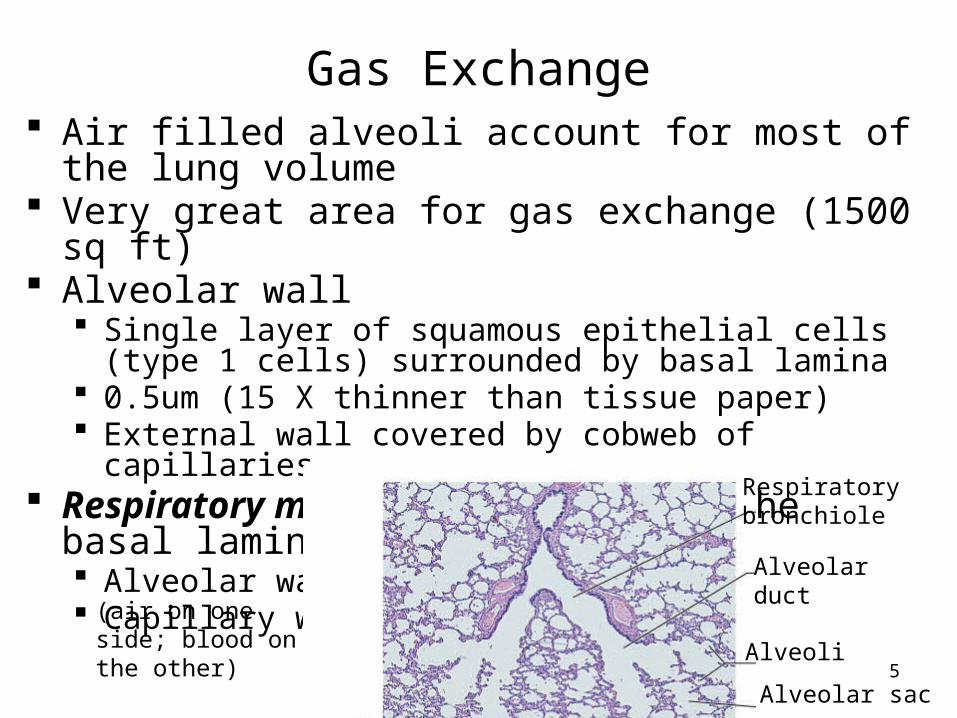
Gas Exchange Air filled alveoli account for most of the lung volume Very great area for gas exchange (1500 sq ft) Alveolar wall
Single layer of squamous epithelial cells (type 1 cells) surrounded by basal lamina
0.5um (15 X thinner than tissue paper) External wall covered by cobweb of capillaries
Respiratory membrane: fusion of the basal laminas of Alveolar wall Capillary wall
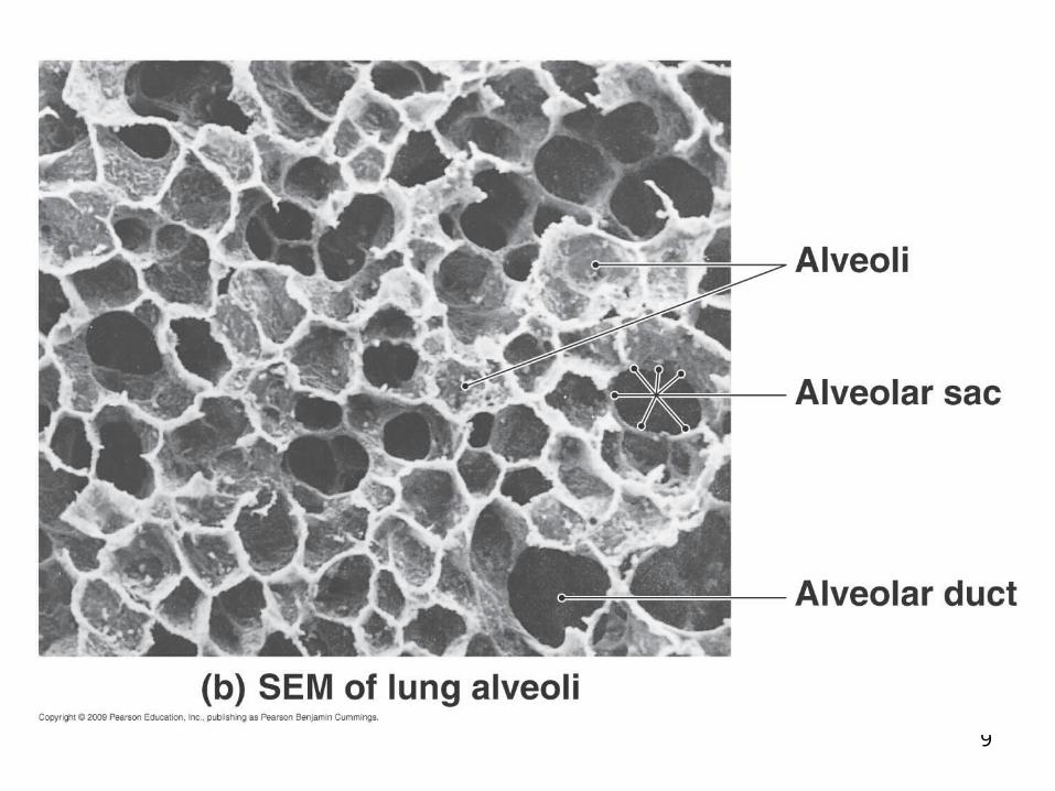
Alveolar sac
Respiratorybronchiole
Alveolarduct
Alveoli
(air on one side; blood on the other)
6
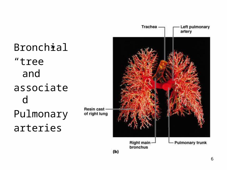
Bronchial
“tree” and
associated
Pulmonary
arteries
7
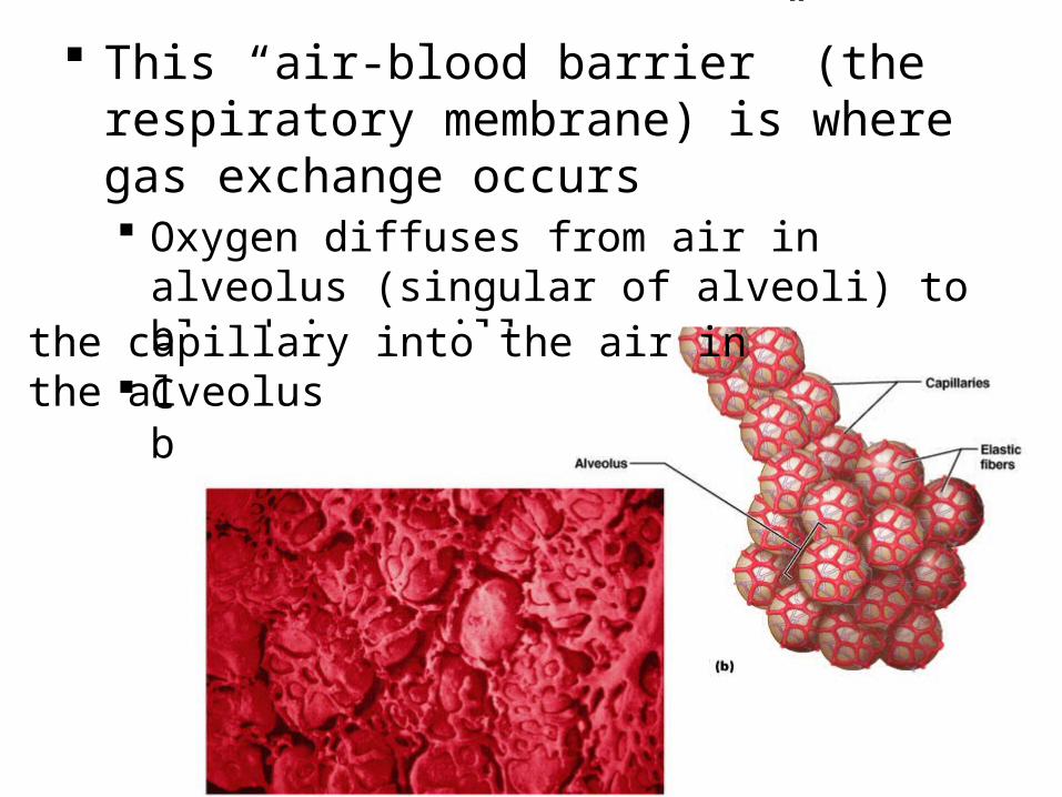
This “air-blood barrier” (the respiratory membrane) is where gas exchange occurs Oxygen diffuses from air in alveolus (singular
of alveoli) to blood in capillary Carbon dioxide diffuses from the blood in
the capillary into the air inthe alveolus
8
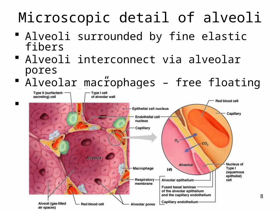
Microscopic detail of alveoli Alveoli surrounded by fine elastic fibers Alveoli interconnect via alveolar pores Alveolar macrophages – free floating “dust cells” Note type I and type II cells and joint membrane
9
10
Ventilation
Breathing = “pulmonary ventilation” Pulmonary means related to the lungs
Two phases Inspiration (inhalation) – air in Expiration (exhalation) – air out
Mechanical forces cause the movement of air Gases always flow from higher pressure to lower For air to enter the thorax, the pressure of the air in it
has to be lower than atmospheric pressure Making the volume of the thorax larger means the air inside it
is under less pressure(the air has more space for as many gas particles, therefore it is under less pressure)
The diaphragm and intercostal muscles accomplish this
11
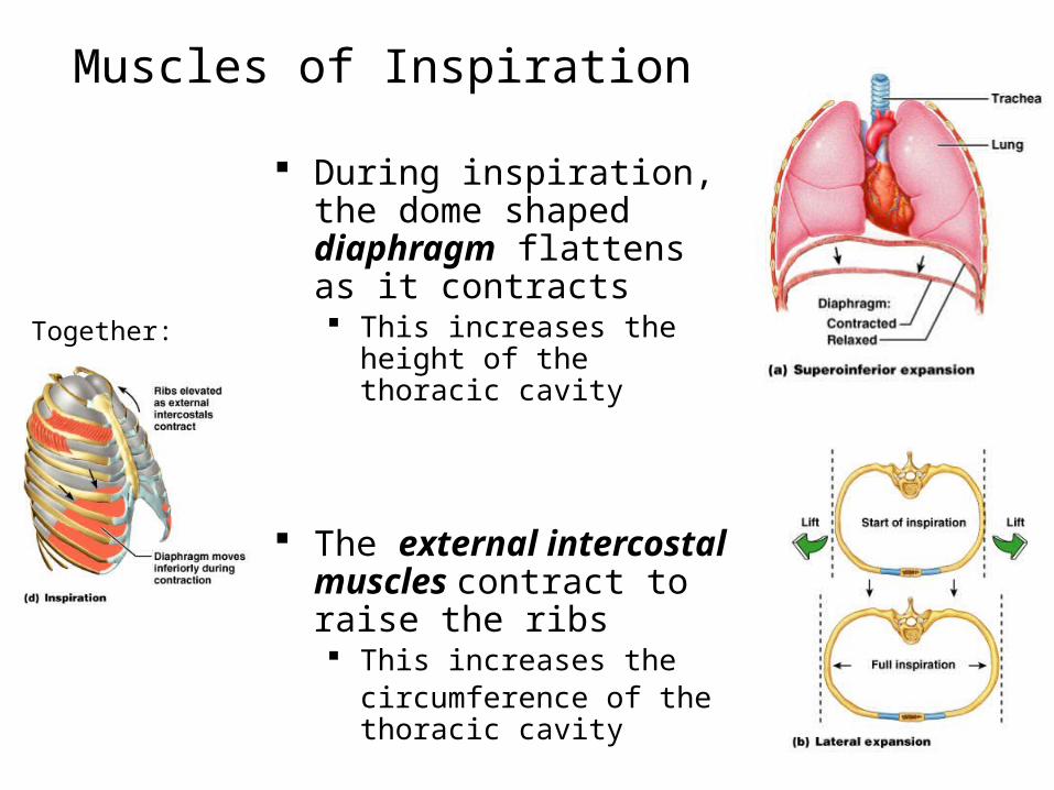
Muscles of Inspiration
During inspiration, the dome shaped diaphragm flattens as it contracts This increases the height of
the thoracic cavity
The external intercostal muscles contract to raise the ribs This increases the
circumference of the thoracic cavity
Together:
12
Inspiration continued
Intercostals keep the thorax stiff so sides don’t collapse in with change of diaphragm
During deep or forced inspiration, additional muscles are recruited: Scalenes Sternocleidomastoid Pectoralis minor Quadratus lumborum on 12th rib Erector spinae
(some of these “accessory muscles” of ventilation are visible to an observer; it usually tells you that there is respiratory distress – working hard to breathe)
13
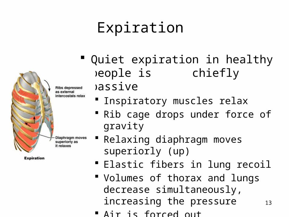
Expiration
Quiet expiration in healthy people is chiefly passive Inspiratory muscles relax Rib cage drops under force of gravity Relaxing diaphragm moves superiorly
(up) Elastic fibers in lung recoil Volumes of thorax and lungs decrease
simultaneously, increasing the pressure Air is forced out
14
Expiration continued
Forced expiration is active Contraction of abdominal wall muscles
Oblique and transversus predominantly Increases intra-abdominal pressure forcing the
diaphragm superiorly Depressing the rib cage, decreases thoracic
volume Some help from internal intercostals and latissimus
dorsi
(try this on yourself to feel the different muscles acting)