Embed Size (px)
Citation preview
HEAD AND NECK TUMORS 39
CHAPTER 4
Head and neck tumorsJohn Andrew Ridge, MD, PhD, Bonnie S. Glisson, MD, Eric M. Horwitz, MD,and Michael O. Meyers, MD
In 2003, it is estimated that head and neck cancers will comprise 2%-3% of allcancers in the United States and account for 1%-2% of all cancer deaths. Thistotal includes 19,400 cases of oral cavity cancers, 9,500 cases of laryngealcancer, and 8,300 cases of pharyngeal cancer. Most patients with head andneck cancer have metastatic disease at the time of diagnosis (regional nodalinvolvement in 43% and distant metastasis in 10%).
Head and neck cancers encompass a diverse group of uncommon tumors thatfrequently are aggressive in their biological behavior. Moreover, patients witha head and neck cancer often develop a second primary tumor. These tumorsoccur at an annual rate of 3%-7%, and 50%-75% of such new cancers occur inthe upper aerodigestive tract or lungs.
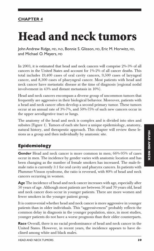
The anatomy of the head and neck is complex and is divided into sites andsubsites (Figure 1). Tumors of each site have a unique epidemiology, anatomy,natural history, and therapeutic approach. This chapter will review these le-sions as a group and then individually by anatomic site.
Epidemiology
Gender Head and neck cancer is more common in men; 66%-95% of casesoccur in men. The incidence by gender varies with anatomic location and hasbeen changing as the number of female smokers has increased. The male-fe-male ratio is currently 3:1 for oral cavity and pharynx cancers. In patients withPlummer-Vinson syndrome, the ratio is reversed, with 80% of head and neckcancers occurring in women.
Age The incidence of head and neck cancer increases with age, especially after50 years of age. Although most patients are between 50 and 70 years old, headand neck cancer does occur in younger patients. There are more women andfewer smokers in the younger patient group.
It is controversial whether head and neck cancer is more aggressive in youngerpatients than in older individuals. This “aggressiveness” probably reflects thecommon delay in diagnosis in the younger population, since, in most studies,younger patients do not have a worse prognosis than their older counterparts.
Race Overall, there is no racial predominance of head and neck cancer in theUnited States. However, in recent years, the incidence appears to have de-clined among white and black males.
HEA
D A
ND
NEC
K
40 CANCER MANAGEMENT: A MULTIDISCIPLINARY APPROACH
Among blacks, head and neck cancer is associated with lower survival for simi-lar tumor stages. The overall 5-year survival rate is 56% in whites and 34% inblacks.
Geography There are wide variations in the incidence of head and neck can-cer among different geographic regions. The risk of laryngeal cancer, for ex-ample, is two to six times higher in Bombay, India, than in Scandinavia. Thehigher incidence of the disease in Asia is thought to reflect the prevalence ofrisk factors, such as betel nut chewing and use of smokeless tobacco. In theUnited States, the high incidence among urban males is thought to reflect ex-posure to tobacco and alcohol. Among rural women, there is an increased riskof oral cancer related to the use of smokeless tobacco (snuff).
HEA
D A
ND
NEC
K
FIGURE 1: Anatomic sites and subsites of the head and neck.

HEAD AND NECK TUMORS 41
Nasopharyngeal carcinoma is another head and neck tumor with a distinct ethnicpredilection. Endemic areas include southern China, northern Africa, and re-gions of the far Northern Hemisphereareas in which the diet of inhabitantsincludes large quantities of salted meat and fish. When people from these regionsmigrate to areas with a lower disease incidence, their risk falls but remains el-evated. Cancer of the nasopharynx in these geographic areas also has been asso-ciated with Epstein-Barr virus (EBV) infection (see “Etiology and risk factors”section).
Disease site The approximate distribution of head and neck cancer follows:oral cavity, 44%; larynx, 31%; and pharynx, 25%.
Etiology and risk factors
Risk factors for head and neck cancer include tobacco and alcohol use, ultra-violet (UV) light exposure, viral infection, and environmental exposures.
Tobacco The incidence of head and neck tumors correlates most closely withthe use of tobacco.
Cigarettes Head and neck tumors occur six times more often among cigarettesmokers than nonsmokers. The age-standardized risk of mortality from laryngealcancer appears to rise linearly with increasing cigarette consumption. For theheaviest smokers, death from laryngeal cancer is 20 times more likely than fornonsmokers. Furthermore, active smoking by head and neck cancer patients isassociated with significant increases in the annual rate of second primary tu-mor development (compared with former smokers or those who have neversmoked). Use of unfiltered cigarettes or dark, air-cured tobacco is associatedwith further increases in risk.
Cigars Total cigar consumption increased by nearly 50% in the United States inthe 1990s. Often misperceived as posing a lower health risk than cigarette smok-ing, cigar smoking results in a change in the site distribution for aerodigestivetract cancer, according to epidemiologic data. Although the incidence of can-cer at some sites traditionally associated with cigarette smoking (eg, larynx,lungs) is decreased in cigar smokers, the incidence of cancer is actually higherat other sites where pooling of saliva and associated carcinogens tends to occur(oropharynx, esophagus).
Smokeless tobacco Use of smokeless tobacco also is associated with an increasedincidence of head and neck cancer, especially in the oral cavity. Smokelesstobacco users frequently develop premalignant lesions, such as oral leukoplakia,at the site where the tobacco quid rests against the mucosa. Over time, theselesions may progress to invasive carcinomas. The use of snuff has been associ-ated with an increase in cancers of the gum and oral mucosa.
Alcohol Alcohol consumption, by itself, is a risk factor for the developmentof pharyngeal and laryngeal tumors, although it is a less potent carcinogenthan tobacco. For individuals who use both tobacco and alcohol, these riskfactors appear to be synergistic and result in a multiplicative increase in risk.

HEAD AND NECK TUMORS 43
Marijuana Smoking marijuana is associated with the development of headand neck cancer, but the degree of risk is unknown.
Anatomy
As mentioned above, the anatomy of the head and neck region is complex.The anatomic sites are illustrated in Figure 1. More detailed descriptions areincluded below in the discussions of specific sites and subsites.
Levels of the neck
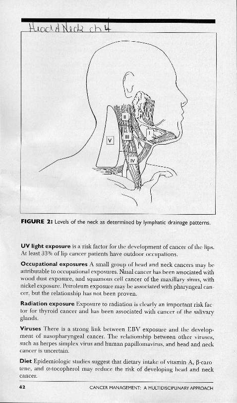
The anatomy of the neck is relevant to the treatment of all head and neckcancers. The neck may be divided into levels (Figure 2). The lymphatic drain-age of the unmanipulated neck is systematic and predictable; knowledge ofthese drainage patterns assists the clinician in locating the primary tumor thathas given rise to a neck metastasis (Table 1).
Signs and symptoms
Head and neck cancer typically produces symptoms referable to the upperaerodigestive tract, including alterations in deglutition, phonation, hearing,and respiration. In particular, patients should be questioned about dysph-agia, odynophagia, globus sensation, hoarseness, a change in the ability toform words, epistaxis, epiphora, otalgia, hemoptysis, stuffiness of the ears,and trismus. (Signs and symptoms of cancer at specific anatomic sites andsubsites can be found in the respective discussions of these tumors.)
It is important to ascertain the duration and course (progression or im-provement) of symptoms. Progression of disease is often noted during theevaluation and worsens prognosis.
Level I includes the submental and submandibular triangles.
Level II includes the superior jugular chain nodes extending from the man-dible down to the carotid bifurcation and posteriorly to the posterior borderof the sternocleidomastoid muscle.
Level III consists of the jugular nodes from the carotid bulb inferiorly to theomohyoid muscle.
Level IV continues from the omohyoid muscle inferiorly to the clavicle.
Level V represents the posterior triangle bounded by the sternocleidomas-toid anteriorly, the trapezius posteriorly, and the omohyoid inferiorly. Fewlesions metastasize to level V without involvement of more central nodes.
44 CANCER MANAGEMENT: A MULTIDISCIPLINARY APPROACH
Screening and diagnosis
SCREENING
Because the cure rates for early-stage head and neck cancers are high, the conceptof screening for the disease has intuitive appeal. Evaluation of asymptomaticindividuals has not been shown to decrease mortality from head and neckcancer, however. The US Preventive Health Service Task Force does not rec-ommend screening for oral cancer due to the lack of evidence supporting screen-ing as a means of decreasing mortality. In countries with a high incidence oforal cavity cancer, such as India, screening may be helpful and is currentlyunder evaluation.
DIAGNOSIS
The need for expeditious diagnosis of head and neck cancer and referral to askilled head and neck specialist cannot be overemphasized, as early diagnosiscan lead to a reduction in mortality. One study suggested that in the 24 monthsprior to the diagnosis of head and neck cancer, patients had a median of 10.5health-care visits. These visits provide an opportunity to evaluate patients’ symp-toms and underscore the important role of dentists and primary care physi-cians in the early diagnosis of head and neck cancer.
TABLE 1: Lymphatic drainage of the head and neckand associated sites of primary tumors
Lymphatic drainage Likely primary sites
Level ISubmental Lower lip, chin, anterior oral cavity (including anterior
one-third of the tongue and floor of the mouth)
Submandibular Upper and lower lips, oral tongue, floor of the mouth,facial skin
Level II Oral cavity and pharynx (including soft palate, base of thetongue, and piriform sinus)
Level III Larynx, hypopharynx, and thyroid
Level IV Larynx, hypopharynx, thyroid, cervical esophagus, andtrachea
Level V Nasopharynx, thyroid, paranasal sinuses, and posteriorscalp
Supraclavicular Infraclavicular sites (including lungs, esophagus, breasts,pancreas, GI tract, GU and gynecologic sources)
HEAD AND NECK TUMORS 45
History
Risk factors as outlined above, including a history of tobacco and alcoholuse and environmental exposures, should be reviewed. Any adult patientwith symptoms referable to the upper aerodigestive tract that have lastedlonger than 2 weeks or with an asymptomatic neck mass should undergo athorough examination with a high index of suspicion for carcinoma.
Physical examination
The physical examination is the best means for detecting lesions of the upperaerodigestive tract. Frequently, the initial assessment also will indicate theseverity and chronicity of the disease. Due to the frequent occurrence ofmultiple primary tumors in patients with a head and neck tumor, carefulevaluation of the entire upper aerodigestive tract is necessary at the time ofdiagnosis. The examination should always follow a systematic approach.
Skin/scalp A search should be made for ulcers, nodules, and pigmented orother suspicious lesions. This part of the evaluation is frequently overlooked.
Cranial nerves A cranial nerve evaluation is essential for any patient witha head and neck tumor or neck mass (which may be a manifestation ofoccult cancer). This evaluation should include assessing eye motion (cra-nial nerve [CN] III, IV, and VI); testing sensation of the face (CN V); ex-amining the muscles of facial expression by having the patient grin, gri-mace, raise eyebrows, close eyes tightly, show teeth, and puff out the cheeks(CN VII); testing of hearing (CN VIII); assessing gag reflex (CN IX); evalu-ating vocal cord mobility (CN X); and having the patient fully abduct theshoulder (CN XI) and protrude the tongue (CN XII). Even the slightestabnormality may be helpful in identifying a primary tumor.
Eyes/ears/nose The eyes, ears, and nose should be evaluated for any sign ofmass effect, abnormal drainage/discharge, bleeding, or effusion.
Oral cavity Halitosis may be the first indication of a lesion in the upperaerodigestive tract. The teeth, gingivae, and entire mucosal surface shouldbe inspected. (Dentures should be removed.) The lymphoid tissue of the tonsil-lar pillars should be inspected and any asymmetry noted. Tongue mobilityalso should be evaluated.
The floor of the mouth, tongue, and cheeks should be palpated using a bi-manual technique (one gloved finger inside the mouth and the second handunder the mandible). Palpation should be the last step of the examinationdue to stimulation of the gag reflex. Worrisome lesions should be biopsied.
Neck A systematic examination of the neck consistently documents the lo-cation of any mass. Palpation is the cornerstone of the examination. It isperformed by grasping the tissue and feeling the nodes between the thumband index and long fingers. The relationship of a mass to major structures,such as the salivary gland, thyroid, and carotid sheath, should be considered.

46 CANCER MANAGEMENT: A MULTIDISCIPLINARY APPROACH
Important qualities of a mass include location, character, tenderness, size,mobility, and associated thrill or bruit. The thyroid should be palpated.
Indirect laryngoscopy The nasopharynx, hypopharynx, and larynx shouldall be examined with care. The vocal cords should be visualized and theirmobility evaluated. Mirror examination provides an overall impression ofmobility and asymmetry, which may point to a hidden tumor.
Direct laryngoscopy Nasopharyngoscopes permit a thorough inspectionof the upper aerodigestive tract in the office setting. Attention should befocused individually on the piriform sinuses, tongue base, pharyngeal walls,epiglottis, arytenoids, and true and false vocal cords. Also, any pooling ofsecretions should be noted.
Endoscopy Approximately 5% of patients with head and neck cancer havea synchronous primary squamous cell cancer of the head and neck, esopha-gus, or lungs. “Triple” endoscopy includes direct laryngoscopy, esophagos-copy, and bronchoscopy with directed biopsy and should be performed inall patients with an occult primary squamous cell cancer and in many pa-tients with a known head and neck primary. Triple endoscopy also can pro-vide information regarding the extent of the tumor.
The most common sites of silent primary tumors are the tonsils, base of thetongue, and piriform sinuses. Tumors of the nasopharynx have become eas-ier to identify with the increased use of flexible nasopharyngoscopy. Biopsiesshould be performed in common areas of silent primaries in addition to theprimary anatomic sites associated with lymphatic drainage of any neck mass.
Laboratory evaluation
There are no specific screening laboratory tests other than preoperativestudies performed in the diagnostic evaluation of most head and neckcarcinomas. EBV, anticapsid antibodies, and serum IgG are tumormarkers for nasopharyngeal carcinomas.
Diagnostic imaging
Plain x-rays PA and lateral chest x-rays should be obtained in all adultpatients to eliminate the possibility of occult lung metastasis or a secondprimary. A Panorex film may be helpful in delineating bony involvementin some cases of oral cavity lesions.
Ultrasonography is of limited use in evaluating squamous cell cancer ofthe head and neck.
CT The CT scan is probably the single most informative test in the assessmentof a head and neck tumor. It may delineate the extent of disease and the pres-ence and extent of lymphatic involvement and will distinguish cystic from solidlesions. CT scans of the chest, abdomen, and pelvis sometimes may identify thesite of an occult primary tumor presenting with a node low in the neck. CT
HEAD AND NECK TUMORS 47
offers high spatial resolution and discriminates among fat, muscle, bone, andother soft tissues and surpasses MRI in the detection of bony erosion.
Dynamic contrast CT provides an increased ability to distinguish blood vesselsfrom enlarged lymph nodes or masses and maintains image quality with theuse of less contrast agent.
Spiral CT is a faster approach than dynamic contrast CT and has the capabil-ity for multiplanar reconstruction while maintaining the quality of the scan.
MRI may provide accurate information regarding the size, location, andextent of tumor. The advantages of MRI over CT include discrimination oftumor from normal tissue, multiplanar imaging without patient reposition-ing, and the ability to depict blood vessels clearly without iodinated con-trast. The main disadvantage of MRI is movement artifact, which is a par-ticular problem in the larynx and hypopharynx. Gadolinium-enhanced MRIis probably superior to CT for imaging tumors of the nasopharynx andoropharynx.
Angiography There are two indications for arteriography: a pulsatile neckmass and clinical evidence of a paraganglioma. High-resolution angiogra-phy is preferred over digital subtraction angiography because the formerprovides more information and permits embolization to be performed.Angiography is not used routinely in evaluating other primary tumors ofthe head and neck. In particular, angiography is not used routinely to assessarterial invasion or determine resectability of primary laryngeal tumors.
Nuclear scans are helpful in evaluating hyperthyroid patients but otherwiseare of limited use.
PET has been evaluated in both primary and recurrent squamous cell carci-noma of the head and neck. Initial results are encouraging, but this tool shouldstill be considered investigational.
Biopsy
Biopsies of the primary tumor often can be performed in an outpatient setting.
Punch or cup forceps biopsy is important in the diagnosis of mucosal le-sions. The biopsy should be obtained at the border of the lesion away fromareas of obvious necrosis.
Fine-needle aspiration (FNA) is a useful diagnostic modality. Multiplepasses are made through the lesion with a fine-gauge (22-gauge) needle whilesuction is applied. Suction should be released before withdrawing the needlethrough surrounding soft tissue of the neck. FNA has an associated false-negative rate as low as 7%. The diagnostic accuracy depends on the physician’sskill and the cytopathologist’s experience.

48 CANCER MANAGEMENT: A MULTIDISCIPLINARY APPROACH
Cytology is particularly useful in distinguishing a metastatic squamous cellcarcinoma from other malignant histologies. However, a negative result shouldnot be interpreted as “absence of malignancy.”
Core biopsy should not be performed on a neck mass, with the rare excep-tion of a proven lymphoma.
Open biopsy should be performed only when a diagnosis has not been madeafter extensive clinical evaluation and FNA is nondiagnostic. The operationshould be performed only by a surgeon prepared to conduct immediate de-finitive surgical treatment at that time (which may entail radical neckdissection).
Pathology
Squamous cell carcinoma
More than 90% of all head and neck cancers are squamous cell carcinomas.
Histologic grade There are three histologic grades based on the amount ofkeratinization: A well-differentiated tumor is characterized by > 75%keratinization, a moderately differentiated tumor by 25%-50%, and a poorlydifferentiated tumor by < 25%.
In general, the more poorly differentiated a lesion, the higher is the inci-dence of regional metastases and the poorer the prognosis. Histologic gradehas not been a consistent predictor of clinical behavior, however. Featuresthat predict aggressive behavior include perineural spread, lymphatic inva-sion, and tumor spread beyond the lymph node capsule.
Morphologic growth patterns Four morphologically distinct growth pat-terns have been recognized. The ulcerative type is the most common formand begins as a round or oval ulcer that is friable. Ulcerative lesions progresstoward infiltration. Infiltrative lesions extend deeply into underlying tissues.The exophytic type tends to grow more superficially and metastasize laterthan the other types. It begins as an area of thickened epithelium.
Verrucous cancer is an uncommon variant that, in the United States, typi-cally occurs in elderly patients with poor oral hygiene or ill-fitting dentures.It is characterized by a warty, bulky, elevated, fungating appearance. Verru-cous cancers seldom metastasize.
Other tumor types
Other less common head and neck cancers include mucoepidermoid carci-noma, adenoid cystic carcinoma, and adenocarcinoma, all of which may arisein the salivary glands. Head and neck cancers with neuroendocrine featuresinclude small-cell undifferentiated cancer and esthesioneuroblastoma (olfac-tory neuroblastoma). Both Hodgkin’s disease and non-Hodgkin’s lymphomamay also be diagnosed as head and neck tumors, often involving the lymphnodes of the neck or Waldeyer’s ring.
HEAD AND NECK TUMORS 49
Sequence of disease progression
There is a sequence of disease progression from atypia/dysplasia throughcarcinoma in situ to frankly invasive cancer. Leukoplakia and erythroplakiaare terms applied to clinically identifiable lesions that may harbor invasivecancer or undergo malignant transformation.
Leukoplakia results from chronic irritation of mucous membranes bycarcinogens; this irritation stimulates the proliferation of white epithelial andconnective tissue. Histopathologic examination reveals hyperkeratosisvariably associated with underlying epithelial hyperplasia. In the absenceof underlying dysplasia, leukoplakia rarely (< 5%) is associated with progres-sion of disease to malignancy.
Erythroplakia is characterized by superficial, friable, red patches adjacentto normal mucosa. It is commonly associated with underlying epithelial dys-plasia and has a much greater potential for malignancy than leukoplakia.Carcinoma is found in nearly 40% of erythroplakia.
Dysplasia is characterized by cellular atypia, loss of normal maturation, andloss of normal epithelial stratification. It is graded as mild, moderate, or se-vere, based on the degree of nuclear abnormality present. In the transitionfrom mild to severe dysplasia, nuclear abnormalities become more marked,mitoses become more apparent, and these changes involve increasing depthof epithelium. The likelihood of developing a carcinoma relates to the de-gree of dysplasia. In the case of severe dysplasia, as many as 24% of patientsmay develop invasive squamous cell cancer.
Carcinoma in situ is characterized by the presence of atypical changesthroughout the epithelium with complete loss of stratification. It is estimatedthat approximately 75% of invasive squamous cell carcinomas have an asso-ciated in situ component. Specific DNA mutations have also been identifiedin the sequence of disease progression from mild dysplasia to atypia to carci-noma in situ to invasive carcinoma.
Regional and distant metastases
The incidence of lymph node metastases is related to the size and thicknessof the primary tumor. If the primary site is near the midline, contralateral orbilateral metastases should be anticipated. In the presence of lymph nodemetastases, extracapsular spread of tumor is an important prognostic factor.
Staging and prognosis
Staging system The TNM staging system of the American Joint Committeeon Cancer (AJCC) maintains uniformity in the staging of head and neck tu-mors. The staging of primary mucosal tumors of the head and neck varies withthe anatomic location and will be covered later by site. However, the stagingsystem for metastases and stage groupings are nearly uniform for all mucosalsites.
50 CANCER MANAGEMENT: A MULTIDISCIPLINARY APPROACH
Prognosis correlates strongly with stage at diagnosis. For many head and neckcancer sites, survival for patients with stage I disease exceeds 80%. For pa-tients with locally advanced disease at the time of diagnosis, stages III andIV disease, survival drops below 40%. Development of nodal metastases re-duces survival of a patient with a small primary tumor by ~50%. Involve-ment of even a single lymph node is associated with a marked decline insurvival. Most patients with head and neck cancer have stage III or IV dis-ease at diagnosis.
Pattern of relapse Despite aggressive primary treatment, the majority ofrelapses that occur following a head and neck cancer are within the head andneck. Locoregional relapse accounts for ~80% of primary treatment failures.Distant metastases increase as the disease progresses and most often involvethe lungs, bones, and liver. By the time of death, 10%-30% of patients willhave clinically detected distant metastases.
Field cancerization is an important concept related to the natural history ofhead and neck cancer. This term describes the diffuse epithelial injury through-out the head and neck, lungs, and esophagus that results from chronic expo-sure to carcinogens.
Clinically, field cancerization is manifested by the frequent occurrence of (1)mucosal abnormalities, such as leukoplakia and dysplasia, beyond the mar-gins of a head and neck cancer and (2) second primary tumors within thisexposed field. The lifetime risk of a head and neck cancer patient developinga new cancer is 20%-40%. Over time, as the risk of relapse of the initial can-cer declines, the development of a new cancer represents the greatest risk forthese patients.
Treatment approaches
Head and neck tumors may be treated with curative intent using surgery,radiation therapy, or a combination of the two modalities. Chemotherapymay be combined with irradiation (chemoradiation) in the management ofadvanced (stage III/IV) lesions of the oropharynx, hypopharynx, and lar-ynx and for nasopharyngeal cancers more advanced than stage T2b.
Stages I and II disease at most sites may be treated with either resection orradiation therapy. The best therapeutic approach for the primary tumor de-pends on the anatomic site. The approach to treatment of the neck also var-ies with the site and treatment of the primary tumor. A neck dissection in aclinically negative neck might be considered optional for primary tumors ofthe oral cavity but would typically be performed in association with pharynxoperations since resection would require incision/dissection in the neck. Neckdissections should remain standardized (ie, complete anatomic dissections,as opposed to “berry picking” or random biopsy) in these settings so as toavoid incomplete surgery.
HEAD AND NECK TUMORS 51
Preoperative assessment Before surgical resection, preoperative assess-ment of the extent of disease is essential. Complete physical examinationand appropriate radiologic evaluation are necessary. Triple endoscopy (laryn-goscopy, bronchoscopy, and esophagoscopy) or “quad scopes” (adding na-sopharyngoscopy) with an examination with the patient under anesthesiamay be helpful to assess the full extent of disease and to search for con-comitant primaries. Biopsy for histologic confirmation may also be performedin this setting.
Surgical principles Classic principles of surgical oncology apply to headand neck cancer. Complete resection is necessary. Securing sufficient mar-gins may be challenging due to the many structures in this area. Reconstruc-tion is complex after resection of head and neck tumors, as the surgery mayhave an impact on appearance, speech, and swallowing. Decisions regardingthe extent of resection should be made by experienced surgeons.
Surgery plus radiation therapy
The combination of radical surgery and radiation therapy has been used forseveral decades to reduce the rate of locoregional recurrence in patients withadvanced head and neck cancers.
Postoperative vs preoperative radiation therapy Postoperative radiationtherapy (60-70 Gy in 6-7 weeks) reduces the rate of locoregional recurrencefrom ~50% to 15% for tumors with pathologic features predictive of locoregionalrecurrence (positive margins, extracapsular nodal disease, and multiple involvedlymph nodes) that are resected using radical extirpative surgery.
Preoperative radiotherapy (45-50 Gy in 4-5 weeks) has been used for pa-tients with advanced primary tumors, but rates of locoregional recurrenceappear to be lower and complications fewer with postoperative radiationtherapy. Preoperative radiotherapy is indicated for marginally resectable tu-mors, such as those with fixed cervical lymph nodes. In this setting, preop-erative irradiation often permits resection of an otherwise unresectable tumor.
Postoperative chemotherapy/radiation therapy The indications for post-operative radiation therapy are well established and include a large primarytumor (T3/T4), close or positive margins, an involved lymph node > 3 cm ormultiple involved lymph nodes, extracapsular extension, tumor fixation, andconnective tissue invasion. The addition of postoperative radiation therapyreduces the risk of locoregional failure but does not decrease the risk of de-veloping distant metastases or change the overall survival rate.
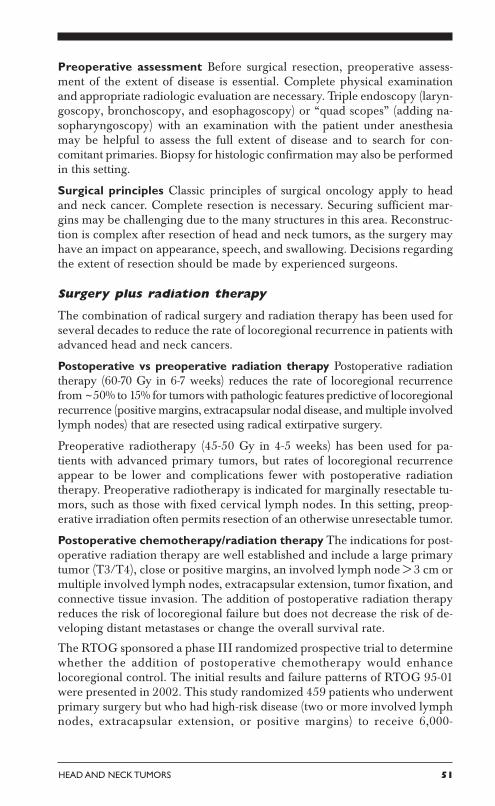
The RTOG sponsored a phase III randomized prospective trial to determinewhether the addition of postoperative chemotherapy would enhancelocoregional control. The initial results and failure patterns of RTOG 95-01were presented in 2002. This study randomized 459 patients who underwentprimary surgery but who had high-risk disease (two or more involved lymphnodes, extracapsular extension, or positive margins) to receive 6,000-
52 CANCER MANAGEMENT: A MULTIDISCIPLINARY APPROACH
6,600 cGy of postoperative irradiation with or without cisplatin (Platinol)(given days 1, 22, and 43). The median follow-up was 26.6 months and thelocoregional control was 74% vs 79% for the radiotherapy and radiotherapyand chemotherapy arms respectively (P = .16). Two-year overall survival was57% and 63% (P = .51) and disease-free survival was 43% and 54%(P = .049), respectively. The incidence of grade 3 and higher acute toxicitywas 33% for the radiotherapy alone arm and 75% for the radiotherapy andcisplatin arm (P < .0001). The authors concluded that the addition of postop-erative chemotherapy increased acute toxicity but did not significantly im-prove clinical end points compared with radiotherapy alone.
Curative radiation therapy
Radiation therapy with curative intent usually involves daily treatment for 6-7 weeks (total dose, 60-70 Gy). Although there is no tissue loss with radiationtherapy, complications include dry mouth, tissue fibrosis, trismus, bone ne-crosis, and hypothyroidism. Some problems are common and sufficientlydebilitating to warrant significant concern in treatment planning for headand neck cancer. Surgery often produces less morbidity.
Radiation fractionationThe RTOG 90-03 trial was conducted to determine the efficacy of variousfractionation schemes in the treament of locally advanced head and neckcancer. Four schedules were tested: (1) standard fractionation at 2 Gy/frac-tion/day, 5 days/week, to 70 Gy/35 fractions/7 weeks; (2) hyperfractionationat 1.2 Gy/fraction, twice daily, 5 days/week to 81.6 Gy/68 fractions/7 weeks;(3) accelerated fractionation with split at 1.6 Gy/fraction, twice daily, 5 days/week, to 67.2 Gy/42 fractions/6 weeks, including a 2-week rest after 38.4 Gy;or (4) accelerated fractionation with concomitant boost at 1.8 Gy/fraction/day, 5 days/week, and 1.5 Gy/fraction/day to a boost field as a second dailytreatment for the last 12 treatment days to 72 Gy/42 fractions/6 weeks. Atotal of 1,113 patients were entered in the study, with a median follow-up of23 months. Patients treated with both hyperfractionation and accelerated frac-tionation with a concomitant boost had significantly increased locoregionalcontrol rates compared with patients on the other two arms. All three groupstreated with the altered fractionation schemes had more acute, but not late,side effects. The study concluded that hyperfractionation and accelerated frac-tionation with a concomitant boost are the optimal treatment schemes.
Intensity-modulated radiation therapyIntensity-modulated radiation therapy (IMRT) is a new approach to obtain-ing highly conformal radiation dose distributions needed to irradiate com-plex targets positioned near sensitive normal structures. In the case of headand neck cancer, these sensitive normal structures include the parotid glands,spinal cord, and eyes. Treatment planning for IMRT (also known as inverseplanning) is different from that of conventional or three-dimensional cranial

HEAD AND NECK TUMORS 53
radiation therapy. The starting point with IMRT is a description of the de-sired dose distribution rather than the application of traditional fields andbeam modifiers to generate an acceptable plan. Conventional radiation treat-ment utilizes relatively uniform beams of radiation (typically between 2-4beams), whereas IMRT, instead of using 4 beams of 50 cGy each, could use50 beams of 4 cGy each. Each beam direc-tion is divided into multiple segments tomodulate the radiation dose. The use ofIMRT in the treatment of head and neck can-cer has focused primarily on sparing the pa-rotid glands and preserving salivary function.
Various groups have examined dosimetricand quality-of-life differences between IMRTand conventional radiation techniques. Thegroup at Memorial Sloan-Kettering CancerCenter compared its IMRT planned treat-ment for nasopharyngeal cancer with con-ventional treatment with a conformal boost.Locoregional control was 97% vs 78% at 2years. The University of Michigan and Wash-ington University have reported reductionsin xerostomia.
Chemotherapy
Induction (neoadjuvant) chemotherapyThe high response rates achieved with induction chemotherapy (clinical re-sponse rates of 70% with cisplatin-based regimens, including complete histo-logic responses) (Table 2) have not resulted in improved locoregional controlor survival when compared with radiotherapy alone. However, several stud-ies have documented a decreased rate of distant metastasis with inductionchemotherapy.
Although response to chemotherapy is a strong predictor of response to radia-tion therapy, chemotherapy does not confer an additional survival benefit.Whether or not chemotherapy may allow a reduction in the type of surgeryor radiation therapy that must be given to cure these patients is disputed andremains open to study.
Concomitant chemotherapy and radiation therapy The major goal ofadministering chemotherapy concurrently with irradiation is to “radiosensi-tize” the tissue in the radiation field and increase the likelihood of locore-gional control. This approach has been associated with improvements inlocoregional control and survival in several randomized trials, and its bene-fits have been confirmed in recent meta-analyses. The optimal way in whichto combine these two modalities is the focus of much ongoing research inhead and neck oncology.
A recent meta-analysis evaluatingthe role of chemotherapy insquamous cell carcinoma of thehead and neck was reported. Thisstudy compared treatment of10,741 patients in 63 trials with orwithout chemotherapy. The pooleddata showed an absolute survivalbenefit of 4% at 2 and 5 years infavor of chemotherapy. This reportalso evaluated 861 patients in 6trials comparing inductionchemotherapy followed byirradiation with concomitant oralternating chemoradiotherapy.These data revealed a 3% absolute(but not statistically significant)survival benefit at 2 and 5 yearsfavoring concomitant chemo-therapy (Pignon JP, Bourhis J,Domenge C, et al: Lancet355[9208]:949-955, 2000).
54 CANCER MANAGEMENT: A MULTIDISCIPLINARY APPROACH
Preliminary results from a Head and Neck Intergroup study confirm the su-periority of concurrent chemoradiation therapy compared with irradiationalone for patients with unresectable squamous cell cancer. A total of 295patients with stage III and IV head and neck were randomized to participatein one of three arms: (A) radiotherapy alone to 70 Gy in 35 fractions;(B) 70 Gy in 35 fractions plus concurrent cisplatin on days 1, 22, and 43; and(C) split-course radiotherapy and three cycles or concurrent cisplatin/5-FUchemotherapy with 30 Gy given with cycle 1 and 30-40 Gy given with cycle3. Grade 3 or worse toxicity occurred in 53% of arm A patients; 86% in armB patients, and 77% of arm C patients. The 2- and 3-year Kaplan-Meier pro-jected survivals for arm A are 30% and 20%, compared with 43% and 37%for arm B (P = .016), and 40% and 27% for arm C (P = .13). Median survivalis 12.6 months for arm A, 19.1 months for arm B, and 14.0 months for arm C.The addition of concurrent high-dose single-agent cisplatin to conventionalradiotherapy significantly improves survival with acceptable toxicity. Addi-tionally, concurrent multiagent chemotherapy did not offset the loss of effi-cacy resulting from split-course irradiation.
Induction chemotherapy followed by radiation therapy has been shownin a Veterans Affairs (VA) trial to produce survival comparable to that at-tained with primary surgery in patients with laryngeal cancer. The advantageof this approach is preservation of the larynx. Combinations of chemother-
TABLE 2: Chemotherapy regimens for headand neck tumors
Drug/combination Dose and schedule
Cisplatin + fluorouracil
Cisplatin 100 mg/m2 IV on day 1
Fluorouracil 1 mg/m2 IV infused continuously over 4 days
Repeat cycle every 3-4 weeks
Jacobs C, Lyman G, Velez-Garcia E, et al: J Clin Oncol 10:257–263, 1992.
Carboplatin + paclitaxel
Carboplatin Dose calculated by the Calvert formula to anarea under the curve between 6 and 7.5mg/mL/min infused over 1-2 hours on day 1after paclitaxel
Paclitaxel 175 mg/m2 IV infused continuously over 3 hours
Repeat cycle every 21 days
PREMEDICATIONS: Dexamethasone, 20 mg PO 12 and 6 hours prior to paclitaxel; aswell as diphenhydramine, 50 mg IV, and ranitidine, 50 mg IV, both 30 to 60 minutes prior topaclitaxel
Dang TP, Murphy BA, Cmelak A, et al: Proc Am Soc Clin Oncol 17:393a, 1998.
Table prepared by Ishmael Jaiyesimi, DO

HEAD AND NECK TUMORS 55
apy and radiation therapy are being activelystudied, and other approaches, such as con-comitant treatment, appear to be effective.
Adjuvant chemotherapy following sur-gery or irradiation Adjuvant chemotherapyhas been given following initial surgery orradiation therapy in an attempt to eliminatemicroscopic residual disease and distant me-tastases. Although this approach has resultedin a reduced rate of distant metastasis, it hasnot been associated with improvedlocoregional control or survival. As con-comitant approaches evolve andlocoregional control for advanced diseasebecomes the rule rather than the exception,the value of additional chemotherapy (as in-duction or adjuvant therapy) will need to bereexplored to address the problem of distantmetastasis.
Locally advanced head and neck cancerData from prospective trials continue to sup-port the use of concurrent chemotherapy andirradiation as an alternative to surgery or ir-
radiation alone for locally advanced cancers of the head and neck. RTOG90-03 randomized 1,113 patients with stage III and IV squamous cell carci-noma of the oral cavity, oropharynx, or supraglottic larynx and stage II-IVsquamous cell carcinoma of the base of the tongue or hypopharynx to receiveone of four different schedules of irradiation alone. They included 7,200 cGyin 42 fractions (180 cGy/fraction initially, followed by 150 cGy/fraction to aboost field for the last 12 fractions bid). Locoregional control, disease-freesurvival, and overall survival rates were 54.5%, 39.3%, and 50.9%, respec-tively, which were superior to those obtained with single daily fractions of2.0 Gy over 7 weeks.
RTOG 99-14 was a phase II study designed to integrate this altered fraction-ated irradiation regimen with chemotherapy. Cisplatin was given during weeks1 and 3. When results on 77 evaluable patients were reported, 82% of pa-tients were alive at the time of analysis. One-year overall survival was 81.3%;grade 3 and 4 chronic toxicity were 23% and 8%, respectively. The nextRTOG phase III trial for advanced head and neck cancer will incorporatethis regimen as the “experimental” arm.
Chemotherapy for recurrent or metastatic disease The combinationof cisplatin/fluorouracil (5-FU) produces overall response rates of approxi-mately 30% and survival rates of 6 months. In randomized trials compar-ing this combination with single-agent cisplatin, 5-FU, or methotrexate,
ECOG (Eastern CooperativeOncology Group) 1393 random-ized patients with recurrentsquamous cell cancer of the headand neck to receive cisplatin (100mg/m2 day 1) and 5-FU (1 g/m2 days1-4; arm 1) or cisplatin (75 mg/m2
day 1) and paclitaxel (75 mg/m2 day1; arm 2). Patients could have hadno prior treatment for recurrentdisease and were required to haveECOG performance status of 0 or1. A total of 194 patients werestudied, with 96 and 92 patientseligible on arms 1 and 2,respectively. Overall response andmedian survival rates wereequivalent (arm 1: 22% and 8months; arm 2: 28% and 9 months).The toxicity analysis favored arm 2,which was associated with fewercases of stomatitis, diarrhea, myelo-suppression, and infection. Qualityof life did not favor either arm(Murphy B, Li Y, Cella D, et al: Proc AmSoc Clin Oncol [abstract] 20:224a,2001).
56 CANCER MANAGEMENT: A MULTIDISCIPLINARY APPROACH
response rates with the single agents are lower but survival is equivalent.However, because many practitioners believe response is a surrogate for pal-liation, cisplatin/5-FU has been used widely in this setting.
The taxanes are the most active cytotoxins yet identified in head and neckcancer, with overall response rates of approximately 35% in patients withrecurrent or incurable disease. In a recent randomized trial, the combinationof paclitaxel (Taxol)/cisplatin has been compared with cisplatin/5-FU in pa-tients with recurrent disease (see box on previous page). Although primaryefficacy outcomes were equivalent for the two arms, paclitaxel/cisplatin wasless toxic overall and can now be considered a safer, more convenient alter-native to cisplatin and infusional 5-FU in recurrent disease. The evaluationsof taxane-based neoadjuvant therapy and concurrent therapy with irradia-tion are ongoing.
Other drugs receiving recent attention are those targeting growth factors andtheir receptors. The epidermal growth factor receptor (EGFR) is particularlynotable, since nearly 100% of head and neck tumors overexpress this recep-tor. A number of EGFR inhibitors are currently being evaluated alone andin combination with other drugs.
Photodynamic therapyPhotodynamic therapy may have some promise in the treatment of mu-cosal dysplasia and small head and neck tumors.
Small studies of photodynamic therapy, performed at several institutions,suggest that widespread areas of carcinoma in situ or severe dysplasia, aswell as cancer, are often extirpated after photodynamic therapy. Althoughsome patients have experienced durable remissions, the long-term efficacyof this modality remains uncertain.
Chemoprevention
The area of chemoprevention has received a great deal of attention in recentyears, and the concept of “field cancerization” is important in this context.As mentioned previously, this concept refers to the diffuse epithelial injuryincurred by upper aerodigestive tract mucosa due to chronic exposure tocarcinogens (most commonly alcohol and tobacco). These mucosal changesincrease the risk of developing premalignant lesions (leukoplakia anderythroplakia), as well as multiple primary lesions.
The retinoids (vitamin A analogs) have shown the greatest promise in achiev-ing effective chemoprevention. The mechanism of action involves changesin gene expression mediated by nuclear retinoic acid receptors, which ap-pear to function as transcription factors. In the mouse skin tumor and ham-ster buccal pouch models, retinoids have been shown to significantly preventtumor growth and development. They have also been shown to significantlydecrease the prevalence of premalignant lesions in humans in both random-ized and nonrandomized clinical trials. Prospective trials have demonstrated
HEAD AND NECK TUMORS 57
that serum levels of retinoids are lower in patients who subsequently developupper aerodigestive tract tumors.
Ongoing clinical trials are investigating the possible role of retinoids in theprevention of second primary tumors in both the upper aerodigestive tractand lungs. However, a recent phase III ECOG (Eastern Cooperative On-cology Group) trial showed no benefit in prevention of recurrence withlow-dose cis-retinoic acid (10 mg/d) in stage I/II patients. The ECOG trialused very low doses (7.5-10 mg/d). A study by Hong et al using high doses(1-2 mg/kg/d) produced positive results, but morbidity and compliance wereproblems. Results are pending from a trial employing intermediate dosing(30 mg/d).
RehabilitationRehabilitation also is very important in the perioperative care of head andneck cancer patients. It includes physical and occupational therapy, speechand swallowing rehabilitation, and nutritional support. For example, resec-tion of the spinal accessory nerve, which innervates the trapezius muscle,leads to scapular winging, inability to abduct the arm fully, and, eventually,to severe pain around the shoulder. These symptoms may be amelioratedwith appropriate physical therapy.
Adapting to the loss of the larynx also requires intensive rehabilitation andpatient motivation. Voice rehabilitation options include esophageal speech,artificial larynges (portable, battery-operated devices), and tracheoesopha-geal shunts.
Nutritional support is facilitated by temporary nasoduodenal tubes orgastrostomy tubes (which impose added morbidity but are more sociallyacceptable and ease the patient’s transition to normal activities).
Management of symptoms and treatment side effects
Eating problems At the time of diagnosis, many head and neck cancerpatients will have lost a significant amount of weight. Maintaining adequatenutrition is a major problem for these patients, as both the tumor and treat-ment side effects, such as mucositis from chemotherapy and radiation ther-apy, may be contributory. For patients who are unable to eat or who arebeing treated with aggressive concomitant chemotherapy and radiationtherapy protocols, placement of a gastrostomy tube is often desirable in or-der to maintain caloric intake and adequate hydration.
Pain Clinicians must also be aware of the significant pain associated withthese lesions and use narcotic analgesics appropriately to relieve discomfort.
Mucositis The use of chemotherapy concomitantly with radiation therapyincreases the occurrence of mucositis.
58 CANCER MANAGEMENT: A MULTIDISCIPLINARY APPROACH
Nephrotoxicity and ototoxicity For patients treated with cisplatin-con-taining regimens, renal insufficiency and ototoxicity are potential serious sideeffects.
Xerostomia Following radiation therapy of a head and neck cancer, xero-stomia may be a significant long-term side effect. In some patients, pilocarpinehydrochloride (Salagen) has been useful in stimulating the production of saliva.
The use of organic thiophosphates, such as amifostine (Ethyol), in patients under-going radiotherapy for head and neck cancer may reduce the severity of acute andlate xerostomia without compromising the antitumor activity of the irradiation.
Gastroesophageal reflux Often asymptomatic or “silent” gastroesophagealreflux disease (GERD) is a common finding in patients treated forpharyngolaryngeal squamous cell carcinoma. In addition, cisplatin-containingchemotherapy may aggravate GERD.
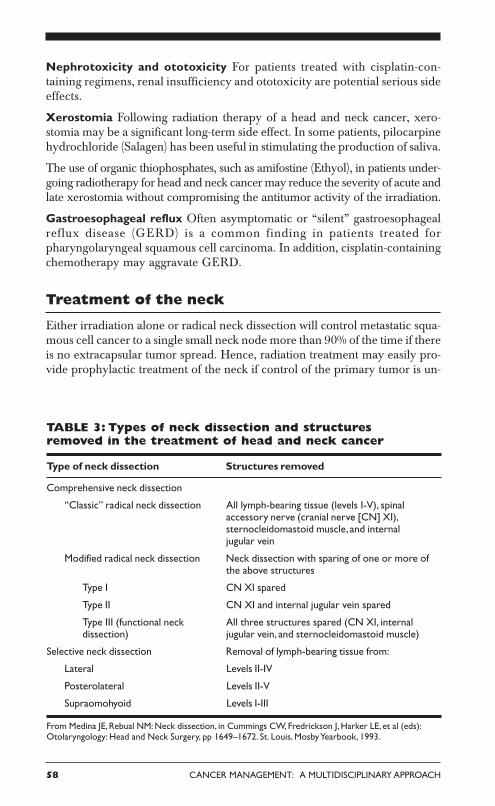
Treatment of the neck
Either irradiation alone or radical neck dissection will control metastatic squa-mous cell cancer to a single small neck node more than 90% of the time if thereis no extracapsular tumor spread. Hence, radiation treatment may easily pro-vide prophylactic treatment of the neck if control of the primary tumor is un-
TABLE 3: Types of neck dissection and structuresremoved in the treatment of head and neck cancer
Type of neck dissection Structures removed
Comprehensive neck dissection
“Classic” radical neck dissection All lymph-bearing tissue (levels I-V), spinalaccessory nerve (cranial nerve [CN] XI),sternocleidomastoid muscle, and internaljugular vein
Modified radical neck dissection Neck dissection with sparing of one or more ofthe above structures
Type I CN XI spared
Type II CN XI and internal jugular vein spared
Type III (functional neck All three structures spared (CN XI, internaldissection) jugular vein, and sternocleidomastoid muscle)
Selective neck dissection Removal of lymph-bearing tissue from:
Lateral Levels II-IV
Posterolateral Levels II-V
Supraomohyoid Levels I-III
From Medina JE, Rebual NM: Neck dissection, in Cummings CW, Fredrickson J, Harker LE, et al (eds):Otolaryngology: Head and Neck Surgery, pp 1649–1672. St. Louis, Mosby Yearbook, 1993.
HEAD AND NECK TUMORS 59
dertaken with irradiation. Traditionally, if the tumor in the neck was N2 orgreater, or if there was tumor beyond the confines of a node, radical neckdissection and irradiation were combined for optimal control of the neck tu-mor. More recently, evidence suggests that N2-N3 disease that has a completeclinical and radiologic response to induction chemoradiotherapy may not re-quire a complete neck dissection. This concept continues to evolve.
Types of dissection
There are several approaches to the surgical treatment of the neck nodes inpatients with head and neck cancer (Table 3). This discussion will be limitedto two types of neck dissection: comprehensive and selective dissection.
Comprehensive neck dissection entails complete removal of all lymphatictissue from the neck (levels I-V). A radical neck dissection includescomprehensive node dissection with removal of the sternocleidomastoid muscle,jugular vein, and spinal accessory nerve. Modified radical neck dissection wasdeveloped to diminish the morbidity of the classic operation. The most im-portant structure to preserve is the spinal accessory nerve.
Selective neck dissection consists of the removal of lymph node groups athighest risk of containing metastases from a primary cancer. In such proce-dures, the lymph nodes removed correspond to the most significant drainagebasins of specific head and neck tumor sites. These are staging operations usu-ally performed in patients with a clinically N0 neck cancer. If metastases areidentified, further treatment to the neck will be required. A selective neck dis-section should not be employed as the sole treatment of clinically palpabledisease.
Sentinel lymph node biopsy for oral cavity lesions has been evaluated. Fortypatients with clinically N0 necks underwent sentinel lymph node biopsy fol-lowed by complete neck dissection. A sentinal node was identified in 90% ofnecks, with a 97% accuracy rate in predicting the nodal status of the remainderof the neck. This finding corresponded to a sensitivity of 94% and a specificityof 100%. Although these results are encouraging, they need to be validated ina larger trial. An ongoing American College of Surgeons Oncology Groupstudy (Z0360) is examining this technique in patients with T1 or T2, N0 oralcavity cancer. Sentinel lymph node biopsy may prove useful in small lesionswithout deep penetration, but it remains investigational.
Follow-up of long-term survivors
As mentioned, head and neck cancers are aggressive tumors. The majority(80%) of recurrences will develop within 2 years. Since many recurrences aretreatable with curative intent, patients should be followed closely during themonths following their treatment. This period coincides with the time of great-est need from the standpoint of rehabilitation.

60 CANCER MANAGEMENT: A MULTIDISCIPLINARY APPROACH
After 2 years, second primary tumors of the head and neck and lungs becomeimportant causes of death and morbidity. Late complications of treatment suchas radionecrosis, radiation-induced fibrosis, and hypothyroidism, as well assequelae of spinal accessory nerve sacrifice or injury, may develop even afteryears. Complications and second primary cancers are more common in pa-tients who continue to smoke.
Timing of follow-up evaluations Follow-up evaluations at regular intervalsshould be complete and should include a focused history and examination, asoutlined above. Physicians who are able to perform a head and neckexamination (including laryngoscopy) should direct follow-up. After surgicaltreatment, this evaluation will usually require visits with the head and necksurgeon. Patients treated with irradiation should be followed by both their ra-diation oncologist and a head and neck surgeon or otolaryngologist.
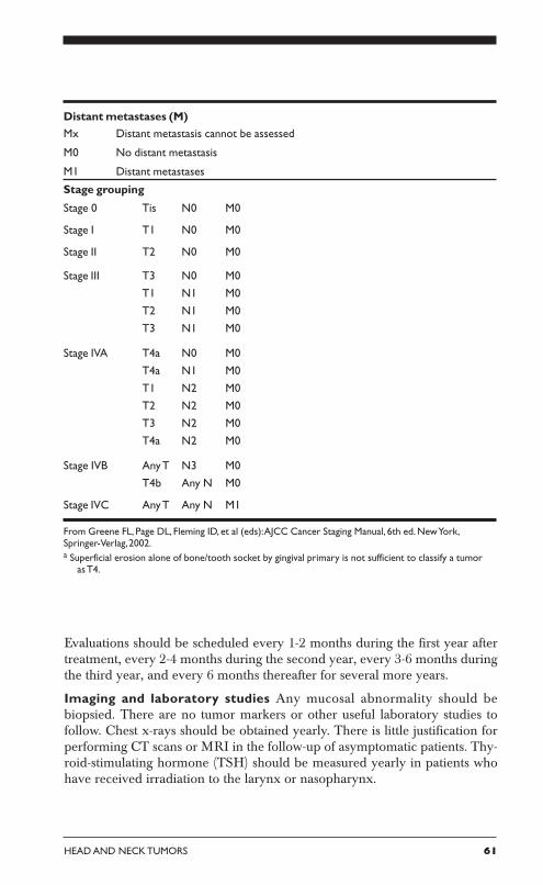
TABLE 4: TNM staging systemfor cancers of the lips and oral cavity
Primary tumor (T)
Tx Primary tumor cannot be assessedT0 No evidence of primary tumorTis Carcinoma in situT1 Tumor 2 cm or less in greatest dimensionT2 Tumor more than 2 cm but not more than 4 cm in greatest dimensionT3 Tumor more than 4 cm in greatest dimensionT4 (lip) Tumor invades through cortical bone, inferior alveolar nerve, floor of
mouth, or skin of face, ie, chin or nosea
T4a (oral cavity) Tumor invades adjacent structures (eg, through cortical bone,into deep [extrinsic] muscle of the tongue, maxillary sinus, or skin of face)
T4b Tumor involves masticator space, pterygoid plates, or skull base and/orencases internal carotid artery
Regional lymph nodes (N)
Nx Regional nodes cannot be assessedN0 No regional lymph node metastasisN1 Metastasis in a single ipsilateral lymph node, 3 cm or less in greatest dimensionN2 Metastasis in a single ipsilateral lymph node, more than 3 cm but not more
than 6 cm in greatest dimension; or in multiple ipsilateral lymph nodes, nonemore than 6 cm in greatest dimension; or in bilateral or contralateral lymphnodes, none more than 6 cm in greatest dimension
N2a Metastasis in single ipsilateral lymph node, more than 3 cm but not more than6 cm in greatest dimension
N2b Metastasis in multiple ipsilateral lymph nodes, none more than 6 cm ingreatest dimension
N2c Metastasis in bilateral or contralateral lymph nodes, none more than 6 cm ingreatest dimension
N3 Metastasis in a lymph node, more than 6 cm in greatest dimension
HEAD AND NECK TUMORS 61
Distant metastases (M)Mx Distant metastasis cannot be assessed
M0 No distant metastasis
M1 Distant metastases
Stage grouping
Stage 0 Tis N0 M0
Stage I T1 N0 M0
Stage II T2 N0 M0
Stage III T3 N0 M0
T1 N1 M0
T2 N1 M0
T3 N1 M0
Stage IVA T4a N0 M0
T4a N1 M0
T1 N2 M0
T2 N2 M0
T3 N2 M0
T4a N2 M0
Stage IVB Any T N3 M0
T4b Any N M0
Stage IVC Any T Any N M1
From Greene FL, Page DL, Fleming ID, et al (eds): AJCC Cancer Staging Manual, 6th ed. New York,Springer-Verlag, 2002.a Superficial erosion alone of bone/tooth socket by gingival primary is not sufficient to classify a tumor
as T4.
Evaluations should be scheduled every 1-2 months during the first year aftertreatment, every 2-4 months during the second year, every 3-6 months duringthe third year, and every 6 months thereafter for several more years.
Imaging and laboratory studies Any mucosal abnormality should bebiopsied. There are no tumor markers or other useful laboratory studies tofollow. Chest x-rays should be obtained yearly. There is little justification forperforming CT scans or MRI in the follow-up of asymptomatic patients. Thy-roid-stimulating hormone (TSH) should be measured yearly in patients whohave received irradiation to the larynx or nasopharynx.
62 CANCER MANAGEMENT: A MULTIDISCIPLINARY APPROACH
HEAD AND NECK TUMOR REGIONS
As mentioned above, tumors occurring at different anatomic sites and sub-sites of the head and neck vary considerably with regard to epidemiology,risk factors, anatomy, natural history, staging of the primary tumor, and ther-apy. The following sections highlight these differences.
ORAL CAVITY
Sites of the oral cavity include the lips, hard palate, floor of the mouth, buc-cal mucosa, and tongue. Cancers at these sites comprise < 5% of all malignan-cies in the United States.
Etiology and risk factors
Tobacco and alcohol As with other head and neck tumors, there is a cor-relation between the use of tobacco and development of oral cavity cancers.There is a clear dose-response relationship between tobacco exposure andtumor development. A full 90% of patients with oral cancers use tobacco,and many drink alcohol.
Vitamin A Patients with vitamin-A deficiency seem to be at higher risk,whereas diets high in fruits and vegetables seem to be protective.
Chronic irritants, including mouthwash and poor oral hygiene, are associ-ated with tumor development.
Viruses Herpes simplex virus type 1 and human papillomavirus have beenimplicated in the development of oral cancer.
Anatomy
The oral cavity extends from the cutaneous vermilion junction of the lips tothe junction of the hard and soft palate above and to the line of thecircumvallate papillae below. It includes the lips, buccal mucosa, upper andlower alveolar ridges, retromolar trigone, floor of the mouth, hard palate,and anterior two-thirds of the tongue (the “oral” tongue). The primary lym-phatic drainage is to the submental triangle, submandibular nodes, and up-per deep jugular nodes.
Natural history
The most common presenting complaint is a sore in the mouth or on the lips.One-third of patients present with a neck mass.
The differential diagnosis includes other malignancies and benign diseases orlesions. Other malignancies to be considered include salivary gland tumors,

HEAD AND NECK TUMORS 63
sarcoma, lymphoma, and melanoma. Benign diseases include pyogenic granu-loma, tuberculous disease, aphthous ulcers, and chancres.
Benign mucosal lesions include papillomas and keratoacanthomas, which maybe exophytic or infiltrative. The exophytic lesions are less aggressive. Theinfiltrative papillomas and keratoacanthomas are more often associated withdestruction of surrounding tissues and structures. These lesions may progressto malignancy. The TNM staging system for cancers of the lips and oral cav-ity is outlined in Table 4. T4 lesions have been divided into T4a (resectable)and T4b (unresectable) in the sixth edition of the AJCC Cancer Staging Manual.
Treatment
Radiation therapy has been widely used in patients with cancers of the oraltongue and floor of the mouth. Interstitial radiation therapy alone may beused for early (T1-T2) tumors, with local control rates of 80%-95%. A com-bination of external-beam radiation therapy (60 Gy in 6 weeks) plus inter-stitial radiotherapy provides excellent local control for early tumors.
Cancers arising at other sites within the oral cavity, such as the gingiva or buc-cal mucosa, usually are best treated with a primary surgical approach, butpostoperative radiation therapy is added when poor pathologic features arepresent.
Surgical approaches to the primary cancer include peroral, transcervical,or combined operations. A comprehensive neck dissection should be per-formed in all patients with palpable cervical metastases. When an N0 lesionapproaches the midline, bilateral supraomohyoid dissection should be con-sidered. If the lymph nodes from the contralateral side of the neck containcancer, contralateral neck treatment is needed. If there are bilateral, palpablenodes, both sides of the neck should be dissected.
Combined-modality treatment, including surgical resection and radiationtherapy (60-70 Gy in 6-7 weeks), is advised for the treatment of advanced(stage III-IV) disease.
ORAL CAVITY SITE: LIPS
The lips are the most common site of oral cavity cancer. There are approxi-mately 3,600 new cases per year in the United States. The lower lip is af-fected most often. The vast majority (90%) of patients with lip cancer aremen, and 33% have outdoor occupations.
Natural history
The most frequent presentation is a slow-growing tumor of the lower lip thatmay bleed and hurt. Physical examination must include assessment of hypo-

64 CANCER MANAGEMENT: A MULTIDISCIPLINARY APPROACH
esthesia in the distribution of the mental nerve (cutaneous sensation of chinarea). Currently, fewer than 10% of American patients with squamous cellcarcinoma of the lower lip have cervical metastases.
Treatment
Treatment of the primary tumor The primary tumor may be treated withradiation therapy (60-70 Gy in 6-7 weeks) or surgical resection. Currently, opera-tions are more common than irradiation in the United States.
Resection involves excision with at least 0.5 cm of normal tissue circumferen-tially beyond the recognized border of the tumor. After the resection of largerlesions, reconstruction may pose a major challenge. Small tumors are excisedwith a V incision.
Patients with advanced disease (stage III or IV) are usually managed witha combination of surgery and postoperative radiation therapy (60-70 Gy in6-7 weeks).
Treatment of the neck Elective treatment of the neck is seldom recommendedfor patients with squamous cell carcinoma of the lower lip and a clinicallynegative neck because few of these patients have cervical metastases. Neckdissection is recommended only in patients with palpable cervical metastases.In these individuals, the recommended approach often includes neck dissec-tion and radiation therapy.
Results The cure rate for T1-T3 tumors is 90% with surgical excision alone.Smaller lesions (T1-T2) may be treated equally well with radiation therapy.Survival rates for patients with T1 and T2 lesions are 90% and 80%, respec-tively. Overall, younger patients have a poorer prognosis, as do those withinvolvement of the mandible and extension of the tumor within the oral cavity.
ORAL CAVITY SITE: TONGUE
The oral tongue (anterior two-thirds) is the site of 75% of all tongue cancers.There are approximately 7,100 new cases of oral tongue cancer each year inthe United States.
Epidemiology and risk factors
Gender and age The male-female ratio is 3:1. Although the median age ofonset is 60 years, tongue cancer may occur in patients younger than 30 yearsof age. Young patients may have no recognized risk factors.
Risk factors Tongue cancers are associated with poor oral hygiene and withalcohol and tobacco use.
HEAD AND NECK TUMORS 65
Natural history
The most common presenting symptom in patients with cancer of the tongueis pain. Other symptoms include difficulty with deglutition and speech. Theremay be a history of leukoplakia, especially in younger women.
Rate of growth Cancer of the tongue seems to grow more rapidly than otheroral cavity cancers. Tongue cancers may grow in an infiltrative or exophyticfashion. The infiltrative tumors may be quite large at presentation.
Lesion thickness Thicker lesions have a worse prognosis than thin cancers,and lesion thickness is a more important prognostic factor than is simple tumorstage.
Cervical metastases occur more frequently from tongue cancer than fromany other tumor of the oral cavity. At initial evaluation, 40% of patients havenode metastases. Bilateral and contralateral metastases from lateral tongue can-cers are uncommon.
Treatment
Early-stage disease Treatment usually entails partial glossectomy. Marginsshould be assessed at the time of resection, as the disease spreads along musclebundles, leading to more extensive tumor than is appreciated grossly.
Radiation therapy (60-70 Gy in 6-7 weeks) is a suitable option for small orminimally infiltrating tumors. Large, infiltrative lesions should be treated withcombined-modality therapy (radiation therapy and surgical resection).
Advanced disease More advanced tumors with mandibular involvementrequire composite resection, including a partial glossectomy, mouth floor re-section, and partial mandibulectomy.
Treatment of the neck A selective neck dissection is often recommended forthe clinically N0 disease neck. Comprehensive neck dissection is required inthe presence of palpable cervical metastases.
Results Control of disease closely correlates with the extent of the primarytumor and presence of metastases. Rates of local control using radiation therapyor surgery are similar for T1 (~85%) and T2 (~80%) tumors. T3 tumors shouldbe treated using surgery and radiation therapy. Only 10%-15% of local recur-rences are amenable to repeat resection.
Overall survival is approximately 50%. Rates of survival at 5 years by stageare stage I, 80%; stage II, 60%; and stage III-IV, 15%-35%. For equivalentprimary cancers, the presence of lymph node metastases decreases the sur-vival rate by 50%.

66 CANCER MANAGEMENT: A MULTIDISCIPLINARY APPROACH
ORAL CAVITY SITE: FLOOR OF THE MOUTH
There are approximately 1,500 cases of floor of the mouth cancer in the UnitedStates annually. Mouth floor cancer accounts for 10%-15% of all oral cavitycancers.
Epidemiology and risk factors
Gender and age The male-female ratio is 3:1, but the incidence amongwomen is increasing. The median age at presentation is 60 years.
Tobacco and alcohol Approximately two-thirds of patients with cancer ofthe mouth floor are heavy smokers and 50% are alcoholics. Alcohol actssynergistically with tobacco. Alcohol may act as a promoter, a direct irritant,or a solvent to increase the solubility of carcinogens from tobacco.
Smokeless tobacco There is a strong association between the use of smoke-less tobacco and oral carcinogenesis. Data from the southern United Statesreveal a 50-fold increased risk of oral cavity cancer in women who use smoke-less tobacco.
Pathology
Most lesions are moderately differentiated to well-differentiated squamouscell cancers and are exophytic in character.
Natural history
Patients usually present with a painful mass located near the oral tongue.Since these lesions do not cause pain until they are deep, they are frequentlyadvanced at presentation.
Extension of disease into the soft tissues of the submandibular triangle is notuncommon. Fixation of the tumor to bone suggests possible mandibularinvolvement. A Panorex film of the mandible may reveal invasion of themandible via direct extension (through a tooth socket) or via perineural in-vasion spreading along the mental nerve through the mental foramen.Changes in the mental foramen can be distinct or demonstrate slight asym-metry when compared with the contralateral anatomy. Restricted tonguemobility reflects invasion into the root of the tongue. Palpation demonstratesthe depth of infiltration much better than does inspection alone.
Tumors near the midline may obstruct the duct of the submandibular gland,leading to swelling and induration, which may be difficult to distinguish fromlymph node metastases. Level I nodes are the first-echelon metastatic sites.
Multifocality Multifocal cancers are more common in the floor of the mouththan in other oral cavity sites. Approximately 20% of patients with mouth
HEAD AND NECK TUMORS 67
floor tumors have a second primary tumor, half of which are in the head andneck.
Treatment
Early invasive lesions (T1-T2) involving the mucosa alone may be treatedwith either surgery or irradiation (60-70 Gy in 6-7 weeks) alone, with compa-rable results. Primary tumors with mandibular involvement should be surgi-cally resected.
Cancer invades the mandible through tooth sockets. Hence, if the tumormerely abuts the mandible, a marginal mandibulectomy (which removes thebone margin but preserves continuity) may be performed. Otherwise, a seg-mental resection is needed.
Selective neck dissection for treatment planning is advisable for thick stage Ior II cancers.
Advanced disease The treatment of choice for advanced disease is com-bined-modality therapy with surgery and radiation therapy. Complete surgi-cal resection may require a composite resection of the mandible, including apartial glossectomy and neck dissection for advanced primary cancers.
Lesions near the midline with a clinically positive lymph node require ipsi-lateral comprehensive neck dissection with a contralateral selective (supra-omohyoid) neck dissection. Otherwise, both sides of the neck should be treatedwith irradiation. If there is clinical evidence of bilateral involvement, bilat-eral comprehensive neck dissection should be performed.
Results Overall, ~40% of patients are cured of their disease; 80% of recur-rences appear within the first 2 years. Survival rates at 5 years by stage are asfollows: stage I, 85%; stage II, 75%; stage III, 66%; and stage IV, 30%. Signsof poor prognosis include involvement of both the tongue and mandible andextension of the tumor beyond the oral cavity.
NASOPHARYNX
Nasopharyngeal carcinoma is uncommon in most of the world. Endemicareas include southern China, northern Africa, and regions of the far North-ern Hemisphere. The incidence (per 1,000 population) ranges from 25.6 inmen and 10.2 in women in Hong Kong to 0.6 in men and 0.1 in women inConnecticut.
Epidemiology and risk factors
Gender and age The incidence of nasopharyngeal cancer peaks in the fourthto fifth decades of life, and the male-female ratio is 2.2:1. Both patient age atdisease onset and male-female ratio are lower for nasopharyngeal cancer thanfor other head and neck malignancies.

68 CANCER MANAGEMENT: A MULTIDISCIPLINARY APPROACH
TABLE 5: TNM staging system for cancers of the pharynx(including base of tongue, soft palate, and uvula)
Primary tumor (T)
Tx Primary tumor cannot be assessedT0 No evidence of primary tumorTis Carcinoma in situNasopharynx
T1 Tumor confined to the nasopharynxT2 Tumor extends to soft tissue of oropharynx and/or nasal fossa T2a Without parapharyngeal extensiona
T2b With parapharyngeal extensiona
T3 Tumor invades bony structures and/or paranasal sinusesT4 Tumor with intracranial extension and/or involvement of cranial nerves,
infratemporal fossa, hypopharynx, or orbitOropharynx
T1 Tumor 2 cm or less in greatest dimension
T2 Tumor more than 2 cm but not more than 4 cm in greatest dimension
T3 Tumor more than 4 cm in greatest dimension
T4a Tumor invades the larynx, deep/extrinsic muscle of tongue, medial pterygoid,hard palate, or mandible (resectable)
T4b Tumor invades lateral pterygoid muscle, pterygoid plates, lateral nasopharynx,or skull base or encases carotid artery (unresectable)
Hypopharynx
T1 Tumor limited to one subsite of hypopharynx and 2 cm or less in greatestdimension
T2 Tumor involves more than one subsite of hypopharynx or an adjacent siteor measures more than 2 cm but not more than 4 cm in greatest diameterwithout fixation of hemilarynx
T3 Tumor measures more than 4 cm in greatest dimension or with fixation ofhemilarynx
T4a Tumor invades thyroid/cricoid cartilage, hyoid bone, thyroid gland, esophagus, orcentral compartment soft tissueb (resectable)
T4b Tumor invades prevertebral fascia, encases carotid artery, or involvesmediastinal structures (unresectable)
Risk factors Nasopharyngeal carcinoma appears to have different deter-minants than other head and neck cancers. They include diet, viral agents,and genetic susceptibility. Populations of endemic areas have a diet charac-terized by high consumption of salt-cured fish and meat. Studies reveal anassociation between EBV and nasopharyngeal carcinoma. Anti-EBV anti-bodies have been found in the sera and saliva of patients with this type ofcarcinoma. Major histocompatibility (MHC) profiles associated with increasedrelative risk include H2, BW46, and B17 locus antigens.

HEAD AND NECK TUMORS 69
Regional lymph nodes (N): Nasopharynx
Nx Regional nodes cannot be assessedN0 No regional lymph node metastasisN1 Unilateral metastasis in lymph node(s), 6 cm or less in greatest dimension,
above the supraclavicular fossac
N2 Bilateral metastasis in lymph node(s), 6 cm or less in greatest dimension,above the supraclavicular fossac
N3 Metastasis in a lymph node(s) > 6 cm and/or to supraclavicular fossa N3a Greater than 6 cm in dimension N3b Extension to the supraclavicular fossac
Regional lymph nodes (N): Oropharynx and hypopharynx
Nx Regional lymph nodes cannot be assessedN0 No regional lymph node metastasisN1 Metastasis in a single ipsilateral lymph node, 3 cm or less in greatest dimensionN2 Metastasis in a single ipsilateral lymph node, more than 3 cm but not more
than 6 cm in greatest dimension; or in multiple ipsilateral lymph nodes, nonemore than 6 cm in greatest dimension; or in bilateral or contralateral lymphnodes, none more than 6 cm in greatest dimension
N2a Metastasis in a single ipsilateral lymph node, more than 3 cm but not morethan 6 cm in greatest dimension
N2b Metastasis in multiple ipsilateral lymph nodes, none more than 6 cmin greatest dimension
N2c Metastasis in bilateral or contralateral lymph nodes, none more than 6 cm ingreatest dimension
N3 Metastasis in a lymph node, more than 6 cm in greatest dimension
Distant metastases (M)
Mx Distant metastasis cannot be assessedM0 No distant metastasisM1 Distant metastasesa Parapharyngeal extension denotes posterolateral infiltration of tumor beyond the pharyngobasilar fasciab Central compartment soft tissue includes prelaryngeal strap muscles and subcutaneous fatc Midline nodes are considered ipsilateral nodes
Anatomy and pathology
The nasopharynx communicates anteriorly with the nasal cavity and inferiorlywith the oropharynx. The superior border is the base of the skull. The lateraland posterior pharyngeal walls are composed of muscular constrictors. Poste-riorly, the nasopharynx overlies the first and second cervical vertebrae. Theeustachian tubes open into the lateral walls. The soft palate divides the na-sopharynx from the oropharynx.
continued on following page

70 CANCER MANAGEMENT: A MULTIDISCIPLINARY APPROACH
Cancers arising in the nasopharynx are classified using World Health Organization(WHO) criteria: type 1 denotes differentiated squamous cell carcinoma; type 2,nonkeratinizing carcinoma; and type 3, undifferentiated carcinoma. The TNMstaging system for cancers of the pharynx is outlined in Table 5.
TABLE 5: TNM staging system for cancers of the pharynx(including base of tongue, soft palate, and uvula) (continued)
Stage grouping: Nasopharynx
Stage 0 Tis N0 M0Stage I T1 N0 M0Stage IIA T2a N0 M0Stage IIB T1 N1 M0
T2 N1 M0T2a N1 M0T2b N0 M0T2b N1 M0
Stage III T1 N2 M0T2a N2 M0T2b N2 M0T3 N0 M0T3 N1 M0T3 N2 M0
Stage IVA T4 N0 M0T4 N1 M0T4 N2 M0
Stage IVB Any T N3 M0Stage IVC Any T Any N M1
Stage grouping: Oropharynx and hypopharynx
Stage 0 Tis N0 M0Stage I T1 N0 M0Stage II T2 N0 M0Stage III T3 N0 M0
T1 N1 M0T2 N1 M0T3 N1 M0
Stage IVA T4a N0 M0T4a N1 M0T1 N2 M0T2 N2 M0T3 N2 M0T4a N2 M0
Stage IVB T4b Any N M0Any T N3 M0
Stage IVC Any T Any N M1
From Greene FL, Page DL, Fleming ID, et al (eds): AJCC Cancer Staging Manual, 6th ed. New York,Springer-Verlag, 2002.

HEAD AND NECK TUMORS 71
Natural history
A mass in the neck is the presenting complaint in 90% of patients. Otherpresenting symptoms include a change in hearing, sensation of ear stuffiness,tinnitus, nasal obstruction, and pain.
Cranial nerve involvement Invasion of disease into the base of the skull isseen in ~25% of cases and may lead to cranial nerve involvement. CN VI isthe first cranial nerve to be affected, followed by CN III and CN IV. Deficitsare manifested by changes in ocular motion. Involvement of CN V may alsooccur; this is manifested by pain or paresthesia high in the neck or face.
Level V metastases Unlike malignancies of the oral cavity and oropharynx,nasopharyngeal cancers often metastasize to level V lymph nodes. Bilateralmetastases are common.
Treatment
Treatment of nasopharyngeal cancer usually involves radiation therapy(65-70 Gy) for the primary tumor and draining lymph nodes. Overall sur-vival is 50% at 5 years. Surgical resection has very high morbidity and isseldom entertained, even after recurrence of a small cancer.
Nasopharyngeal cancer is distinguished from other sites of head and neckcancer by its radiosensitivity and chemosensitivity. Although advanced nodaldisease can be controlled by irradiation alone in ~50% of patients, eventualdistant metastasis remains a problem.
The final report of the intergroup trial 0099 confirmed that for patients withlocally advanced nasopharyngeal cancer, concurrent cisplatin chemotherapywith radiation therapy (followed by systemic chemotherapy) provided a clearsurvival benefit compared with treatment with irradiation alone. At 5 years,patients who received combined-modality therapy had overall survival ratesof 67%, compared with 37% if they received radiation thrapy alone (P = .001).Disease-free survival at 5 years was 74% for the chemoradiation therapy armvs 46% for the radiation therapy-alone arm.
OROPHARYNX
Carcinoma of the oropharynx affects 4,000 patients in the United Statesannually.
Epidemiology and risk factors
Gender and age Oropharyngeal cancer usually occurs in the fifth to sev-enth decades of life. The male-female ratio is 3-5:1.

72 CANCER MANAGEMENT: A MULTIDISCIPLINARY APPROACH
Tobacco and alcohol The most significant risk factors are tobacco andalcohol use.
Anatomy and pathology
The opening to the oropharynx is a ring bounded by the anterior tonsillarpillars (faucial arch), extending upward to blend with the uvula and inferi-orly across the base of the tongue (behind the circumvallate papillae). Thewalls of the oropharynx are formed by the pharyngeal constrictor muscles,which overlie the cervical spine posteriorly. The superior boundary is thesoft palate, which separates the oropharynx from the nasopharynx.
Inferiorly, the oropharynx is divided conceptually from the hypopharynx(laryngopharynx) at the level of the epiglottis. Subsites include the base ofthe tongue, soft palate, tonsillar area, and posterior pharyngeal wall. Theextent of a primary tumor may be difficult to assess due to its location.
The jugulodigastric nodes (levels II and III) constitute the first echelon oflymphatic drainage. Metastases may also appear in the parapharyngeal andretropharyngeal nodes and may be detected only through imaging studies.
Premalignant lesions occur in the oropharynx but are less common than inthe oral cavity.
Treatment
Radiation therapy External-beam radiation therapy (65-70 Gy over 7weeks) and interstitial irradiation have been used in the curative treatmentof oropharyngeal carcinomas for over 70 years. Radiation therapy repre-sents a reasonable alternative to surgery and may also be required followingradical resection of tumors with poor pathologic features to reduce the like-lihood of local recurrence.
Altered fractionation schedules (accelerated and/or hyperfractionated) havegained interest in the past several years based on both theoretical groundsand the results of mainly retrospective data. One prospective, randomizedtrial in patients with oropharyngeal cancer (excluding base of the stonguecancer) documented a 20% increase in locoregional control and a 14%survival benefit at 5 years in patients who receive 8,050 cGy in ahyperfractionated schedule, as opposed to 7,000 cGy in a conventionallyfractionated schedule. This improvement in outcome was not offset by anysignificant increase in acute or late tissue toxicity. Adoption of this strategy asa standard of care awaits confirmation of its superiority in a large, four-armcooperative group trial that has completed accrual and is under analysis.
Local control rates with radiation therapy for all primary sites (including thetonsil, soft palate, base of the tongue, and posterior oropharyngeal wall) areas follows: T1, 90%; T2, 80%; T3, 65%; and T4, 55%. Cancers of the tonsillar

HEAD AND NECK TUMORS 73
fossa are better controlled with irradiation than cancers arising in other sub-sites of the oropharynx.
Chemoradiation Recent additions to the literature support the use of con-current chemotherapy with irradiation as an alternative to surgery or radia-tion alone for locally advanced cancers of the oropharynx. Brizel et al re-ported the results from a phase III randomized prospective study from DukeUniversity. Patients were randomized to receive either hyperfractionated ir-radiation alone to a total dose of 7,500 cGy or hyperfractionated irradiationto a dose of 7,000 cGy with concurrent cisplatin and 5-FU chemotherapyduring irradiation and two cycles of the drugs after radiation therapy. At 3years, the rate of overall survival was 55% vs 34% in favor of the combined-modality arm; locoregional control was also superior in the combined-therapyarm, 70% vs 44%. Complications were similar between the two treatmentarms.
A second study by the RTOG compared three different chemo/radiationtherapy regimens for patients with stage III/IV squamous cell carcinoma ofthe head and neck. RTOG 97-03 randomized patients to receive 70 Gy in 7weeks with either cisplatin and 5-FU during the last 10 days of treatment,hydroxyurea (Hydrea) and 5-FU with radiation therapy delivered on alter-nating weeks, or weekly cisplatin and paclitaxel. Two-year survival rates forall three arms ranged from 60%-67%.
Surgery Excision through the open mouth of all but small palatal and ton-sillar lesions is generally inadequate. Substantial dissection is needed to pro-vide exposure, and reconstruction is often required. A mandibulotomy,composite resection of the mandible, and/or total laryngectomy areoccasionally required.
OROPHARYNGEAL SITE: BASE OF THE TONGUE
Cancer of the base of the tongue is far less common than that of the oraltongue.
Anatomy
The base of the tongue is bordered anteriorly by the circumvallate papillaeand posteriorly by the epiglottis. There is a rich lymphatic network, withmetastases frequently seen in levels II-V.
Natural history
The base of the tongue is notorious for lesions that infiltrate deeply into mus-cle and are advanced at diagnosis. This finding is probably due to the relativelyasymptomatic anatomic location. Thus, bimanual oral examination with digi-tal palpation is a critical part of the physical examination.
74 CANCER MANAGEMENT: A MULTIDISCIPLINARY APPROACH
Most patients present with pain and dysphagia. Other symptoms include aneck mass, weight loss, otalgia, and trismus.
All oropharyngeal cancers have a strong propensity to spread to the lymphnodes, and base of the tongue tumors are no exception. Approximately 70%of patients with T1 primary base of the tongue tumors have clinically palpa-ble disease in the neck and 20%-30% have palpable, bilateral lymph nodemetastases. The risk of nodal metastases increases with increasing T stageand approaches 85% for T4 lesions.
Treatment
Early-stage disease Stage I or II cancers may be treated equally effectivelywith either surgical resection or radiation therapy (65-70 Gy in 6-7 weeks)alone. Radiation therapy often results in a lesser functional deficit.
With surgery, lymphadenectomy is recommended due to the high frequencyof metastatic spread. If irradiation of the primary is employed, both sides of theneck should be treated, even if the nodes do not seem to be involved.
Advanced disease More advanced disease may require total resection ofthe tongue base with supraglottic or total laryngectomy to ensure completeremoval of disease. Limited laryngeal resection may result in recurrent aspi-ration, which is not tolerated by many patients. Total laryngectomy may bethe only way to isolate the airway from oral secretions and eliminate the riskof aspiration. Chemoradiation via external-beam irradiation or irradiationalone combined with an implant can be curative for patients with advancedtumors of the tongue base and is often the treatment of choice.
Results In general, the prognosis of cancers of the tongue base is poor due totheir advanced stage at presentation. The extent of nodal disease predictssurvival. For T1 and T2 cancers, local control rates approach 85%. The ma-jor determinant of treatment failure is the tumor’s growth pattern, with ahigh local control rate for exophytic lesions and a far worse rate for infiltra-tive tumors.
OROPHARYNGEAL SITE:TONSIL AND TONSILLAR PILLAR
The tonsil and the tonsillar pillar are the most common locations for tumorsin the oropharynx.
Natural history
Tonsillar fossa tumors tend to be more advanced and more frequently metas-tasize to the neck than do tonsillar pillar cancers. At presentation, 55% ofpatients with fossa tumors have N2 or N3 disease, and contralateral metasta-ses are common. Symptoms include pain, dysphagia, weight loss, a mass inthe neck, and trismus.
HEAD AND NECK TUMORS 75
Treatment
Single-modality therapy (irradiation or surgery alone) is acceptable for T1and T2 tumors. Irradiation alone (60-70 Gy in 6-7 weeks) may be curative formore advanced tumors, although many of these lesions will require surgerycombined with irradiation. The neck should always be included in treatmentplanning. More advanced disease usually requires surgery combined withirradiation (60-70 Gy in 6-7 weeks).
HYPOPHARYNX
Hypopharyngeal cancers are approximately one-third as common as laryngealcancers.
Epidemiology and risk factors
Approximately 80% of cases occur in men. Plummer-Vinson syndrome is aknown risk factor, and, in endemic areas, the male-female ratio is 1:4, revers-ing the otherwise 4:1 male-female ratio.
Anatomy
The hypopharynx (or laryngopharynx) is the entrance to the esophagus. Thesuperior aspect (above the plane of the hyoid bone) communicates with theoropharynx, and the inferior border is situated in the plane of the lowest partof the cricoid cartilage (the esophageal inlet). The anterior surface (postcri-coid area) is contiguous with the posterior surface of the larynx. Thepharyngeal musculature forms the lateral and posterior walls. The piriformsinuses are within the hypopharynx on each side of the larynx.
The hypopharynx contains three subsites: the paired piriform sinuses (lat-eral, pear-shaped funnels); the posterior pharyngeal wall, from the level ofthe vallecula to the level of the cricoarytenoid joints; and the postcricoidarea (pharyngoesophageal junction), which begins just below the arytenoidsand extends to the inferior border of the cricoid cartilage. Seventy percent ofhypopharyngeal cancers occur in the piriform sinuses.
Natural history
Hypopharyngeal tumors produce few symptoms until they are advanced(~70% are stage III at presentation). They may cause a sore throat, otalgia, achange in voice, odynophagia, or an isolated neck mass. Subtle changes onphysical examination, including pooling of secretions, should be regardedwith concern.
Nodal metastases Diffuse local spread is common and is due to tumor exten-sion within the submucosa. Abundant lymphatic drainage results in a higherincidence of lymph node metastases than with other head and neck tumors. At

76 CANCER MANAGEMENT: A MULTIDISCIPLINARY APPROACH
presentation, 70%-80% of patients with hypopharyngeal tumors have palpablecervical lymph node metastases; in half of these patients, palpable cervicalnodes are the presenting complaint. Levels II and III are most commonlyinvolved. Bilateral metastases are seen in only 10% of patients with piriformsinus cancers but in 60% of those with postcricoid tumors.
TABLE 6: TNM staging system for cancers of the larynx
Primary tumor (T)
Tx Primary tumor cannot be assessedT0 No evidence of primary tumorTis Carcinoma in situSupraglottis
T1 Tumor limited to one subsite of supraglottis with normal vocal cord mobility
T2 Tumor invades mucosa of more than one adjacent subsite of supraglottis orglottis or region outside the supraglottis (eg, mucosa of base of tongue,vallecula, medial wall of piriform sinus) without fixation of the larynx
T3 Tumor limited to larynx with vocal cord fixation and/or invades any of thefollowing: postcricoid area, preepiglottic tissues
T4a Tumor invades through the thyroid cartilage and/or invades tissues beyondthe larynx (eg, trachea, soft tissues of neck [resectable], including deepextrinsic muscle of the tongue, strap muscles, thyroid, or esophagus)
T4b Tumor invades prevertebral space, encases carotid artery, or invadesmediastinal structures (unresectable)
Glottis
T1 Tumor limited to the vocal cord(s) (may involve anterior or posteriorcommissure) with normal mobility
T1a Tumor limited to one vocal cord
T1b Tumor involves both vocal cords
T2 Tumor extends to supraglottis and/or subglottis, or with impaired vocalcord mobility
T3 Tumor limited to larynx with vocal cord fixation
T4a Tumor invades through the thyroid cartilage and/or invades tissues beyondthe larynx (eg, trachea, soft tissues of neck, including deep extrinsic muscle ofthe tongue, strap muscles, thyroid, or esophagus) (resectable)
T4b Tumor invades prevertebral space, encases carotid artery, or invadesmediastinal structures (unresectable)
Subglottis
T1 Tumor limited to the subglottis
T2 Tumor extends to vocal cord(s) with normal or impaired mobility
T3 Tumor limited to larynx with vocal cord fixation
T4a Tumor invades cricoid or thyroid cartilage and/or invades tissues beyond thelarynx (eg, trachea, soft tissues of neck, including deep extrinsic muscles ofthe tongue, strap muscles, thyroid, or esophagus) (resectable)
T4b Tumor invades prevertebral space, encases carotid artery, or invadesmediastinal structures (unresectable)
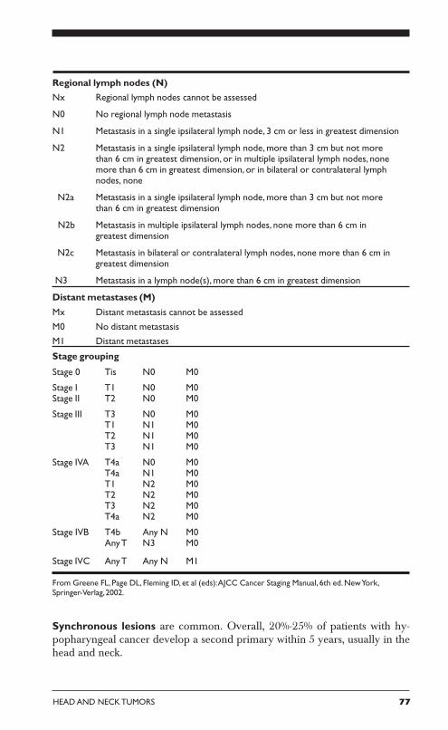
HEAD AND NECK TUMORS 77
Regional lymph nodes (N)
Nx Regional lymph nodes cannot be assessed
N0 No regional lymph node metastasis
N1 Metastasis in a single ipsilateral lymph node, 3 cm or less in greatest dimension
N2 Metastasis in a single ipsilateral lymph node, more than 3 cm but not morethan 6 cm in greatest dimension, or in multiple ipsilateral lymph nodes, nonemore than 6 cm in greatest dimension, or in bilateral or contralateral lymphnodes, none
N2a Metastasis in a single ipsilateral lymph node, more than 3 cm but not morethan 6 cm in greatest dimension
N2b Metastasis in multiple ipsilateral lymph nodes, none more than 6 cm ingreatest dimension
N2c Metastasis in bilateral or contralateral lymph nodes, none more than 6 cm ingreatest dimension
N3 Metastasis in a lymph node(s), more than 6 cm in greatest dimension
Distant metastases (M)
Mx Distant metastasis cannot be assessed
M0 No distant metastasis
M1 Distant metastases
Stage grouping
Stage 0 Tis N0 M0
Stage I T1 N0 M0Stage II T2 N0 M0
Stage III T3 N0 M0T1 N1 M0T2 N1 M0T3 N1 M0
Stage IVA T4a N0 M0T4a N1 M0T1 N2 M0T2 N2 M0T3 N2 M0T4a N2 M0
Stage IVB T4b Any N M0Any T N3 M0
Stage IVC Any T Any N M1
From Greene FL, Page DL, Fleming ID, et al (eds): AJCC Cancer Staging Manual, 6th ed. New York,Springer-Verlag, 2002.
Synchronous lesions are common. Overall, 20%-25% of patients with hy-popharyngeal cancer develop a second primary within 5 years, usually in thehead and neck.

78 CANCER MANAGEMENT: A MULTIDISCIPLINARY APPROACH
Chemotherapy and external-beam radiotherapy Similar in design to theVA Cooperative Laryngeal Cancer Study, a randomized trial of the EuropeanOrganization for Research on the Treatment of Cancer (EORTC) has shownthat initial therapy with cisplatin and 5-FU, followed by definitive irradiationin patients with complete remissions (or, alternatively, surgical salvage) resultsin at least equivalent survival relative to immediate pharyngolaryngectomy.Of patients treated with initial chemotherapy, 28% retained a functional lar-ynx at 3 years.
Results In general, even when treatment includes both surgery and radiationtherapy, hypopharyngeal cancer is difficult to control. The overall 5-year survivalrate is 25%; this rate decreases by 50% in the presence of lymph node metastases.
LARYNX
Laryngeal cancers constitute approximately 1.2% of all new cancer diagno-ses in the United States. Approximately 9,500 new cases are expected inthe year 2003.
Epidemiologyand risk factors
Laryngeal cancer most commonly affectsmiddle-aged to older men who smoke tobaccoand drink alcohol. There is no racialpredominance,s but, in the past, blacks havepresented at a younger age with a poorer prog-nosis. Most laryngeal squamous cell carcinomasresult from exposure to carcinogens that causediffuse mucosal changes (field effect).
Anatomy and pathology
Laryngeal anatomy is complex and includescartilages, membranes, and muscles. Thethree subsites of the larynx are the glottis, ortrue vocal cords; the supraglottis, which in-cludes the false cords, epiglottis, and aryepi-
glottic folds; and the subglottis, which is the region below the glottis andwithin the cricoid cartilage. The TNM staging system for cancers of the lar-ynx is outlined in Table 6.
Treatment
Laryngectomy All or part of the larynx may need to be removed to achievesurgical control of laryngeal cancer. Decision-making for partial laryngectomyis complex and depends on the patient’s wishes, the extent of local disease, and
To evaluate nonsurgical manage-ment of patients with stage III/IVlaryngeal cancer, a phase III studytested three arms: (A) inductioncisplatin/5-FU followed byradiation therapy; (B) concurrentcisplatin and radiation therapy; (C)radiation therapy alone. Totalradiation dose was 70 Gy in 7weeks with 2 Gy/fractions in allarms. A total of 507 patients wereentered in the study, with 65% ofthem having stage III disease. Two-year laryngectomy-free survivalwas 58% for arm A, 66% for arm B,and 55% for arm C. The studyconcluded that concurrentchemoradiotherapy significantlyincreased the time to laryngec-tomy (Forastiere AA, Berkey B, MaorM, et al: Proc Am Soc Clin Oncol[abstract] 20:2a, 2001).

HEAD AND NECK TUMORS 79
the skills of the surgeon and radiation oncologist. Pharyngocutaneous fistulaoccurs in < 15% of patients but remains the most common complication fol-lowing total laryngectomy. Spontaneous closure with conservative treatmentand local wound care occurs in the majority of patients.
External-beam radiation therapy and chemoradiation With improvementsin techniques and fractionation schedules, external-beam radiation therapy,which allows for laryngeal preservation, is an option for all but the mostadvanced tumors. A combination of induction chemotherapy (cisplatin plus5-FU) and subsequent radiation therapy (6,600-7,600 cGy) may be used topreserve the larynx in the majority of patients with T3 laryngeal cancer with-out compromising survival. This approach is complex and requires carefulattention during all phases of treatment.
Results In patients with T1 and T2 tumors of the glottic or supraglottic larynx,radiation therapy is associated with local control rates of 75%-95%. Patientswith T3 tumors can expect local control rates of 50%-65% with radiation therapyalone.
The VA Cooperative Laryngeal Cancer Study randomized patients withresectable squamous cell carcinoma of the larynx to receive either standard sur-gery followed by radiation therapy or neoadjuvant therapy with cisplatin and 5-FU followed by radiation therapy for those achieving a good response tochemotherapy. Approximately two-thirds of patients survived 2 years followingthe combination of either chemotherapy plus irradiation or resection plus irra-diation. Of those patients initially treated with chemotherapy and irradiation,one-third required a total laryngectomy because of a lack of response to treat-ment; the larynx was successfully preserved in two-thirds of these patients.
As in all head and neck cancer patients, the development of second cancerscontinues to result in mortality for several years following successful therapy.
LARYNGEAL SITE: SUPRAGLOTTIS
Supraglottic tumors occur less frequently than tumors of the true vocalcords. The epiglottis is the most common location for supraglottic cancers.
Natural history
Tumors close to the glottis produce symptoms sooner than tumors at othersubsites. However, nearly 60% of patients with supraglottic tumors have T3 orT4 primary tumors at presentation.
The supraglottis has a rich lymphatic network. There is an associated highincidence of lymph node metastases in early-stage tumors (40% for T1 tumors).The incidence of metastases in a clinically N0 neck is about 15%. The incidenceof bilateral cervical lymph node involvement is about 10%, and this rate in-creases to 60% for anterior tumors. The neck is a frequent site of recurrence inpatients with supraglottic malignancies.
80 CANCER MANAGEMENT: A MULTIDISCIPLINARY APPROACH
Treatment
Treatment of the primary tumor Appropriate cancers may be treatedwith partial laryngectomy. A supraglottic laryngectomy removes the upperportion of the thyroid cartilage and its contents, including the false vocalcords, as well as the epiglottis and aryepiglottic folds. This approach pre-serves speech and swallowing, but more extensive resections increase thedemands on lung function, limiting their utility. Supraglottic laryngectomyis not well tolerated by patients with impaired lung function because ofaspiration.
In patients with T1-T2 tumors, local control rates with radiation therapy (65-70 Gy in 6-7 weeks) exceed 80%. Also, as noted above, T3 tumors may betreated with radiation therapy (same regimen). Supraglottic laryngectomy isseldom appropriate as salvage therapy following irradiation due to complica-tions, including swelling, difficulty swallowing, and poor wound healing. Theusual salvage operation for persistent supraglottic cancer following radiationtherapy is total laryngectomy.
More advanced disease requires combined-modality treatment and often re-quires total laryngectomy. Radiation therapy may be employed with curativeintent, as may induction chemotherapy followed by irradiation, as describedearlier.
Treatment of the neck The high incidence of cervical metastases makestreatment of the neck a necessity. About one-third of clinically negative neckscontain involved nodes, and the incidence of recurrence in the untreated neckis high. In the surgical treatment of T1-T2 primary tumors, bilateral selectiveneck dissection is recommended.
Larynx preservation Data from RTOG 91-11 continue to mature, and anupdate to this trial was reported at the 2002 American Society of TherapeuticRadiology and Oncology (ASTRO) meeting, including rates of locoregionalcontrol, laryngectomy-free survival, and overall survival. This three-arm studyrandomized 517 evaluable patients to receive one of the following regimens:induction chemotherapy (with cisplatin and 5-FU) and conventional irradia-tion of 7,000 cGy, concurrent chemotherapy (cisplatin given days 1, 22, and43), or irradiation therapy alone to 7,000 cGy. Locoregional control for thethree arms was 61%, 78%, and 56%, respectively (P < .01). The number oflaryngectomies was 43, 21, and 49, respectively, for the three arms. Overallsurvival was about 75% for each arm. The authors concluded that concurrentchemoradiation therapy was superior to the other regimens, with the highestlocoregional control rates and laryngectomy-free survival.
Results The cure rate for early cancers of the larynx approaches 80% or more.Half of patients with T3 cancer are cured, whereas more than two-thirds ofpatients with T4 cancer will die of cancer.
HEAD AND NECK TUMORS 81
LARYNGEAL SITE: GLOTTIS
The glottis is the most common location of laryngeal cancer in the UnitedStates, comprising more than half of all cases.
Natural history
The cure rate for tumors of the true vocal cords is high. These cancers producesymptoms early, and, thus, most are small when detected. Approximately 60%are T1 and 20% are T2. Normal cord mobility implies invasion of disease lim-ited to the submucosa. Deeper invasion results in impaired vocal cord motion;this finding is most common in the anterior two-thirds of the vocal cord.
The true vocal cords have very little lymphatic drainage. Cervical metastasesare infrequent with T1 and T2 tumors.
Treatment
Treatment of the primary tumor Carcinoma in situ is highly curable andmay be treated equally well with microexcision, laser vaporization, or radiationtherapy. Treatment decisions should be based on the extent of local disease.Multiple recurrences should heighten suspicion for an invasive component,and a more aggressive approach, such as partial or total laryngectomy, shouldbe employed. T1 and T2 tumors may be treated by partial laryngectomy orradiation therapy (65-70 Gy in 6.5-7 weeks). In selected patients, partial laryn-gectomy can be performed after irradiation failure for T1-T2 glottic cancer,with excellent tumor control, satisfactory laryngeal function, and no increasein morbidity.
Advanced T4 disease is best treated with total laryngectomy. The standardtreatment for T3 glottic cancer has been laryngectomy. However, many T3lesions are now being treated with concomitant chemoradiotherapy, with sal-vage laryngectomy required in ~25% of patients for residual/recurrent diseaseor laryngeal dysfunction. Induction chemotherapy followed by irradiation canalso be used, as described earlier.
Treatment of the neck Due to the sparse lymphatic network and low inci-dence of cervical metastases, elective neck dissection is indicated only fortransglottic lesions. Palpable disease obviously requires neck treatment.
Results Cure rates by tumor size alone are as follows: T1, 90%; T2, 80%; T3,50%; and T4, 40%. Neck involvement worsens the prognosis dramatically.
LARYNGEAL SITE: SUBGLOTTIS
Subglottic cancer is unusual, accounting for fewer than 10% of all laryngealcancers.
82 CANCER MANAGEMENT: A MULTIDISCIPLINARY APPROACH
Natural history
These cancers tend to be poorly differentiated, and, as the region is clinically“silent,” most present as advanced lesions (~70% are T3-T4). The subglottisalso has rich lymphatic drainage, and the incidence of cervical metastases is20%-30%.
Treatment
Partial laryngectomy is not practical for the treatment of tumors in thesubglottis, and, thus, therapy usually includes total laryngectomy plus neckdissection. Combination therapy (surgery plus radiation therapy [60-65 Gyin 6-7 weeks]) is recommended for more advanced disease.
Results The cure rate for the uncommon T1 and T2 tumors is ~70%. Mostfailures occur in the neck. The cure rate for more advanced lesions is ~40%.
UNKNOWN HEAD AND NECK PRIMARY SITE
The cervical lymph nodes are the most common metastatic site at whichsquamous cell carcinoma is found.
Natural history
Most patients who present with squamous carcinoma involving cervical lymphnodes, especially in the upper or middle portion of the cervical chain, willhave a primary site within the head and neck. When the lower cervical orsupraclavicular lymph nodes are involved, a primary lung cancer should besuspected.
In the overwhelming majority of these cases, the primary lesion will bediscovered based on history, physical examination, proper radiographic evalu-ation (CT and/or MRI), and examination with the patient under anesthesiawith endoscopy (direct laryngoscopy, nasopharyngoscopy, esophagoscopy,and bronchoscopy). “Silent” primary tumors are most often discovered inthe base of the tongue or tonsillar fossa.
Treatment
A substantial percentage of patients achieve long-term disease-free survivalafter treatment of the involved side of the neck. Locoregional control andsurvival are diminished by multiple lymph nodes and the presence of extra-capsular extension of disease in the involved neck.
Irradiation alone Patients with early-stage neck disease (N1 disease or small,mobile N2a disease) can often be treated with radiotherapy alone. Radiationtherapy dosages and techniques should be similar to those used in patients
HEAD AND NECK TUMORS 83
with advanced primary head and neck cancer. The nasopharynx, orophar-ynx, and hypopharynx should be included in the irradiated field.
Surgery alone When neck dissection is used at the initial treatment, a pri-mary tumor in the head and neck subsequently becomes obvious in about20% of patients.
Irradiation alone or combined with surgery Combination therapy (sur-gery plus radiation therapy [60-65 Gy in 6-7 weeks]) is recommended forpatients who are found at surgery to have multiple involved nodes or extra-capsular extension or who have suspected residual microscopic disease inthe neck without a clinically detectable tumor. Open nodal biopsy does notappear to compromise outcome as long as adequate radiotherapy is deliv-ered subsequently.
In most modern series utilizing predominantly combination therapy, 5-yearsurvival rates exceed 50%. The volume of tumor in the involved neck influ-ences outcome, with N1 and N2 disease having a significantly higher curerate than N3 disease or massive neck involvement. Regional relapse is usu-ally predicted by extranodal disease.
Chemotherapy The role of chemotherapy for metastatic squamous carci-noma in cervical lymph nodes remains undefined.
RECURRENT HEAD AND NECK CANCER
As mentioned previously, surveillance after treatment of head and neck can-cer is mandatory, as early detection of second primary cancers or locoregionalrecurrence affords the best chance for disease control. Nearly two-thirds ofpatients whose head and neck cancer recurs develop tumor at (or near) theprimary site or in the neck nodes. Eighty percent of head and neck cancerrecurrences eventuate within 2 years. Development of recurrent tumor inthe neck is the single most common type of treatment failure in patients withsquamous cell carcinoma of the upper aerodigestive tract.
Differentiating recurrence from late complications of irradiation Dif-ferentiating between recurrent carcinoma and significant sequelae of radio-therapy is a difficult clinical problem at all sites within the head and neck.Any suspicious mucosal changes, enlarged nodes in the neck, or discretesubcutaneous nodules warrant prompt biopsy.
Candidates for surgery Different choices of first treatment (ie, surgery orradiation therapy) and the intensity of follow-up influence success in treatingrecurrence. Aggressive surgical intervention should be offered to two groupsof patients with recurrent local or regional disease: those whose therapy ischosen with curative intent and those who have the prospect for significantpalliation.

84 CANCER MANAGEMENT: A MULTIDISCIPLINARY APPROACH
The types of recurrence that may be approached surgically with the greatestlikelihood of success include (1) metastases in the neck after initial treatmentlimited to the primary tumor alone and (2) reappearance or persistence ofcancer at a site previously treated with radiotherapy alone. Salvage resectionmay also be considered in other situations, however. They include the ap-pearance of cancer in the neck after prior irradiation or neck dissection, atthe margins after previous resection, and even at the base of the skull.
Although surgery is the standard of care for the treatment of recurrent dis-ease, there is a growing body of evidence suggesting that reirradiation withconcurrent chemotherapy can save selected patients when resection is notpossible. Several institutions have reported experiences retreating patients,and these results led to the development of the first multi-institutionreirradiation study.
A single-arm, phase II study (RTOG 96-10) evaluated toxicity and therapeu-tic results for patients with recurrent squamous cell carcinoma of the headand neck. Eighty-six patients received four weekly courses of 1.5 Gy frac-tions twice daily with concurrent 5-FU and hydroxyurea. Each cycle wasseparated by 1 week of rest. The median survival was 8.1 months and the 1-and 2-year survival rates were 41.7% and 16.2%, respectively. Comparedwith patients who experienced early recurrences, patients whose disease re-curred 3 years after the original irradiation fared better, with 1- and 2-yearsurvival rates of 48.1% and 32.1%, respectively.
Unless a patient cannot tolerate an operation, resection of discrete local orregional recurrent tumors should be entertained as the first course of treat-ment. Management of recurrences involves complex decision-making andrequires familiarity with multidisciplinary care.
SUGGESTED READING
Al-Sarraf M, LeBlanc M, Giri PGS, et al: Superiority of 5-year survival withchemoradiotherapy (CR-RT) vs radiotherapy in patients (pts) with locally advancednasopharyngeal cancer (NPC). Intergroup (0099) (SWOG 8892, RTOG 8817, ECOG2388) phase III study: Final report (abstract). Proc Am Soc Clin Oncol 20:227a, 2001.
Ang KK, Harris J, Garden AS, et al: Concomitant boost radiation and concurrentcisplatin for advanced head and neck carcinomas: Preliminary results of a phase II trialof the RTOG (99-14). Int J Radiat Oncol Biol Phys 54:2–3, 2002.
Cooper JS, Pajak TF, Forastiere AA, et al: Patterns of failure for resected advancedhead and neck cancer treated by concurrent chemotherapy and radiation therapy: Ananalysis of RTOG 95-01/intergroup phase III trial. Int J Radiat Oncol Biol Phys 54:2,2002.
Corry J, Smith JG, Peters LJ: The concept of a planned neck dissection is obsolete.Cancer J 7:472–474, 2001.
Forastiere AA, Berkey B, Maor M, et al: Phase III trial to preserve the larynx: Induc-tion chemotherapy and radiotherapy vs concomitant chemoradiotherapy vs radiotherapyalone, intergroup trial R91-01 (abstract). Proc Am Soc Clin Oncol 20:2a, 2001.

HEAD AND NECK TUMORS 85
Garden AS, Pajak TF, Vokes E, et al: Preliminary results of RTOG 97-03: A phase IIrandomized trial of concurrent radiation (RT) and chemotherapy for advanced squa-mous cell carcinomas (SCC) of the head and neck (abstract). Proc Am Soc Clin Oncol20:223a, 2001.
Lin A, Kim HM, Terrell JE, et al: Head and neck cancer-specific quality of life (HN-QOL) and its correlation with xerostomia following parotid-sparing irradiation (RT) ofhead and neck cancer. Int J Radiat Oncol Biol Phys 54:167, 2002.
Maor MH, Berkey B, Forastiere AA, et al: Larynx preservation and tumor control instage III and IV laryngeal cancer: A three-arm randomized intergroup trial: RTOG 91-11. Int J Radiat Oncol Biol Phys 54:2–3, 2002.
NCCN practice guidelines for head and neck cancer. Oncology 14(11A):163–194,2000.
Ozyigit G, Chao CK: Quality of life assessment in head and neck cancer patients treatedwith definitive or postoperative IMRT. Int J Radiat Oncol Biol Phys 54:167–168, 2002.
Parliament MB, Scrimger R, Kurien E, et al: Preservation of oral health-related qualityof life and salivary flow rates after inverse-planned intensity modulated radiotherapy(IMRT) for head and neck cancer (HNC). Int J Radiat Oncol Biol Phys 54:165–166,2002.
Shoaib T, Souta DS, MacDonald DG, et al: The accuracy of head and neck carcinomasentinel lymph node biopsy in the clinically N0 neck. Cancer 91:2077–2083, 2001.
Wheeler RH, Harris J, Spencer S, et al: RTOG 96-10: Phase II study of reirradiation(RRT) with concurrent hydroxyurea (HU) and 5-fluorouracil (FU) in patients (pts) withrecurrent squamous cell cancer of the head and neck: Survival results (abstract). Proc AmSoc Clin Oncol 20:222a, 2001.
Wolden S, Pfister D, Zelefsky M, et al: Intensity modulated radiation therapy improveslocoregional control for nasopharyngeal carcinoma (abstract). Proc Am Soc Clin Oncol21:240a, 2002.

86 CANCER MANAGEMENT: A MULTIDISCIPLINARY APPROACH