Embed Size (px)
Citation preview

Part I – Chapter 5 - 1
`
INTRODUCTION • Epithelial cells form sheets of cells that are tightly joined to one another. Main
functions are: a. Cover surfaces. b. Line cavities. c. Form the secretory portions of many glandular structures.
• Epithelium is derived from all germ layers:
a. Ectoderm forms epidermis. b. Mesoderm forms mesothelium. c. Endoderm forms the lining of gastrointestinal tract.
• Major features of epithelia:
1. Epithelial cells line surfaces: Epithelia line and protect all free surfaces in the human body except joint cavities and the anterior surface of the iris:
§ The epithelial epidermis of the skin covers the outer surface of the body. § The hair follicles and glands in the skin are also epithelial. § Lining digestive system and its diverticula. § Lining respiratory system. § Lining cardiovascular system called endothelium. § Body cavities derived from intraembryonic coelom (pericardial cavity,
thoracic cavity and peritoneal cavity) are lined by mesothelium. § The urogenital system
2. Epithelial cells have tight lateral adhesions: An epithelium is one or more
layers of cells that are tightly joined together. The adhesions hold the epithelial cells together into a coherent barrier tissue. The apical junctions between cells in many epithelia have a sealing and adhesive structure called the Junctional complex, which isolated the internal milieu of the organism and tightly joins epithelial cells together.
3. Epithelial cells are polarized: The apical epithelial cells face a free surface of
the body or lumen of an organ or blood vessels and may be covered by microvilli or cilia. The basal surface rests on an extracellular layer of fibrils and glycoproteins called the basement membrane, or basal lamina, which is the boundary between the epithelium and the underlying connective tissue.
4. Epithelia are avascular: the epithelium is nourished by diffusion from blood
vessels present connective tissue layer surrounded the epithelium contains which.
Chapter 5 Epithelial Tissues
Part I – Chapter 5 - 2
Classification of Epithelia • Epithelial tissues are subdivided into:
1. Non-Glandular Epithelia 2. Glandular Epithelia 3. Modified Epithelia
Non-Glandular Epithelia
• Non glandular epithelia are classified by the number of cell layers they contain: I. Simple Epithelia: have one cell layer. All cells rest on the basement membrane
and reach the apical surface. II. Stratified Epithelia: have more than one cell layer, consequently not all cells
rest on the basement membrane or reach the apical surface. III. Pseudostratified Epithelia: are simple epithelia that appear to be stratified.
In these epithelia, all cells rest on the basement membrane; however, not all cells reach the apical surface. The stratified appearance occurs because nuclei lie at different levels in the epithelia.
Part I – Chapter 5 - 3
• Epithelia are further classified by cell thickness: a. Squamous cells are flat. b. Cubical or cuboidal cells; equal height and width. c. Columnar cells are taller than they are wide.
• In stratified epithelia, the apical layer is diagnostic:
a. Stratified Squamous epithelia; apical cells are squamous cells. b. Stratified Cubical epithelia; apical cells are cubical cells. c. Stratified Columnar epithelia; apical cells are columnar cells.
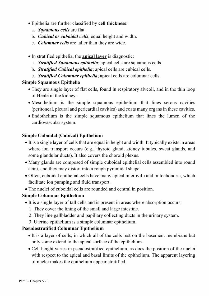
Simple Squamous Epithelia • They are single layer of flat cells, found in respiratory alveoli, and in the thin loop
of Henle in the kidney. • Mesothelium is the simple squamous epithelium that lines serous cavities
(peritoneal, pleural and pericardial cavities) and coats many organs in these cavities. • Endothelium is the simple squamous epithelium that lines the lumen of the
cardiovascular system.
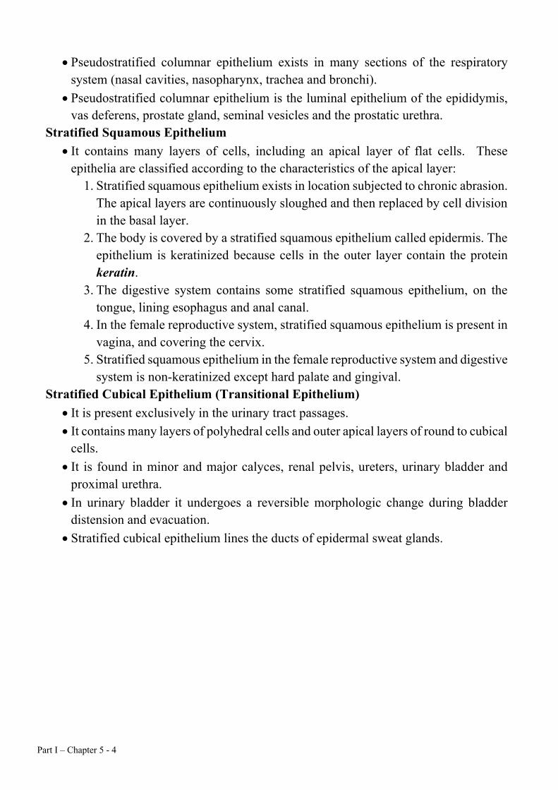
Simple Cuboidal (Cubical) Epithelium • It is a single layer of cells that are equal in height and width. It typically exists in areas
where ion transport occurs (e.g., thyroid gland, kidney tubules, sweat glands, and some glandular ducts). It also covers the choroid plexus.
• Many glands are composed of simple cuboidal epithelial cells assembled into round acini, and they may distort into a rough pyramidal shape.
• Often, cuboidal epithelial cells have many apical microvilli and mitochondria, which facilitate ion pumping and fluid transport.
• The nuclei of cuboidal cells are rounded and central in position. Simple Columnar Epithelium • It is a single layer of tall cells and is present in areas where absorption occurs:
1. They cover the lining of the small and large intestine. 2. They line gallbladder and papillary collecting ducts in the urinary system. 3. Uterine epithelium is a simple columnar epithelium.
Pseudostratified Columnar Epithelium • It is a layer of cells, in which all of the cells rest on the basement membrane but
only some extend to the apical surface of the epithelium. • Cell height varies in pseudostratified epithelium, as does the position of the nuclei
with respect to the apical and basal limits of the epithelium. The apparent layering of nuclei makes the epithelium appear stratified.
Part I – Chapter 5 - 4
• Pseudostratified columnar epithelium exists in many sections of the respiratory system (nasal cavities, nasopharynx, trachea and bronchi).
• Pseudostratified columnar epithelium is the luminal epithelium of the epididymis, vas deferens, prostate gland, seminal vesicles and the prostatic urethra.
Stratified Squamous Epithelium • It contains many layers of cells, including an apical layer of flat cells. These
epithelia are classified according to the characteristics of the apical layer: 1. Stratified squamous epithelium exists in location subjected to chronic abrasion.
The apical layers are continuously sloughed and then replaced by cell division in the basal layer.
2. The body is covered by a stratified squamous epithelium called epidermis. The epithelium is keratinized because cells in the outer layer contain the protein keratin.
3. The digestive system contains some stratified squamous epithelium, on the tongue, lining esophagus and anal canal.
4. In the female reproductive system, stratified squamous epithelium is present in vagina, and covering the cervix.
5. Stratified squamous epithelium in the female reproductive system and digestive system is non-keratinized except hard palate and gingival.
Stratified Cubical Epithelium (Transitional Epithelium) • It is present exclusively in the urinary tract passages. • It contains many layers of polyhedral cells and outer apical layers of round to cubical
cells. • It is found in minor and major calyces, renal pelvis, ureters, urinary bladder and
proximal urethra. • In urinary bladder it undergoes a reversible morphologic change during bladder
distension and evacuation. • Stratified cubical epithelium lines the ducts of epidermal sweat glands.
Part I – Chapter 5 - 5
Simple squamous epithelium lateral and top view Simple Columnar with microvilli Pseudostratified columnar ciliated with Goblet cell Transitional epithelium Stratified squamous epithelium with keratin
Part I – Chapter 5 - 6
Epithelial Polarity and Cell Replacement A- Apical modifications
1. Apical surfaces are not adhesive: • Most epithelial apices are not adhesive, even in the smallest tubules in the body. • In small intestines, the apical portion of the cell is coated with a thick,
glycoconjugate-rich layer external to the outer leaflet of the plasma membrane called the glycocalyx, which ensure non-adhesive feature of apices.
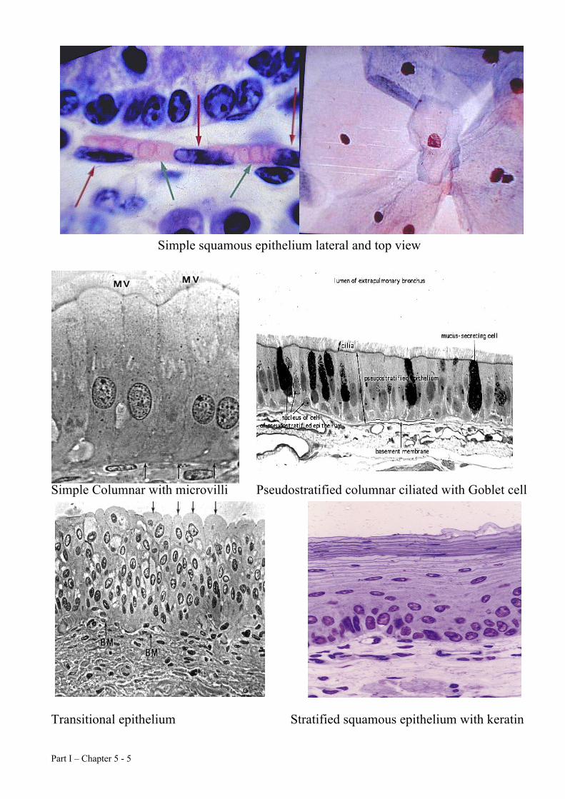
2. Apical protrusions: The epithelial apex often contains protrusions from the cell surface. • The protrusion may be scattered, as in mesothelium and endothelium, and may
take the form of microvilli: MICROVILLI
§ Microvilli are 80nm wide and 1-2µm long small finger-like foldings of cell membrane emitted from the free surface of the cell into the lumen.
§ They increase the cell surface area for absorbing materials from the lumen. § Microvilli can be few (as in mesothelium), numerous (as in the
syncytiotrophoblast), or very dense as in the brush border of kidney and intestinal epithelia.
§ Each microvillus contains a core of 25 to 30 actin filaments, cross-linked by villin, attached to an amorphous region at its tip and extending into the cytoplasm, where the actin filaments are embedded in the terminal web.
§ The terminal web is a complex of actin and spectrin molecules as well as intermediate filaments located at the cortex of the epithelial cells.
§ At regular intervals, myosin-I and calmodulin connect the actin filaments to the plasma membrane of the microvillus, giving it support.
§ Epithelia not functioning in absorption or transport may exhibit microvilli without cores of actin filaments.
Part I – Chapter 5 - 7
2) Stereocilia: are long microvilli present in the male reproductive tract and in the membranous labyrinth of the inner ear.
§ They are similar to microvilli except that they are longer and constricted at the point where they join the cell apex.
§ Sensory stereocilia contain many actin-containing microfilaments and large amounts of myosin.
3) Cilia: Many epithelia apices are ciliated as in: § In the trachea to expel mucous outside. § In the uterine tubes to move the fertilized ova. § In the apical portions of many sensory epithelial cells and they are not
motile and involved in energy transduction function of the cell. 3. Apical-basal polarity: Many epithelial cells have a polarized ultrastructure
that reflects their apical specialization: • Many ciliated cells have numerous mitochondria in the cell apex, close to the
basal bodies. B- Basal modifications
Basement membranes • Most epithelia have a basement membrane (lymphatic capillaries are a notable
exception), which ranges from scant to thick, depending on the epithelium: (1) Small capillaries have a thin or fenestrated basement membrane. (2) The basement membrane in the trachea is quite thick and serves as a defense
against bacterial invasion. (3) The epidermal basement membrane is thick and rugged to endure the
constant stress sustained by the epidermis. • Some basement membranes have an amorphous basement lamina adjacent to
the epithelium and a fibrous reticular lamina below the basement lamina. • The fine structure of the basement membrane varies considerably with the
location and function of the epithelia: (1) Capillary basement membranes are very thin and have no obvious
substructure. (2) In contrast, the glomerular basement membrane for the double epithelium
in the Bowman's capsule is thick but lacks a reticular lamina. Basement membrane composition • Basement membranes contain highly glycosylated Type IV collagen, which is rich
in the amino acid hydroxylysine and is composed of three α1(IV) subunits. Type IV collagen appears to be highly cross-linked and rich in carbohydrate units.
• Many basement membranes contain other glycoproteins such as laminin and proteoglycans such as heparan sulfate proteoglycan.
Adhesion of basement membranes and epithelial cells • Basal cells in the stratum germinativum of the epidermis are firmly anchored to
the basement membrane by intracellular specializations called hemidesmosomes.
Part I – Chapter 5 - 8
• Hemidesmosomes are similar to the desmosomes present in many junctions' complexes.
• Desmosomes and Hemidesmosomes seem to be sites where a cell strongly adheres to another cell or to an extracellular basement membrane.
Basement membrane functions • The basement membrane is the basal limit of an epithelium. In many luminal
epithelia, the basement membrane is the boundary between the epithelium and subjacent connective tissue.
• The basement membrane anchors the epithelium and may be a substratum for epithelial cell and connective tissue cell attachment.
• The basement membrane maintains the shape of acini and branched ducts and tubules.
• In some instances, the basement membrane is a selective barrier concerning the function of the glomerular basement membrane.
Other basal modifications: • The basal membrane of cells where ion transport is occurring often is elaborately
folded into numerous intercalated, finger-like projections, which increase the surface area of the basal plasma membrane.
• The many mitochondria that produce the ATP necessary for ion transport are located near these projections.
The Junctional complex: General characteristics:
• Epithelial tissue defines boundaries and separates functional compartments in the body, For example, the lumen of the small intestines contains a complex mixture of digestive enzymes capable of turning the gastrointestinal tract wall into a soup of amino acids. However, the junctional complex, an apical and lateral membrane specialization, isolates the gastrointestinal tract lumen from the sensitive wall of the gut.
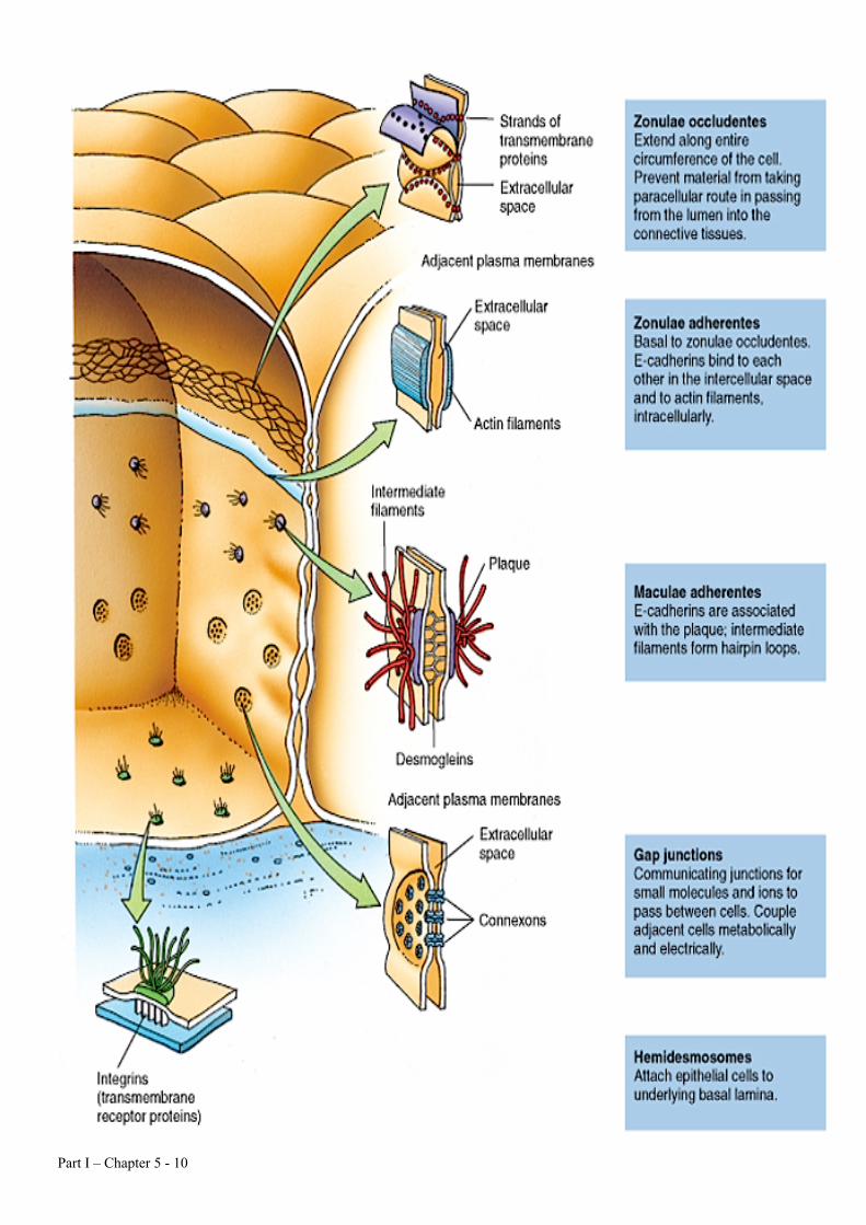
Ultrastructure: • Short sections of the membrane outer leaflets appear to fuse, forming an
occluding junction called the zonula occludens (tight junction), which extends around the apex of the columnar epithelial cells in a belt, creating a seal between the lumen and the lateral extracellular fluid environment.
• In freeze-fracture-etch, the zonula occludens in some epithelial tissues appears as an anastomosing network of ridges (points of membrane fusion) that impede the movement of molecules from the lumen to the lateral extracellular compartment.
• Below the zonula occludens, plasma membranes diverge to form a separation of 10-15nm called the zonula adherens (intermediate junction), a structure thought to be an adhesive junction. 1. The zonula adherens is a simple membrane apposition with varying
amounts of electron-dense material in the intervening gap.
Part I – Chapter 5 - 9
2. Intermediate (10nm) filaments radiate from the zonula adherens into the cytoplasmic matrix of apposed cells.
• The macula adherens (desmosome) is located below the zonula adherens. As its name implies, the macula adherens is thought to be a structure that holds cells together.
• Each of the apposed cells contributes half of the desmosome. An epithelial cell abutting on a basement membrane sometimes forms a hemidesmosome.
• At the macula adherens, the plasma membranes separate to 25-30 nm. An intermediate dense line runs between the cells in this separation.
• The inner aspect of apposed plasma membranes is covered with a punctate electron-dense material. Long bundles of Tonofilaments (a kind of 10nm intermediate filament) radiate from the plaque of electron-dense material.
• Many epithelia contain a structure called the gap junction (nexus), which is a specialized region where the outer leaflets approach each other, leaving a small (2nm) but definite gap.
• Gap junctions appear to be composed of hexagonal arrays of barrel-shaped structures, composed of six subunits arranged around an electron lucid central core. The central core is an aqueous channel between closely apposed cells. Ions and other small molecules pass freely between closely apposed cells, presumably through the aqueous channels in the gap junction.
Part I – Chapter 5 - 10
Epithelial cell replacement:
1. Some epithelial cells are replaced as they are lost by the division of the other cells within the epithelium.
2. This is especially true in stratified squamous epithelia such as the epidermis, where apical squamous cells are lost to friction. Without replacement, the entire epithelium would soon wear away. Epidermal basal cells constantly divide to produce the daughter cells that replace sloughed apical cells.
3. Intestinal epithelium: A slightly different process occurs in the gastrointestinal tract. Here are constantly subjected to the destructive influence of digestive enzymes, and intestinal villi are highly motile.
1. As intestinal villi move up and down, cells on the villi tips are sloughed into the lumen.
2. Cell division occurs at the base of intestinal villi in deep pits called the crypts of Lieberkuhn.
3. Daughter cells migrate up the crypt walls to the top of the villi where they are lost from the epithelium.
4. Stem cells:
Part I – Chapter 5 - 11
• In instances of epithelial turnover, the basal portion of the epithelium invariably contains a population of stem cells.
• Like all mitotic cells, stem cells divide into two cells. One daughter cell remains as a stem while the other differentiates and moves toward the apex.
• Proliferative stem cells are undifferentiated and usually rest on the basement membrane.
• The cell cycle varies tremendously among stem cell populations in different epithelia, ranging from days to years. For example, the epidermis turns over every 27 days, while seminiferous epithelia turn over every 64 days.
Modified Epithelial Cells
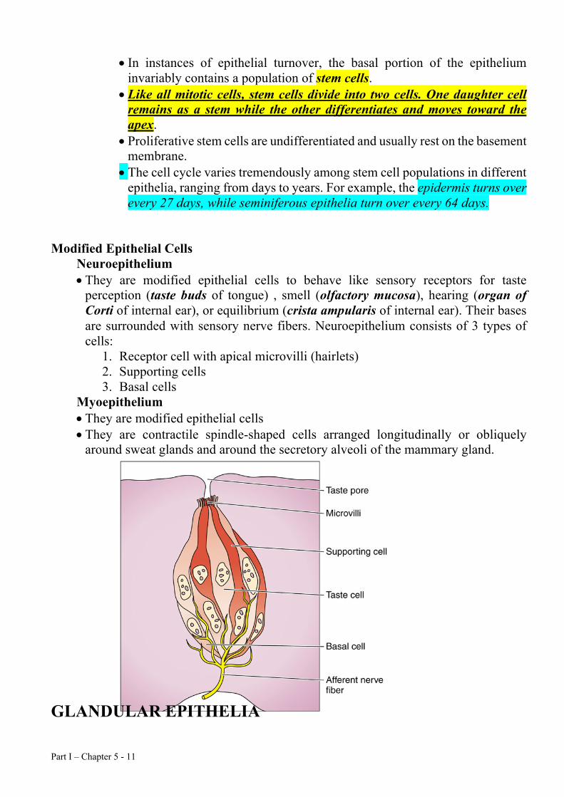
Neuroepithelium • They are modified epithelial cells to behave like sensory receptors for taste
perception (taste buds of tongue) , smell (olfactory mucosa), hearing (organ of Corti of internal ear), or equilibrium (crista ampularis of internal ear). Their bases are surrounded with sensory nerve fibers. Neuroepithelium consists of 3 types of cells:
1. Receptor cell with apical microvilli (hairlets) 2. Supporting cells 3. Basal cells
Myoepithelium • They are modified epithelial cells • They are contractile spindle-shaped cells arranged longitudinally or obliquely
around sweat glands and around the secretory alveoli of the mammary gland. GLANDULAR EPITHELIA
Part I – Chapter 5 - 12
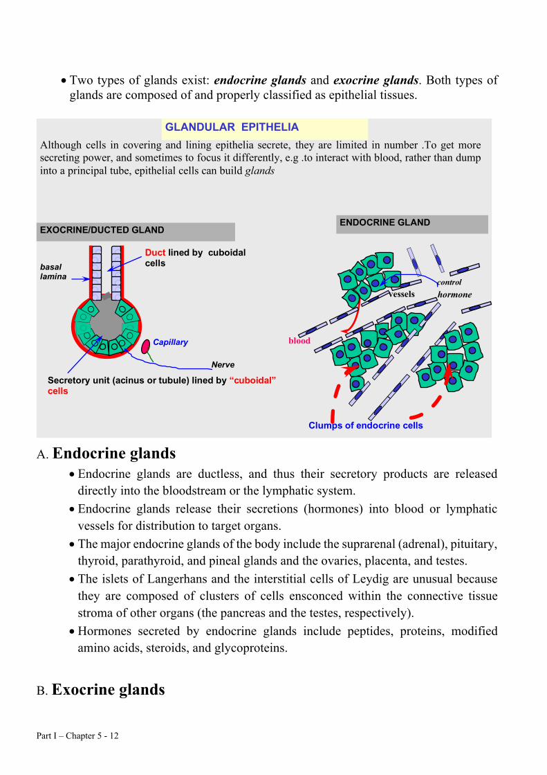
Although cells in covering and lining epithelia secrete, they are limited in number . To get more secreting power, and sometimes to focus it differently, e.g . to interact with blood, rather than dump into a principal tube, epithelial cells can build glands
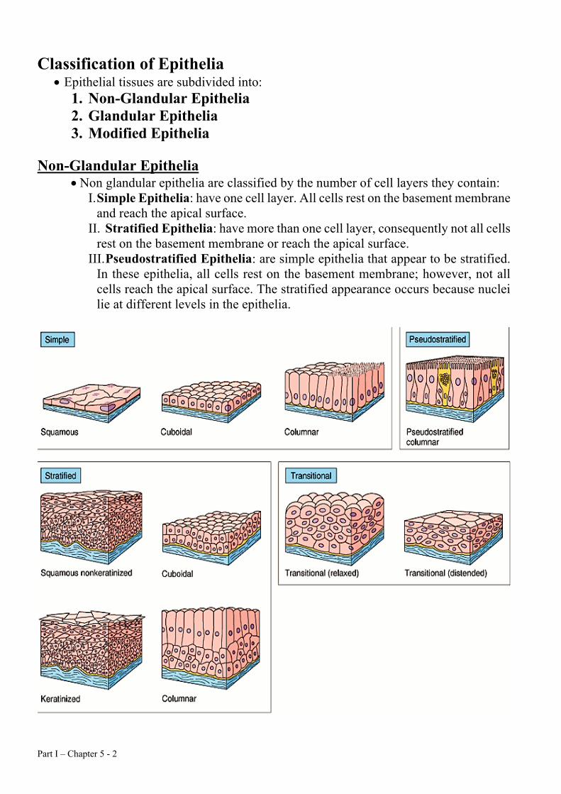
GLANDULAR EPITHELIA
vessels
ENDOCRINE GLAND
Clumps of endocrine cells
hormone
blood
Duct lined by cuboidal cells
Secretory unit (acinus or tubule) lined by “cuboidal” cells
EXOCRINE/DUCTED GLAND
Capillary
basal lamina
Nerve
control
• Two types of glands exist: endocrine glands and exocrine glands. Both types of
glands are composed of and properly classified as epithelial tissues. A. Endocrine glands
• Endocrine glands are ductless, and thus their secretory products are released directly into the bloodstream or the lymphatic system.
• Endocrine glands release their secretions (hormones) into blood or lymphatic vessels for distribution to target organs.
• The major endocrine glands of the body include the suprarenal (adrenal), pituitary, thyroid, parathyroid, and pineal glands and the ovaries, placenta, and testes.
• The islets of Langerhans and the interstitial cells of Leydig are unusual because they are composed of clusters of cells ensconced within the connective tissue stroma of other organs (the pancreas and the testes, respectively).
• Hormones secreted by endocrine glands include peptides, proteins, modified amino acids, steroids, and glycoproteins.
B. Exocrine glands
Part I – Chapter 5 - 13
• Exocrine glands secrete products onto the body surface through ducts. • The skin and digestive tract, which are continuous with each other, receive
exocrine secretions from glands such as sweat glands (skin) and the liver and pancreas (digestive tract).
• Exocrine gland may be unicellular gland (Goblet cell) or multicellular.
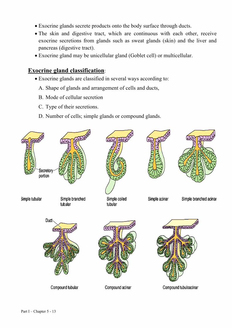
Exocrine gland classification: • Exocrine glands are classified in several ways according to:
A. Shape of glands and arrangement of cells and ducts,
B. Mode of cellular secretion
C. Type of their secretions.
D. Number of cells; simple glands or compound glands.
Part I – Chapter 5 - 14
• According to shape of glands are classified: 1. Straight tubular glands consist of a simple straight tubule and exist in the
small intestine. 2. Coiled tubular glands consist of a coiled tubule. Sweat glands are one
example of coiled tubular glands. 3. Branched tubular glands have branches deep in the gland. This type of gland
exists in the stomach and endometriurn. 4. Simple tubuloalveolar glands have a single duct leading to a cluster of alveoli,
or acini. Examples of this type of gland include the small salivary glands in the oral cavity and Brunner's glands in the duodenum.
5. Simple alveolar glands have several acini attached to a single duct (The sebaceous glands of the skin).
6. Compound tubular glands have numerous tubules connected to multiple ducts. These glands exist in the testes.
7. Compound tubuloalveolar glands have numerous secretory acini drain into numerous efferent ducts. These ducts typically merge into a smaller number of main ducts. The parotid salivary glands and the pancreas are example of this type of gland.
8. Compound alveolar glands are compound glands because they have numerous draining ducts; they terminate in the acini that have flat squamous cells rather than acini with cuboidal or pyramidal cells. The lungs are compound alveolar glands.
• According to modes of secretion:
1. Merocrine secretion involves the release of membrane-bound packets of
secretion product. The packets are formed as membranes derived from the ER
and Golgi apparatus surround the secretion product. The thyroid gland and
pancreas exhibit merocrine secretion.
2. Apocrine secretion products include a portion of the apical cytoplasm from the
secretory cell. Mammary glands exhibit apocrine secretion.
3. Holocrine secretion is characterized by whole cells bursting open to become the
secretory product. Epidermal sebaceous glands exhibit holocrine secretion.
• According to types of secretion:
Part I – Chapter 5 - 15
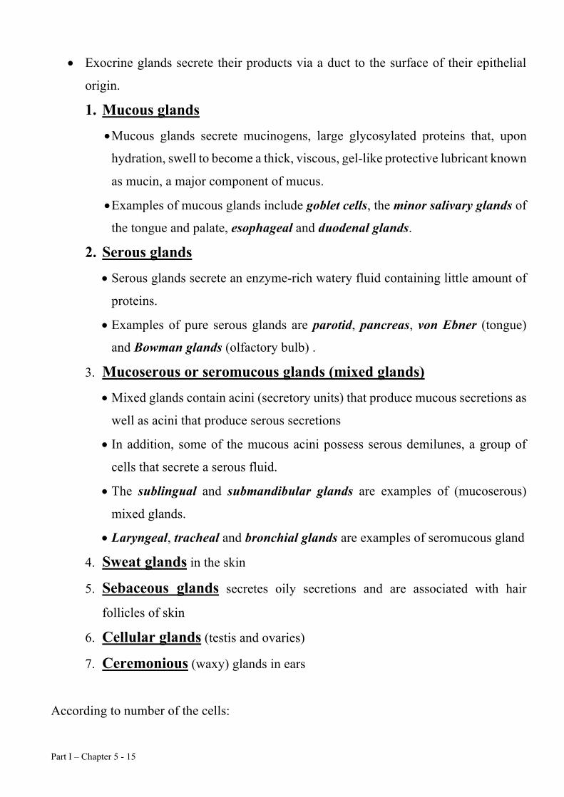
• Exocrine glands secrete their products via a duct to the surface of their epithelial
origin.
1. Mucous glands
• Mucous glands secrete mucinogens, large glycosylated proteins that, upon
hydration, swell to become a thick, viscous, gel-like protective lubricant known
as mucin, a major component of mucus.
• Examples of mucous glands include goblet cells, the minor salivary glands of
the tongue and palate, esophageal and duodenal glands.
2. Serous glands
• Serous glands secrete an enzyme-rich watery fluid containing little amount of
proteins.
• Examples of pure serous glands are parotid, pancreas, von Ebner (tongue)
and Bowman glands (olfactory bulb) .
3. Mucoserous or seromucous glands (mixed glands)
• Mixed glands contain acini (secretory units) that produce mucous secretions as
well as acini that produce serous secretions
• In addition, some of the mucous acini possess serous demilunes, a group of
cells that secrete a serous fluid.
• The sublingual and submandibular glands are examples of (mucoserous)
mixed glands.
• Laryngeal, tracheal and bronchial glands are examples of seromucous gland
4. Sweat glands in the skin
5. Sebaceous glands secretes oily secretions and are associated with hair
follicles of skin
6. Cellular glands (testis and ovaries)
7. Ceremonious (waxy) glands in ears
According to number of the cells:
Part I – Chapter 5 - 16
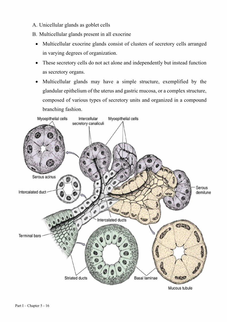
A. Unicellular glands as goblet cells
B. Multicellular glands present in all exocrine
• Multicellular exocrine glands consist of clusters of secretory cells arranged
in varying degrees of organization.
• These secretory cells do not act alone and independently but instead function
as secretory organs.
• Multicellular glands may have a simple structure, exemplified by the
glandular epithelium of the uterus and gastric mucosa, or a complex structure,
composed of various types of secretory units and organized in a compound
branching fashion.