Embed Size (px)
Citation preview

CHARACTERIZATION OF THE MYOSIN ADENOSINE
TRIPHOSPHATE (M.ATP) CROSSBRIDGE IN RABBIT AND
FROG SKELETAL MUSCLE FIBERSMARK SCHOENBERGLaboratory ofPhysical Biology, National Institute ofArthritis and Musculoskeletal and Skin Diseases,Bethesda, Maryland 20892
ABSTRACT In the presence of ATP and absence of Ca2", muscle crossbridges have either MgATP or MgADP . Pibound at the active site (S. B. Marston and R. T. Tregear, Nature [Lond.], 235:22:1972). The behavior of these myosinadenosine triphosphate (M * ATP) crossbridges, both in relaxed skinned rabbit psoas and frog semitendinosus fibers,was analyzed. At very low ionic strength, T = 50C, ,u = 20 mM, these crossbridges spend a large fraction of the timeattached to actin. In rabbit, the attachment rate constants at low salt are 104 - 105 s-', and the detachment rateconstants are 104 S-l. When ionic strength is increased up to physiological values by addition of 140 mM potassiumpropionate, the major effect is a weakening of the crossbridge binding constant -30-40-fold. This effect occurs becauseof a large decrease, -100-fold, in the crossbridge attachment rate constants. The detachment rate constants decreaseonly 2-3-fold. The effect of ionic strength on crossbridge binding in the fiber is very similar to the effect of ionic strengthon the binding of myosin subfragment-1 to unregulated actin in solution. Thus, the effect of increasing ionic strength infibers appears to be a direct effect on crossbridge binding rather than an effect on troponin-tropomyosin. The findingthat crossbridges with ATP bound at the active site can and do attach to actin over a wide range of ionic strengthsstrongly suggests that troponin-tropomyosin keeps a muscle relaxed by blocking a step subsequent to crossbridgeattachment. Thus, rather than troponin-tropomyosin serving to keep a muscle relaxed by inhibiting attachment, itseems quite possible that the main way in which troponin-tropomyosin regulates muscle activity is by preventing theweakly-binding relaxed crossbridges from going on through the crossbridge cycle into more strongly-binding states.
INTRODUCTIONDuring the steady state hydrolysis of ATP by actomyosin,many distinct actin-myosin-nucleotide complexes areformed (Lymn and Taylor, 1971; Bagshaw and Trentham,1974). These include the moieties formed immediatelyupon ATP binding, myosin adenosine triphosphate(M * ATP) and AM * ATP, the myosin-products com-plexes formed immediately upon hydrolysis, M * ADP Piand AM * ADP * Pi and the moieties formed as productsare released, M * ADP, AM * ADP, M, and AM. It hasbeen shown that in solution the M * ATP species, as well asthe M * ADP- Pi species formed immediately upon hydrol-ysis, bind weakly to actin (Stein et al., 1979). TheM * ADPand M states have been shown to bind strongly to actin(Highsmith, 1976; Marston et al., 1979; Greene andEisenberg, 1980).Over the years, considerable evidence has accumulated
that states similar to those seen in solution exist in musclefibers (Eisenberg and Greene, 1980; Hibberd and Tren-tham, 1986; Schoenberg, 1988). Previously it was shownthat a crossbridge state having-many of the properties ofthe M*ATP AM*ATP and MADP - PiAMADP * Pi states seen in solution could be isolated andstudied by examining relaxed fibers at very low ionicstrength (Brenner et al., 1982; Schoenberg et al., 1984;Brenner et al., 1986; Schoenberg, 1988). That crossbridge
BIOPHYS. J. © Biophysical Society * 0006-3495/88/07/135/14Volume 54 July 1988 135-148
state has alternatively been referred to as the weakly-binding state, the rapid-equilibrium state, or simply, theM * ATP state. It seems quite likely that the crossbridgestate in the fiber that corresponds to the M * ATP state insolution is the species that forms the initial attachmentwith actin and it may even be the state that precedes thecrossbridge power stroke. Thus, a detailed understandingof its kinetic properties would greatly enhance our under-standing of the contractile cycle.
Recently (Schoenberg, 1985), several different ways forextracting the fiber crossbridge rate constants frommechanical measurements were discussed in detail. Thepresent work applies one of these techniques to relaxedskinned rabbit psoas and also frog semitendinosus fibers inorder to measure both the relative number of attachedcrossbridges and the crossbridge detachment rate con-stants at different ionic strengths. From this, the cross-bridge attachment rate constants are also estimated. Inagreement with previous studies (Brenner et al., 1982;Schoenberg et al., 1984), it was found that the crossbridgeattachment and detachment rate constants are such that,at low ionic strength, the crossbridges in a relaxed fiberspend a large fraction of the time attached to actin. It wasfound further that increasing ionic strength to physiolog-ical values causes a large decrease in the crossbridgeattachment rate constants. This results in the crossbridges
$2.00 135

spending significantly less time attached to actin at physio-logical ionic strength. The number of crossbridges attachedunder different conditions correlates well with the bio-chemically-measured binding constant of myosin subfrag-ment-1 to actin in solution. It does not correlate with theresting tension of the fiber. This suggests that both at lowand normal ionic strength, the rapid stiffness of relaxedfibers at normal sarcomere length is due to weakly-bindingrapid-equilibrium crossbridges. It suggests further thatthese relaxed crossbridges are in states which correspond tothe weakly-binding SI - ATP and SI * ADP. Pi statesseen in solution, and that they attach to and detach fromthe actin filament over a wide range of ionic strengths.Since the crossbridges continually attach to actin in arelaxed fiber even at normal ionic strength, a key step inmuscle regulation must be subsequent to crossbridgeattachment.
This work was presented, in part, at a symposium on theMolecular Mechanism of Muscle Contraction, held inHakone, Japan, October 27-31, 1986 (Schoenberg,1988).
THEORY
Since, in this paper, the magnitude of the attachment anddetachment rate constants of the relaxed M * ATP cross-bridge and how they vary with ionic strength is explored, itwill be useful to briefly summarize the theoretical resultsupon which the analysis is based. One of the importantresults that came from previous modeling of rapid-equilib-rium crossbridges (Schoenberg, 1985) is that the responseof the crossbridges to mechanical strain is dominated bythe crossbridge detachment rate constants. The attach-ment rate constants mainly affect the number of cross-bridges attached. Intuitively, this occurs because themechanical response to strain is due to crossbridgesdetaching and reattaching in positions of lesser strain. It isthe most highly strained crossbridges that most oftendetach and reattach, and it is the detachment of thesehighly strained (high force-producing) bridges that tendsto dominate the force response. The reattaching cross-bridges tend not only to attach to the actin filament inpositions of lesser strain but also to attach in an equilib-rium distribution that produces little net force.
Previously I defined the chord stiffness of a muscle fiberas the amount of force generated by a given stretch dividedby the size of the stretch. If the duration of the stretch issuch that half of the crossbridges detach before the stretchis complete, the force induced at the end of the stretch willbe about half that that would have been induced if nocrossbridges detached (Schoenberg, 1985). What thismeans is that if one plots chord stiffness, S,, versus thelogarithm of the duration of stretch, td, the position of thecurve on the logarithmic axis will depend upon the cross-bridge detachment rate constants, and the amplitude of thecurve will depend upon the number of crossbridgesattached. It is this important result that enables us to
determine whether a particular intervention affects theattachment rate constants, the detachment rate constants,or both.
Previously (Schoenberg, 1985), using a relatively simpleone-attached-state model, it was shown that if crossbridgesare detaching with only a single rate constant, the relation-ship between chord stiffness and duration of stretch is
S' = (nbK/kbtd) [1 - exp (-kbtd)], (1)
where Sd is the chord stiffness measured over a stretchdistance d, nb is the total number of attached crossbridges,K is the stiffness of a single bridge, kb is the crossbridgedetachment rate constant, and td is the duration of thestretch. It is straightforward to show that if the cross-bridges are detaching with i different rate constants, onehas, if nj crossbridges detach with rate constant kjI,
c= (K/td) Z (ni/kj) [1 - exp (-kjtd)]j-l
(2)
Eqs 1 and 2 will be useful in estimating the crossbridgedetachment rate constants from an Sc - log td plot. Eq 2serves to approximate the case where different populationsof crossbridges detach with different rate constants or thedetachment rate constants vary with crossbridge strain.Note that both equations are independent of the attach-ment rate constant except in so far as the attachment rateconstant influences the total number of attached cross-bridges.
METHODS
Solutions and Fiber PreparationSkinned rabbit psoas fibers were isolated and prepared as reportedpreviously (Schoenberg and Eisenberg, 1985). The rabbit fibers wereskinned (made permeable to the bathing solution) by keeping them in askinning solution for over 30 min. The rabbit skinning solution, fashionedafter that of Eastwood et al. (1979), contained 150 mM KPropionate, 3mM MgAcetate, 3 mM Na2ATP, 5 mM ethylenebis (oxyethylene-nitrilo) tetraacetic acid (EGTA), 0.5 mM dithiothreitol (DTT), and 5mM KH2PO4, pH 6.8 at 50C. The frog fibers were skinned using amodification of the method of Julian et al., 1971. Ventral heads of thesemitendinosus muscle from American grass frogs (rana pipiens) wereisolated and tied, via the tendons, to a Sylgard (Dow Corning Corp.,Midland, MI) foundation. Enough chilled frog Ringer's solution (Schoen-berg and Wells, 1984) to barely cover the whole muscle was applied, andto this, chilled frog skinning solution was added. The frog skinningsolution was three parts rabbit skinning solution as described above andone part glycerol added to facilitate skinning. The skinning solution wasadded in aliquots initially as small as 100 Ml in order to prevent thepotassium in the solution from causing a contracture (Hodgkin andHorowicz, 1960). After a more than 100-fold excess of skinning solutionwas added, effectively diluting out the Ringer's solution, the muscle wasincubated for 1 h. Subsequently, a single fiber was isolated, the glycerol-containing skinning solution was exchanged for glycerol-free relaxingsolution, and the fiber was transferred to the experimental chamber formounting as reported previously (Schoenberg and Eisenberg, 1985).Low ionic strength relaxing solution contained 1 mM EGTA, 3 mM
MgCl2, 1 mM Na2H2ATP, 0.5 mM DTT, and 10mM imidazole, pH 7.0.An additional 4 mM KCI was present because of separate pH adjustmentof the individual constituents, making the total ionic strength of the
BIOPHYSICAL JOURNAL VOLUME 54 1988136
solution just under 20 mM. Higher ionic strength relaxing solutions weremade by adding KPropionate. All rabbit fiber experiments were done at50C at a sarcomere length of 2.5 ,um. The frog fiber experiments weredone at 40C and a sarcomere length of 2.4 ,um.
Force Transducer. The force transducer was made from anAE801 force gage (Sensonor, Horten, Norway). The beam of thetransducer was shortened 2.5-3 mm by gently grinding it against a pieceof #600 emery paper rotating on a lathe. A 200 ,um x 200 ,um x 700 ,mcarbon-epoxy fiber was epoxied at the tip perpendicular to the siliconbeam. It was to this that the muscle fiber was attached using a smallamount of cyanoacrylic glue applied through a glass micropipette. Afterthe experiment, the fiber and glue could be removed by applying acetone.The body of the transducer was held in a brass housing. Both the brasshousing, the brass parts of the AE801, and the active elements of thetransducer were pretreated with SS4004 primer and coated with RTV 60silicone rubber compound (General Electric Co., Waterford, NY). Thismade them impervious to saline solution and insensitive to light.The frequency response of the transducer was tested by releasing the
tension on a small length of silk thread glued between the force transducerand displacement generator. For the different transducers used, thefrequency response in water was generally 20-35 kHz and the dampingtime-constant was between 300-600 gs. The sensitivity was typically 10mV/dyn, the high frequency noise was -0.5 dyn, and the drift over 1 hwas -2 dyn. The total compliance of the system, including the displace-ment generator described below, was <2 x 10⁻³ ,m/dyn.
Displacement Generator. The displacement generator wasmade from a 1.5 inch diameter speaker coil kindly provided by AcousticsResearch (Cambridge, MA). The cloth dome covering the speaker coilwas reinforced with epoxy and an 8-cm long, 2.5 mm diameter stainlesssteel extension epoxied in place. The movement of the dome and stainlesssteel extension was guided by two Y-shaped plastic leafsprings located 10and 60 mm from the dome. Each arm of the leafspring measured 0.75 x6 x 45 mm. When positioned in place, the stainless steel extension satabove the experimental chamber. Three 10 mm carbon-epoxy struts in aninverted tripod configuration formed a rigid vertical extension to the bath.At the tip of this, a very small horizontal carbon-epoxy fiber, similar tothe one at the tip of the force transducer, was epoxied in place formounting the muscle fiber. The displacement of the speaker was detectedas described previously (Schoenberg et al., 1974). The half-time of thedetector response was <3 5s.
Driving the Displacement Generator. The signal from thedisplacement transducer, in addition to being displayed, was fed forwardto a LF356N operational amplifier (National Semiconductor, SantaClara, CA) which calculated the difference between the speaker displace-ment and a command signal. This difference signal was then summedwith a signal proportional to the first derivative of the displacement signalin order to generate the negative feedback signal to control the drivingcurrent to the speaker. The driving current, up to 7 A, was supplied by anHC2500 power operational amplifier (RCA, Somerville, NJ). Althoughwith this setup the speaker could move as much as 150 ,um in 150 Ps, thelargest displacement applied to the fiber was more typically 30 Am in
M50ps.
Sarcomere Length Detection. The sarcomere length detect-ing system consisted of a 10 mW 633 nm HeNe laser (Jodon Engineering,Ann Arbor, MI), three cylindrical focusing lenses, and a Schottky barrierposition sensitive light detector (SC-10, United Detector Technology,Santa Monica, CA). The components were aligned in such a way that,with the fiber in place and illuminated, either the right or left first orderdiffraction line fell upon the center of the Schottky barrier photodetector.Defining the x axis as the axis of the horizontal muscle, the y axis as theaxis of the horizontal laser beam, the z axis as the vertical axis, and y = 0asethe location of the fiber, a 200-mm focal length cylindrical lens at y =
-335 mm compressed the beam in the z-direction and a 300-mm focallength cylindrical lens at y = -110 mm compressed the beam in thex-direction. This lens system caused the 1.2-mm laser beam to measure,at the plane of the fiber, -0.25 mm in the z-direction and 0.3 mm in thex-direction. A 30-mm focal length cylindrical lens at y = + 55 mmcompressed the first order diffraction line in the z-direction onto thephotodetector located at y = + 120 mm.
Changes in position of the first order diffraction line were detected byusing three terminals of the Schottky barrier photodetector. The p-terminal was reverse-biased by 15 V and the two n-terminals, which carriedthe currents collected at opposite ends of the detector active area, wereconnected to two AD51 l's (Analog Devices, Norwood, MA) wired in acurrent-to-voltage converting configuration. Each feedback loop of theAD51 l's had a 100 kOhm resistor and 47 pF capacitor in parallel. Inaddition, each AD511 had circuitry for subtracting out the dark andbackground currents collected at each terminal. After background cur-rent cancellation, the signals from the AD511's were electronicallysubtracted to produce a signal proportional to the position of the centroidof the incident light and to intensity. When the difference signal from theAD51 l's was divided by the sum of two signals using an AD463B divider(Analog Devices, Norwood, MA), a signal proportional only to positionwas generated. A 44% decrease in light intensity produced a spuriouslength signal of <1 nm/half-sarcomere. The detector was positioned sothat the incident light fell close to the center of the active area. Thisminimized the sensitivity of the detector to movements of light in adirection perpendicular to the one being sensed. With this precaution, a1-mm motion in the z-direction produced <1/20 the signal of a 1-mmmotion in the x-direction.The noise in the sarcomere length detection system depended upon the
incident light intensity. With the intensity usually available from focusingthe first order diffraction line from a single fiber, the noise of thesarcomere length detector was <0.1 nm/half-sarcomere. With a fiber inplace, mechanical vibrations in the system raised the noise level to -0.3nm/half-sarcomere. A sarcomere length change of 1 nm/half-sarcomereproduced a signal of 25 mV. The measured half-time for the response ofthe sarcomere length detector was 9 ,s.
Setting up the Fiber. Since the displacement generator,fiber, and force transducer were mounted on a Unislide translation device(Velmex, East Bloomfield, NY) which was positioned on a rotary table,they could be translated, as well as rotated, relative to the laser beam. Theamount of translation was measured with a noncontact eddy currentdisplacement sensor (KD-23 10-1 5U, Kaman Instrument Corp., ColoradoSprings, CO). The amount of rotation was measured from the resistanceof a 1-turn potentiometer. A 50-mm diameter wheel, attached to the shaftof the potentiometer, rubbed along the edge of the rotary table androtated as it rotated.
After the fiber was mounted, it was translated so that the laser beamfor measuring changes in sarcomere length shined on the fiber 0.5 - 1mm from the force transducer end. The exact spot was chosen to be justbeyond the region of fiber affected by gluing to the transducer. Recordingthe sarcomere displacement near the force transducer permitted determi-nation of the fiber's force-displacement relationship with a minimum ofartifact related to series compliance or the finite time required formechanical signals to propagate along the length of the fiber (Schoenberget al., 1974).Once the fiber was axially positioned, it was then rotated relative to the
laser beam so as to maximize the intensity of the first order diffractionline. This not only maximized the light signal, it enabled us to samplefrom the largest subpopulation of myofibrils in the event that allmyofibrils were not identical (Rudel and Zite-Ferenczy, 1979; Brenner,1985; Sundell et al., 1986).
Measuring Chord Stiffness as a Function of Stretch Dura-tion. Chord stiffness is defined as the force generated by a givenduration stretch divided by the size of the stretch. In obtaining therelationship between chord stiffness and the duration of stretch, the force
SCHOENBERG Crossbridges in Relaxed Muscle 137

and sarcomere length signals caused by the stretch were fed into twochannels of a digital storage oscilloscope (Model 4094, Nicolet Instru-ment Corp., Madison, WI). To reduce noise, for each duration stretch,from 10-50 repeat traces were averaged. The multiple traces wereobtained at a repeat interval of 3 s, unless the stretch duration exceeded300 ms, in which case, the repeat interval was 10 times the stretchduration. Typically, after averaging, the noise in the traces was <0.05nm/half-sarcomere and 0.05 dyn. After the averaged traces werecollected, they were transmitted, via an RS-232 interface, to a Masscomp68010-based supermicrocomputer (Model 5500, Masscomp, Maynard,MA).The first step of the data analysis was to correct the force signal for the
finite frequency response of the force transducer. This was done using theprocedure outlined in Appendix B of Ford et al. (1977). Fig. 1 showsseveral typical averaged traces for three different velocity stretchesapplied to a single, skinned, relaxed rabbit psoas fiber, at 5°C, bathed in asolution having a total ionic strength of -20 mM. For the fastest stretchapplied, the figure shows both the original and corrected force signals. Itis seen that because of the high natural frequency of the force transducer,the magnitude of the force correction is small.From records like those shown in Fig. 1, chord stiffness was measured
for stretch distances of 0.5, 1, 2, and 4 nm/half-sarcomere. This was doneby having the computer analyze the time it took to stretch the fiber eachdistance, having it calculate the amount of force generated at the preciseinstant the fiber had been stretched by that distance, and having it dividethe force generated by the distance stretched. The total processing,including display of the force-displacement relationship, took -2 min,during which time the next averaged record, obtained with a differentspeed of stretch, could be recorded. In this way, four full chordstiffness-duration of stretch relationships, one for each chord distance,were relatively quickly obtained.
RESULTS
One important conclusion that comes from examining thethermodynamics of rapid-equilibrium crossbridges(Schoenberg, 1985) is that the horizontal position of thechord stiffness - log (duration of stretch) relationshipshould depend upon the crossbridge detachment rate con-stants. An important test of this is whether the horizontalposition of the Sc - log td relationship indeed shifts underconditions where the detachment rate constants change.Fig. 2 shows the Sc -log td relationships for a fiber bathedin 2 mM Mg-adenyl-5'-yl imidodiphosphate (MgAMP-PNP) and in 2 mM magnesium pyrophosphate (MgPP;).
3
>11
0 0.4 0.6 1.2
'W1 4in 6f.j.441-E E 2-0 0
Un 0 0 0.4 0.6 1.2rime [a]
A
EC
l-,C
0
CUtin
0
14
12
10
4
2
-3
PP,
I
-2 -1 0 1 2 3
-log (duration of stretch) [s]
FIGURE 2 Chord stiffness versus logarithm of duration of stretch for asingle skinned rabbit psoas fiber bathed in 2 mM MgAMP-PNP or 2 mMMgPP1 solution. Other ionic constituents: Imidazole, 10 mM; EGTA, 3mM; MgCl2 added in excess of PP; or AMP-PNP, 2 mM; KPropionate,70 mM; DTT, 0.5 mM; pH = 7.0; T = 50C. The chord distance was 2nm/half-sarcomere. In this and subsequent figures, chord stiffness, S,, isalways plotted versus (- log td) in order that speed of stretch increase tothe right. Experiment 070787: Fiber cross-section, 107 x 107 ,um; P0 =94 dyn. A second experiment on one other fiber gave similar results. Asmentioned in the text, scatter on the order of 10% for measurements madein the presence of nucleotide analogues was not uncommon. For thisreason, in this figure only, the MgPPi stiffness was multiplied by 0.9 inorder to make it, as seen previously on average, equal to the MgAMP-PNP stiffness for td = 10-' s.
The Sc - log td relationships are precisely of the typeexpected for an equilibrium population of crossbridgeswith continual crossbridge attachment and detachment.With short-duration, high-speed stretches the chord stiff-ness is high; with slower, longer-duration stretches thechord stiffness is considerably reduced. In solution (RogerGoody, personal communication; Jose Biosca and EvanEisenberg, personal communication), replacing AMP-PNP with PP; causes -a 10 - 20 fold increase in the rateof myosin subfragment-1 (SI) detachment from actin. AsFig. 2 shows, in the fiber this causes a horizontal shift ofthe Sc-log td relationship just a little over a decade to theright. This is precisely the result expected from the '85modeling. However, while Fig. 2 nicely illustrates the '85theory, it is not a perfect confirmation of it since replacing
10
,0
0 1 2 3
4-
2-
0
0 1 2 3
Time [me]B
40
00 125 250
4-
2-
o 1250 -250Time [ge]
c
FIGURE 1 Typical sarcomere length and force traces from a single relaxed skinned rabbit psoas fiber stretched in = 20 mM ionic strengthrelaxing solution, with different velocity stretches. A is the slowest speed stretch, C the fastest. Note different time scales and forcesensitivities. The dashed curve in the force panel of C shows the force trace corrected for the finite frequency response of the transduceraccording to the procedure of Ford et al., 1977. This particular force transducer had a natural period of 60 jus and a damping time constant of-450 ,us. Note that the correction is quite small, even for this transducer, the slowest one used. Experiment 092286: Fiber cross-section, 67 x107 ,um; Po, the isometric tension measured at 50C in ,u170 mM contracting solution (see Schoenberg and Eisenberg, 1985), was 67 dyn.
BIOPHYSICAL JOURNAL VOLUME 54 1988138

AMP-PNP with PPi in solution affects the SI attachmentrate constants just about as much as it does the detachmentones.
In theory, chord stiffness can depend not only upon thecrossbridge detachment rate constants and the duration ofstretch, but also upon the stretch amplitude (Schoenberg,1985). Fig. 3 A shows the cord stiffness - log td relation-ships of a relaxed, skinned rabbit psoas fiber, bathed in asolution with an ionic strength of 20 mM, for four stretchamplitudes, 0.5, 1, 2, and 4 nm/half-sarcomere. It is seenthat the SC - log td relationship is more or less independentof chord amplitude. The curves for the shorter chorddistances deviate slightly leftward at intermediate speedsof stretch and slightly to the right for faster speeds.However, the observed deviations are small and maysimply reflect systematic reading errors caused by a smallspurious 275-,us period oscillation in the original records(see Fig. 1 B). Most of the subsequent figures display the4-nm/half-sarcomere chord stiffness since, in theory, thisis the one least susceptible to any errors arising fromuncertainties in estimation of the exact time of the start ofstretch, water waves set up by the rapid speaker motion,electrical noise in the transducer traces caused by the largecurrent driving the speaker, and residual transmission timeeffects. Fig. 3 B shows the excellent reproducibility of suchdata.
Having seen that it is possible to obtain a precise Sc -log td relationship and also that the horizontal position ofthe curve should give us information about the crossbridgedetachment rate constants, we are now in a position toexamine the influence of ionic strength on the Sc - log tdrelationship of relaxed rabbit psoas fibers. Single fiberswere isolated and mounted as described in the Methodssection. They were then bathed in either low ionic strengthrelaxing solution (,u = 20 mM) or in low ionic strengthrelaxing solution with 20, 60, or 140 mM added KPropion-
i
I1
12
aI
I.-
00
12A a
01S A
a0
.°.BoI .... -. t
I 2 3 4
-boe (durtonm of frtoh) [.1
S
a
a A
AP0
a
. .!=. I *-_ . - I0 1 2 3 4
-io (dron ofSObvtc) Is)
FIGURE 3. Chord stiffness versus logarithm of duration of stretch for askinned rabbit psoas fiber bathed in 20 mM ionic strength relaxingsolution. (A) Chord stiffness measured with different chord distances.(B) Reproducibility of the 4-nm/half-sarcomere chord stiffness. In A,chord distances, in nm/half-sarcomere, were 0.5 (0), 1 (A), 2 (E), and 4,(0). In B, (0) is the low ionic strength 4-nm/half-sarcomere relationshiprecorded at start of experiment; (A) is the same relationship recordedsome 5 h, 500-1,000 stretches, and 6 solution changes later. Experiment043087: Fiber cross-section, 73 x 102 ,m. P0 not measured. Experimentson five additional fibers gave similar results.
PI.E ._
:CEa
6L00
20 mU
40 mm
0 ,nM
1S0 mm
1 2 3
-log (duration of atetch) [a]4
FIGURE 4 Chord stiffness versus duration of stretch for single relaxedskinned rabbit psoas fibers in solutions of different ionic strength. T =50C, 4 nm/half-sarcomere chord distance. The ionic strength value isgiven next to each curve. The significance of the solid and dashed curves isgiven in the legend to Fig. 5. Experiment 052086: Fiber cross-section,78 x 97 ,um. Po =63 dyn.
ate. An S,, - log td relationship for stretch durationsbetween 10-4 and 1 s was determined for the fiber in eachof the solutions. As a reference, the rigor stiffness, whichwas more or less independent of stretch duration between10-3 and 1 s, was also measured. Fig. 4 shows the data.
It is seen that as ionic strength increases, there is a bigdecrease in the chord stiffness for a given duration of
A
4 .4
3
o 1 2 84 o 1 2 3 4-log (duraion if titetch) [*1 -tdg (diurtion of -strech) [el
FIGURE 5. Fitting of the higher ionic strength Sc - log td data assum-ing either (A), that ionic strength affects only the attachment rateconstants, or (B), that ionic stength affects only the detachment rateconstants. Symbols (A, 0, 0), are data obtained at ionic strengths of 40,80, and 160 mM respectively. The dotted curves show the S,, - log tdcurves expected with either of the assumptions above. It is seen thatassumption B is a rather poor one. The dotted curves were obtained asfollows: First the ,u = 20 mM ionic strength data were fitted to a sum of 2or 3 exponentials (solid curve, Fig. 4). Expressing the relationshipdescribing the ,u = 20 mM data as Sc = f (log td), the dotted curves wereobtained by fitting the higher ionic strength data to the relationship, S, =aOf(log [tdl- a,), where a, was fixed at 0 for A and ao was fixed at 1.0for B. The absolute best fit to the data, obtained by allowing both ao and a,to vary, is shown by the dotted curves in Fig. 4. In all cases the fitting wasdone with a nonlinear least squares alogrithm based upon Marquardt'scompromise. To avoid overweighting points at either end of the scale,instead of using S, and td as the fitted variables, the log of S" was fitted tolog ao + log f(log [td] - a,). The amount of shifting or scaling wasdirectly determined from ao and a,. The 95% confidence limit of thedetermined parameters was typically 10-25%. The average parametervalues and S.E.M.'s for five complete experiments on rabbit psoas fibersare shown in Fig. 6.
SCHOENBERG Crossbridges in Relaxed Muscle
I
139

stretch. It is important to know whether this decrease inchord stiffness is due to a real decrease in the number ofattached crossbridges or whether, alternatively, it is due toan increase in the detachment rate constants without achange in crossbridge number. The latter might possiblyoccur if detachment became so rapid that our finite speedapparatus was no longer capable of detecting the attachedcrossbridges (Brenner et al., 1986). From the arguments inthe Theory section (see also Schoenberg, 1985), we see thatif increasing ionic strength is decreasing the number ofattached crossbridges without affecting the detachmentrate constants, it will simply cause vertical scaling of theSc - log td relationship. If it is increasing the detachmentrate constants without decreasing the number of attachedcrossbridges, it will cause the S, - log td relationship toshift to the right horizontally. Fig. 5 shows an attempt to fitthe data with one or the other of these two extremepossibilities. Fig. 5 B shows it is not at all possible to fit thedata with the assumption that increasing ionic strengthsimply causes a big increase in the detachment rateconstants. This is seen from the big divergences betweendata and fit for td < 10' s and the smaller but none-the-less significant deviations for 10-3 < td < 10' s. Incontrast, Fig. 5 A shows that the assumption that increas-ing ionic strength affects only the attachment rate con-stants is a reasonable one. The best fit to the data, thatshown by the dotted lines in Fig. 4, suggests that as ionicstrength is increased from very low values up to physiolog-ical values there is actually a very small decrease, -two-threefold, in the detachment rate constants, with a bigdecrease, -100-fold, in the attachment rate constants (seeDiscussion).The complete analysis illustrated in Figs. 4-5 was
repeated on five separate fibers. Fig. 6 shows the averagedresults, Fig. 6 A showing the relative number of attachedcrossbridges (determined from the degree of vertical scal-
ing) and 6 B showing the change in detachment rateconstant (determined from the amount of horizontal shift)as ionic strength is increased from 20 up to 160 mM. Wewill see later that at ,u = 20 mM the crossbridge detach-ment rate constants are in the neighborhood of I04 s'- andthe crossbridges spend -2/3 of the time attached to actin.The data suggest that at the same time that increasingionic strength from very low values up to physiologicalvalues by adding KPropionate causes only a smalldecrease, two-threefold, in the crossbridge detachmentrate constants, it appears to cause a big decrease, -20-foldin the amount of time the crossbridges spend attached tothe actin filament.One problem in calculating the relative fraction of time
crossbridges spend attached to the actin filament simply bycomparing the,u = 20 mM and, = 160 mM stiffnesses isthat the interfilament spacing of a relaxed skinned fiber at,u = 160 mM is considerably swollen relative to that at ,u =20 mM (Brenner et al., 1984). This must be taken intoaccount because resting stiffness has been shown to dependupon interfilament spacing (Berman and Maughan, 1982;Goldman and Simmons, 1986). One way of decreasing theinterfilament spacing of a relaxed fiber at normal ionicstrength to approximately the spacing at ,u = 20 mM is byadding 3% of the high molecular weight polymer DextranT500 to the bathing solution (see Godt and Maughan,1977; Brenner, et al., 1984). The effect this has on theresting chord stiffness is shown in Figs. 7 A and B. Fig. 7 Ashows that adding 3% Dextran T500 to a relaxed rabbitpsoas fiber at physiological ionic strength increases theresting chord stiffness -twofold. In a total of three experi-ments, the average increase was 120 ± 10%. It is interest-ing that adding 3% Dextran T500 has little effect on the,u = 20 mM chord stiffness (Fig. 7 B, average ratio fromthree experiments, 1.07 ± 0.03). It also has little effect on
3
20"' 401/2 801/i 160'/2 20" 40""2Ionic strength (mM)1/2
801/2 160'/2
to(o
FIGURE 6 Summary of the calculated change in the fraction ofattached crossbridges (A) and the crossbridge detachment rate constants(B) with increasing ionic strength. Average of five fibers. Error bars showS.E.M. At ui = 20 mM, the best estimate of the absolute fraction ofattached crossbridges (equal to the fraction of time an individual cross-
bridge spends attached to actin) is 70 ± 20% (see Discussion). Thecrossbridge detachment rate constants at A = 20 mM are 104 S-1 (seeDiscussion). The data displayed are the data uncorrected for interfila-ment spacing changes. Correction for interfilament spacing changeswould make the relative number of attached crossbridges at g = 160 mMincrease 2.2-fold (see Text and Fig. 7).
c0
I
0.C)
12 r
A
p - 160 mM
6
I
l E~~~~~~~~~~~~~~l
o 1 2 3 4-log (duration of stretch) [a]
U)3I
S$0
C.
B
p -20 mM
Ia
S0
a
o 1 2 3 4
-tog (duration of stretch) [a]
FIGURE 7. Effect of 3% Dextran T500 on the resting chord stiffness-duration of stretch relationship. (e) standard solutions; (0) standardsolution plus 3% Dextran T500. (A), ionic strength = 160 mM; (B), ionicstrength = 20 mM. The two curves in A show almost perfect scaling, thedashed curve being equal to the solid curve x 2.4. In a total of threeexperiments, the average scaling was 2.2 ± 0.1. Experiment 043087:Fiber cross-section, 73 x 102; PO not measured. From the data of Brenneret al., 1984, the interfilament spacing of a relaxed fiber at ,u = 160 mMwith 3% Dextran is estimated to be about the same as that of the relaxedfiber at g = 20 mM without Dextran.
BIOPHYSICAL JOURNAL VOLUME 54 1988
a)0
4.1aL.
-aEc
>._Ia)-i1k
2
I
00
a
140
9
.E
6C
0
n
cn
n.50 _o
6
A
0A00
A0
A0
A O'A 0A
1 2 3
-log (duration of stretch) [a]
4
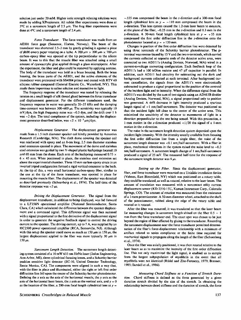
FIGURE 8 The resting chord stiffness-duration of stretch relationship attwo different ATP concentrations. (0) ATP = 1 mM; (A) ATP - 0.25mM. The 1 mM ATP solution was our standard solution; the 0.25 mMATP solution had 0.75 mM MgATP replaced with 5 mM KPropionate.T =4.80C. Experiment 110387: Fiber cross-section, 73 x 107 Am; P0 = 79dyn. Experiments on three additional fibers gave similar results.
the rigor stiffness (data not shown). Thus, when interfila-ment spacing changes are compensated for, there is - a10-fold, rather than a 20-fold, reduction in the amount oftime the crossbridges spend attached to actin at normal saltcompared with low salt.
It was important in our experiments to be certain wewere looking at the behavior of the M * ATP crossbridgeand not that of a partial rigor crossbridge. Fig. 8 shows theSc - log td relationship obtained from a fiber bathed in ourstandard low ionic strength relaxing solution (having[ATP] = 1 mM) and the same relationship obtained fromthe same fiber bathed in a 20 mM ionic strength relaxingsolution having an ATP concentration of 0.25 mM. It isseen that the two curves are identical. Additionally, theSc - log td relationship obtained in a 40 mM ionic strengthrelaxing solution having a MgATP concentration of 5 mMwas similar to that obtained in our standard, 1 mMMgATP, ,u = 40 mM relaxing solution (data not shown).Thus the Sc -log td relationship is insensitive to IATP] inthe mM range and all the crossbridges presumably haveMgATP (or MgADP * Pi) at the nucleotide binding site.The question arises, particularly at physiological ionic
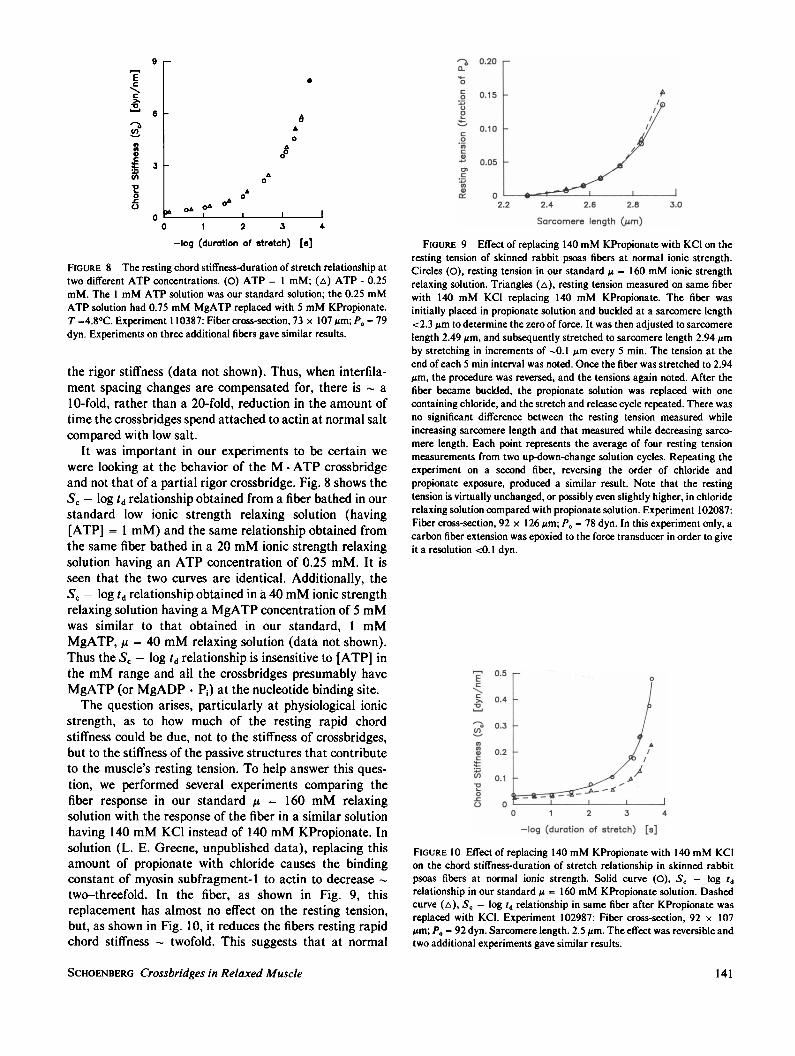
strength, as to how much of the resting rapid chordstiffness could be due, not to the stiffness of crossbridges,but to the stiffness of the passive structures that contributeto the muscle's resting tension. To help answer this ques-tion, we performed several experiments comparing thefiber response in our standard ,u = 160 mM relaxingsolution with the response of the fiber in a similar solutionhaving 140 mM KCI instead of 140 mM KPropionate. Insolution (L. E. Greene, unpublished data), replacing thisamount of propionate with chloride causes the bindingconstant of myosin subfragment- 1 to actin to decrease -
two-threefold. In the fiber, as shown in Fig. 9, thisreplacement has almost no effect on the resting tension,but, as shown in Fig. 10, it reduces the fibers resting rapidchord stiffness twofold. This suggests that at normal
0.200Le0
c 0.15
._2U0
ED%.,0.10c0
W 0.05co
0
1cr_ 0-2.2 2.4 2.6 2.8 3.0
Sarcomere length (/Am)
FIGURE 9 Effect of replacing 140 mM KPropionate with KCl on theresting tension of skinned rabbit psoas fibers at normal ionic strength.Circles (0), resting tension in our standard ,u = 160 mM ionic strengthrelaxing solution. Triangles (A), resting tension measured on same fiberwith 140 mM KCI replacing 140 mM KPropionate. The fiber wasinitially placed in propionate solution and buckled at a sarcomere length<2.3 gm to determine the zero of force. It was then adjusted to sarcomerelength 2.49,m, and subsequently stretched to sarcomere length 2.94 zmby stretching in increments of -0.1 ,um every 5 min. The tension at theend of each 5 min interval was noted. Once the fiber was stretched to 2.94jm, the procedure was reversed, and the tensions again noted. After thefiber became buckled, the propionate solution was replaced with onecontaining chloride, and the stretch and release cycle repeated. There wasno significant difference between the resting tension measured whileincreasing sarcomere length and that measured while decreasing sarco-mere length. Each point represents the average of four resting tensionmeasurements from two up-down-change solution cycles. Repeating theexperiment on a second fiber, reversing the order of chloride andpropionate exposure, produced a similar result. Note that the restingtension is virtually unchanged, or possibly even slightly higher, in chloriderelaxing solution compared with propionate solution. Experiment 102087:Fiber cross-section, 92 x 126,m; P. = 78 dyn. In this experiment only, acarbon fiber extension was epoxied to the force transducer in order to giveit a resolution <0.1 dyn.
E05Ec
> 0.4
a 0.3U)
*2In 0.2
U) 0.1o
0C- 0
0 1 2 3 4
-log (duration of stretch) [8]
FIGURE 10 Effect of replacing 140 mM KPropionate with 140 mM KC1on the chord stiffness-duration of stretch relationship in skinned rabbitpsoas fibers at normal ionic strength. Solid curve (0), Sc - log tdrelationship in our standard ,u = 160 mM KPropionate solution. Dashedcurve (A), Sc - log td relationship in same fiber after KPropionate wasreplaced with KCI. Experiment 102987: Fiber cross-section, 92 x 107Mum; P0 = 92 dyn. Sarcomere length. 2.5 um. The effect was reversible andtwo additional experiments gave similar results.
SCHOENBERG Crossbridges in Relaxed Muscle 141

sarcomere length (-2.5 ,um in the rabbit) a good fraction ofthe resting chord stiffness measured in response to rapidstretches is due to crossbridges, not to the structurescausing the fiber's resting tension (see also Discussion).
All the above data are from skinned rabbit psoas fibersat 50C. The resting stiffness of single, skinned, frogsemitendinosus fibers was also examined and typical Sr -log td plots at different ionic strengths are shown in Fig. 11.Again we see the kind of speed dependence of chordstiffness that is characteristic of rapid-equilibrium cross-bridges. However, normalized either to rigor stiffness orcross-sectional area, in the frog, the chord stiffness at agiven ionic strength and speed of stretch is two to threetimes lower than in the rabbit. In rabbit, the stiffnessmeasured at ,u = 20 mM with our fastest stretch was 51 +
3% of the rigor stiffness (n = 8). In frog, (n = 5), thecorresponding number was 28 ± 7%. Normalized to cross-sectional area, the maximum,u = 20 mM resting stiffnessin the rabbit was 1.17 ± 0.14 x 10⁻³ dyn/(nm/half-sarcomere)/(,gm)2 (n = 7) and in the frog, (n = 5), it was0.386 ± 0.13 x 10⁻³ dyn/(nm/half-sarcomere)/(gm)2.The data from frog, obtained over two different seasons,seemed somewhat more variable than that from rabbit.Larger fibers, as noted before by Ford et al., 1977, tendedto have significantly less resting stiffness per cross-sectional area. It is possible this might represent a truedependence of the frog's resting stiffness on season, fibertype, or diameter, but it was not possible to ascertain thiswith the limited number of fibers studied.
DISCUSSION
GeneralA significant advance in understanding muscle behaviorhas been the realization that, in many regards, the muscleproteins in the fiber behave like proteins in solution. It isnow quite clear that, just as for noncovalent binding ofproteins in solution, when we speak of the crossbridges asbeing bound to actin, we mean not that they are immutably
1.6Ec
C 12.
CO0.8
0.4
fE0U.
0
20 mlJ
40 mM
so mm
160 rnm
0 1 2 3
-log (duration- of stretch) Es]4
FIGURE 11 Chord stiffness versus duration of stretch for a single frogsemitendinosus fiber. Conditions like Fig. 4, except T = 40C, sarcomerelength = 2.4 ,m. Experiment 091486: Fiber cross-section, 87 x 97 Am,P. = 138 dyn. Two additional complete experiments on frog fibers gavesimilar results. Figure reprinted with permission from Schoenberg, 1988.
fixed to the actin filament, but only that the attachmentand detachment rate-constants are such so as to favorattachment over detachment. Even under conditions wherethe equilibrium is well shifted towards attachment (such ascrossbridge binding to actin in the presence of MgPP; orMgAMP-PNP at moderate or low ionic strength) themyosin crossbridges continually detach from and rebind tothe actin filament (see Fig. 2; also Schoenberg and Eisen-berg, 1985).
In a fiber, when a crossbridge detaches from a site on theactin filament, it need not necessarily reattach back to thesame site. When the crossbridges have been strained bystretch of the fiber, the detaching crossbridges will tend toreattach back to actin sites in positions of lesser strain. Ifthe stretch can be completed on a time scale shortcompared with the crossbridge detachment rate constants,one may determine the detachment rate constants simplyfrom the rate at which the force generated by the stretchdecays. We used this technique before to study cross-bridges having either MgAMP-PNP or MgPPi at theactive nucleotide binding site, finding that the detachmentrate constants in the presence of PP; are - 15 times fasterthan in the presence of AMP-PNP (Schoenberg andEisenberg, 1985). However, this technique cannot beapplied to crossbridges having ATP at the nucleotidebinding site because with ATP, the crossbridges detach ona time scale too fast for the fiber stretch to be completed.An alternative method developed for obtaining the
crossbridge detachment rate constants involves measuringthe chord stiffness of a fiber with different duration rampstretches (Schoenberg, 1985). Since the chord stiffness willbe maximal when measured with a stretch duration tooshort to allow any crossbridge detachment and very smallwhen measured with a stretch duration that allows multi-ple detachment and reattachment, the horizontal positionof a plot of chord stiffness versus the logarithm of thestretch duration will provide information about the cross-bridge detachment rate constants. We saw this illustratedin Fig. 2 and in the present study, this technique wasapplied to study the relaxed crossbridge.
It was possible, in resting rabbit psoas fibers, to makeaccurate and quite reproducible measurements of thechord stiffness-duration of stretch relationship. It was notclear that this would necessarily be the case since ourmethod rests heavily on the use of laser diffraction, atechnique known to be fraught with difficulty (Riidel andZite-Ferenczy, 1979). The excellent reproducibility of ourresting S, -log td curves (Fig. 3 B) is undoubtedly relatedto the fact that our measurements of sarcomere lengthchanges during stretch were both reproducible and alsoinsensitive to small variations in either angle or horizontalposition of the laser beam (see Brenner, et al., 1986).The reproducibility of data from relaxed frog semitendi-
nosus fibers was nearly as good as illustrated in Fig. 3 B,but that from rabbit and frog rigor fibers was not. The bestrabbit rigor fibers behaved reproducibly to within a few
BIOPHYSICAL JOURNAL VOLUME 54 1988142

percent but fibers showing scatter on the order of 10 oroccasionally even 20% were not uncommon. The differencein precision of measurements on relaxed fibers comparedwith rigor fibers may be related to differences in distortionof the orientation of the myofibrils with stretch. Thus,possibly in the rigor case, larger changes in the angularorientation of the myofibrils with stretch may amplifyartifacts related to Bragg angle effects (Riidel and Zite-Ferenczy, 1979; Goldman, 1987).
The Fiber Rate and Binding Constants
From our results, it is possible to make reasonable esti-mates of the detachment rate constants, and also the fiberbinding constants. From these, indirectly the attachmentrate constants of the M * ATP crossbridge may also beevaluated. The detachment rate constants are obtained byfitting the data of Fig. 4 to either Eq. 1 or 2. Fig. 12 Ashows ,u = 20 mM the fitted data to Eq. 1. The fact that thetheoretical one-rate constant curve is steeper than the datasuggests that the M * ATP crossbridge does not detachwith a single rate constant but with a somewhat widerrange of rate constants (see Schoenberg, 1985). We canestimate what this range is by fitting the data to Eq. 2using two or more rate constants. The 3-rate constant fit isshown in Fig. 12 B. While the assumption that 100% of thecrossbridges detach with a single rate constant does notgive an exceptionally good fit to the data (Fig. 12 A), thebest 3-exponential fit (Fig. 12 B), which fits the datanearly perfectly, has -90% of the crossbridges detachingwith only one of the rate constants. This situation, whichwas seen for all five experiments analyzed in detail, is quitedifferent from what is seen for crossbridges with MgAMP-PNP or MgPPi at the active site. With AMP-PNP orMgPP1, a 3-exponential fit gives three equally-weightedrate constants, the fastest and slowest separated by almostthree decades (Schoenberg and Eisenberg, 1985). Sincethe single-exponential fit for the M * ATP crossbridge at
A6
o1O 0° X ,o 1 2 3 4
-log (duration of stretch) [8]
0
B
0 1 2 3 4
-log (duration of stretch) [s]
FIGURE 12 Fitting the chord stiffness-duration of stretch relationshipof a relaxed skinned rabbit psoas fiber in 20 mM ionic strength relaxingsolution either (A), to Eq. 1, or (B), Eq. 2. Open circles, data; solid lines,fitted curves. For solid line in A, n = 1.0, k, = 4.1 x 13 s-'. In (B), n, =
0.91, k, = 2.3 x 10⁴ s-', n2 = 0.06, k2 = 390 s-', n3 = 0.03, k3 = 3.7 s-'.Same fiber as Fig. 4. For the five rabbit fibers summarized in Fig. 6, n,
averaged 0.88 +/- 0.02 and k, averaged (1.74 +/-0.20) x 104 S-1.
very low ionic strength gives a detachment rate constant of4 x 103 s-', while the 3-exponential fit has 90% of thecrossbridges detaching with a rate constant of -2 x 10⁴s- 1, it is clear that in contrast to the behavior of theM - AMP-PNP or M - PP1 crossbridge, the majority ofthe M * ATP crossbridges detach, at low ionic strength,over a narrow range of rate constants centered in theneighborhood of 104 S- .
The detachment rate constants at normal ionic strengthare somewhat more difficult to estimate since the substan-tially lower amplitude of the , = 160 mM data makes theprocedure outlined above less reliable. Another way ofapproaching the problem is to use the low ionic strengthdata as a reference condition and consider the relativeeffect of increasing ionic strength. To do this, we make useof the finding discussed above that the horizontal positionof the SC - log td relationship is determined mainly by thecrossbridge detachment rate constants. A maneuver suchas increasing ionic strength, if it affects the detachmentrate constants, should cause a horizontal shift in the Sc-log td curve. If changing ionic strength affects the numberof attached crossbridges without affecting the detachmentrate constants, i.e., if it affects only the attachment rateconstants, it should simply cause the Sc - log td curve toscale. As we saw from Fig. 5 A, the attempt to fit the ionicstrength data for the rabbit assuming only scaling gives afairly reasonable fit. However, the fact that the actual dataare slightly more concave upward than the scaled curvessuggests that, in addition to causing scaling, increasingionic strength also causes a slight leftward shift in the Sc-log td curve. It was found, doing a best least-squares fit tothe data, that increasing ionic strength from ,u = 20 mM to,u = 160 mM causes a shift in the Sc-log td relationship of-0.33 ± 0.30 decades to the left. This corresponds to only- a 2.1 ± 1.4-fold decrease in the crossbridge detachmentrate constants (see Fig. 6 B). The corresponding decreasegoing to 80 mM ionic strength was 3.0 + 0.6-fold. Thus atphysiological ionic strength, the detachment rate constantsof the M - ATP crossbridge are on the order of 3 x 10³ to104s-1.The attachment rate constants must be deduced in a
somewhat less direct fashion. Since the attachment rateconstants do not greatly influence the Sc - log td relation-ship except as they modulate the number of crossbridgesattached, it is necessary first to estimate the latter in orderto know the former (see Eqs. 1 and 2; also Schoenberg,1985). Although we do not know for sure the exact numberof attached M * ATP crossbridges in a relaxed rabbitpsoas fiber, there is now quite a bit of information concern-ing this. This information may be summarized as follows:(a) X-ray diffraction studies that attempt to estimate thenumber of crossbridges attached by estimating the amountof mass in the vicinity of the thin filaments (L. C. Yu andB. Brenner, manuscript in preparation) estimate thatbetween 60 and 80% of the crossbridges are attached atvery low ionic strength. (b) Stiffness measurements such as
SCHOENBERG Crossbridges in Relaxed Muscle
c 6
4)
'a2
2
03
13
2
143

those reported here show that the stiffness in low ionicstrength relaxing solution, measured with the fasteststretches is just over 50% of the rigor stiffness (see alsoBrenner et al., 1982, 1986). Since it is possible that themaximum chord stiffness measured in the relaxed fibermight be higher if we could stretch faster, if we assumethat the stiffness per bridge is the same in a relaxed fiber asit is in rigor, and that all the bridges are attached in rigor(Lovell and Harrington, 1981; Cooke and Franks, 1980;Cooke and Thomas, 1980), then 50% becomes a reasonablelower estimate of the number of crossbridges attached in,u = 20 mM relaxing solution. The binding constant of theM * ATP crossbridge at low ionic strength must thereforebe >1. (c) Causing a slight weakening of the bindingconstant at low ionic strength by increasing the ionicstrength only slightly results in a significant decrease in thenumber of crossbridges attached. The binding constant atlow ionic strength must therefore be <10, because other-wise a small decrease in its value would not cause cross-bridge detachment. (d) When the crossbridge bindingconstant is substantially weakened by the addition of 140mM KPropionate to very low ionic strength relaxingsolution, there is - a 10-fold decrease in the number ofattached crossbridges.
Since these observations argue that the crossbridgebinding constant is between 1 and 10 at very low ionicstrength, the crossbridge attachment rate constants at verylow ionic strength must be 1-10 times the detachmentones. That is, they must be 104 - 105 s-'. In addition, abinding constant of 1-10 means that at low ionic strength,the M * ATP crossbridges spend 50-90% of the timeattached to actin. Since our data suggest that at normalionic strength the crossbridges spend '/Ao as much timeattached to actin, at normal ionic strength the crossbridgesmust spend 5-9% of the time attached to actin. This means
that the binding constant at normal ionic strength is in therange of 0.05-0.1 and the attachment rate constants musttherefore be on the order of 100-1000 s'-. The attachmentand detachment rate constants of the M - ATP cross-bridge for skinned rabbit psoas fibers, and also the fractionof time the crossbridges spend attached to actin in arelaxed fiber, are summarized in Table I.
For frog semitendinosus fibers, the rapid resting stiff-ness for a given ionic strength and duration of stretch isonly -'/2 to 1/3 that of rabbit psoas fibers. This could meaneither that in the relaxed frog fewer weakly bindingcrossbridges are attached, or alternatively, that the samenumbers are attached but the detachment rate constantsare faster. The finding from the x-ray data of Xu et al.(1987) that the mass transfer at low ionic strength issignificantly smaller in the frog compared with the rabbitsupports the first hypothesis.
It is important to ask whether the relatively small restingchord stiffnesses measured at physiological ionic strengthcould be mainly due, not to rapid equilibrium crossbridges,but to the passive structural elements responsible for amuscle fiber's resting tension. Several lines of evidencesuggest it is not. First of all, at the sarcomere length wework at in the rabbit, the resting tension is very, very small,-1% of the maximum isometric tension. Secondly,although the rapid resting stiffness at normal ionicstrength is also somewhat small, its stretch duration depen-dence is practically identical to that of the much larger,obviously crossbridge related stiffness, which is measured(Brenner et al., 1982) at low ionic strength. Thirdly, andmost importantly, a strong suggestion that the rapid rest-ing stiffness is mainly due to rapid-equilibrium cross-bridges comes from the observation that when one replacespropionate with chloride, there is about a twofold decreasein the rapid resting stiffness (Fig. 10). The fact that
TABLE ISUMMARY OF THE CROSSBRIDGE ATTACHMENT AND DETACHMENT RATE CONSTANTS
OF RELAXED SKINNED RABBIT PSOAS FIBERS AT 50C*
Parameter Low salt (,u = 20 mM) Normal salt (M = 160 mM)
Detachment rate constants (s-') 104 -3 x I03(5 x 10³- 5 x 10'4)t (2 x 10- 2 x0)
Attachment rate constants (s-') -2 x 10⁴ -250(5 x 10³ - 5 x 10⁵)11 (100 - io00)t,#
Percent attached to actin 70 7(50 - 90)1 (5 - 9)**#
*Most likely values, with estimated range of uncertainty given below in parentheses.$Obtained from the horizontal position of the Sr - log td relationship, or equivalently, by fitting relationship to Eq. 1 or 2.5Obtained either by assuming that the stiffness of a relaxed crossbridge is similar to that of a rigor crossbridge or from x-ray diffraction results.'Obtained from the horizonal shift of the S, - log td relationship with increased salt.**Obtained from the vertical scaling of the S, - log td relationship with increased salt.1iCalculated from the detachment rate constants and fraction attached according to the approximation, nb = fl(f + f'), where nb is the number ofattached crossbridges andfandf' are the attachment and detachment rate constants respecively.44With interfilament spacing changes compensated for.
BIOPHYSICAL JOURNAL VOLUME 54 1988144

replacing propionate with chloride has virtually no effecton the resting tension (Fig. 9), while in solution it causes atwo-threefold decrease in the binding constant of myosinsubfragment-1 to actin (L. E. Greene, manuscript submit-ted for publication), strongly suggests the conclusion thatthe rapid stiffness is due mainly to crossbridges, not restingtension structures.One may actually estimate the contribution of the
resting tension structures to the rapid chord stiffness atphysiological ionic strength. At a sarcomere length of 3.5,um, where we are looking primarily at the stiffness of thestructures responsible for the fiber's resting tension, theresting rapid chord stiffness is ~-/io of the rigor stiffnessmeasured at 2.5 ,um (Brenner et al., 1982). If we assumethat the component of the resting chord stiffness due to thestructures responsible for the fiber's passive resting tensionis proportional to the slope of the resting tension-sarcomerelength relationship, then, since the slope of that relation-ship at 2.5 ,m is - '/lo that at 3.5,um (Fig. 9; also Horowitsand Podolsky, 1988), the contribution of the resting tensionstructures to the resting rapid chord stiffness at sarcomerelength 2.5,tm is estimated to be --/Aoo of the rigor stiffness.Since the measured resting rapid chord stiffness underthese conditions is about l/20 of the rigor stiffness, thissuggests that the noncrossbridge contribution to the rapidresting stiffness of rabbit psoas fibers in physiological ionicstrength propionate solution is probably on the order ofonly 1 part in 5.
Relationship to the Mechanism ofMuscle Relaxation
From the data presented, it is clear that, in fully relaxedfibers, M - ATP crossbridges continually attach to anddetach from the actin filament over a wide range of ionicstrengths. Thus the current results strongly support ourprevious contention, based on our experiments at low ionicstrength (Brenner et al., 1982; Schoenberg et al., 1984),that muscle relaxation cannot be due solely to troponin andtropomyosin blocking attachment as envisioned by theclassic steric blocking model for muscle relaxation (Hux-ley, 1972, Haselgrove, 1972; Parry and Squire, 1973). Ourevidence strongly suggests that blockage of a step subse-quent to attachment is essential and necessary for musclerelaxation.The finding that keeping a muscle relaxed depends upon
blockage of a step subsequent to attachment raises anumber of questions. The first of these concerns which stepis blocked. It is clear from the work of D. K. Hill (1968),Lannergren (1971), Haugen and Sten-Knudsen (1981),our own work, and that of others, that only very fewstrongly-binding crossbridges exist in a relaxed fiber. Thusthe step subsequent to attachment which is blocked to keepa muscle relaxed is one that prevents the weakly-bindingM * ATP crossbridges from going on through the cross-
bridge cycle onto the more strongly-binding states.'Whether the step blocked is the actual force producingpower stroke step, or one before it, remains to be seen. Insolution, the step blocked during inhibition of the actosub-fragment- I ATPase appears to be at or near the phosphaterelease step (Chalovich and Eisenberg, 1982; Rosenfeldand Taylor, 1987).
Another question unanswered by the current data arewhether Ca2' has any effect on the binding of theM - ATP crossbridge. In solution, the biochemical evi-dence suggests that the effect of Ca2, on the binding ofsubfragment 1 or heavy meromyosin to regulated actin is<5-10-fold (Chalovich and Eisenberg, 1982; Wagner andGiniger, 1981; Wagner, 1984; Chalovich and Eisenberg,1986; Rosenfeld and Taylor, 1987). Presently, unfortu-nately, there is very little fiber evidence concerning this.The finding that mass shifts towards the actin filamentupon activation at physiological ionic strength does notshed light on this question because it is not yet knownwhether any of this mass shift is due to an increase in thebinding of the M * ATP crossbridge or whether most of itis due to the continually attaching and detaching M * ATPcrossbridges going on through the crossbridge cycle (afterattachment) to more strongly-binding states. To resolvethis question, it will be necessary to develop techniques fordetecting weakly-binding bridges under conditions wherethey coexist with more strongly-binding states.
Separate from the question of whether the troponin-tropomyosin regulatory system has much influence on thebinding of the M * ATP crossbridge, another question thatremains is whether the weak binding of the M - ATPcrossbridge at physiological ionic strength contributes tomuscle regulation. The finding that muscle fibers areperfectly well regulated at low ionic strength where thebinding constant of the M * ATP crossbridge is greatlyincreased suggests it does not. On the other hand, thefinding that fully relaxed muscle fibers at low ionicstrength can become partially activated by small changesin temperature or free Mg,2 (Gulati, 1983) suggests thatperhaps it does. Resolution of this question will clearlyrequire a more detailed knowledge of the regulatory mech-anism.When a fiber becomes activated by Ca2 , there will be a
mixture of weakly- and strongly-binding crossbridgestates. The overall fraction of weakly-binding crossbridgestates in an activated fiber will be determined by the rateconstant governing the transition from the weakly-bindingto strongly-binding states and also that which governs thetransition back to the weakly-binding states. The finding
'Calling the weakly-binding relaxed crossbridges M * ATP crossbridges issimply a matter of nomenclature and is not meant to imply that hydrolysisis the step that is blocked in relaxation; the resting crossbridges may infact have ADP- Pi, rather than ATP, at the nucleotide binding site(Marston and Tregear, 1972).
SCHOENBERG Crossbridges in Relaxed Muscle 145

that the stiffness of an activated fiber measured with amoderate velocity stretch is 60-80% that of a rigor fiber(M. Schoenberg, manuscript submitted for publication;Goldman and Simmons, 1977; Yamamoto and Herzig,1978), along with the fact that nearly 100% of the cross-bridges appear to be attached in rigor, suggests that in anactivated fiber -20-40% of the crossbridges are inM * ATP-like states. If Ca2' has no effect on the strengthof binding of the M *ATP crossbridge (Chalovich andEisenberg, 1982), then in an activated fiber at normal ionicstrength, <4% of the M * ATP-like crossbridges (10% of40%) will actually be attached. If Ca2+ increases thebinding of the M . ATP crossbridge 5-10-fold as morerecent evidence suggests (Wagner, 1984; Chalovich andEisenberg, 1986), then perhaps the number of weakly-binding crossbridges attached in an active fiber will be inthe 10-20% range.From the above arguments, it would appear that the
attached M * ATP crossbridge state most likely is differentfrom any of the force producing states postulated byHuxley and Simmons, 1971. Those states were postulatedto be more strongly binding and also to have slowerattachment and detachment rate constants. The M * ATPstate may, however, be similar to the crossbridge state thatwas postulated by Huxley, 1972, in order to explain betterthe energetics of muscle contraction.
Comparison to Solution and Other SystemsThe first suggestion that possibly there might be cross-
bridges attached in a relaxed fiber came from the biochem-ical studies of Chalovich et al. (1982) It is interesting howclosely the behavior of the weakly-binding crossbridges infibers actually mirrors the behavior of S1 * ATP in solu-tion. The interaction of S1 with actin in the presence ofATP at 200C, 0.1 M KCl, and pH 7 is described (Geeves etal., 1986) by the reaction scheme
K- 10'M-' k2 5,000s-'AM+T. AMT-
k2 - 1 s-'
k3>, 5,000 s-'A-MT A + MT
where AM and A-M represent two conformations of theactin myosin linkage and T represents ATP. Virtually thesame scheme apparently applies to the fiber. Thus, Gold-man and colleagues (Goldman et al., 1984), starting fromAM and adding ATP, measured a second order rateconstant for ATP binding, K,k2, of _106 M-1 s'-. Theyfurther found, in agreement with a K1 value of 103 M-1,that the rate of crossbridge detachment in their experi-ments varied with ATP concentrations up to 1 mM. In ourexperiments, where presumably we are starting from anA-MT -like state, we find a crossbridge detachment rateconstant, k3, of 104 s-1. As expected from the abovescheme, this is independent of ATP concentration in the
0.25-5 mM range. Thus, both with regard to the rateconstant for ATP binding, and also the rate constant forthe actual detachment step, there appears to be goodagreement between fiber and solution.
Another area of agreement between fiber and solutionconcerns the effect of ionic strength on the binding con-stant of the M * ATP crossbridge. We have already dis-cussed that replacing 140 mM propionate with chloride,which in solution causes a two-threefold decrease in thebinding constant of myosin subfragment-1 to regulatedactin, in the fiber, causes a twofold decrease in the rapidresting stiffness. Additionally, increasing ionic strength insolution from , = 20 to,u = 160 mM by adding 140 mMKCl causes - a 100-fold weakening of the binding constantof myosin subfragment-1 to unregulated actin (Greene etal., 1983). If we assume, based upon the stiffness and x-raymeasurements, that -70% of the crossbridges are attachedat ,u = 20 mM, then that same maneuver of adding 140mM KCl in the fiber causes the fiber binding constant tobe reduced from -2 to -0.03, - a 70-fold reduction. Thus,increasing ionic strength in the fiber, where all the regula-tory proteins are present, produces about the same weaken-ing of binding as occurs in solution in the absence ofregulatory proteins. This is particularly important becauseit shows that even if ionic strength has some effect on thetroponin-tropomyosin regulatory system, the effect is notone that greatly influences the strength of binding of theM * ATP crossbridge.A final similarity between fiber and solution is the fact
that in both cases the major effect of ionic strength is onthe attachment rate constants. These similarities serve toreinforce the idea that the weakly-binding crossbridgestates seen in the fiber closely resemble the weakly-bindingM * ATP state seen in solution. M * ATP-like crossbridgeshave now been found in rabbit, frog, crab and perhaps evenone type of smooth muscle (Brenner et al., 1982; Xu et al.,1987; Wakabayashi et al., 1984; K. Wakabayashi, per-sonal communication; Siegman et al., 1976). Although theexact strength of binding may not be the same in all species(this paper, Xu et al., 1987, Brenner and Squire, 1987),none-the-less, the phenomenon of weakly-binding cross-bridges, like the phenomenon of equilibrium rather thanirreversible attachment, may very well be a rather generalone. It is thus interesting to speculate what the role of theM * ATP crossbridge might be.One of the great mysteries with regard to muscle
contractions is how the power stroke of a 20-nm cross-bridge can produce motions believed to be as large as 10nm (Ford et al., 1977). If one assumes that only strongly-binding crossbridge states attach to actin and that thesebind to actin as stereospecifically as the rigor crossbridgedoes, then, from a knowledge of the conformationalchanges in other proteins, it is hard to imagine how suchlarge scale motions are generated. On the other hand, theconfigurations of S1 - ATP bound to actin, and alsoM * ATP crossbridges bound to actin, have been shown to
BIOPHYSICAL JOURNAL VOLUME 54 1988146

be rather nonstereospecific and quite different from thebinding configuration in rigor (Craig et al., 1985; Brenneret al., 1984; Matsuda and Podolsky, 1984). With theknowledge that there are two types of attached cross-bridges, it is tempting to speculate that only by having theM * ATP or a similar type crossbridge as the first attachedstate in the crossbridge cycle is it possible for muscle toproduce the uniquely large motions that are thought toaccompany the power stroke.
The author thanks the many scientists who gave him permission toreference their unpublished work. The ramp generator used in thesestudies to control the loudspeaker motion was fabricated by NationalInstitutes of Health's Biomedical Engineering and InstrumentationBranch and expertly designed by Mr. Horace Cascio.
Receivedfor publication 27 November 1987 and infinalform 14 March1988.
REFERENCES
Bagshaw, C. R., and D. R. Trentham. 1974. The characterization ofmyosin-product complexes and of product-release steps during themagnesium ion-dependent adenosine triphosphatase reaction. Bio-chem. J. 141:331-349.
Berman, M. R., and D. W. Maughan. 1982. Axial elastic modulus as afunction of relative fiber width in relaxed skinned muscle fibers.Pfluegers Arch. Eur. J. Physiol., 393:99-103.
Brenner, B. 1985. Sarcomere domain organization within single skinnedrabbit psoas fibers and its effects on laser diffraction studies. Biophys.J. 48:967-982.
Brenner, B., J. M. Chalovich, L. E. Greene, E. Eisenberg, and M.Schoenberg. 1986. Stiffness of skinned rabbit psoas fibers in MgATPand MgPP; solution. Biophys. J. 50:685-691.
Brenner, B., M. Schoenberg, J. Chalovich, L. Greene, and E. Eisenberg.1982. Evidence for crossbridge attachment in relaxed muscle at lowionic strength. Proc. Natl. Acad. Sci. USA. 79:7288-7291.
Brenner, B., and J. M. Squire. 1987. Rapid stiffness of single relaxedskinned fish fibres: no detectable crossbridge attachment at low ionicstrength. J. Muscle Res. Cell Motil. 8:67.
Brenner, B., L. C. Yu, and R. J. Podolsky. 1984. X-ray evidence forcrossbridge formation in relaxed muscle fibers at various ionicstrengths. Biophys. J. 46:299-306.
Chalovich, J. M., and E. Eisenberg. 1982. Inhibition of actomyosinATPase activity by troponin-tropomyosin without blocking the bindingof myosin to actin. J. Biol. Chem. 252:2432-2437.
Chalovich, J. M., and E. Eisenberg. 1986. The effect of troponin-tropomyosin on the binding of heavy meromyosin to actin in thepresence of ATP. J. Biol. Chem. 261:5088-5093.
Cooke, R., and K. Franks. 1980. All myosin heads form bonds with actinin rabbit rigor skeletal muscle. Biochemistry. 19:2265-2269.
Cooke, R., and D. Thomas. 1980. Spin label studies of the structure anddynamics of glycerinated muscle fibers: applications. Fed. Proc.39:1962.
Craig, R., L. E. Greene, and E. Eisenberg. 1985. Structure of theactin-myosin complex in the presence of ATP. Proc. Natl. Acad. Sci.USA. 82:3247-3251.
Eastwood, A. B., D. S. Wood, K. L. Bock, and M. M. Sorenson. 1979.Chemically skinned mammalian skeletal muscle. 1. The structure ofskinned rabbit psoas. Tissue and Cell 11:553-566.
Eisenberg, E., and L. E. Greene. 1980. The relation of muscle biochemis-try to muscle physiology. Annu. Rev. Physiol. 42:293-309.
Ford, L. E., A. F. Huxley, and R. M. Simmons. 1977. Tension responsesto sudden length change in stimulated frog muscle fibres near slacklength. J. Physiol. (Lond.). 269:441-515.
Geeves, M. A., T. E. Jeffries, and N. C. Millar. 1986. ATP-induceddissociation of rabbit skeletal actomyosin subfragment. 1. Character-ization of an isomerization of the ternary acto-S1-ATP complex.Biochemistry. 25:8454-8458.
Godt, R. E., and D. W. Maughan. 1977. Swelling of skinned muscle fibersof the frog. Experimental observations. Biophys. J. 19:103-116.
Goldman, Y. E. 1987. Measurement of sarcomere shortening in skinnedfibers from frog muscle by white light diffraction. Biophys. J. 52:57-68.
Goldman, Y. E., M. G. Hibberd, and D. R. Trentham. 1984. Relaxationof rabbit psoas muscle fibres from rigor by photochemical generation ofadenosine-5'-triphosphate. J. Physiol. (Lond.). 354:577-604.
Goldman, Y. E., and R. M. Simmons. 1977. Active and rigor musclestiffness. J. Physiol (Lond.). 269:55P-57P.
Goldman, Y. E., and R. M. Simmons. 1986. The stiffness of frog skinnedmuscle fibres at altered lateral filament spacing. J. Physiol. (Lond.).378:175-194.
Greene, L. E., and E. Eisenberg. 1980. Dissociation of the actin - sub-fragment 1 complex by adenyl-5'-yl imidodiphosphate, ADP, and PP;.J. Biol. Chem. 255:543-548.
Greene, L. E., J. R. Sellers, E. Eisenberg, and R. S. Adelstein. 1983.Binding of gizzard smooth muscle myosin subfragment I to actin in thepresence and absence of adenosine 5'-triphosphate. Biochemistry.22:530-535.
Gulati, J. 1983. Magnesium ion-dependent contraction of skinned frogmuscle fibers in calcium-free solution. Biophys. J. 44:113-121.
Haselgrove, J. C. 1972. X-ray evidence for a conformational change inthe actin-containing filaments of vertebrate striated muscle. ColdSpring Harbor Symp. Quant. Biol. 37:341-352.
Haugen, P., and 0. Sten-Knudsen. 1981. The dependence of the short-range elasticity on sarcomere length in resting isolated frog musclefibres. Acta. Physiol. Scand. 112:113-120.
Hibberd, M. G., and D. R. Trentham. 1986. Relationships betweenchemical and mechanical events during muscular contraction. Annu.Rev. Biophys. Biophys. Chem. 15:119-161.
Highsmith, S. 1976. Interactions of the actin and nucleotide binding siteson myosin subfragment 1. J. Biol. Chem. 251:6170-6172.
Hill, D. K. 1968. Tension due to interaction between sliding filaments inresting striated muscle. The effect of'stimulation. J. Physiol. (Lond.).199:637-684.
Hodgkin, A. L., and P. Horowicz. 1960 Potassium contractures in singlemuscle fibres. J. Physiol. (Lond.). 153:386-403.
Horowits, R., and R. J. Podolsky. 1988. Thick filament movement andisometric tension in activated skeletal muscle. Biophys. J. 54:00-00.
Huxley, H. E. 1972. Structure changes in the actin- and myosin-containing filaments during contraction. Cold Spring Harbor Symp.Quant. Biol. 37:361-376.
Julian, F. J. 1971. The effect of calcium on the force-velocity relation ofbriefly glycerinated frog muscle fibres. J. Physiol. (Lond.). 218:117-145.
Lannergren, J. 1971. The effect of low-level activation on the mechanicalproperties of isolated frog muscle fibers. J. Gen. Physiol. (Lond.).58:145-162.
Lovell, S. J., and W. F. Harrington. 1981. Measurement of the fraction ofmyosin heads bound to actin in rabbit skeletal myofibrils in rigor. J.Mol. Biol. 149:659-674.
Lymn, R. W., and E. W. Taylor. 1971. Mechanism of adenosinetriphosphate hydrolysis by actomyosin. Biochemistry. 10:4617-4624.
Marston, S. B., and R. T. Tregear. 1972. Evidence for a complex betweenmyosin and ADP in relaxed muscle fibres. Nature (Lond.). 235:23-24.
Marston, S. B., R. T. Tregear, C. D. Rodger, and M. L. Clarke. 1979.Coupling between the enzymatic site of myosin and the mechanicaloutput of muscle. J. Mol. Biol. 128:111-126.
Matsuda, T., and R. J. Podolsky. 1984. X-ray evidence for two structuralstates of the actomyosin cross-bridge in muscle fibers. Proc. Natl.Acad. Sci. USA. 81:2364-2368.
Parry, D. A. P., and J. M. Squire. 1973. The structural role of
SCHOENBERG Crossbridges in Relaxed Muscle 147

tropomyosin in muscle regulation: analysis of x-ray diffraction patternsfrom relaxed and contracting muscles. J. Mol. Biol. 75:33-55.
Rosenfeld, S. S., and E. W. Taylor. 1987. The mechanism of regulation ofactomyosin subfragment 1 ATPase. J. Biol. Chem. 262:9984-9993.
Ruidel, R., and F. Zite-Ferenczy. 1979. Do laser diffraction studies onstriated muscle indicate stepwise sarcomere shortening? Nature(Lond.). 278:573-575.
Schoenberg, M. 1985. Equilibrium muscle crossbridge behavior: theoreti-cal considerations. Biophys. J. 48:467-475.
Schoenberg, M. 1988. The kinetics of weakly- and strongly-bindingcrossbridges: implications for contraction and relaxation. In TheMolecular Mechanism of Muscle Contraction. H. Sugi and G. H.Pollack, editors. Plenum Publishing Corp., New York. 189-202.
Schoenberg, M., B. Brenner, J. M. Chalovich, L. E. Greene, and E.Eisenberg. 1984. Cross-bridge attachment in relaxed muscle. In Con-tractile Mechanisms in Muscle. G. H. Pollack and H. Sugi, editors.Plenum Publishing Corp., New York 269-284.
Schoenberg, M, and E. Eisenberg. 1985. Muscle cross-bridge kinetics inrigor and in the presence of ATP analogues. Biophys. J. 48:863-871.
Schoenberg, M., and J. B. Wells. 1984. Stiffness, force and sarcomereshortening during a twitch in frog semitendinosus muscle bundles.Biophys. J. 45:389-397.
Schoenberg, M., J. B. Wells, and R. J. Podolsky. 1974. Musclecompliance and the longitudinal transmission of mechanical impulses.J. Gen. Physiol. (Lond.). 64:623-642.
Siegman, M. J., T. M. Butler, S. U. Mooers, and R. E. Davies. 1976.
Crossbridge attachment, resistance to stretch, and viscoelasticity inresting mammalian smooth muscle. Science (Wash. DC). 191:383-385.
Stein, L. A., R. P. Schwarz, P. B. Chock, and E. Eisenberg. 1979.Mechanism of actomyosin adenosine triphosphatase. Evidence thatadenosine 5' triphosphate hydrolysis can occur without dissociation ofthe actomyosin complex. Biochemistry. 18:3895-3909.
Sundell, C. L., Y. E. Goldman, and L. D. Peachey. 1986. Fine structure innear-field and far-field laser diffraction patterns from skeletal musclefibers. Biophys. J. 49:521-530.
Wagner, P. D. 1984. Effect of skeletal muscle myosin light chain 2 on theCa2"-sensitive interaction of myosin and heavy meromyosin withregulated actin. Biochemistry. 23:5950-5956.
Wagner, P. D., and E. Giniger. 1981. Calcium-sensitive binding of heavymeromyosin to regulated actin in the presence of ATP. J. Biol. Chem.256:12647-12650.
Wakabayashi, K., K. Namba, and T. Mitsui. 1984. Configurations ofmyosin heads in the crab striated muscle as studied by X-ray diffrac-tion. In Contractile Mechanisms in Muscle. G. H. Pollack and H. Sugi,editors. Plenum Publishing Corp., New York. 237-250.
Xu, S., M. Kress, and H. Huxley. 1987. X-ray diffraction studies of thestructural state of crossbridges in skinned frog sartorius muscle at lowionic strength. J. Muscle Res. Cell Motil. 8:39-54.
Yamamoto, T., and J. W. Herzig. 1978. Series elastic properties ofskinned muscle fibres in contraction and rigor. Pfluigers Arch. Eur. J.Physiol. 373:21-24.
148 BIOPHYSICAL JOURNAL VOLUME 54 1988