Embed Size (px)
Citation preview
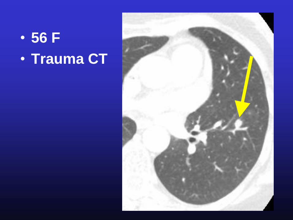
• 56 F
• Trauma CT
Incidental Lung Nodule
• Small, incidental lung nodules very
common in smokers and
nonsmokers
• ~ ½ smokers > age 50 have small
nodules on thin section CT
• Vast majority are benign
• Many nodules represent
granulomas, esp in histoplasmosis
endemic areas, e.g. central USA
MacMahon H. Radiology 2005;237:395
Incidental Lung Nodule
• chance of malignancy with size
• <1% of nodules <4 mm in smokers
turn into lethal cancers
• ~10-20% of nodules ~8mm in
smokers turn into lethal cancers
• Most small incidental nodules are
benign
MacMahon H. Radiology 2005;237:395
Incidental Lung Nodule
• age correlates with chance of
malignancy
• Lung cancer uncommon < age 40,
rare < 35
MacMahon H. Radiology 2005;237:395
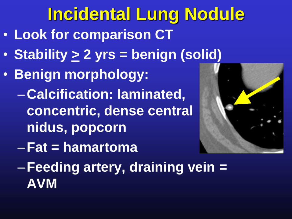
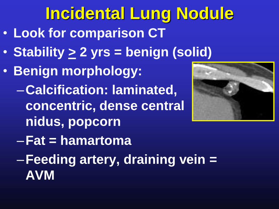
Incidental Lung Nodule• Look for comparison CT
• Stability > 2 yrs = benign (solid)
• Benign morphology:
–Calcification: laminated,
concentric, dense central
nidus, popcorn
–Fat = hamartoma
–Feeding artery, draining vein =
AVM
Incidental Lung Nodule• Look for comparison CT
• Stability > 2 yrs = benign (solid)
• Benign morphology:
–Calcification: laminated,
concentric, dense central
nidus, popcorn
–Fat = hamartoma
–Feeding artery, draining vein =
AVM
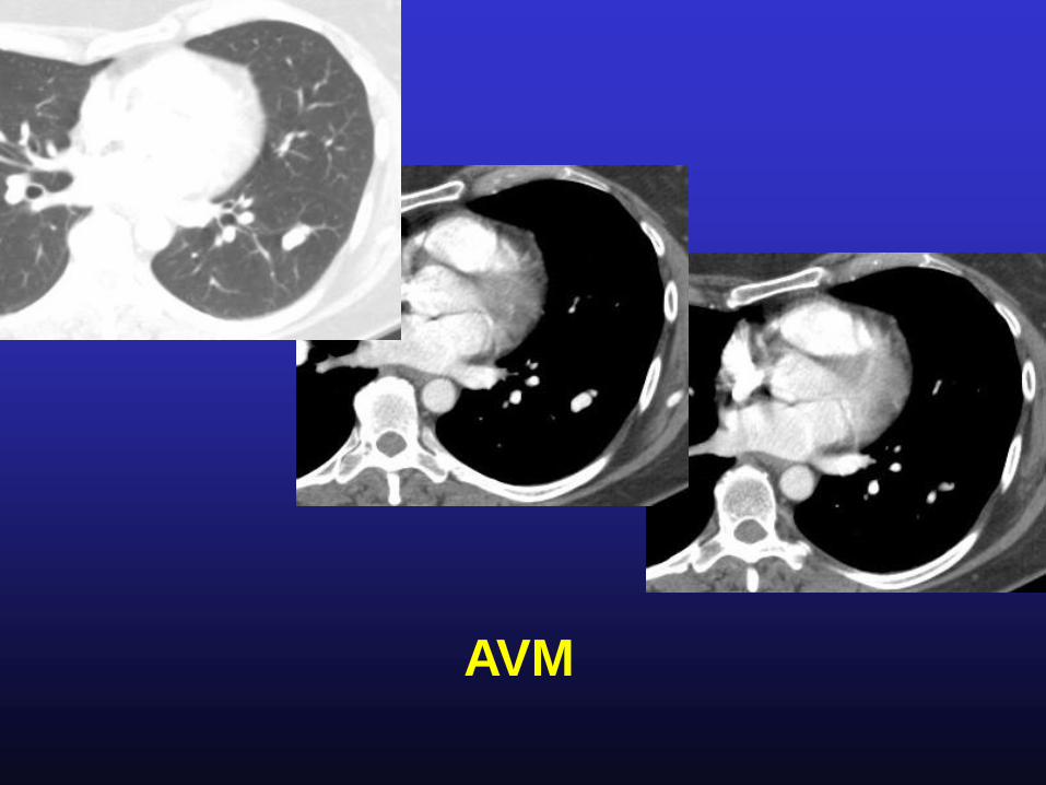
AVM
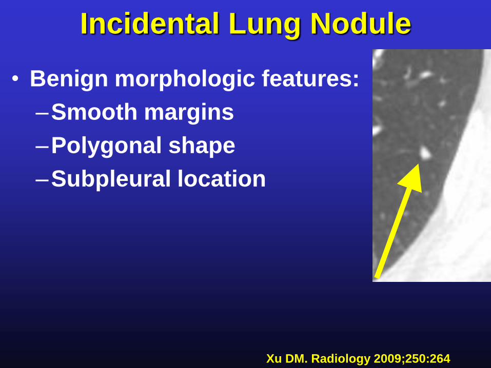
Incidental Lung Nodule
• Benign morphologic features:
–Smooth margins
–Polygonal shape
–Subpleural location
Xu DM. Radiology 2009;250:264
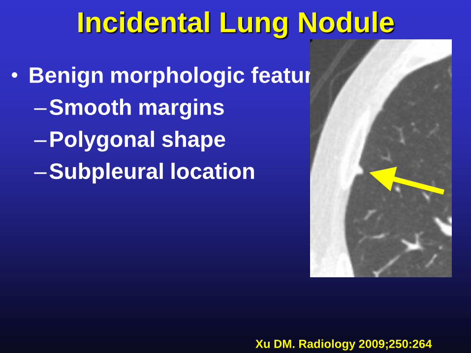
Incidental Lung Nodule
• Benign morphologic features:
–Smooth margins
–Polygonal shape
–Subpleural location
Xu DM. Radiology 2009;250:264
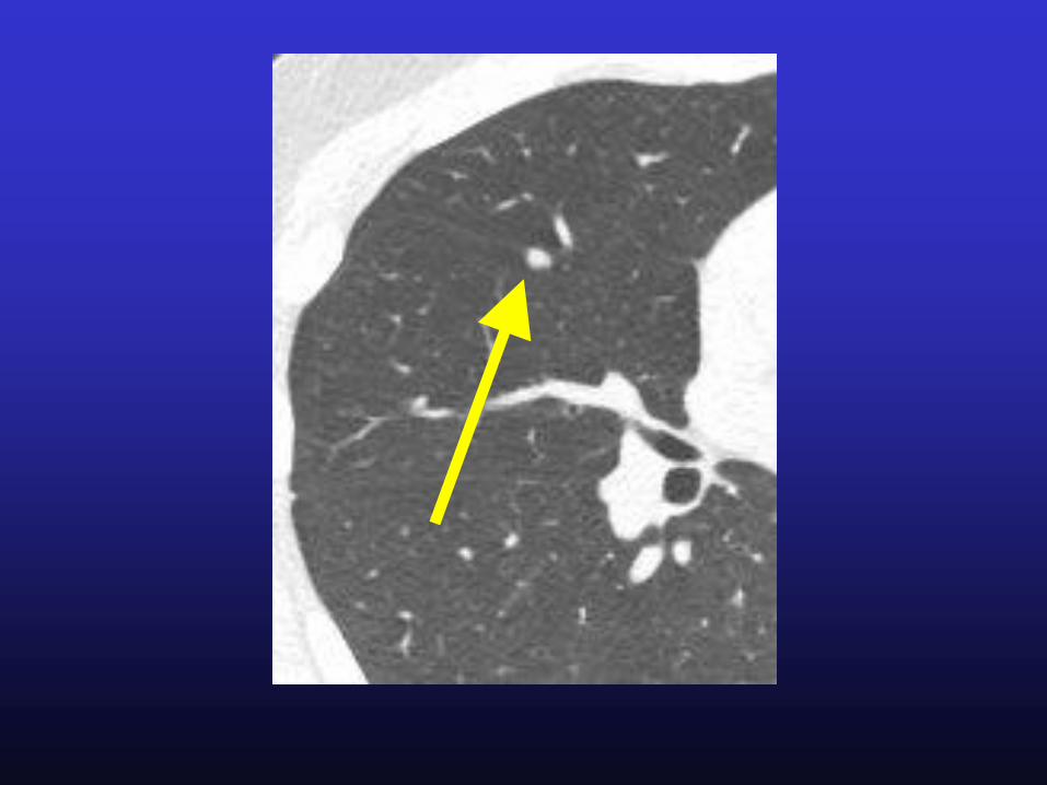
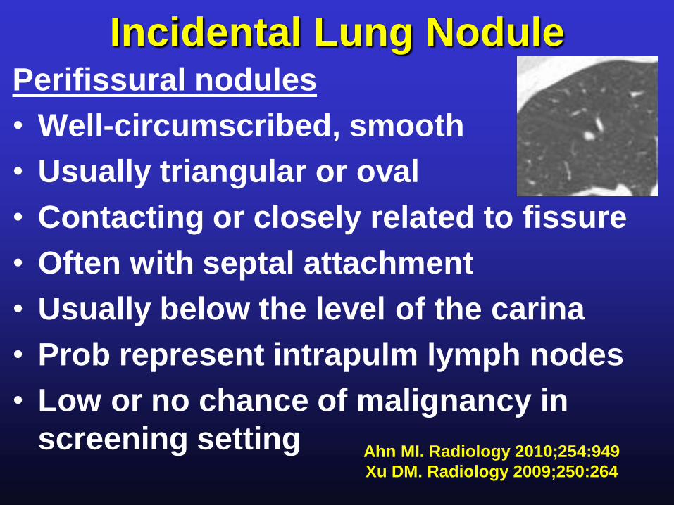
Incidental Lung NodulePerifissural nodules
• Well-circumscribed, smooth
• Usually triangular or oval
• Contacting or closely related to fissure
• Often with septal attachment
• Usually below the level of the carina
• Prob represent intrapulm lymph nodes
• Low or no chance of malignancy in
screening setting Ahn MI. Radiology 2010;254:949
Xu DM. Radiology 2009;250:264
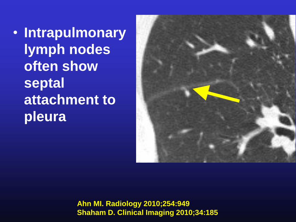
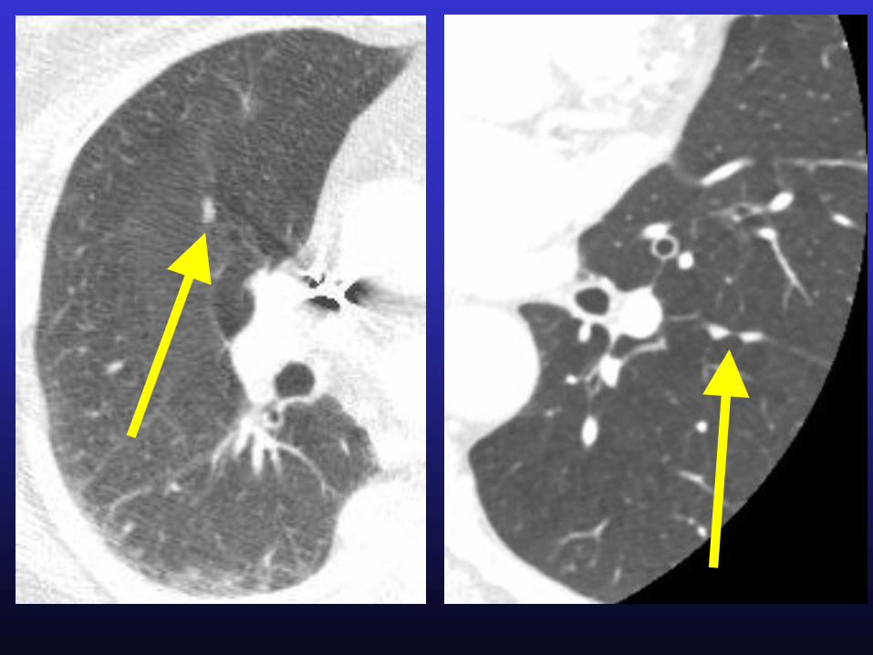
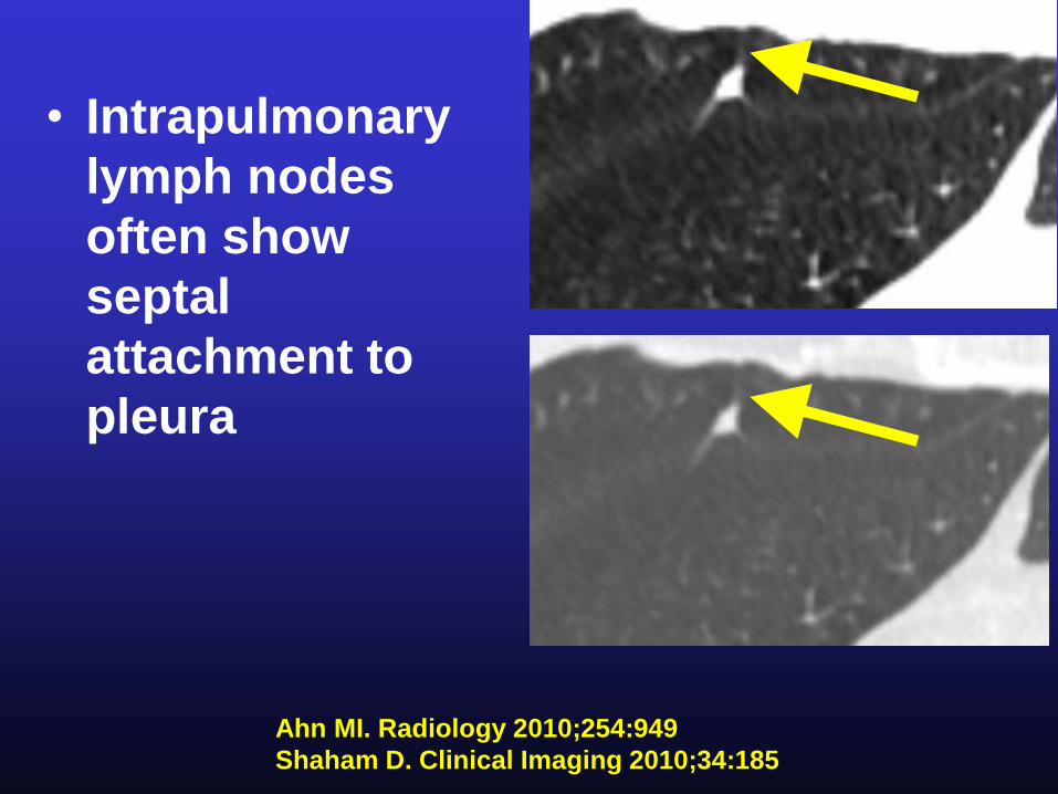
• Intrapulmonary
lymph nodes
often show
septal
attachment to
pleura
Ahn MI. Radiology 2010;254:949
Shaham D. Clinical Imaging 2010;34:185
• Intrapulmonary
lymph nodes
often show
septal
attachment to
pleura
Ahn MI. Radiology 2010;254:949
Shaham D. Clinical Imaging 2010;34:185
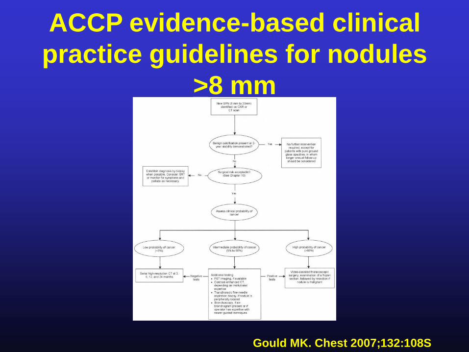
Gould MK. Chest 2007;132:108S
ACCP evidence-based clinical
practice guidelines for nodules
>8 mm
Incidental Lung Nodule
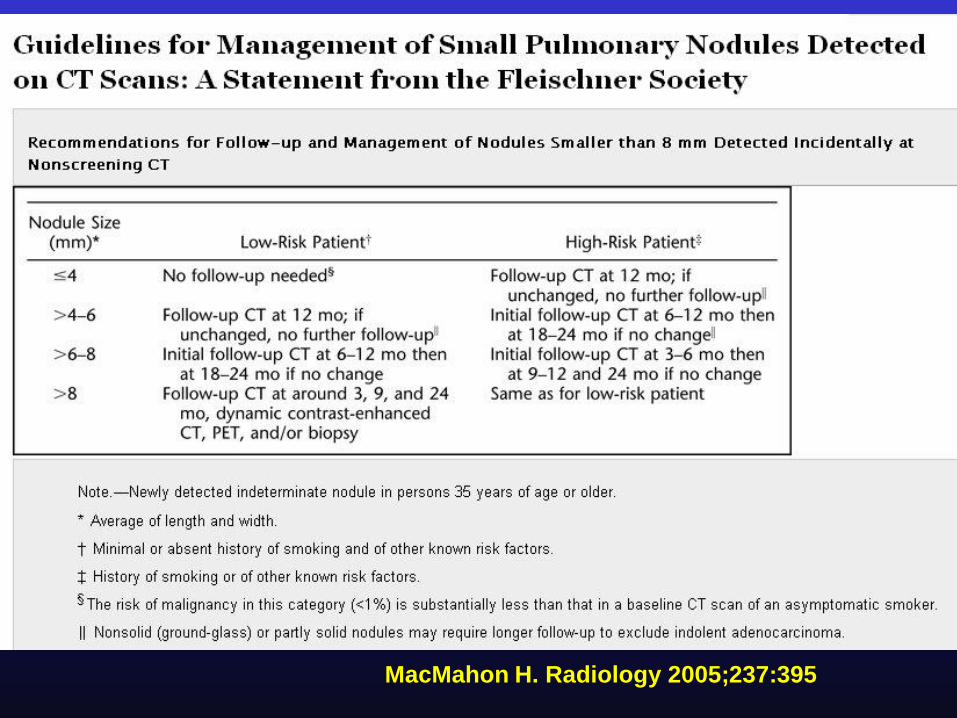
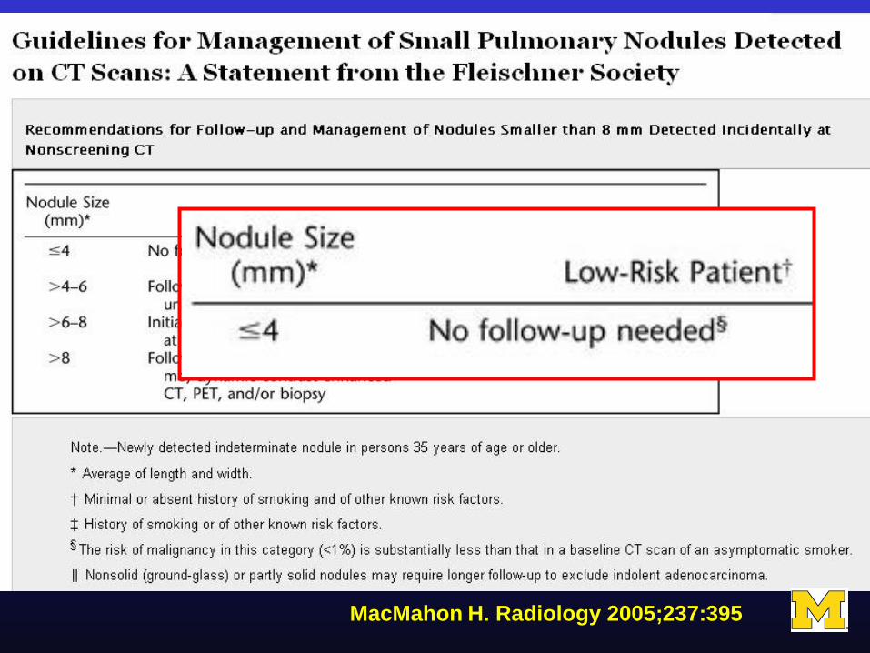
• Fleischner Society guidelines 2005
–Based on pt risk and nodule size
–Existence well known among rads
–Poor compliance with
recommendations
• ? Unfamiliarity with details
• ? Disagreement with guidelines
• ? Medicolegal concernsMacMahon H. Radiology 2005;237:395
Eisenberg RL. Radiology 2010;255(1):218
Quint LE. Academic Radiology, in press
MacMahon H. Radiology 2005;237:395
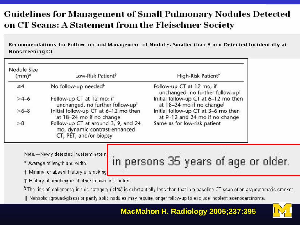
MacMahon H. Radiology 2005;237:395
MacMahon H. Radiology 2005;237:395
MacMahon H. Radiology 2005;237:395
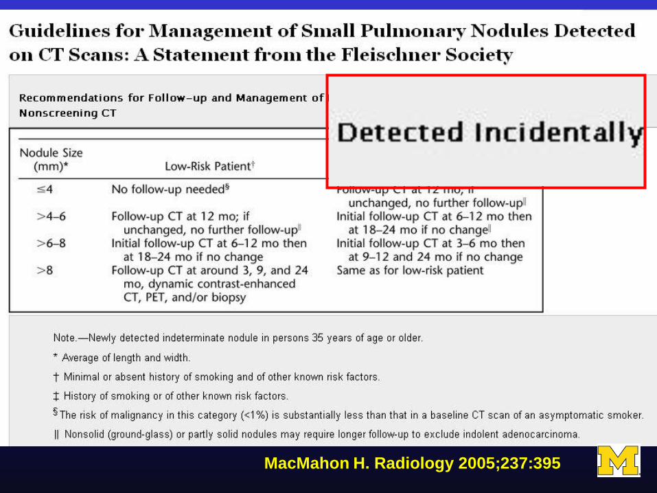
Incidental Lung Nodule
• Fleischner society guidelines apply
only to INCIDENTAL nodules:
unrelated to known underlying disease
• Do NOT apply:
–Known or suspected malignancy that
may metastasize to the lungs
–Unexplained fever
MacMahon H. Radiology 2005;237:395
Incidental Lung Nodule
• Conservative management often
appropriate for:
–very elderly pts
–pts with major comorbid
disease
• More tests, biopsies, surgery not
always indicated
MacMahon H. Radiology 2005;237:395
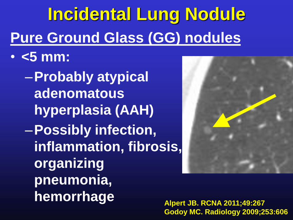
Incidental Lung Nodule
Pure Ground Glass (GG) nodules
Alpert JB. RCNA 2011;49:267
Godoy MC. Radiology 2009;253:606
• <5 mm:
–Probably atypical
adenomatous
hyperplasia (AAH)
–Possibly infection,
inflammation, fibrosis,
organizing
pneumonia,
hemorrhage
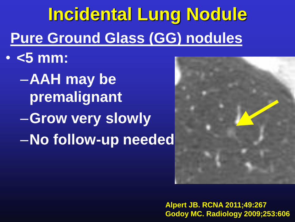
Incidental Lung Nodule
Pure Ground Glass (GG) nodules
Alpert JB. RCNA 2011;49:267
Godoy MC. Radiology 2009;253:606
• <5 mm:
–AAH may be
premalignant
–Grow very slowly
–No follow-up needed
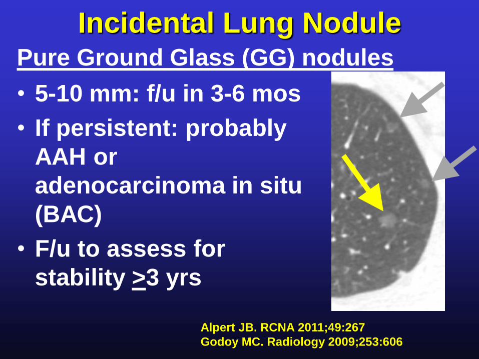
Incidental Lung Nodule
• 5-10 mm: f/u in 3-6 mos
• If persistent: probably
AAH or
adenocarcinoma in situ
(BAC)
• F/u to assess for
stability >3 yrs
Pure Ground Glass (GG) nodules
Alpert JB. RCNA 2011;49:267
Godoy MC. Radiology 2009;253:606
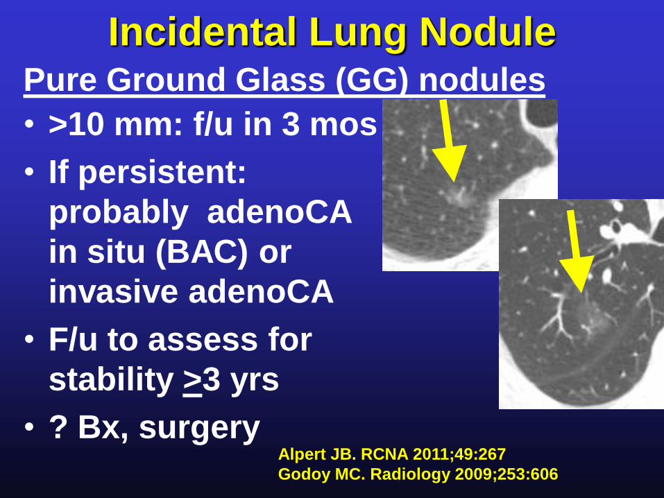
Incidental Lung Nodule
• >10 mm: f/u in 3 mos
• If persistent:
probably adenoCA
in situ (BAC) or
invasive adenoCA
• F/u to assess for
stability >3 yrs
• ? Bx, surgery
Pure Ground Glass (GG) nodules
Alpert JB. RCNA 2011;49:267
Godoy MC. Radiology 2009;253:606
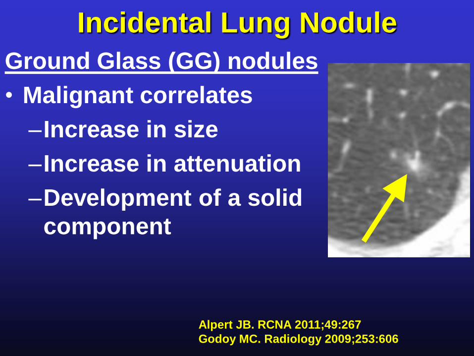
Incidental Lung Nodule
Ground Glass (GG) nodules
• Malignant correlates
–Increase in size
–Increase in attenuation
–Development of a solid
component
Alpert JB. RCNA 2011;49:267
Godoy MC. Radiology 2009;253:606
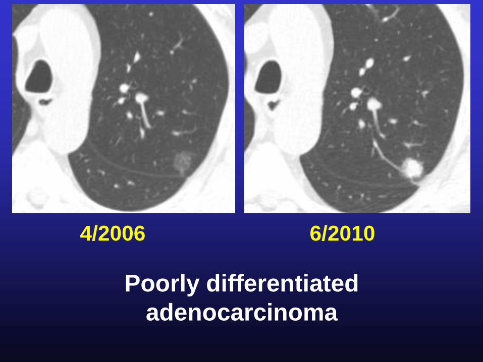
4/2006 6/2010
Poorly differentiated
adenocarcinoma
Incidental Lung Nodule
• Follow-up CT technique:
–Low dose, e.g. 40 mAs
–Thin section , e.g. 1-2.5 mm,
50% overlap
–No intravenous contrast
MacMahon H. Radiology 2005;237:395
Incidental Lung Nodule
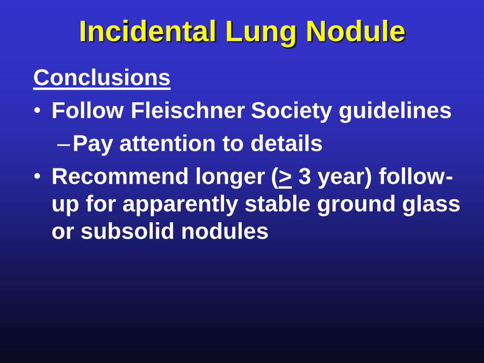
Conclusions
• Follow Fleischner Society guidelines
–Pay attention to details
• Recommend longer (> 3 year) follow-
up for apparently stable ground glass
or subsolid nodules