Embed Size (px)
Citation preview
www.chcmj.ac.in
- Definition of Oligozoospermia- A Commentary
- The Frequency of Medically Compromised Patients Visiting Chettinad Dental College and Research Institute : A Retrospective Study
- Antioxidants in Health and Disease: Review of Clinical Trials
- Management of Severe Traumatic Brain Injury in Adults
- Nutraceuticals in Sperm Abnormalities
- An Innovative Combined Three Dimensional Augmentation of Alveolar Ridge Using Titanium Mesh, PRF and Autogenous Bone Graft with Implant Placement
- From the Pages of History : Harvey Cushing (1869 – 1939)
pISSN NO. 2277 - 8845
eISSN NO. 2278 - 2044
Chettinad Health CityMEDICAL JOURNAL
In this issue
Chettinad Health City
Indexed in INDEX COPERNICUS
GENAMICS JOURNAL SEEK
GOOGLE SCHOLAR
RESEARCH BIBLEVolume - 2, Number - 4, October - December 2013
pISSN NO. 2277 - 8845
eISSN NO. 2278 - 2044
IT Support & Design
Mr. S. M. Michael
Mr. Ramesh Palaniappan
Mr. S. T. Manigandan (Designs)
Shri M.A.M.R. Muthiah Trustee - CARE
Mr. SpK. ChidambaramRegistrar - CARE
Dr. M. Narayana Reddy
Dr. R. Pandurangan
Dr. D. Rajasekaran
Dr. Ramnath Shyamala
Dr. R. Ravi Kumar
Dr. A. Ruckmani
Dr. B. Srinivasan
Dr. M. S. Srinivasan
Dr. Vasantha N Subbiah
Mrs. Veena M Joseph
Dr. L. Uma Devi
All rights are reserved
Chettinad Health City Medical Journal is published by the Chettinad Academy of Research and Education.
Apart from the fair dealing for the purposes of research or private study, or criticism or review, no part of the publication can be reproduced, stored, or transmitted in any form or by any means without prior permission.
Chettinad Health City Medical Journal and /or its publisher cannot be held responsible for errors or for any consequences arising from the use of the information contained in this journal. The appearance of advertising or product information in the various sections of the journal does not constitute an endorsement or approval by the journal and / or its publisher of the quality or the value of the said product or of claims made for it by its manufacturer.
EDITORIAL OFFICE
Dr. N. Pandiyan
Chief Consultant,
Department of Reproductive Medicine,
Chettinad Health City,
Rajiv Gandhi Salai, (OMR, Chennai),
Kelambakkam, Kanchipuram Dist.,
Tamil Nadu - 603 103 India
T. +91 (0)44 4742 8300
F. +91 (0)44 4741 3343
Email:[email protected]
PUBLISHED BY
Chettinad Academy of Research and Education
WEBSITEwww.chcmj.ac.in
All disputes within the jurisdiction of the Madras High Court only
Legal Support
Mr. Balaji
Chettinad Academy of Research and Education (CARE)
Dr. M.A.M.Ramaswamy Chancellor - CARE
Dr. V. RajiVice Chancellor - CARE
Editorial Advisors
Dr. V. Raji
Dr. K.Ravindran
Dr. R.M.Pitchappan
Dr. P. Rajesh
Mrs. L. Lakshmi
Chief Editor
Dr. N. Pandiyan
Editors
Dr. K. Ramesh Rao
Dr. R. Murugesan
Dr. Pradeep G. Nayar
Dr. Ramesh V G
Dr.R.Arun Kumar
Dr. Ashok Palaniappan
Dr. Balaji.T.K
Dr. R. Ganesan
Dr. M. Jeya
Dr. S. B. Jothi Ramalingam
Dr. Lailu Mathews
Dr. E. Malligai
Dr. C. Manohar
Dr. R. Murali
Dr. Nagajothi
Section Editors
Associate Editors
Dr. D. C. Mathangi
Dr. V. Anitha
Dr. Thilaka Muthiah
Dr. K. Senthil Kumar
National Editors
Dr. Dalim Kumar BaidyaAIIMS, New Delhi.
Dr. Suresh NairSri Chitra Institute of Medical Sciences & Technology, Thiruvananthapuram.
Dr. Vedantam RajshekharCMC,Vellore.
Dr. K.A.AbrahamApollo Hospitals, Chennai.
Dr. P.B.SeshagiriIISc, Bangalore.
Dr. Satish Kumar AdigaManipal University, Manipal.
Assistant Editors
Dr. Shah Dupesh Khan
Ms. Prathima
International Editors
Dr. Jayant G Mehta,
BHR University Hospitals NHS Trust, UK
Dr. Ram Dhillon, Middlesex University, UK
Chettinad Health CityMEDICAL JOURNAL
pISSN NO. 2277 - 8845
eISSN NO. 2278 - 2044
Contents
EditorialPandiyan N
Commentary
Definition of Oligozoospermia- A CommentaryPrathima T, Pandiyan N
Original Article
The Frequency of Medically Compromised Patients Visiting Chettinad Dental College and Research Institute : A Retrospective StudyAnitha V, Shivakumar V, Rajesh P, Shanmugam M, Meenapriya B, Amritha, Priyadarshini G
Invited ArticleAntioxidants in Health and Disease: Review of Clinical TrialsNamrata Sanjeevi
Review Article
Management of Severe Traumatic Brain Injury in AdultsGopalakrishnan Raman, Ramesh V G, Mohanarangam T, K ArunThevaraja, Karthikeyan K V
Nutraceuticals in Sperm AbnormalitiesRanjani S, Asha Benziger, Pandiyan N
Case ReportAn Innovative Combined Three Dimensional Augmentation of Alveolar Ridge Using Titanium Mesh, PRF and Autogenous Bone Graft with Implant PlacementVinayak S Gowda, Meena Priya B
From the Pages of History
Harvey Cushing (1869 – 1939)Ramesh V G
Author Index
Subject Index
Thank You Reviewers
107
108
110
113
123
133
137
142
143
145
148

19
20&
ITC GRAND CHOLA, CHENNAI
www.trendo2014.com
TRENDO 2014 Emerging trends in Endocrinology & Diabetes
Mid-Term Conference of Endocrine
Society of India &
2nd Annual Conference of Endocrine Society of
Tamil Nadu and Puducherry July
2014
Symposia
• New vistas in Diabetes
• Recent Advances in Thyroid disease
• Translational Research in Diabetes
• Endocrine Disorders in pregnancy
•Advances in Osteoporosis
•Advances in Pituitary disease
• Newer Diabetic Therapies
• Endocrine Cancers
Clinical case seminars
• Diagnosis and management of GDM
• Starting and Intensifying Insulin
• Diabetes in Children
•Management of Thyroid nodules and cancer
Meet the Expert
• Obesity in clinical care
• Positioning newer diabetic therapies in
clinical care
•Management of Thyrotoxicosis
•Approach to Short stature
Debates
• Non calcemic effects of Vitamin D
• The ADA guideline Vs the Defronzo
algorithm
• Statin up and forget it - Does it work for
Indians
• BMD based Osteoporosis therapy : Are we
treating a number or outcome?
For Enquiries: Dr. Muthukumaran Jayapaul ,Organizing Secretary
Email: [email protected], Mobile : 098411 42606
Conference Secretariat: Arka Center for Hormonal Health Pvt Ltd
5/2, First Avenue, Sastri Nagar, Adyar, Chennai-600 020. Phone: 044-24900050/51/52
You are invited...
Physicians, Diabetologists, Obstetricians, Paediatricians
and Post-graduates
Editorial
107
Vanakkam.
This issue of the journal carries an original article, several interesting review articles and a case report, besides the usual columns.
An original article reports on the prevalence of medical disorders in a dental population; some patients presented with the medical disorder and in others, the disorder was picked up at a routine dental examination thereby indicating that even the unspoken mouth often tells about the underlying medical condition.
Traumatic brain injury is increasing worldwide more so in India, road traffic accidents being the major cause. Unruly traffic, poorly maintained roads, unregulated drivers, drunken driving, negligent riding and an absolute disregard for the law, all contribute in no small measure to this avoidable epidemic. A review article outlines the intensive care management of traumatic brain injury.
Food is the primary source of nourishment to all human beings. Food both in excess and in the deficient state can cause disease; Thiruvalluvar has very well stressed the importance of food in the famous couplet –
Neutraceuticals is a 68 billion dollar industry. However, the role of such nutritional supplements – nutraceuticals and antioxidants in health and disease is not so clear.
An invited article on ‘Antioxidants in health and disease’ reviews the different trials in different disease conditions and gives a candid view of the role of antioxidants. Neutraceuticals claim to enhance male fertility. Nutraceuticals are primarily antioxidants given to men with infertility based on the assumption that reactive oxygen species found in the semen sample of these men are detrimental. A review article discusses the role of Neutraceuticals in male infertility.
A Medical update emphasizes the beneficial role of walking in the prevention of fractures and another one highlights the situation where informed consent may not be required. A case report where a three-dimensional augmentation of a deficient alveolar ridge is proposed as a treatment is an interesting read.
Pages of history outline the life and time of Harvey Cushing. Interesting informative medical updates complete the issue and volume. Hope you enjoy going through this issue. Please do give us your valuable feedback.
Dr. N. Pandiyan
Chief Editor : Chettinad Health City Medical Journal
E-mail : [email protected]
Couplet 942
No need of medicine to heal your body's pain,If, what you ate before digested well, you eat again
Explanation
No medicine is necessary for him who eats after assuring (himself) that what he has (already) eaten has been digested.
jpUf;Fws; 942
kUe;njd Ntz;lhthk; ahf;iff; fUe;jpa jw;wJ Nghw;wp Ùzpd;

Chettinad Health City Medical Journal
Definition of Oligozoospermia- A Commentary
Commentary
Prathima. T*, Dr.Pandiyan. N**
Corresponding author - Prathima. T ([email protected])
Chettinad Health City Medical Journal 2013; 2(4): 108-109
Ms.Prathima graduated from Bangalore University, Bangalore. She finished her Masters in Molecular Biology coming second in the university. She worked in the field of embryology for 3 years in Bangalore. She is currently working as the Assistant Embryologist in the Dept. of Reproductive Medicine, Chettinad Health City.
Volume 2, Number 4
*Assistant Embryologist, **Professor & HOD, Dept of Reproductive medicine, Chettinad Super Specialty Hospital, Kelambakkam, Chennai, India.
the world) for identifying the boundary between normal and oligozoospermic samples.Semen analysis is an important tool in male infertility
investigation. Through its cellular and chemical components, human semen can provide information on the functional properties of the organs producing this fluid, i.e., the testes, epididymis and accessory glands1.
The history of semen analysis dates back to 1677 when Anton von Leeuwenhoek made a remarkable discovery of the spermatozoa, which he called animalcules or spermatozoon. In his letter to The Royal Society of London, he described the structure of spermatozoa so accurately that in retrospect, his illustrations with the help of such a primitive microscope seem incredible. Leeuwenhoek was also the first to discover the presence of spermatozoa in the fallopian tubes and uterus of an animal apart from demonstrating that the sperm are produced in the testicles2.
Upon discovery of the sperm, analysis of semen entered a more scientific realm. Semen analysis was developed by pioneers in the field like Lode, MacLeod Heim and Hotchkiss, not to forget Eliasson and Gold3. It was Edward Martin, in the year 1902, who first put forth the inclusion of semen analysis in male infertility investigation4. Even with all the efforts put in by such brilliant scientists to standardize semen analysis, it still is, as Christopher De Jonge rightly said, the subject of both commendation and condemnation5. Semen analysis remains a numbers game6.
In 1980, the WHO published its first manual on semen analysis thereby establishing standards internationally. It has been updated periodically; five manuals have been published over the last three decades. It has undergone numerous changes over the years, the initial ones being more consensus-based while the last one seems evidence-based, despite its discrepancies.
Oligozoospermia is the nomenclature given when the sperm concentration is less than 15 million/ml, according to the WHO manual 5th edition. However, the values for this nomenclature have varied quite significantly over the last few decades (Table-1). A normal sample, in the 1940’s, was considered to have a sperm count of 60 mil/ml or more7. This, over the years, was lowered to 20 mil/ml based on a consensus by international andrologists of yesteryears. While nobody knows exactly as to why this value was chosen, it was published by the WHO in its’ last 4 manuals and had become a gold standard (still is, in a few labs across
Semen parameters
WHO 1980
WHO 1987
WHO 1992
WHO 1999
WHO 2010
Volume (ml) -- ≥ 2 ≥ 2 ≥ 2 ≥1.5
Sperm concentration
(106
/ml)
20 - 200
≥ 20 ≥ 20 ≥ 20 ≥15
Total sperm concentration
(106
)
-- ≥ 40 ≥ 40 ≥ 40 ≥39
Total motility
(% motile)
≥ 60 ≥ 50 ≥ 50 ≥ 50 ≥40
Progressive motility
≥ 25 ≥ 25% ≥ 25%
(grade A)
≥ 25%
(grade A)
≥32% (A+B)
Vitality (% alive) -- ≥ 50 ≥ 75 ≥ 75 ≥58
Morphology
(% normal)
80.5 ≥ 50 ≥ 30 14 ≥4
Table 1 - Variations in values of semen across the years
The current edition value of 15 mil/ml for sperm concentration also seems arbitrary though evidence-based. It’s anybody’s guess as to how this can result in 39 million/ejaculate with a volume of 1.5 ml8. The expression of sperm concentration per ml and not per ejaculate seems incorrect as it is the output of sperm in the semen that is of interest. A 2 ml sample of 30 million per ejaculate will have 15 million per ml but a 5 ml sample of 30 million per ejaculate will have only 6milllion per ml. So to report the former as ‘normal’ and the latter as ‘subnormal’ or ‘abnormal’ seems unjustified.
It is imperative to remember that reference ranges given by the WHO manual are not absolute and definitely not diagnostic cut-off values but only results obtained out of an observation of a fertile population, which reflects an ‘approximate’ probability that the fertility potential could be high9.
Also, the reference values for the latest edition of the WHO manual were based on a single sample from each participant10. The WHO contradicts itself with such an evidence when it has clearly mentioned in the very same manual that, “a man’s semen quality cannot be characterized from a single semen sample”8. A semen sample shows high intra-individual variation and therefore categorizing a sample as oligozoospermic based on a single analysis is incorrect.
108
There are multiple factors which can result in a low sperm count. Loss of portion of sample during collection, days of abstinence, infection, partial obstruction of genital tract, drugs, environmental pollutants and other toxic factors due to unhealthy lifestyle. There are numerous papers stating that the use of cell phones, long periods of watching television and stress could result in decline of sperm count and motility11. It has also been reported that oligozoospermia and azoospermia are caused by micro-deletions in the AZF region in the long arm of the Y chromosome, which is related to spermatogenesis12.
A man’s sperm count is determined by the number of Sertoli cells in his testes, whose number is ascertained early in development i.e. six months before and after birth. As the germ cells develop they depend on Sertoli cells for support, physically and metabolically. However, each Sertoli cell is restricted to support only a certain number of germ cells. So the number of Sertoli cells, which by itself cannot increase after puberty, in each testis determines the overall sperm output. If the testis is exposed to any adverse factors later in life, like toxins, pollutants, disease or drugs, it might result in a drop in the sperm count but nothing can increase it13.
Oligozoospermia is not an isolated condition; it is frequently associated with compromised sperm quality, including reduced motility and abnormal morphology, as in oligoasthenoteratozoospermia (OATS). The cause of this is still unknown thereby making the efficacy of any treatment for the same very doubtful.
Male infertility cannot be determined solely on the result of a semen analysis as there is no evidence stating what number and quality of sperm are required for a man to be considered fertile. Oligozoospermia also has to be seen in the context of female fertility as infertility involves the couple and not just the male or female. There is no definite number below which pregnancy is impossible nor is there a number above which pregnancy is certainly possible.
Oligozoospermia (except in extreme cases of occasional sperm in the ejaculate), even in 2014, remains undefined.
Acknowledgements
We thank the faculty of the Dept. of Reproductive Medicine for their support through important discussions and valuable comments on this topic.
Commentary Definition of Oligozoospermia- A commentary Volume 2, Number 4
References
1)
2)
3)
Eliasson R. Semen analysis with regard to sperm number, sperm morphology and functional aspects. Asian J of Andrology.2010;12:26-32
Karamanou M, Poulakou-Rebelakou E, Tzetis M, et al. Anton van Leeuwenhoek(1632- 1723): Father of micromorphology and discoverer of spermatozoa. Revista Argentina de Microbiologia. 2010;42:311-314
Menkveld R. The basic semen analysis. Male
4)
5)
6)
7)
8)
9)
10)
11)
12)
13)
infertility-Diagnosis and treatment. First edition. CRC Press. Chapter 9:141-170.
Jequier A.M. Edward Martin (1859-1938):The founding father of modern clinical embryology. Intl Journal of Andrology. 1991; 14:1-10.
Christopher De Jonge. Semen analysis; looking for an upgrade in class. Fertil Steril.2012; 97(2):260-266.
Pandiyan N. Semen analysis- A numbers game. Chettinad Health City Medical J. 2012, 1(1):2-3.
Andersson M.A, Jorgensen N, Main K.M, et al, Adverse trends in male reproductive health: we may have reached a crucial ‘tipping point’. Intl J of Andrology.2008; 31:74-80.
World Health Organisation. WHO Laboratory Manual for the Examination and Processing of Human Semen. 5th edn. 2010. http://whqlibdoc.who.int/publications/2010/9789241547789_eng.pdf
Jequier A.M. Is quality assurance in semen analysis still really necessary? A clinician’s viewpoint. Human Reproduction.2005; 20(8):2039-2042.
Cooper,T.G., Noonan,E., von Eckardstein,S, et al. World Health Organization reference values for human semen characteristics. Human Reprod Update; 2010,16:231–245
Gaskins AJ, Mendiola J, Afeiche M, et al. Physical activity and television watching in relation to semen quality in young men. Br J Sports Med.2013,0: 1-7
O’Flynn O’Brien K.L, Varghese A.C, Agarwal A. The genetic causes of male factor infertility- A review. Fertil Steril.2010, 93(1):1-12.
Sharpe R.M.Sperm counts and fertility in men: a rocky road ahead. EMBO Rep; Science and Society.2012; 13(5):398-403.
109
Chettinad Health City Medical Journal
The Frequency of Medically Compromised Patients Visiting Chettinad Dental College and Research Institute : A Retrospective Study
Original Article
Dr. V.Anitha***, Dr. V.Shivakumar**, Dr. P.Rajesh*, Dr. M.Shanmugam****, Dr. B.Meenapriya*****, Ms.Amritha & Ms. G.Priyadarshini******
Dr. V. Anitha completed Bachelor of Dentistry in 1997 from RV. Dental College, Bangalore and Post Graduation from Meenakshi Ammal Dental College, Chennai in 2004. She has three international publications and 15 national publications to her credit. She is presently working as Professor of Periodontics in Chettinad Dental College and Research Institute.
Volume 2, Number 4
***Professor,**Professor and Head of the Dept., *Principal, ****Associate Professor, *****Lecturer, ******Compulsory Rotatory Internship, Chettinad Dental College and Research Institute, Chennai, India.
Introduction
“Mouth is the mirror of the body”. The incidence of systemic diseases has increased presently due to changes in the life style of an individual. There is a rapid advancement in the treatment modality of systemic disease which has resulted in the enhanced life span of an individual. A two way relationship exists between oral diseases and systemic diseases which is the prime focus today.
Hence a good oral health is necessary to maintain a healthy systemic condition. The knowledge of the importance of focal infection causing systemic diseases resulted in increased number of medically compromised patients visiting the dental college. Hence this study analyzes the frequency of systemic problems among patients visiting the Chettinad Dental College and Research Institute (CDCRI).
Materials and Methods
In this retrospective study medical records of patients visiting CDCRI was analyzed for the year 2011, 2012, and 2013 for the presence of systemic disease. The list of extracted data from each patient’s documents contained the history of cardiovascular, respiratory,
renal, endocrine diseases like diabetes mellitus, thyroid, hematological disorders, and gastrointestinal diseases .This data was tabulated and frequency distribution of each disease was analyzed using a SPSS software.
Results
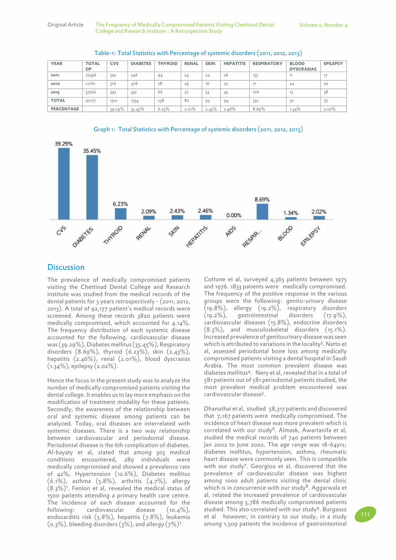
Around 92,177 patients’ medical records visiting the Chettinad Dental College and Research Institute were analyzed retrospectively for 3 years (2011, 2012, and 2013). 3,820 patients had medically compromised conditions which accounted for 4.14% of the total population with oral disease who had visited the Chettinad Dental College and Research Institute. Between 4.14% of the medically compromised patients visiting Chettinad Dental College and Research Institute the frequency of each systemic problem is mentioned below as a percentage. The percentage of systemic disease accounted are as follows: cardiovascular disease (39.29%), Diabetes mellitus (35.45%), Respiratory disorders (8.69%), thyroid (6.23%), Hepatitis (2.46%), skin (2.43%), Epilepsy (2.02%), Renal disorders (2.01%), Blood dyscrasias (1.34%), which is depicted in the tabulation and graph (Table 1), (Graph- 1).
Abstract
“Mouth Is The Mirror Of The Body”. The incidence of systemic diseases has increased presently due to changes in the life style of an individual. There is a rapid advancement in the treatment modality of systemic disease which has resulted in the enhanced life span of an individual. A two way relationship exists between oral diseases and systemic diseases which is the prime focus today. Hence this study analyzes the frequency of systemic problems among patients visiting the Chettinad Dental College and Research Institute. Materials and Methods: In this retrospective study medical records of patients visiting CDCRI was analyzed for the year 2011, 2012, and 2013 in the presence of systemic disease. The list of extracted data from each patient’s documents contained the history of cardiovascular diseases, respiratory, renal diseases, endocrine diseases like diabetes mellitus and thyroid, hematological disorders, gastrointestinal diseases. This data was tabulated and frequency distribution of each disease was analyzed using SPSS software. Results: Around 92,177 patients’ medical records were analyzed retrospectively for 3 years (2011, 2012, and 2013). 3,820 patients had medically compromised conditions which accounted for 4.14%.The percentage of systemic disease accounted are the following: cardiovascular disease (39.29%), Diabetes mellitus (35.45%), Respiratory disorders (8.69%), thyroid (6.23%), Hepatitis (2.46%), Skin (2.43%), Epilepsy (2.02%), Renal disorders (2.01%), Blood dyscrasias (1.34%), which is depicted in the tabulation and graph. Conclusion: As there is increased prevalence of systemic disorders, primarily, a modification in the treatment modality can be done only with a perfect medical charting. Secondly, more emphasis should be given to annual dental checkups as a "healthy mouth leads to healthy body".
Key words : Systemic disorders, Prevalence, Focal infection.
Chettinad Health City Medical Journal 2013; 2(4):110 - 112
Corresponding author - Anitha. V ([email protected])
110
Discussion
The prevalence of medically compromised patients visiting the Chettinad Dental College and Research institute was studied from the medical records of the dental patients for 3 years retrospectively - (2011, 2012, 2013). A total of 92,177 patient’s medical records were screened. Among these records 3820 patients were medically compromised, which accounted for 4.14%. The frequency distribution of each systemic disease accounted for the following, cardiovascular disease was (39.29%), Diabetes mellitus (35.45%), Respiratory disorders (8.69%), thyroid (6.23%), skin (2.43%), hepatitis (2.46%), renal (2.01%), blood dyscrasias (1.34%), epilepsy (2.02%).
Hence the focus in the present study was to analyze the number of medically compromised patients visiting the dental college. It enables us to lay more emphasis on the modification of treatment modality for these patients. Secondly, the awareness of the relationship between oral and systemic disease among patients can be analyzed. Today, oral diseases are interrelated with systemic diseases. There is a two way relationship between cardiovascular and periodontal disease. Periodontal disease is the 6th complication of diabetes. Al-bayaty et al, stated that among 303 medical conditions encountered, 289 individuals were medically compromised and showed a prevalence rate of 42%, Hypertension (12.6%), Diabetes mellitus (6.1%), asthma (5.8%), arthritis (4.7%), allergy (8.3%)1. Fenlon et al, revealed the medical status of 1500 patients attending a primary health care centre. The incidence of each disease accounted for the following: cardiovascular disease (10.4%), endocarditis risk (5.8%), hepatitis (7.8%), leukemia (0.3%), bleeding disorders (3%), and allergy (7%)2.
Original Article The Frequency of Medically Compromised Patients Visiting Chettinad Dental College and Research Institute : A Retrospective Study
Volume 2, Number 4
Cottone et al, surveyed 4,365 patients between 1975 and 1976. 1833 patients were medically compromised. The frequency of the positive response in the various groups were the following: genito-urinary disease (19.8%), allergy (19.2%), respiratory disorders (19.2%), gastrointestinal disorders (17.9%), cardiovascular diseases (15.8%), endocrine disorders (8.3%), and musculoskeletal disorders (15.1%). Increased prevalence of genitourinary disease was seen which is attributed to variations in the locality3. Natto et al, assessed periodontal bone loss among medically compromised patients visiting a dental hospital in Saudi Arabia. The most common prevalent disease was diabetes mellitus4. Nery et al, revealed that in a total of 581 patients out of 581 periodontal patients studied, the most prevalent medical problem encountered was cardiovascular disease5.
Dhanuthai et al, studied 58,317 patients and discovered that 7,167 patients were medically compromised. The incidence of heart disease was more prevalent which is correlated with our study6. Almask, Awartanifa et al, studied the medical records of 740 patients between Jan 2002 to June 2002. The age range was 18-64yrs; diabetes mellitus, hypertension, asthma, rheumatic heart disease were commonly seen. This is compatible with our study7. Georgiou et al, discovered that the prevalence of cardiovascular disease was highest among 1000 adult patients visiting the dental clinic which is in concurrence with our study8. Aggarwala et al, related the increased prevalence of cardiovascular disease among 3,786 medically compromised patients studied. This also correlated with our study9. Burgausz et al however, in contrary to our study, in a study among 1,509 patients the incidence of gastrointestinal
Table-1: Total Statistics with Percentage of systemic disorders (2011, 2012, 2013)
YEAR TOTAL
OP CVS DIABETES THYROID RENAL SKIN HEPATITIS RESPIRATORY BLOOD
DYSCRASIAS EPILEPSY
2011 27456 592 546 94 24 24 26 155 11 17
2012 27161 516 476 78 29 16 25 71 24 20
2013 37560 393 332 66 27 53 43 106 15 38
TOTAL 92177 1501 1354 238 80 93 94 332 50 75
PERCENTAGE 39.29% 35.45% 6.23% 2.01% 2.43% 2.46% 8.69% 1.34% 2.02%
Graph 1: Total Statistics with Percentage of systemic disorders (2011, 2012, 2013)
111
disease was more prevalent10. The results of the present study showed increased incidence of cardiovascular disease followed by diabetes mellitus, and respiratory disease. All the above mentioned disorders are associated with a medical emergency. Hence, further studies are required to assess the etiology, the interlinked pathogenesis and risk factors of the disease. One of the most prevalent dental causes of these systemic diseases is periodontal disease. Periodontal disease can precipitate cardiovascular disease, respiratory disease, and it’s the 6th complication of diabetes. Hence it’s advisable to have regular dental check up annually as “prevention is better than cure”.
Conclusion
Based on the results of the study there seems to be an increased incidence of cardiovascular disease followed by diabetes and respiratory disorder which is attributed to environmental changes, life style and age. As there is increased prevalence of systemic disorders, primarily a modification in the treatment modality can be done only when a perfect medical charting is done. Secondly, more emphasis should be given to annual dental checkups as a "healthy mouth leads to healthy body". Hence further studies are required to analyze the etiology of these disorders.
Volume 2, Number 4
References1)
2)
Al-bayaty HF, Murti PR, Naidu RS, Matthews R. Medical problems among dental patients at the school of dentistry, the university of West Indies. Journal of Dental Education, 2009; 73(12):1408-14.
Fenlon., MR, Cartan., MC, Medical status of a patients attending a primary care practice in Ireland. Journal of Ireland Dental association, 1991; 37(3-4):75-77
3)
4)
5)
6)
7)
8)
9)
10)
Cottone., JA, Kafrawy., AH, Medications and health history survey of 4,365 dental patients. Journal of Am Dent Assoc, 1979; 98(5):713-8.
Nattozs., AL, Zahranim., Periodontal bone loss and self reported medical conditions in a dental school patient population. Journal of International Academy of Periodontics, 2010; 12(4):104-9.
Nery., EB, Meister., F, Ellinger., RF, Isalami . Prevalence of medical problems in periodontics patients obtained from 3 different populations. Journal of Periodontics, 1987; 58(8):564-8.
Dhanuthai., K, Sappayatosok., K, Bijaphala., P, Kulvitit., S. Prevalence of medically compromised conditions in dental patients. Med oral patol oral cir buccal, 2009; 14(16):287-91.
Almas., K, Awartani., FA. Prevalence of medically compromised patients referred for periodontics treatment to teaching hospital in central Saudi Arabia. Saudi Med Journal, 2003; 24(11):12-24-5.
Georgiouto, Marshallri, Bartold., PM, The prevalence of systemic diseases in Brisbane in general and periodontal practice patients. Australian dental journal 2004; dec 49(4):(177-87).
Aggarwala., Panatsr., Talukder., Self reported medical problems among dental patients in western Uttar Pradesh, India, Journal of dental education, 2011; dec75(12):1635-40
Burgausz, Allseed., O, Khader., YS, Prevalence of medical conditions among patients attending dental teaching clinics in northern Jordan. Journal of Contemporary Dental Practice, 2007; 8(1):60-7).
If it is Alzheimer's, your judgment is as good as your physician's!
Have you noticed significant memory lapses that you felt are worthy of being reported in the recent past? Then you are potentially a candidate for developing Alzheimer's later in your life. That appears to be the conclusion of a study conducted in Sanders-Brown Center on Aging, University of Kentucky. In that study, the investigators asked 3701 men aged over 60 a simple question: "Have you noticed any change in your memory since you last came in?". The responses obtained led the investigators to conclude that the subjective memory complaints are early markers of Alzheimer disease. This has also been the observation of several other epidemiologists. The researchers consider that encouraging as it is now possible to intervene early to reduce the effects of cognitive memory impairment. (Science Daily, 21 February 2014. (www.sciencedaily.com/releases/2014/02/140221114124.htm)
- Dr. K. Ramesh Rao
112
Original Article The Frequency of Medically Compromised Patients Visiting Chettinad Dental College and Research Institute : A Retrospective Study

Chettinad Health City Medical Journal
Antioxidants in Health and Disease: Review of Clinical Trials
Invited Article
Namrata Sanjeevi
Corresponding author - Namrata Sanjeevi ([email protected])
Namrata Sanjeevi graduated from PSG College of Technology with a degree in Biotechnology. Currently, she is a doctoral candidate in Nutritional Sciences at the University of Texas, Austin with a solid foundation in research methods and fundamentals of nutrition. Research interests include the design and implementation of a multifaceted approach to identify factors that influence suboptimal diets in Supplemental Nutrition Assistance Program (SNAP) participants.
Volume 2, Number 4
Doctoral candidate, University of Texas, USA
Introduction
Oxygen is an element that is crucial for the sustenance of life on earth. It is paradoxical that this indispensable element can cause harmful effects in humans under certain circumstances. Much of the detrimental consequences of oxygen are attributed to its ability to form free radicals1. A free radical is a reactive molecule that contains at least one unpaired electron in its outer orbit, and is capable of independent existence2. Accumulation of these molecules in the body results in oxidative stress, a process by which physiologically important molecules such as carbohydrates, proteins and lipids are damaged3. However, the body can employ antioxidants to impede the threat of free radical attack4.Antioxidants are potent scavengers of free radicals1. They function by donating an electron to a free radical or by eliminating initiators of free radicals5. Antioxidants may be classified as endogenous or exogenous depending on their mode of acquisition by the body1. Endogenous antioxidants are naturally produced by the body1.Superoxide dismutase, catalase, glutathione peroxidase and glutathione reductase are enzymatic systems within the body that function as antioxidants1. Lipoid acid, glutathione, L-arginine, coenzyme Q10, melatonin, uric acid and bilirubin are examples of non-enzymatic antioxidants that are produced during metabolism3,4. On the other hand, exogenous antioxidants cannot be synthesized biologically, and must be supplied through the diet and/or supplements1. Vitamin E (�alpha - tocopherol), vitamin C (ascorbate), �beta-carotene (provitamin A carotenoid), trace elements such as selenium, manganese, zinc, flavonoids, lycopene, omega-3 and omega-6 fatty acids are some antioxidants that can be obtained from the diet1. Fruits, vegetables, nuts, herbs, spices and beverages are identified as natural sources of
such exogenous antioxidants6. A delicate balance between prooxidant and antioxidant substances is achieved by the production and scavenging of free radicals3. Under optimal physiological conditions, this equilibrium marginally shifts to favor a prooxidant status and maintains mild oxidative stress within the body3. Antioxidants perform a dual role; of scavenging free radicals while still allowing a sufficient amount to persist and carry out vital functions7. Some of the important biological functions of free radicals are cell signaling and redox regulation3. However, an acute shift towards prooxidant status will lead to oxidative damage7. Additionally, lack of regulation of free radicals is implicated in the pathogenesis of several disease states such as cancer, cardiovascular disease, neurological disease, pulmonary disease, rheumatoid arthritis, nephropathy, ocular disease and pre-eclampsia1.The purpose of this paper is to review some of the clinical trials that have explored the influence of antioxidant intake from food and supplements on the prevention of initiation and progression of certain chronic diseases. Additionally, the paper will discuss about the use of antioxidants in health maintenance.
Cancer
Mortality and morbidity associated with cancer is a major public health problem. It was estimated that 12.7 million people were affected by cancer worldwide in 20088. Furthermore, about 7.6 million deaths were attributed to cancer8. Breast cancer and lung cancer are the leading causes of cancer death among females and males, respectively8. The development of cancer is a multistage process that involves initiation, progression
Abstract
Free radicals play an important role in several biological processes such as cell signaling and redox regulation. However, prolonged exposure to free radicals leads to oxidative damage. Subsequently, it has been implicated in the progression of several diseases like cancer, cardiovascular disease, neurological disease, pulmonary disease, rheumatoid arthritis, nephropathy, ocular disease and pre-eclampsia. The antioxidant defense system within the body may confer protection to oxidative damage by scavenging free radicals. Antioxidants also may be obtained from dietary sources/ supplements. The efficacy of antioxidant intake on initiation and progression of chronic diseases will be reviewed.
Chettinad Health City Medical Journal 2013; 2(4): 113 - 122
113
and promotion of the tumor9. Free radicals can wreck havoc at all stages of cancer development9. The influence of free radical induced- deoxyribonucleic acid (DNA) alterations on carcinogenesis may be mediated by epigenetic effects on gene expression, mutations and chromosomal rearrangements10. Tobacco smoke, ultraviolet (UV) radiation, consumption of red meat and alcohol, and obesity have been identified as risk factors for various types of cancer owing to their ability to generate oxidative stress11. The antioxidant status of cancer patients has been found to be significantly lower than that of normal individuals, as demonstrated by reduced levels of glutathione, glutathione peroxidase, superoxide dismutase, vitamin C and E in cancer patients12. Observational studies have reported that the intake of fruits and vegetables confer protection against cancers of the lung, breast, stomach, pharynx, esophagus and pancreas13. Given that fruits and vegetables are rich sources of antioxidants, it can be reasoned that antioxidant supplementation reduces the risk of cancer. On the contrary, clinical trials testing the efficacy of antioxidant supplementation for cancer prevention have yielded limited success. The �alpha-tocopherol and �beta-carotene (ATBC) study sought to determine the effectiveness of �alpha-tocopherol and �
beta-carotene supplementation in reducing the occurrence of lung cancer among male smokers, aged 50-69 years. Results of this trial demonstrated that the incidence of lung cancer was significantly higher among individuals receiving �beta-carotene14. The �
Beta-Carotene and Retinol Efficacy Trial (CARET) also found that the relative risk of lung cancer was greater in participants who received �beta-carotene and retinol supplementation when compared to placebo15.
Participants for this trial included men who were substantially exposed to asbestos due to their occupation. The Linxian study was conducted in Linxian, a rural county belonging to north-central China. The population in this region was reported to be disproportionately affected by high rates of esophageal and gastric cancers16, as well as subclinical deficiencies of retinol, carotenoids, tocopherols and other vitamins. The following combination of supplements were used in the Linxian study: Supplement A consisting of retinol palmitate and zinc; Supplement B including riboflavin and niacin, Supplement C comprising of vitamin C and molybdenum, and Supplement D consisting of �
beta-carotene, selenium and �alpha-tocopherol. The eight intervention groups received AB, AC, AD, BC, BD, CD, ABCD, or placebo.The only significant outcome was that the group which received supplement D had a lower risk for stomach cancer mortality when compared to the other groups17. The Women's Antioxidant Cardiovascular Study (WASC) indicated that vitamin C, �alpha-d-tocopherol acetate and �beta-carotene did not reduce total cancer incidence in women with a history of cardiovascular disease (CVD) or three or more risk factors for CVD18. A reduction in non-Hodgkin’s lymphoma risk was observed in women receiving �beta-carotene18. However, lung cancer incidence was higher among women receiving vitamin C18.
Results from the Selenium and Vitamin E Cancer Prevention Trial (SELECT) established that selenium,
Invited Article Antioxidants in Health and Disease: Review of Clinical Trials Volume 2, Number 4
vitamin E or selenium and vitamin E combination did not reduce prostate cancer risk in a group of healthy men19. Thus, results from human clinical trials that explore the efficacy of antioxidant supplementation on cancer incidence have been inconclusive.
Cardiovascular disease (CVD)
CVD is the leading cause of death worldwide, and accounted for approximately 17 million deaths in 200820. Diseases of the cardiac muscle tissue and the vascular system, such as atherosclerosis, ischemic heart disease (IHD), stroke, congestive heart failure (CHF), are responsible for CVD. An inappropriate diet, obesity, physical inactivity, alcohol abuse and cigarette smoking are some of the modifiable risk factors of CVD21. The effects of risk factors on CVD may be mediated by oxidative stress, which in turn can cause oxidation of low-density lipoprotein (LDL) and disruption of vascular homeostasis. Thus, a diet rich in antioxidants may protect against the risk of CVD. A greater intake of fruits and vegetables has been associated with a lower risk of CVD22. The Heart Outcomes Prevention Evaluation (HOPE) was a large scale trial that reported no significant differences in myocardial infarction (MI), stroke and death from cardiovascular causes between vitamin E supplemented and placebo groups23. In contrast to the HOPE Trial, other studies that have researched the influence of antioxidant supplementation on CVD risk illustrate the beneficial effects of vitamin E. The Secondary Prevention using Antioxidants of Cardiovascular Disease in Endstage renal disease (SPACE) trial showed a significant decrease in fatal and non-fatal AMI, ischemic stroke, peripheral vascular disease, and unstable angina in CVD patients receiving vitamin E supplement when compared to the placebo group24. Similarly, Cambridge Heart Antioxidant Study (CHAOS)study indicated that vitamin E supplementation decreased the incidence of CVD death and non-fatal MI in coronary artery disease (CAD) patients25. The Nurses Healthy Study (NHS) determined vitamin A, vitamin C and vitamin E intake obtained from food sources and supplements in a population of middle-aged women. Results from this study reported no association between vitamin A, vitamin C and coronary disease risk26. However,intake of vitamin E from food sources was inversely related to risk of death from major coronary events26. The use of vitamin E supplements for more than 2 years was associated with a reduction in coronary disease risk27. The Physician’s Health Study (PHS) showed that �
beta-carotene did not reduce cardiovascular mortality in the supplemented group versus the placebo group28. Results from the CARET trial indicated an increased risk for cardiovascular death in individuals treated with �beta-carotene and retinol than the control group15. Additionally, the ATBC study did not establish a beneficial effect of �beta-carotene on cardiac-related mortality in male smokers with a history of MI29.Thus, vitamin E supplements have shown a greater therapeutic potential for CVD when compared to other antioxidants.
Pre-eclampsia
Pre-eclampsia during pregnancy is marked by high
114

blood pressure and proteinuria30. Clinical manifestations of pre-eclampsia include birth of small-for-gestational-age infant, poor growth of the infant and premature birth, neonatal morbidity and mortality, and conditions that affect liver, kidneys, brain or blood clotting system for the woman31. The presence of free radicals may lead to injury of endothelial cells that line the inside surfaces of blood vessels, which in turn results in the clinical symptoms of pre-eclampsia32. Predisposition for LDL, resistance to oxidative stress, and antioxidant intake are the major determinants of a woman’s response to oxidative stress33. Since a dietary deficiency of antioxidants is associated with this disorder, antioxidant supplementation may be employed as a potential measure to help prevent and treat this condition. Interventional studies have evaluated the use of vitamin C and E combination34,36, vitamin C and E in combination with allopurinol37,vitamin C alone38, red palm oil39, lycopene40 and selenium41 in pre-eclampsia. Pregnant women at low, moderate or high risk of developing pre-eclampsia were included for participation in these studies. Women with pre-eclampsia were excluded from participation. The primary outcomes examined were pre-eclampsia, severe pre-eclampsia, preterm birth, small-for-gestational age infants, and infant mortality. No significant differences were reported in the risk of any of the primary outcomes between the antioxidant supplemented and control group for the trials. Women allocated to lycopene had a greater reduction in the relative risk of pre-eclampsia. However, results from this study were based on a small group of women. The existing body of literature does not favor the use of antioxidants during pregnancy to reduce pre-eclamptic risk.
Diabetes mellitus
Diabetes mellitus is a chronic, multiorgan disease that can severely damage the eyes, kidneys, nerves, heart and/ or blood vessels42. Globally, it is estimated that the number of adults affected by diabetes was 285 million in the year 2010, and will increase to 439 million by 203043. India and China are expected to be disproportionately burdened by the increase in diabetes prevalence43. This condition is characterized by hyperglycemia that arises out of abnormalities in insulin secretion or insulin action42. Hyperglycemia can trigger generation of free radicals, thereby creating a state of oxidative stress that is involved in pathogenesis of diabetes and its related complications43. Small scale trials have shown the beneficial effects of antioxidants on diabetes-related complications. Supplementation with vitamin E44 and vitamin E plus C45 positively influenced endothelial-dependent vasorelaxation in Type I diabetic patients. However, a positive effect was not observed for Type 2 diabetic patients supplemented with vitamin E plus C45. In another study, significant improvement in renal function was observed in Type 2 diabetic patients supplemented with vitamin E plus C46. The Primary Prevention Project (PPP) trial demonstrated no beneficial effect of vitamin E for diabetic subjects47. However, the population for the PPP trial was not restricted to diabetic patients. In contrast, the �alpha-lipoic acid in Diabetic Neuropathy (ALADIN)48, ALADIN II49,
Volume 2, Number 4
ALADIN III50, DEKAN (Deutsche kardialeautonomeneuropathie)51 and SYDNEY52 trials were limited to a population of diabetic subjects. ALADIN, ALADIN II and ALADIN III studies demonstrated significant improvements in patient symptoms, nerve function, and neuropathy impairment score, respectively, in diabetic patients who were supplemented with �alpha-lipoic acid48,50. In the DEKAN study, cardiac autonomic neuropathy was found to be improved in the alpha�-lipoic acid treated group versus placebo group51. The SYDNEY trial showed advancements in sensory symptoms of diabetic polyneuropathy upon �alpha-lipoic treatment52. In summary, large scale clinical trials that involve �
alpha-lipoic acid treatment have proved to be more effective than trials involving vitamin E treatment.More basic and clinical research is required to test the efficacy of antioxidants, such as �alpha-lipoic acid, in improving the prognosis of diabetes.
Chronic obstructive pulmonary disease (COPD)
COPD is a common lung disease characterized by airflow limitation attributed to disrupted alveolar attachment, mucus hypersecretion and inflammatory obstruction of the airway53. It is a major public health burden worldwide, and is estimated to affect about 14 million people in the United States53. Smoking and environmental pollution are two major risk factors for COPD53. Oxidative stress from exposure to tobacco and air pollutants may deplete plasma antioxidant capacity, thereby leading to inflammation and mucus secretion54. Intake of antioxidants via diet/supplements has been suggested as an ideal way to boost the lung antioxidant system55. Moreover, it has been associated with improved lung function, and is suggested as a strategy to enhance COPD outcomes56. The Women’s Health Study (WHS) established that vitamin E supplementation lead to a decrease in the risk of chronic lung disease in women57. Lykkesfeldt et al, have shown that supplementation of vitamin C, vitamin E and �beta-carotene enabled repletion of ascorbic acid in smokers58. N-acetyl-L-cysteine (NAC) is a nutritional supplement that has been used to strengthen antioxidant defense system in patients with COPD. NAC has been found to decrease oxidative stress in airways of COPD patients59, and alleviate bronchial hypersecretion60. Although the use of antioxidants in COPD shows potential, more research is required to formulate recommendations on antioxidant supplementation for COPD management.
Antioxidants in health maintenance
A prolonged exposure to free radicals may occur as a consequence of normal physiological processes, such as aging and intense exercise, thereby disrupting the delicate balance that exists within our body7. Normal individuals can incorporate antioxidant rich foods into their diet to protect themselves from oxidative damage, and thus maintain their health. Anlasik et al, reported a positive association between fruit and vegetable intake and antioxidant status in a group of healthy elderly subjects61. On the other hand, intake of antioxidant supplements is recommended only when a reduced antioxidant status is identified7. For example, several
115
Invited Article Antioxidants in Health and Disease: Review of Clinical Trials

micronutrient deficiencies have been associated with increased morbidity and mortality in children belonging to Africa and Asia62. Some of the deficient micronutrients include vitamin A and zinc62, which also function as antioxidants. In such cases, supplementation may markedly improve clinical manifestations of the deficiency63,64. It is vital to apply caution when using supplements for healthy individuals since intake of high amounts of antioxidant supplements may lead to antioxidative stress65. Hence, precise determination of individual's free radical and antioxidant levels is required before prescribing antioxidant supplements 7.
Conclusions
Several observational studies have demonstrated the beneficial effect of antioxidant rich diets on disease outcomes. However, discrepancy exists between observational studies and clinical trials that test the efficacy of antioxidants in disease prevention. The lack of adequate success in clinical trials can be viewed from different perspectives. Firstly, several physiological factors influence nutrient bioavailability. Some of the factors include age, gender, ethnicity, body weight,
Volume 2, Number 4
genetic composition and stage of the disease. These factors affect the extent to which antioxidants are utilized by the body, as well as the ability of an individual to respond to antioxidant supplementation. Knowledge of physiological variables that influence antioxidant bioavailability is essential to determine critical aspects of clinical trials, such as effective supplement dosage and duration of treatment. Moreover, a host of lifestyle behaviors are responsible for determining the health of individuals. Antioxidant intake in combination with physical activity, alcohol and tobacco moderation may yield profound benefits in disease management. Thus, multifactorial interventions may serve as alternative strategies in disease management. Finally, investigations on the effects of nutrients in isolation may provide valuable information regarding its mode of action, but do not elucidate the phenomenon of total diet. The intrinsic nature of diet is characterized by several interactions between bioactive dietary components, some of which still remain unexplained. Hence, antioxidant supplements must be prescribed with caution and the use of antioxidant rich foods as disease prevention agents may hold promise in future clinical trials.
Table1. Summary of selected clinical trials testing efficacy of antioxidants in cancer66
Name of study
Trial Primary outcome
Study population Relative Risk(Confidence Interval)
Interpretation of results
ATBC g-tocopherol(50mg), or く-carotene (20 mg), or both versus placebo
Lung cancer incidence
29,133 male smokers 50–69 years of
Age, with a history of MI, followed for 5–8 years
Lung:
0.98 (0.81–1.19) g-tocopherol vs placebo
1.16 (0.97–1.38) く-carotene vs placebo
1.15 (0.96–1.38) both vs placebo
A significant increase in incidence of lung cancer for く-carotene supplemented group. No significant decrease in incidence of lung cancer for any of the other supplemented groups
CARET く-carotene (30 mg) plus retinol (25000 IU) vs placebo
Lung cancer incidence
14,254 smokers+4060 asbestos
workers followed for 4 years
Lung:
1.36 (1.07–1.73) く-carotene plus retinol
vs placebo
A significant increase in lung cancer incidence in く-carotene plus retinol supplemented group
Linxian Study
Intervention groups: AB, AC, AD, BC, BD, CD, ABCD, or placebo. Supplement A:
retinol (5000 IU), zinc (22.5 mg). Supplement B:
riboflavin (3.2 mg)
Supplement C: vitamin C (120 mg), molybdenum
(30 たg). Supplement D: く-carotene (15 mg),
selenium (50 たg), g-tocopherol (30 mg)
Gastric and esophageal cancer
mortality
29,584 adults ages 40–69
followed for 6 years
Esophagus:
0.97 (0.81–1.17) A vs no A
0.90 (0.75–1.08 B vs no B
1.06 (0.88–1.28) C vs no C
1.00 (0.84–1.21) D vs no D
Stomach:
1.05 (0.86–1.27) A vs no A
1.08 (0.89–1.31) B vs no B
1.06 (0.87–1.28) C vs no C
0.81 (0.66–0.98) D vs no D
A significant decrease in stomach cancer mortality for group supplemented with D.
WASC Vitamin C
(500 mg), vitamin E (600 IU qOD), く-carotene
(50 mg qOD), 3 combinations of 2 agents,
And all 3 vs placebo.
CVD incidence
7627 women at least 40 years of age who did not have cancer.
Average follow-up 9.4 years
Lung:
1.84 (1.14–2.97) any vitamin C vs placebo
1.25 (0.79–1.97) any vitamin E vs placebo
1.26 (0.80–1.99) any く-carotene vs placebo
Lung cancer incidence was significantly higher in women receiving vitamin C. No significant decrease in incidence of total cancer/ specific cancer in any other supplemented group.
SELECT selenium (200 たg),
vitamin E (400 IU), or both vs placebo
Prostate cancer incidence
35,533 men age ≥50 years without any suspicion for prostate cancer followed for 7-12 years
Prostate:
1.13 (0.99–1.29) vitamin E vs placebo
1.04 (0.90–1.18) selenium vs placebo
1.05 (0.91–1.20) both vs placebo
No significant decrease in incidence of prostate cancer in any of the supplemented groups
116
Invited Article Antioxidants in Health and Disease: Review of Clinical Trials

Volume 2, Number 4
Table 2. Summary of selected clinical trials testing efficacy of antioxidants in CVD
Name of
study
Trial Primary
outcome
Study population Relative Risk (Confidence
Interval)
Interpretation of results
HOPE Vitamin E (400 IU) vs
placebo and ramipril
(10 mg/day) vs
placebo
Myocardial
infarction,
stroke and
death from
cardiovascula
r causes
Patients aged ≥ 55 with
a high risk for CVD.
1838 and 1816 subjects
were diabetic in the
treatment and control
arm, respectively
CVD death:
1·05 (0.90-1.22) vitamin E vs
placebo
MI:1.02 (0.90-1.15) vitamin
E vs placebo
Stroke:
1.17 (0.95-1.42) vitamin E vs
placebo
No apparent beneficial
effect of vitamin E
SPACE Vitamin E (800 IU)
versus
Placebo
Total CVD
endpoints
196 patients with
CVD and undergoing
chronic hemodialysis
Total CVD endpoints
(including sudden death):
0·54 (0·33–0·89)vitamin E vs
placebo
MI(including sudden
death):
0·45 (0·20–0·99) vitamin E vs
placebo
Significant reductions in
total CVD endpoints and
MI in subjects receiving
vitamin E
CHAOS Vitamin E supplements
(400-800 IU) vs
placebo
Incidence of
cardiovascula
r death and
non-fatal MI
2,002 patients with
coronary artery
disease (CAD)
Cardiovascular death and
non-fatal MI:
0.53 (0.34-0.83) vitamin E
vs placebo
A significant reduction in
cardiovascular death and
non-fatal AMI in vitamin E
supplemented group
NHS Classification of
participants into
quintiles based on
vitamin A, vitamin C,
vitamin E intake from
food and supplements
Incidence of
cardiovascula
r death
34,486
postmenopausal
women with no
cardiovascular disease
followed for 7 years
Coronary Heart Disease
(CHD) death:
0.38 (0.18–0.80) vitamin E
vs placebo
A significant decrease in
vitamin E intake from food
and death from CHD
PHS aspirin
(325 mg on alternate
days),く-carotene (50
mg on alternate days),
both active agents vs
placebo
Incidence of
CVD death,
MI and stroke
22,071 male
physicians, aged
40 to 84 years with no
history of CVD events
CVD death:
1.09 (0.93–1.27) く-carotene
vs placebo
MI:0.96 (0.84–1.09) く-
carotene vs placebo
Stroke:0.96 (0.83–1.11) く-
carotene vs placebo
No significant decrease in
CVD endpoints in the
supplemented group.
CARET く-carotene (30 mg)
plus retinol
(25000 IU) vs placebo
Lung cancer
incidence
14,254 smokers+4060
asbestos workers
followed for 4 years
CVD death:
1.26 (0.99- 1.61) く-carotene
plus retinol vs placebo
Increased risk in incidence
of cardiovascular death in
supplemented group
ATBC g-tocopherol (50mg),
or く-carotene (20 mg),
or both versus
placebo
Lung cancer
incidence
29,133 male smokers,
aged 50–69 years with
a history of MI,
followed for 5–8 years
CHD deaths:
1.75 (1.16-2.64)
く-carotene vs placebo
1.33 (0.86-2.05) g –
tocopherol vs placebo
1.58 (1.05-2.40) both vs
placebo
Significant increase in
incidence of fatal deaths
from CHD in く-carotene
and く-carotene plus g-
tocopherol groups.
117
Invited Article Antioxidants in Health and Disease: Review of Clinical Trials

Volume 2, Number 4
Table 3. Summary of selected clinical trials testing efficacy of antioxidants in pre-eclampsia
Author of study
Trial Main outcome Study population Relative Risk (Confidence Interval)
Interpretation of results
Beazley D et al.
Vitamin C (1000 mg) and vitamin E (400 IU) vs placebo
Rate of pre-eclampsia
100 women pregnant at 14 weeks 0 days to 20 weeks 6 days with a history of pre-eclampsia, chronic hypertension, insulin-requiring diabetes mellitus, or multiple gestation
Rate of pre-eclampsia:
0.92 (0.4-2.13)
treatment vs placebo
No significant reduction in the incidence of main outcomes in the supplemented group when compared to the placebo group
Rumbold AR et al.
Vitamin C (1000 mg) and vitamin E (400 IU) vs placebo
Incidence of pre-eclampsia, death of infant and small-for gestational age infants
1877 nulliparous women pregnant between 14 and 22 weeks with normal blood pressure at the first measurement in pregnancy, and at trial entry
Pre-eclampsia:
1.20 (0.82–1.75) treatment vs placebo
Death of infant:
0.79 (0.61–1.02) treatment vs placebo
Small for gestational age infants:
0.87 (0.66- 1.16) treatment vs placebo
No significant reduction in the incidence of main outcomes in the supplemented group when compared to the placebo group
Spinnato JA et al
Vitamin C (1,000 mg) and vitamin E (400 IU)
Incidenceof pre-eclampsia
739 women diagnosed with pre-eclampsia or with a history of pre-eclampsia
Pre-eclampsia:
0.87 (0.61-1.25)
treatment vs placebo
No significant reduction in incidence of pre-eclampsia in supplemented group when compared to the placebo.
Gülmezoğlu AM et al.
Vitamin E (800 IU), vitamin C (1000 mg), and allopurinol (200 mg)
Prolongation of pregnancy and assessment of lipid peroxides
56 women with severe pre-eclampsia between 24 and 32 weeks of gestation
Delivery within 14 days:
0.68 (0.45-1.04)
treatment vs placebo
No significant differences in prolongation of pregnancy in study group when compared to placebo. Furthermore, there were no differences in lipid peroxide levels
Steyn PS et al.
Vitamin C (250 mg) two times a day until 34 weeks’ gestation
Incidence of pre-eclampsia, preterm labor
200 women less than 26 weeks’ gestation and with a history of a previous mid-trimester abortion or previous preterm labor
Pre-eclampsia:
1.00 (0.21-4.84) vitamin C vs placebo
Preterm birth:
1.43 (1.03-1.99)
vitamin C vs placebo
The incidence of preterm birth was higher in women supplemented with vitamin C
Merchant AT et al.
Multivitamin containing thiamine (20 mg), riboflavin (20 mg), B-6 (25mg), B-12 (50 microg), C (500 mg), E (30 mg), and folic acid (0.8 mg),
く-carotene (30 mg) plus preformed vitamin A (5000 IU)versus
placebo
Hypertension during pregnancy
1078 HIV-positive pregnant Tanzanian women between 12 and 27 week gestation
Hypertension during pregnancy:
0.62 (0.40–0.94) multivitamin vs placebo
1.00 (0.66–1.51) vitamin A vs placebo
Women supplemented with multivitamin were less likely to develop hypertension during pregnancy
Mahdy ZA et al.
Tocotrienol-rich fraction (TRF) of palm
oil (100 mg) vs placebo
Hypertension during pregnancy
Healthy women pregnant between 12 and 16 weeks gestation
Pregnancy induced hypertension:
0.36 (0.12–1.09) palm oil vs placebo
No benefits of palm oil in reducing the risk of pregnancy induced hypertension
118
Invited Article Antioxidants in Health and Disease: Review of Clinical Trials

Volume 2, Number 4
Table 4. Summary of selected clinical trials testing efficacy of antioxidants in diabetes
Table 5. Summary of selected clinical trials testing efficacy of antioxidants in COPD
References1)
2)
Pham-Huy LA, He H, Pham-Huy C. Free radicals, antioxidants in disease and health. Int J Biomed Sci. 2008;4:89-96.
Halliwell B, Gutteridge JMC. Free radicals in biology and medicine. 4th. Oxford, UK: Clarendon Press; 2007.
3)
4)
Dröge W. Free radicals in the physiological control of cell function. Physiol Rev. 2002;82:47-95.
Willcox JK, Ash SL, Catignani GL. Antioxidants and prevention of chronic disease. Crit Rev Food Sci Nutr. 2004;44:275-95.
Name of study
Trial Primary outcome Study population Relative Risk (Confidence Interval)
Interpretation of results
PPP Aspirin (100 mg) vs placebo and vitamin E (300 mg) vs placebo
CVD deaths 2062 diabetic patients aged ≥50 years
Cardiovascular death:
1.23 (0.69-2.19) aspirin
vs placebo
Cardiovascular death:
1.07 (0.61-1.90) vitamin E
vs placebo
Vitamin E supplementation did not significantly reduce incidence of CVD death in diabetic subjects
ALADIN
g-lipoic acid (1200mg, 600 mg, or 100 mg) vs
Placebo for 3 weeks
Symptoms of diabetic peripheral neuropathy
328 non-insulin-
dependent diabetic patients with symptomatic peripheral neuropathy
- Significant improvement in symptoms in g-lipoic acid supplemented group
ALADIN II
g-lipoic acid (1200mg or 600 mg) vs
placebo for 24 months
Neuropathic symptoms
65 diabetic patients - Statistically significant improvements in peripheral nerve function parameters
ALADIN III
g-lipoic acid (600 mg) vs placebo for 6 months
Neuropathy impairment score
509 Type II diabetic patients aged 18-65 years
- Improvements in neuropathy
impairment score after 19 days of treatment, which was
maintained for up to 7 months
DEKAN g-lipoic acid (800 mg) vs placebo for 4 months
Cardiac autonomic neuropathy, as indicated by heart rate variability
73 non-insulin-
dependent diabetes mellitus patients
- Improved cardiac autonomic neuropathy in g-lipoic acid treated group
SYDNEY
g-lipoic acid (600 mg) vs placebo for 14 treatments
Neuropathic sensory symptoms
120 diabetic patients with symptomatic diabetic sensorimotor polyneuropathy
- Improvement in sensory symptoms such as
pain, prickling and numbness in g-lipoic acid treated group
Name/ author of study
Trial Primary outcome
Study population Hazard ratio (Confidence Interval)
Interpretation of results
WHS Vitamin E (600 IU every other day) and aspirin (100 mg every other day) vs placebo
Incidence of chronic lung disease
38,597 women aged ≥ 45 without chronic lung disease followed for 10 years
Chronic lung disease:
0.90 (0.81-0.99) vitamin E vs placebo
Vitamin E supplementation lead to reduction in risk of chronic lung disease
Lykkesfeldt et al.
Vitamin cocktail containing vitamin C (272 mg), g-tocopherol acetate (31 mg), and folic acid (400 ´g) vs placebo for 3 months
Plasma antioxidant status
37 smokers and 38 nonsmokers with self-reported low fruit and vegetable intake
- Ascorbic acid was depleted in smokers, and increased after supplementation.
De Benedetto et al.
NAC (600 mg)vs
placebo for 2months
H2O2 content in the exhaled air condensate (EAC)
55 males and females, aged, 41–75 years, non-smokers/ ex-smokers for at least 5 years, and affected by moderate COPD
- NAC decreased oxidant stress in airways, as indicated by H2O2 content in EAC
119
Invited Article Antioxidants in Health and Disease: Review of Clinical Trials

Volume 2, Number 4
5)
6)
7)
8)
9)
10)
11)
12)
13)
14)
15)
16)
17)
18)
Rice-Evans CA, Diplock AT. Current status of antioxidant therapy. Free Radic Biol Med. 1993;15:77-96.
Sikora E, Cieslik E, Topolska K. The sources of natural antioxidants. Acta SciPolTechnol Aliment. 2008;7:5-17.
Poljsak B, Šuput D, Milisav I. Achieving the balance between ROS and antioxidants: when to use the synthetic antioxidants. Oxid Med Cell Longev. 2013;2013:956792.
Jemal A, Bray F, Center MM, Ferlay J, Ward E, Forman D. Global cancer statistics. CA Cancer J Clin. 2011;61:69-90.
Rahman K. Studies on free radicals, antioxidants, and co-factors. Clin Interv Aging. 2007;2:219-36.
Donkena KV, Young CY, Tindall DJ. Oxidative stress and DNA methylation in prostate cancer. Obstet Gynecol Int. 2010;2010:302051.
Stein CJ, Colditz GA. Modifiable risk factors for cancer. Br J Cancer. 2004;90:299-303.
Manju V, Kalaivani Sailaja J, Nalini N. Circulating lipid peroxidation and antioxidant status in cervical cancer patients: a case-control study. Clin Biochem. 2002;35:621-5.
World Cancer Research Fund. Food, nutrition and the prevention of cancer: a global perspective. Washington, DC: American Institute for Cancer Research, 1997.
The effect of vitamin E and beta carotene on the incidence of lung cancer and other cancers in male smokers. The Alpha-Tocopherol, Beta Carotene Cancer Prevention Study Group. N Engl J Med. 1994;330:1029-35.
Omenn GS, Goodman GE, Thornquist MD, Balmes J, Cullen MR, Glass A, Keogh JP, Meyskens FL, Valanis B, et al. Effects of a combination of beta carotene and vitamin A on lung cancer and cardiovascular disease. N Engl J Med. 1996;334:1150-5.
Cheng SJ, Sala M, Li MH, Chouroulinkov I. Esophageal cancer in Linxian county, China: a possible etiology and mechanism (initiation and promotion). Carcinog Compr Surv. 1982;7:167-74.
Qiao YL, Dawsey SM, Kamangar F, Fan JH, Abnet CC, Sun XD, Johnson LL, Gail MH, Dong ZW, et al. Total and cancer mortality after supplementation with vitamins and minerals: follow-up of the Linxian General Population Nutrition Intervention Trial. J Natl Cancer Inst. 2009;101:507-18.
Cook NR, Albert CM, Gaziano JM, Zaharris E, MacFadyen J, Danielson E, Buring JE, Manson JE. A randomized factorial trial of vitamins C and E
19)
20)
21)
22)
23)
24)
25)
26)
27)
28)
and beta carotene in the secondary prevention of cardiovascular events in women: results from the Women's Antioxidant Cardiovascular Study. Arch Intern Med. 2007;167:1610-8.
Lippman SM, Klein EA, Goodman PJ, Lucia MS, Thompson IM, Ford LG, Parnes HL, Minasian LM, Gaziano JM, et al. Effect of selenium and vitamin E on risk of prostate cancer and other cancers: the Selenium and Vitamin E Cancer Prevention Trial (SELECT). JAMA. 2009;301:39-51.
Kelly BB, Narula J, Fuster V. Recognizing global burden of cardiovascular disease and related chronic diseases. Mt Sinai J Med. 2012;79:632-40.
Buttar HS, Li T, Ravi N. Prevention of cardiovascular diseases: Role of exercise, dietary interventions, obesity and smoking cessation. Exp Clin Cardiol. 2005;10:229-49.
Genkinger JM, Platz EA, Hoffman SC, Comstock GW, Helzlsouer KJ. Fruit, vegetable, and antioxidant intake and all-cause, cancer, and cardiovascular disease mortality in a community-dwelling population in Washington County, Maryland. Am J Epidemiol. 2004;160:1223-33.
Yusuf S, Dagenais G, Pogue J, Bosch J, Sleight P. Vitamin E supplementation and cardiovascular events in high-risk patients. The Heart Outcomes Prevention Evaluation Study Investigators. N Engl J Med. 2000;342:154-60.
Boaz M, Smetana S, Weinstein T, Matas Z, Gafter U, Iaina A, Knecht A, Weissgarten Y, Brunner D, et al. Secondary prevention with antioxidants of cardiovascular disease in endstage renal disease (SPACE): randomised placebo-controlled trial. Lancet. 2000;356:1213-8.
Stephens NG, Parsons A, Schofield PM, Kelly F, Cheeseman K, Mitchinson MJ. Randomised controlled trial of vitamin E in patients with coronary disease: Cambridge Heart Antioxidant Study (CHAOS). Lancet. 1996;347:781-6.
Kushi LH, Folsom AR, Prineas RJ, Mink PJ, Wu Y, Bostick RM. Dietary antioxidant vitamins and death from coronary heart disease in postmenopausal women. N Engl J Med. 1996;334:1156-62.
Stampfer MJ, Hennekens CH, Manson JE, Colditz GA, Rosner B, Willett WC. Vitamin E consumption and the risk of coronary disease in women. N Engl J Med. 1993;328:1444-9.
Hennekens CH, Buring JE, Manson JE, Stampfer M, Rosner B, Cook NR, Belanger C, LaMotte F, Gaziano JM, et al. Lack of effect of long-term supplementation with beta carotene on the incidence of malignant neoplasms and cardiovascular disease. N Engl J Med. 1996;334:1145-9.120
Invited Article Antioxidants in Health and Disease: Review of Clinical Trials
Volume 2, Number 4
29)
30)
31)
32)
33)
34)
35)
36)
37)
38)
39)
40)
41)
Rapola JM, Virtamo J, Ripatti S, Huttunen JK, Albanes D, Taylor PR, Heinonen OP. Randomised trial of alpha-tocopherol and beta-carotene supplements on incidence of major coronary events in men with previous myocardial infarction. Lancet. 1997;349:1715-20.
Report of the National High Blood Pressure Education Program Working Group on High Blood Pressure in Pregnancy. Am J Obstet Gynecol. 2000;183:S1-S22.
Backes CH, Markham K, Moorehead P, Cordero L, Nankervis CA, Giannone PJ. Maternal preeclampsia and neonatal outcomes. J Pregnancy. 2011;2011:214365.
Anyaegbunam A, Edwards C. Hypertension in pregnancy. J Natl Med Assoc. 1994 Apr;86:289-93.
Roberts JM, Cooper DW. Pathogenesis and genetics of pre-eclampsia. Lancet. 2001;357:53-6.
Beazley D, Ahokas R, Livingston J, Griggs M, Sibai BM. Vitamin C and E supplementation in women at high risk for preeclampsia: a double-blind, placebo-controlled trial. Am J Obstet Gynecol. 2005;192:520-1.
Rumbold AR, Crowther CA, Haslam RR, Dekker GA, Robinson JS, Group AS. Vitamins C and E and the risks of preeclampsia and perinatal complications. N Engl J Med. 2006;354:1796-806.
Spinnato JA, Freire S, Pinto E Silva JL, Cunha Rudge MV, Martins-Costa S, Koch MA, Goco N, Santos CeB, Cecatti JG, et al. Antioxidant therapy to prevent preeclampsia: a randomized controlled trial. Obstet Gynecol. 2007;110:1311-8.
Gülmezoğlu AM, Hofmeyr GJ, Oosthuisen MM. Antioxidants in the treatment of severe pre-eclampsia: an explanatory randomised controlled trial. Br J Obstet Gynaecol. 1997;104:689-96.
Steyn PS, Odendaal HJ, Schoeman J, Stander C, Fanie N, Grové D. A randomised, double-blind placebo-controlled trial of ascorbic acid supplementation for the prevention of preterm labour. J Obstet Gynaecol. 2003;23:150-5.
Mahdy ZA, Siraj HH, Azwar MH, Wahab MA, Khaza’ai H, Mutalib MSA, et al. Does palm oil vitamin E reduce the risk of pregnancy induced hypertension?Acta Medica. 2013;56:104-9.
Sharma JB, Kumar A, Malhotra M, Arora R, Prasad S, Batra S. Effect of lycopene on pre-eclampsia and intra-uterine growth retardation in primigravidas. Int J Gynaecol Obstet. 2003;81:257-62.
Han L, Zhou SM. Selenium supplement in the prevention of pregnancy induced hypertension. Chin Med J (Engl). 1994;107:870-1.
42)
43)
44)
45)
46)
47)
48)
49)
50)
51)
52)
Maritim AC, Sanders RA, Watkins JB. Diabetes, oxidative stress, and antioxidants: a review. J Biochem Mol Toxicol. 2003;17:24-38.
Shaw JE, Sicree RA, Zimmet PZ. Global estimates of the prevalence of diabetes for 2010 and 2030. Diabetes Res Clin Pract. 2010;87:4-14.
Skyrme-Jones RA, O'Brien RC, Berry KL, Meredith IT. Vitamin E supplementation improves endothelial function in type I diabetes mellitus: a randomized, placebo-controlled study. J Am Coll Cardiol. 2000;36:94-102.
Beckman JA, Goldfine AB, Gordon MB, Garrett LA, Keaney JF Jr, Creager MA. Oral antioxidant therapy improves endothelial function in Type 1 but not Type 2 diabetes mellitus. Am J Physiol Heart Circ Physiol. 2003;285:H2392-8.
Gaede P, Poulsen HE, Parving HH, Pedersen O. Double-blind, randomised study of the effect of combined treatment with vitamin C and E on albuminuria in Type 2 diabetic patients. Diabet Med. 2001;18:756-60.
Sacco M, Pellegrini F, Roncaglioni MC, Avanzini F, Tognoni G, Nicolucci A, Group PC. Primary prevention of cardiovascular events with low-dose aspirin and vitamin E in type 2 diabetic patients: results of the Primary Prevention Project (PPP) trial. Diabetes Care. 2003;26:3264-72.
Ziegler D, Hanefeld M, Ruhnau KJ, Meissner HP, Lobisch M, Schütte K, Gries FA. Treatment of symptomatic diabetic peripheral neuropathy with the anti-oxidant alpha-lipoic acid. A 3-week multicentre randomized controlled trial (ALADIN Study). Diabetologia. 1995;38:1425-33.
Reljanovic M, Reichel G, Rett K, Lobisch M, Schuette K, Möller W, Tritschler HJ, Mehnert H. Treatment of diabetic polyneuropathy with the antioxidant thioctic acid (alpha-lipoic acid): a two year multicenter randomized double-blind placebo-controlled trial (ALADIN II). Alpha Lipoic Acid in Diabetic Neuropathy. Free Radic Res. 1999;31:171-9.
Ziegler D, Hanefeld M, Ruhnau KJ, Hasche H, Lobisch M, Schütte K, Kerum G, Malessa R. Treatment of symptomatic diabetic polyneuropathy with the antioxidant alpha-lipoic acid: a 7-month multicenter randomized controlled trial (ALADIN III Study). ALADIN III Study Group. Alpha-Lipoic Acid in Diabetic Neuropathy. Diabetes Care. 1999;22:1296-301.
Ziegler D, Schatz H, Conrad F, Gries FA, Ulrich H, Reichel G. Effects of treatment with the antioxidant alpha-lipoic acid on cardiac autonomic neuropathy in NIDDM patients. A 4-month randomized controlled multicenter trial (DEKAN Study). Deutsche Kardiale Autonome Neuropathie. Diabetes Care. 1997;20:369-73.
Ametov AS, Barinov A, Dyck PJ, Hermann R,
121
Invited Article Antioxidants in Health and Disease: Review of Clinical Trials

Volume 2, Number 4
53)
54)
55)
56)
57)
58)
59)
Kozlova N, Litchy WJ, Low PA, Nehrdich D, Novosadova M, et al. The sensory symptoms of diabetic polyneuropathy are improved with alpha-lipoic acid: the SYDNEY trial. Diabetes Care. 2003;26:770-6.
Barnes PJ. Chronic obstructive pulmonary disease. N Engl J Med. 2000;343:269-80.
Bowler RP, Barnes PJ, Crapo JD. The role of oxidative stress in chronic obstructive pulmonary disease. COPD. 2004;1:255-77.
Rahman I. Antioxidant therapies in COPD. Int J Chron Obstruct Pulmon Dis. 2006;1:15-29.
Enescu O. Antioxidants rich foods may influence chronic obstructive pulmonary disease evolution. Maedica (Buchar). 2010;5:308.
Agler AH, Kurth T, Gaziano JM, Buring JE, Cassano PA. Randomised vitamin E supplementation and risk of chronic lung disease in the Women's Health Study. Thorax. 2011;66:320-5.
Lykkesfeldt J, Christen S, Wallock LM, Chang HH, Jacob RA, Ames BN. Ascorbate is depleted by smoking and repleted by moderate supplementation: a study in male smokers and nonsmokers with matched dietary antioxidant intakes. Am J Clin Nutr. 2000;71:530-6.
De Benedetto F, Aceto A, Dragani B, Spacone A, Formisano S, Pela R, Donner CF, Sanguinetti CM. Long-term oral n-acetylcysteine reduces exhaled hydrogen peroxide in stable COPD. Pulm Pharmacol Ther. 2005;18:41-7.
60)
61)
62)
63)
64)
65)
66)
Aylward M, Maddock J, Dewland P. Clinical evaluation of acetylcysteine in the treatment of patients with chronic obstructive bronchitis: a balanced double-blind trial with placebo control. Eur J Respir Dis Suppl. 1980;111:81-9.
Anlasik T, Sies H, Griffiths HR, Mecocci P, Stahl W, Polidori MC. Dietary habits are major determinants of the plasma antioxidant status in healthy elderly subjects. Br J Nutr. 2005;94:639-42.
Black RE, Allen LH, Bhutta ZA, Caulfield LE, de Onis M, Ezzati M, Mathers C, Rivera J, Group MaCUS. Maternal and child undernutrition: global and regional exposures and health consequences. Lancet. 2008;371:243-60.
Bhutta ZA, Black RE, Brown KH, Gardner JM, Gore S, Hidayat A, Khatun F, Martorell R, Ninh NX, et al. Prevention of diarrhea and pneumonia by zinc supplementation in children in developing countries: pooled analysis of randomized controlled trials. Zinc Investigators' Collaborative Group. J Pediatr. 1999;135:689-97.
Fawzi WW, Chalmers TC, Herrera MG, Mosteller F. Vitamin A supplementation and child mortality. A meta-analysis. JAMA. 1993;269:898-903.
Poljsak B, Milisav I. The neglected significance of "antioxidative stress". Oxid Med Cell Longev. 2012;2012:480895.
Goodman M, Bostick RM, Kucuk O, Jones DP. Clinical trials of antioxidants as cancer prevention agents: past, present, and future. Free Radic Biol Med. 2011;51:1068-84.
Sexual Selection is Safe Selection
Why are the women more attracted to slimmer and fitter men? If you accept Darwin's ideas, it is due to sexual selection: certain bodily or facial features are indicators of better health and better genes. The findings of a new study published in American Journal of Human Biology (news release, Feb. 18, 2014), apparently supports this. The study, which the authors claim to be the first of its kind, was carried out in University of Wroclaw in Poland, on 90 healthy men and 103 healthy women. Nose and throat swabs were collected from them to find out who among them were colonised by six potentially harmful bacterial species including staphylococci and streptococci. It was found that men with lean body mass and low fat content were less likely to be colonised by bacteria than their fatty cohorts. They were not only carrying less fat but also less germs. The authors feel that the lean, fit men are likely to be more immunocompetent. However, similar association was not observed in women. (http://as.wiley.com/WileyCDA/PressRelease/pressReleaseId-110307.html)
- Dr. K. Ramesh Rao
122
Invited Article Antioxidants in Health and Disease: Review of Clinical Trials
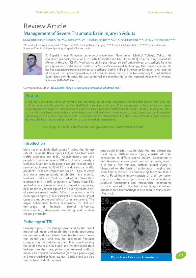
Chettinad Health City Medical Journal
Management of Severe Traumatic Brain Injury in Adults
Review Article
Dr.Gopalakrishnan Raman*, Prof V.G. Ramesh**, Dr. T. Mohanarangam***, Dr. K. ArunThevaraja ***, Dr. K.V. Karthikeyan ****
Corresponding author - Dr.Gopalakrishnan Raman ([email protected])
Dr.Gopalakrishnan Raman is an undergraduate from Government Medical College, Calicut. He completed his post graduation [D.A, MD (Anaesth) and DNB (Anaesth)] from the King Edward VII Memorial Hospital (KEM), Mumbai. He did his post-doctoral certification in Neuroanaesthesia from the prestigious Sree ChitraTirunal Institute for Medical Sciences and Technology, Thiruvananthapuram. He has had extensive experience in Neuroanaesthesia, both in India and the United Kingdom, over a period of 20 years. He is presently working as a Consultant Anaesthetist, in the Neurosurgery OT, in Chettinad Super Speciality Hospital. He was conferred the membership of the National Academy of Medical Sciences (MNAMS) in 2013.
Volume 2, Number 4
*Consultant Neuro-anaesthetist, ** Prof. & HOD, Dept. of Neuro Surgery, *** Consultant Anaesthetist, ****Consultant Neuro Surgeon, Chettinad Super Speciality Hospital, Chennai, India.
Introduction
India has unenviable distinction of having the highest rate of Traumatic Brain Injury (TBI) in Asia from road traffic accidents and falls1. Approximately ten lakh people suffer from severe TBI out of which nearly 2 lakh die. Over ten lakh people require rehabilitation services each year. 60% of TBI are due to road traffic accidents. Falls are responsible for 20 – 25% of cases and occur predominantly in children and elderly. Violence results in 10 % of cases. Alcoholic intoxication is present in 15 – 20% of patients suffering from TBI. 40% of cases are seen in the age group of 21 – 35 years, 20% under 15 years of age and 5% over 65 years. 80% of cases are seen in males. 66% of cases occur in the evening and nights. 71% of cases of TBI are mild, 15% of cases are moderate and 13% of cases are severe. The major behavioural factors responsible for TBI are non-usage of helmets, alcohol influence, over-speeding, dangerous overtaking and careless crossing of roads2.
Pathology of TBI
Primary injury is the damage produced by the direct mechanical impact and acceleration deceleration stress on the skull and brain tissue. Skull fractures occur over the cranial vault and may be depressed fractures compressing the underlying brain. Fractures involving the skull base result in blood and cerebrospinal fluid leakage into the nose, pharynx or external auditory meatus. Periorbital haematomas (racoon / panda sign) and retro-auricular haematomas (battle sign) are also seen in base of skull fractures.
Intracranial injuries may be classified into diffuse and focal injury. Diffuse brain injury consists of brain concussion or diffuse axonal injury. Concussion is definite retrograde and post traumatic amnesia, even if it is for a few minutes. Diffuse axonal injury is diagnosed on the basis of radiological imaging and should be suspected in coma lasting for more than 6 hours. Focal brain injury consists of brain contusion (coup or contre coup injuries), extradural haematoma, subdural haematoma and intracerebral haematoma (usually located in the frontal or temporal lobes). Subarachnoid haemorrhage is also seen in many cases. (Fig 1 - 4).
Abstract
Head injury is a major cause of mortality and morbidity in India and responsible for two lakh deaths each year. In addition, over ten lakh people require rehabilitation services each year. The management of head injury has been revolutionised through the introduction of evidence based recommendations by the Brain Trauma Foundation. This article is broadly based around these recommendations and deals with the emergency room, anaesthetic and intensive care management of head injured patients. It recommends the introduction of protocols by each and every institution dealing with head injured patients in order to streamline their management.
Key words : Head injury, Intracranial haematoma, Glasgow coma scale, Intracranial pressure.
Chettinad Health City Medical Journal 2013; 2(4): 123 - 132
Fig 1 - Acute Extradural Haematoma123
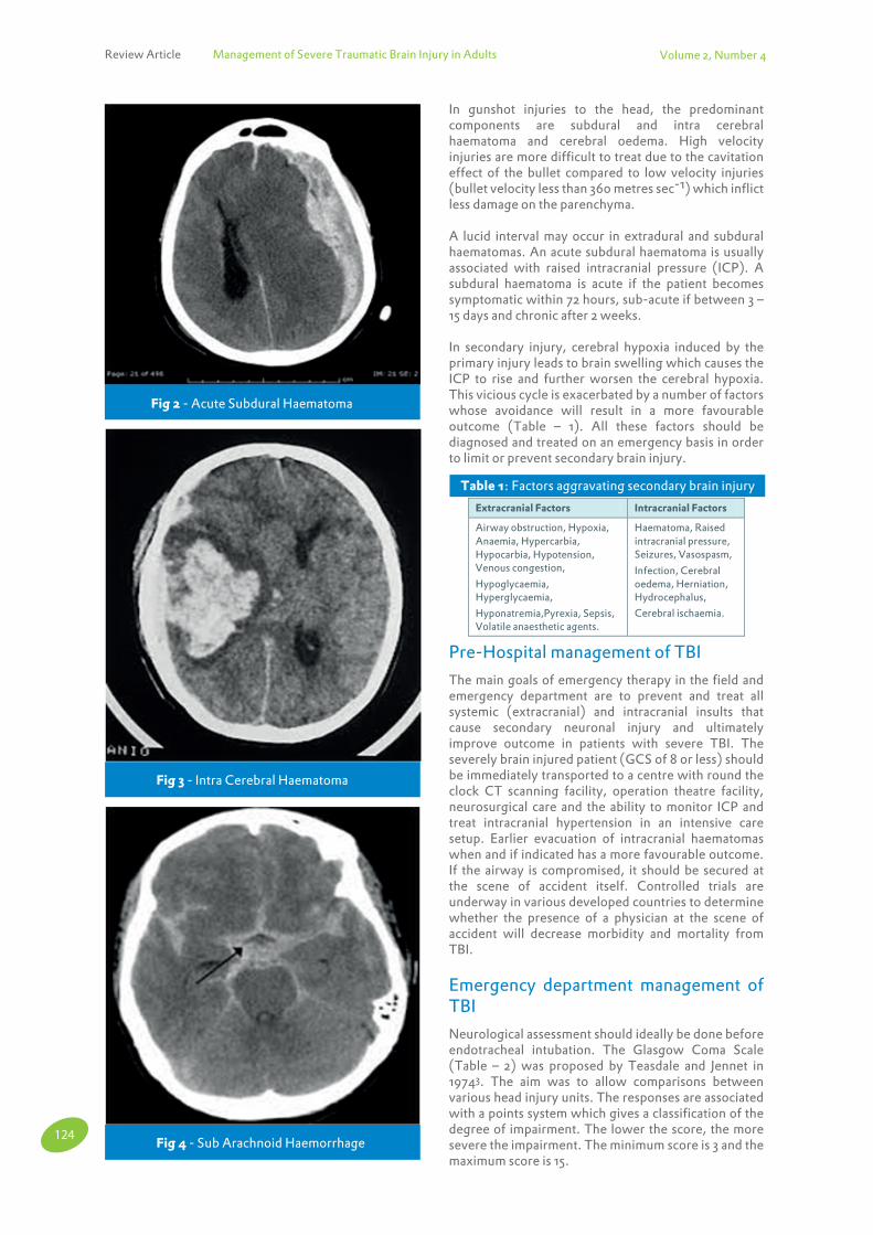
Review Article Management of Severe Traumatic Brain Injury in Adults Volume 2, Number 4
In gunshot injuries to the head, the predominant components are subdural and intra cerebral haematoma and cerebral oedema. High velocity injuries are more difficult to treat due to the cavitation effect of the bullet compared to low velocity injuries (bullet velocity less than 360 metres sec-1) which inflict less damage on the parenchyma.
A lucid interval may occur in extradural and subdural haematomas. An acute subdural haematoma is usually associated with raised intracranial pressure (ICP). A subdural haematoma is acute if the patient becomes symptomatic within 72 hours, sub-acute if between 3 – 15 days and chronic after 2 weeks.
In secondary injury, cerebral hypoxia induced by the primary injury leads to brain swelling which causes the ICP to rise and further worsen the cerebral hypoxia. This vicious cycle is exacerbated by a number of factors whose avoidance will result in a more favourable outcome (Table – 1). All these factors should be diagnosed and treated on an emergency basis in order to limit or prevent secondary brain injury.
Pre-Hospital management of TBI
The main goals of emergency therapy in the field and emergency department are to prevent and treat all systemic (extracranial) and intracranial insults that cause secondary neuronal injury and ultimately improve outcome in patients with severe TBI. The severely brain injured patient (GCS of 8 or less) should be immediately transported to a centre with round the clock CT scanning facility, operation theatre facility, neurosurgical care and the ability to monitor ICP and treat intracranial hypertension in an intensive care setup. Earlier evacuation of intracranial haematomas when and if indicated has a more favourable outcome. If the airway is compromised, it should be secured at the scene of accident itself. Controlled trials are underway in various developed countries to determine whether the presence of a physician at the scene of accident will decrease morbidity and mortality from TBI.
Emergency department management of TBI
Neurological assessment should ideally be done before endotracheal intubation. The Glasgow Coma Scale (Table – 2) was proposed by Teasdale and Jennet in 19743. The aim was to allow comparisons between various head injury units. The responses are associated with a points system which gives a classification of the degree of impairment. The lower the score, the more severe the impairment. The minimum score is 3 and the maximum score is 15.
Fig 2 - Acute Subdural Haematoma
Fig 3 - Intra Cerebral Haematoma
Fig 4 - Sub Arachnoid Haemorrhage
Extracranial Factors Intracranial Factors
Airway obstruction, Hypoxia, Anaemia, Hypercarbia, Hypocarbia, Hypotension, Venous congestion,
Hypoglycaemia, Hyperglycaemia,
Hyponatremia,Pyrexia, Sepsis, Volatile anaesthetic agents.
Haematoma, Raised intracranial pressure, Seizures, Vasospasm,
Infection, Cerebral oedema, Herniation, Hydrocephalus,
Cerebral ischaemia.
Table 1: Factors aggravating secondary brain injury
124

Volume 2, Number 4
A. In Trans-calvarial herniation, the brain protrudes through a defect in the skull. This is often worsened by raised ICP.
B. In Subfalcine herniation, the cingulate gyrus of the frontal lobe is pushed under the falx cerebri. Clinical signs include increased tone or paresis in the contralateral leg.
C. In Uncal herniation, there is displacement of the medial edge of the uncus and the hippocampal gyrus medially and over the ipsilateral edge of the tentorial foramen causing compression of the midbrain. The ipsilateral or contralateral occulomotor nerve may be stretched or compressed. Initially there will be asymmetric pupillary dilatation (anisocoria) which will progress to ipsilateral pupillary dilatation and contralateral hemiparesis.
D. Upward cerebellar herniation is uncommon and is usually due to upward herniation of vermis and cerebellar hemispheres through the tentorial foramen due to infratentorial mass lesions. Radiology usually reveals effacement of the superior vermian cistern, compression of the fourth ventricle, upward and forward displacement of the quadrigeminal plate, mesencephalon and cerebral aqueduct causing supratentorial hydrocephalus. If left untreated, there will be medullary compression leading to bradycardia and ventilatory arrest.
E. Tonsillar herniation is due to a supra or infra tentorial mass lesion and is a rapid and often fatal event unless recognized immediately and treated. The cerebellar tonsils descend through the foramen magnum compressing the lower brain stem resulting in ventilatory arrest. Of all the herniation syndromes, this one has the narrowest window of intervention to prevent death.
F. Cerebral transtentorial herniation is characterised by displacement of the cerebral hemispheres and basal ganglia downwards while the diencephalon and adjacent midbrain are pushed through the tentorial notch. This is usually due to lesions occupying the intracranial vertex or frontal-occipital poles. Clinical presentation will be an impairment of vertical gaze and bilateral extensor posturing.
Tracheal Intubation – Tracheal intubation is indicated in all cases with a GCS of 8 or less. All head injured patients should be considered as having a full stomach. Many of them have an associated cervical spine injury. Rapid sequence intubation with manual in line stabilization (MILS) is preferred in haemodynamically stable patients. The patient is pre-oxygenated with 100 % oxygen and anaesthesia is induced with Thiopentone 3 – 4 mg Kg-1 or Propofol 1 – 2 mg Kg-1. Succinyl Choline 1.5 mg Kg-1 is then administered and the trachea intubated. Applying cricoid pressure if a cervical spine injury is not ruled out is controversial. One recommendation would be to apply cricoid pressure after placing the posterior part of the Philadelphia hard collar.
Routine intravenous induction is carried out in a patient without full stomach and stable haemodynamics by titrating the Thiopentone or Propofol dosage to minimize hypotension. Rocuronium 0.6 - 1.0 mg Kg-1 may be used to facilitate endotracheal intubation since Fig 5 - Brain Herniation Syndromes
Mild TBI – GCS of 14 to 15. Loss of consciousness if present is less than 5 minutes. These cases require close neurological monitoring but not intensive care.
Moderate TBI – GCS of 9 – 13. These patients have a mixed prognosis.
Severe TBI – GCS of 8 or less persisting for 6 hours or more. Virtually all these patients require ICU admission and ICP monitoring.
Pupillary size, any inequalities, reflex response to light and accommodation should also be assessed along with symmetry of motor function in the extremities. Injuries to other organ systems should then be assessed. Intra-thoracic and intra-peritoneal injuries should be treated without delay. Management of bleeding causing haemorrhagic shock takes precedence over neurosurgical procedures.
Brain herniation syndromes are due to mechanical compression by an accumulating mass or a diffusely increased ICP. Almost all cases present with progressive somnolescence and the Cushing’s reflex (hypertension & bradycardia). The following herniation syndromes are described (Figure-5):
Table 2: Glasgow Coma Scale (Adult)Feature Parameter Score
Eye opening
Spontaneous To verbal command To pain None
4 3 2 1
Best verbal
response
Oriented, conversing Disoriented, conversing Inappropriate words Incomprehensible sounds No verbal response
5 4 3 2 1
Best motor
response
Obeys verbal commands Localizes to pain Withdrawal Abnormal flexion (Decorticate) Extension (Decerebrate) No motor response (Flaccid)
6 5 4 3
2
1
125
Review Article Management of Severe Traumatic Brain Injury in Adults

Volume 2, Number 4
Total osmolality is the most important factor that determines the formation of cerebral oedema. Normovolaemia should be maintained in these patients using isotonic and hypertonic solutions. Plasma total osmolality should however be maintained at less than 320 mosm Kg-1.When large volumes of crystalloid are required, use an isotonic solution like Normal saline (308 mosm L-1). Hypertonic saline (3 %) can be beneficial in severe TBI. In addition to being a useful resuscitation fluid, hypertonic saline reduces cerebral oedema and ICP. A rebound phenomenon is not seen following the use of this solution. The dose is 125 to 250 ml every 6 hours. It may also be used as a continuous infusion at 0.5 – 1.0 ml Kg-1 hour-1. Stop hypertonic saline once the serum sodium concentration reaches 160 mEq L-1. Potential adverse effects include central pontine myelinolysis, seizures, congestive heart failure, hypokalemia, hyperchloraemic acidosis and coagulopathy.
Hydroxy Ethyl Starch (HES) and albumin have also been used for resuscitation. The maximum dose of HES is 20 ml / Kg. HES can worsen coagulopathy which is seen in approximately 20 % of patients with TBI. Dextrans should be avoided because of their effect on platelet function.
Blood should be transfused to maintain a haematocrit over 30 % while blood products may be needed to correct coagulation defects such as disseminated intravascular coagulation ( DIC ) induced by the release of brain thromboplastin.
Ringer’s Lactate is hypotonic ( 273mosmKg-1 ) and will result in an increase in cerebral oedema and ICP as well as contribute to lactic acidosis in ischaemic areas of the brain. Dextrose containing solutions should be avoided because hyperglycaemia is associated with a poorer neurological outcome. In addition, the free water load left after the glucose is metabolised will cause a reduction in plasma osmolality and worsen cerebral oedema.
If the blood pressure and cardiac output cannot be restored to normal through fluid resuscitation alone, inotropes may have to be administered. An infusion of dopamine or phenylephrine is preferred over other inotropes.
Radiological Evaluation – CT scanning remains the radiological investigation of choice for evaluating TBI. Indications for scanning TBI patients may be according to the Canadian criteria7 or New Orleans criteria8 (Table – 3 ). Brain imaging helps to distinguish cases that require immediate surgical intervention from those that do not and may also identify those whose protracted recovery requires early tracheostomy and feeding gastrostomy.
Indications for early surgical intervention based on CT findings include open depressed skull fractures with dural laceration and various haematomas. An extra dural haematoma with a volume of over 30 ml should be evacuated regardless of GCS. An acute EDH should be evacuated if associated with a GCS of 8 or less and unequal pupils9.One however has to keep in mind that the acute EDH may be just an incidental finding and not
it has no effect on cerebral dynamics. Lignocaine 1.5 mg Kg-1 given 90 seconds before laryngoscopy will blunt any haemodynamic response and attenuate any rise in ICP. A small dose of propofol or fentanyl or a short acting beta-blocker like esmolol may also be used to blunt the laryngoscopic response.
Ketamine is contraindicated in the routine management of TBI since it elevates the ICP. However, it may have a small role to play in inducing patients in haemorrhagic shock. Midazolam at 0.2 mg Kg-1 may be used for induction. It has minimal haemodynamic effects and hence the cerebral circulation is usually not disturbed. It however interferes with neurological assessments. Fentanyl produces minimal to moderate decreases in MAP and CPP at a dose of 2 – 4 mcg Kg-1. It increases the ICP to a moderate extent4. When larger doses are used, measures to maintain systemic BP may be required.
If facial fractures and soft tissue oedema prevent direct visualization of the larynx, a fibre-optic intubation or intubation over an illuminated stylet may be tried. Avoid nasal intubation if a base of skull fracture is suspected to avoid intracranial placement of the endotracheal tube or introduction of contaminated material into the brain. Nasal intubation should also be avoided in the presence of severe facial bone fractures or a bleeding diathesis. Instruments for an emergency tracheostomy should be available. Once the airway is secured, insert an oro-gastric tube to decompress the stomach.
Antibiotics should be administered at the time of intubation to reduce the incidence of pneumonia. However, prophylactic antibiotic use for ventricular catheter placement to reduce infection is not recommended21.
After securing the airway, mechanical ventilation is initiated and the respiratory rate and tidal volume are adjusted to maintain a PaCO2 of 35 mm Hg. Hyperventilation to a PaCO2 of 30 mm Hg is indicated only if transtentorial herniation is suspected. Prophylactic hyperventilation to a PaCO2 of less than 25 mm Hg is not recommended. Hyperventilation is used only as a temporary measure to reduce ICP and should ideally be avoided during the first 24 hours after injury. If hyperventilation is used, Jugular venous oxygen saturation or brain tissue oxygen tension should be monitored. The Fraction of inspired oxygen (FiO2) and positive end expiratory pressure (PEEP) are adjusted to maintain a PaO2 of 100 mm Hg.Hypoxaemia (PaO2< 60 mm Hg or SpO2 < 90 %) should be avoided5. A PEEP level upto 12 cm H2O is safe in head injury and does not raise the ICP6. Use a fibreoptic bronchoscope to suction out aspirated material from the bronchi.
Fluid resuscitation and haemodynamic stabilization – Fluid resuscitation should be guided by systemic blood pressure, urine output and central venous pressure. Sympathetic over activity in patients with severe TBI along with a reflex response to raised ICP often maintains the blood pressure at normal levels. Induction of anaesthesia in these patients often unmasks hypovolaemia and precipitates hypotension.
126
Review Article Management of Severe Traumatic Brain Injury in Adults
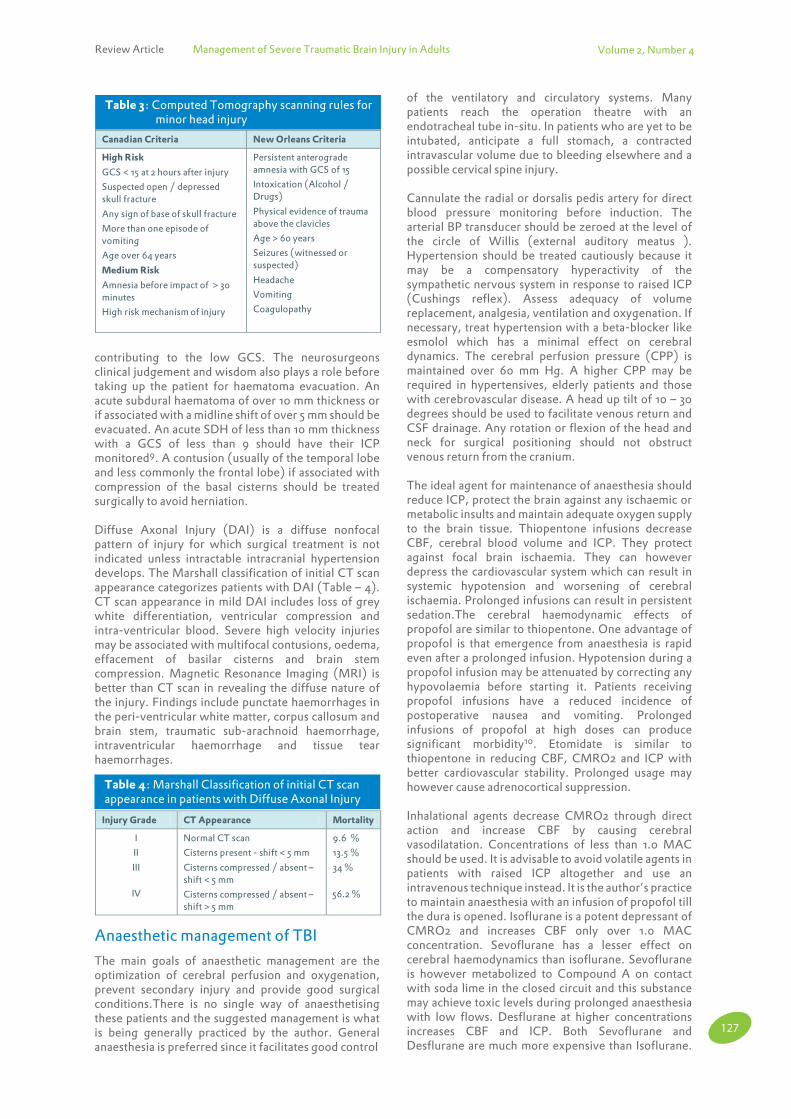
Volume 2, Number 4
of the ventilatory and circulatory systems. Many patients reach the operation theatre with an endotracheal tube in-situ. In patients who are yet to be intubated, anticipate a full stomach, a contracted intravascular volume due to bleeding elsewhere and a possible cervical spine injury.
Cannulate the radial or dorsalis pedis artery for direct blood pressure monitoring before induction. The arterial BP transducer should be zeroed at the level of the circle of Willis (external auditory meatus ). Hypertension should be treated cautiously because it may be a compensatory hyperactivity of the sympathetic nervous system in response to raised ICP (Cushings reflex). Assess adequacy of volume replacement, analgesia, ventilation and oxygenation. If necessary, treat hypertension with a beta-blocker like esmolol which has a minimal effect on cerebral dynamics. The cerebral perfusion pressure (CPP) is maintained over 60 mm Hg. A higher CPP may be required in hypertensives, elderly patients and those with cerebrovascular disease. A head up tilt of 10 – 30 degrees should be used to facilitate venous return and CSF drainage. Any rotation or flexion of the head and neck for surgical positioning should not obstruct venous return from the cranium.
The ideal agent for maintenance of anaesthesia should reduce ICP, protect the brain against any ischaemic or metabolic insults and maintain adequate oxygen supply to the brain tissue. Thiopentone infusions decrease CBF, cerebral blood volume and ICP. They protect against focal brain ischaemia. They can however depress the cardiovascular system which can result in systemic hypotension and worsening of cerebral ischaemia. Prolonged infusions can result in persistent sedation.The cerebral haemodynamic effects of propofol are similar to thiopentone. One advantage of propofol is that emergence from anaesthesia is rapid even after a prolonged infusion. Hypotension during a propofol infusion may be attenuated by correcting any hypovolaemia before starting it. Patients receiving propofol infusions have a reduced incidence of postoperative nausea and vomiting. Prolonged infusions of propofol at high doses can produce significant morbidity10. Etomidate is similar to thiopentone in reducing CBF, CMRO2 and ICP with better cardiovascular stability. Prolonged usage may however cause adrenocortical suppression.
Inhalational agents decrease CMRO2 through direct action and increase CBF by causing cerebral vasodilatation. Concentrations of less than 1.0 MAC should be used. It is advisable to avoid volatile agents in patients with raised ICP altogether and use an intravenous technique instead. It is the author’s practice to maintain anaesthesia with an infusion of propofol till the dura is opened. Isoflurane is a potent depressant of CMRO2 and increases CBF only over 1.0 MAC concentration. Sevoflurane has a lesser effect on cerebral haemodynamics than isoflurane. Sevoflurane is however metabolized to Compound A on contact with soda lime in the closed circuit and this substance may achieve toxic levels during prolonged anaesthesia with low flows. Desflurane at higher concentrations increases CBF and ICP. Both Sevoflurane and Desflurane are much more expensive than Isoflurane.
contributing to the low GCS. The neurosurgeons clinical judgement and wisdom also plays a role before taking up the patient for haematoma evacuation. An acute subdural haematoma of over 10 mm thickness or if associated with a midline shift of over 5 mm should be evacuated. An acute SDH of less than 10 mm thickness with a GCS of less than 9 should have their ICP monitored9. A contusion (usually of the temporal lobe and less commonly the frontal lobe) if associated with compression of the basal cisterns should be treated surgically to avoid herniation.
Diffuse Axonal Injury (DAI) is a diffuse nonfocal pattern of injury for which surgical treatment is not indicated unless intractable intracranial hypertension develops. The Marshall classification of initial CT scan appearance categorizes patients with DAI (Table – 4). CT scan appearance in mild DAI includes loss of grey white differentiation, ventricular compression and intra-ventricular blood. Severe high velocity injuries may be associated with multifocal contusions, oedema, effacement of basilar cisterns and brain stem compression. Magnetic Resonance Imaging (MRI) is better than CT scan in revealing the diffuse nature of the injury. Findings include punctate haemorrhages in the peri-ventricular white matter, corpus callosum and brain stem, traumatic sub-arachnoid haemorrhage, intraventricular haemorrhage and tissue tear haemorrhages.
Anaesthetic management of TBI
The main goals of anaesthetic management are the optimization of cerebral perfusion and oxygenation, prevent secondary injury and provide good surgical conditions.There is no single way of anaesthetising these patients and the suggested management is what is being generally practiced by the author. General anaesthesia is preferred since it facilitates good control
Table 3: Computed Tomography scanning rules for minor head injury
Canadian Criteria New Orleans Criteria
High Risk
GCS < 15 at 2 hours after injury
Suspected open / depressed skull fracture
Any sign of base of skull fracture
More than one episode of vomiting
Age over 64 years
Medium Risk
Amnesia before impact of > 30 minutes
High risk mechanism of injury
Persistent anterograde amnesia with GCS of 15
Intoxication (Alcohol / Drugs)
Physical evidence of trauma above the clavicles
Age > 60 years
Seizures (witnessed or suspected)
Headache
Vomiting
Coagulopathy
Injury Grade CT Appearance Mortality
I
II
III
IV
Normal CT scan
Cisterns present - shift < 5 mm
Cisterns compressed / absent – shift < 5 mm
Cisterns compressed / absent – shift > 5 mm
9.6 %
13.5 %
34 %
56.2 %
Table 4: Marshall Classification of initial CT scan appearance in patients with Diffuse Axonal Injury
127
Review Article Management of Severe Traumatic Brain Injury in Adults
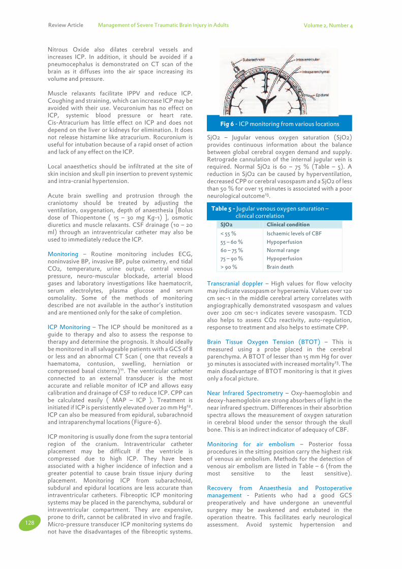
Volume 2, Number 4
SjO2 – Jugular venous oxygen saturation (SjO2) provides continuous information about the balance between global cerebral oxygen demand and supply. Retrograde cannulation of the internal jugular vein is required. Normal SjO2 is 60 – 75 % (Table – 5). A reduction in SjO2 can be caused by hyperventilation, decreased CPP or cerebral vasospasm and a SjO2 of less than 50 % for over 15 minutes is associated with a poor neurological outcome13.
Transcranial doppler – High values for flow velocity may indicate vasospasm or hyperaemia. Values over 120 cm sec-1 in the middle cerebral artery correlates with angiographically demonstrated vasospasm and values over 200 cm sec-1 indicates severe vasospasm. TCD also helps to assess CO2 reactivity, auto-regulation, response to treatment and also helps to estimate CPP.
Brain Tissue Oxygen Tension (BTOT) – This is measured using a probe placed in the cerebral parenchyma. A BTOT of lesser than 15 mm Hg for over 30 minutes is associated with increased mortality13. The main disadvantage of BTOT monitoring is that it gives only a focal picture.
Near Infrared Spectrometry – Oxy-haemoglobin and deoxy-haemoglobin are strong absorbers of light in the near infrared spectrum. Differences in their absorbtion spectra allows the measurement of oxygen saturation in cerebral blood under the sensor through the skull bone. This is an indirect indicator of adequacy of CBF.
Monitoring for air embolism – Posterior fossa procedures in the sitting position carry the highest risk of venous air embolism. Methods for the detection of venous air embolism are listed in Table – 6 (from the most sensitive to the least sensitive).
Recovery from Anaesthesia and Postoperative management - Patients who had a good GCS preoperatively and have undergone an uneventful surgery may be awakened and extubated in the operation theatre. This facilitates early neurological assessment. Avoid systemic hypertension and
Nitrous Oxide also dilates cerebral vessels and increases ICP. In addition, it should be avoided if a pneumocephalus is demonstrated on CT scan of the brain as it diffuses into the air space increasing its volume and pressure.
Muscle relaxants facilitate IPPV and reduce ICP. Coughing and straining, which can increase ICP may be avoided with their use. Vecuronium has no effect on ICP, systemic blood pressure or heart rate. Cis-Atracurium has little effect on ICP and does not depend on the liver or kidneys for elimination. It does not release histamine like atracurium. Rocuronium is useful for intubation because of a rapid onset of action and lack of any effect on the ICP.
Local anaesthetics should be infiltrated at the site of skin incision and skull pin insertion to prevent systemic and intra-cranial hypertension.
Acute brain swelling and protrusion through the craniotomy should be treated by adjusting the ventilation, oxygenation, depth of anaesthesia [Bolus dose of Thiopentone ( 15 – 30 mg Kg-1) ], osmotic diuretics and muscle relaxants. CSF drainage (10 – 20 ml) through an intraventricular catheter may also be used to immediately reduce the ICP.
Monitoring – Routine monitoring includes ECG, noninvasive BP, invasive BP, pulse oximetry, end tidal CO2, temperature, urine output, central venous pressure, neuro-muscular blockade, arterial blood gases and laboratory investigations like haematocrit, serum electrolytes, plasma glucose and serum osmolality. Some of the methods of monitoring described are not available in the author’s institution and are mentioned only for the sake of completion.
ICP Monitoring – The ICP should be monitored as a guide to therapy and also to assess the response to therapy and determine the prognosis. It should ideally be monitored in all salvageable patients with a GCS of 8 or less and an abnormal CT Scan ( one that reveals a haematoma, contusion, swelling, herniation or compressed basal cisterns)11. The ventricular catheter connected to an external transducer is the most accurate and reliable monitor of ICP and allows easy calibration and drainage of CSF to reduce ICP. CPP can be calculated easily ( MAP – ICP ). Treatment is initiated if ICP is persistently elevated over 20 mm Hg12. ICP can also be measured from epidural, subarachnoid and intraparenchymal locations (Figure-6).
ICP monitoring is usually done from the supra tentorial region of the cranium. Intraventricular catheter placement may be difficult if the ventricle is compressed due to high ICP. They have been associated with a higher incidence of infection and a greater potential to cause brain tissue injury during placement. Monitoring ICP from subarachnoid, subdural and epidural locations are less accurate than intraventricular catheters. Fibreoptic ICP monitoring systems may be placed in the parenchyma, subdural or intraventricular compartment. They are expensive, prone to drift, cannot be calibrated in vivo and fragile. Micro-pressure transducer ICP monitoring systems do not have the disadvantages of the fibreoptic systems.
Fig 6 - ICP monitoring from various locations
SJO2 Clinical condition
< 55 %
55 – 60 %
60 – 75 %
75 – 90 %
> 90 %
Ischaemic levels of CBF
Hypoperfusion
Normal range
Hypoperfusion
Brain death
Table 5 - Jugular venous oxygen saturation – clinical correlation
128
Review Article Management of Severe Traumatic Brain Injury in Adults
Volume 2, Number 4
minutes and repeated every 3 – 6 hours. A larger dose of 1.0 – 1.5 gm Kg-1 is used if transtentorial herniation is suspected. It is recommended to restrict usage of mannitol, prior to monitoring ICP, to patients with signs of transtentorial herniation or progressive neurological deterioration not attributable to extracranial causes14. The serum osmolality should be monitored and maintained at less than 320 mosm Kg-1. Frusemide prevents the rebound phenomenon seen with mannitol. Infusions of hypertonic saline and glycerol are also being used in the management of raised ICP.
Mechanical ventilation should be adjusted to maintain a PaCO2 of 35 mm Hg. Lower levels of 30 mm Hg is indicated only if impending herniation is suspected and is used as a bridge to surgical decompression. Levels below 30 mm Hg can produce vasoconstriction and aggravate cerebral ischaemia. Once hyperventilation is instituted, other measures to reduce ICP should be introduced and the PaCO2 allowed to normalize as soon as possible.
Decompressive Craniectomy Decompressive craniectomy is a controversial line of management and is practiced in certain centres only. Post surgical quality of life should be considered before planning this procedure. Decompressive craniectomy decreases ICP and improves outcome in young patients who deteriorate within 48 hours of TBI. It should be considered prior to initiating barbiturate therapy (< 55 years of age is a relative indication). It should be considered in all cases of non-fatal primary brain injury associated with asymmetric or focal brain swelling on CT scan who have refractory intracranial hypertension which has failed to respond to maximal medical therapy, with or without CSF drainage.
Barbiturates – Barbiturates have cerebral protection and ICP lowering effects and must be considered in severe TBI cases in which raised ICP is refractory to medical and surgical management. Barbiturate therapy should be initiated only after the patient is haemodynamically stable. Clinical trials have however not revealed any clear benefit or improved outcome associated with the use of high dose barbiturate therapy for refractory intracranial hypertension. High dose barbiturate therapy is indicated in hemodynamically stable salvageable patients who have raised ICP refractory to the previous steps. Barbiturates reduce ICP by decreasing CBF in parallel with CMRO2. Thiopentone is administered in a loading dose of 15 mg Kg-1 followed by an infusion of 0.2 mg Kg-1min-1. Thiopentone infusions are associated with haemodynamic instability, prolonged recovery after stopping the infusion and difficulty in neurological assessment. Prophylactic administration of barbiturates to induce burst suppression in head injured patients is not recommended15.
Hypothermia – Mild hypothermia (34 – 36 degrees ) has been demonstrated to markedly attenuate ischaemic cerebral injury in animal models. The mechanism may be due to a reduction in metabolic demand, free radical formation and oedema formation. Adverse side effects of hypothermia include hypotension, cardiac arrhythmias, coagulopathies and infection. Rewarming
coughing while emerging from anaesthesia as they are predisposed to cerebral oedema, raised ICP and haematoma formation. If the level of consciousness is depressed preoperatively, ventilate the patient electively in the postoperative period. Poly trauma patients and patients who are hypothermic should also be ventilated electively in the post-operative period. Causes of delayed awakening in the operation theatre include a low preoperative GCS score, elevated ICP, residual drug effects, metabolic and electrolyte disturbances, hypothermia and seizures.
Intensive care management of the TBI patient
The indications for monitoring ICP have been listed in the previous section. An intraventricular catheter is preferred since it provides accurate readings and allows therapeutic drainage of CSF. Loss of autoregulation induced by TBI will result in the CBF becoming dependant on the mean arterial pressure (MAP). Reductions in MAP will then cause cerebral ischaemia while increases in MAP results in hyperaemia, cerebral oedema and rise in ICP. The Lund approach to treat raised ICP works on the assumption that the integrity of the BBB for salts is lost following TBI. The goal of management is to limit the resulting oedema and ICP elevation by using osmotic diuretics and maintaining an ICP of less than 20 mm Hg.
The CPP should be maintained between 50 and 70 mm Hg to maximize brain tissue oxygenation. Arterial hypotension (SBP < 90 mm Hg) should be avoided12. The systolic BP should be maintained between 90 and 160 mm Hg. Adequate MAP is maintained by infusing isotonic fluids to maintain euvolaemia or slight hypervolaemia. Intravascular volume status can be assessed by monitoring the CVP and urine output, respiratory variation in the arterial pulse pressure and ultrasonographic assessment of inferior vena cava size changes with different phases of breathing.
Management of raised ICP – CPP is the difference between MAP and ICP and should be maintained over 60 mm Hg. A head up tilt of 10 – 30 degrees should be used to facilitate venous return and CSF drainage.
The head and neck should be maintained in a neutral position so that venous return from the cranium is not obstructed. Tight endotracheal tube ties should not be used for the same reason. Incremental drainage of CSF through an intraventricular catheter (15-20 ml) will bring down the ICP immediately.
Mannitol and Frusemide may be used as diuretics. Mannitol 0.25 – 1.0 gm Kg-1 is administered over 10 - 20
Table 6 - Methods for detecting air embolism
Trans oesophageal Echocardiography (0.02 ml / Kg ) Precordial Doppler ( 0.05 ml / Kg)
Pulmonary Artery Catheter (0.25 ml / Kg)
Expired CO2 / N2 monitoring (0.5 ml / Kg)
Fall in oxygen saturation
Change in BP and heart sounds (mill wheel murmur)
129
Review Article Management of Severe Traumatic Brain Injury in Adults

Volume 2, Number 4
risk of expansion of intracranial haemorrhage. There is insufficient evidence to support recommendations regarding the preferred agent, dose or timing of prophylaxis22. Any coagulopathy if present should be corrected. Coagulation is impaired in these patients due to release of brain thromboplastin, haemorrhage induced decrease in coagulation factors and increased levels of fibrin degradation products.
Pyrexia (Fever) increases CMRO2 and ICP and may worsen outcome in TBI. It should be treated with cooling blankets and paracetamol. Causes for fever in TBI patients include phenytoin, infection and injury to hypothalamus. The treatment goal should be to maintain normothermia (body temperature of 370C) in these patients.
Complications of TBI
Neurogenic Pulmonary Oedema –The aetiology of neurogenic pulmonary oedema is massive transient central sympathetic discharge due to raised ICP following severe TBI and is particularly associated with hypothalamic lesions. It may rapidly progress to death or resolve completely within a few hours to days. There is systemic vasoconstriction, redistribution of blood from the systemic to the pulmonary circulation,left ventricular failure, pulmonary venoconstriction and increased pulmonary capillary permeability. The treatment is to decrease ICP, reduce systemic blood pressure using diazoxide (1-3 mg IV) every 5 minutes upto 150 mg or phenoxybenzamine (1 mg Kg-1 as an infusion over 2 hours to a maximum of 4 mg Kg-1 day-1), IPPV to support ventilation and inotropes to support the circulation if needed.
Syndrome of Inappropriate ADH secretion – SIADH causes water retention along with continued excretion of sodium. This causes dilutional hyponatremia, decreased serum osmolality, increased urine osmolality and decreased urine output. Water retention and serum hypo-osmolality cause nonspecific signs of water intoxication like nausea, vomiting, headache, irritability, disorientation, seizures and coma. Treatment consists of water restriction, loop diuretics and hypertonic saline. Mild cases are treated with water restriction alone (1-1.5 L day-1). More severe cases are treated with water restriction and loop diuretics which impair the ability of the kidneys to concentrate urine (Frusemide 10 – 20 mg every 6 hours). In severe cases where the serum sodium level is less than 125 mEqL-1, infuse hypertonic saline at 1-2 ml Kg-1 hr-1 for 2 to 3 hours. Correct serum sodium at a rate of 0.5 mEq L-1 hr-1 to avoid central pontine myelinolysis.
Diabetes Insipidus – DI is less commonly seen in TBI. It develops 12-24 hours after injury and lasts for a few days. A decrease in ADH levels results in the excretion of large volumes of dilute urine (4-14 L day-1) that results in dehydration, hypernatremia, increased serum osmolality (over 320 mosmKg-1), decreased urine osmolality (less than 200 mosmKg-1) and decreased urine specific gravity ( < 1.005). Serum sodium levels are often elevated (> 145 mEqL-1). Signs of hypernatremia include decreased level of consciousness, muscle weakness, irritability, spasticity, confusion, ataxia, seizures and coma. Treatment of DI is
should be carried out gradually. Studies indicate that there is a greater reduction in mortality risk when hypothermia is maintained for more than 48 hours16. Hypothermia is not routinely used in clinical practice.
Steroids – The CRASH trial which studied the effect of early administration of methyl prednisolone on outcome after TBI in 10008 patients revealed a higher risk of death within two weeks of injury in the group receiving steroids than in the group receiving placebo17. There was also a higher risk of subsequent death or severe disability. The trial investigators concluded that corticosteroids should not be routinely used in the treatment of TBI. Besides steroid administration also causes hyperglycaemia and is associated with an increased risk of gastrointestinal haemorrhage.
Anticonvulsants – Administration of phenytoin to severe TBI patients prevents early onset seizures, but has no effect on late post traumatic seizures. Seizure prophylaxis reduces the incidence of seizures but has not been shown to improve neurological outcome. Phenytoin should be infused at a rate of less than 50 mg min-1 in adults to avoid cardiovascular depression. Indications for seizure prophylaxis include a GCS of less than 10, cortical contusions, depressed skull fracture, intracranial haematoma, penetrating head injury and seizures occurring during the first 24 hours after TBI. Seizure prophylaxis is recommended during the first week after TBI. Phenytoin at a loading dose of 15 mg Kg-1 over 20 minutes followed by 5 – 7 mg Kg-1 day-1 or Fosphenytoin 15 – 20 mg Kg-1 loading dose followed by 4 – 6 mg Kg-1 day-1 is used. A multicentre prospective study has recently concluded that there is no statistically significant difference between levetiracetam and phenytoin in early TBI seizure prophylaxis18.
Nutrition - Nutritional support should be initiated as soon as possible. Full nutritional support should be achieved by the seventh post injury day. 15 % of total calories should come from proteins. Enteral feeding is preferred and a naso-jejunal tube should be placed to protect against aspiration and gastric intolerance. Stress ulcer prophylaxis is indicated with a H2 inhibitor or proton pump inhibitor (PPI). At least two studies have shown that neurological outcome is poorer if the plasma glucose levels are over 200 mg dl-119,20. Attempts to maintain euglycaemia (80 – 110 mg dl-1) may carry an excessive risk of hypoglycaemia which is not good for the brain. In the absence of clear guidance, maintaining a value between 120 – 160 mg dl-1 is probably the safest course of action.
Early tracheostomy should be performed to reduce the duration of mechanical ventilation. However it does not alter mortality or rate of nosocomial pneumonia. Early extubation in qualified patients may be done without increased risk of pneumonia21.
Deep vein thrombosis (DVT) prophylaxis with TED stockings or pneumatic compression devices should be started as soon as possible. Their use should be continued until patients are ambulatory. Low molecular weight heparin can usually be used in combination with mechanical prophylaxis. There is however an increased
130
Review Article Management of Severe Traumatic Brain Injury in Adults
Volume 2, Number 4
by calculating the free water deficit using the formula [(serum sodium – 140) X body weight(Kg) X 0.6 / 140 ] and replace it with a hypotonic solution like 0.45 % saline or Ringers Lactate. Desmopressin (DDAVP) is used to treat DI. The intravenous or subcutaneous dose is 0.3 mcg Kg-1day-1 in two divided doses. The oral dose is 0.05-1.2 mgday-1 in three divided doses. The intranasal dose is 10-40 mcg day-1 in three divided doses. Once the intravascular volume is restored, persistent hypernatremia may be treated using Hydrochlorothiazide 50-100 mg day-1 IV.
Cerebral salt wasting – CSW is caused by increased secretion of atrial natriuretic peptide, brain natriuretic peptide and c-type natriuretic peptide. Natriuresis, diuresis and vasodilatation occur due to the suppression of aldosterone synthesis by these peptides.Excessive loss of sodium in urine (150-200 mEqL-1) causes hyponatremia. Sodium should be replaced in these cases along with fluid administration. Hypertonic saline may be infused in these patients with close monitoring of serum sodium levels. The rate of correction should not exceed 0.5 mEq L-1 hr-1.
Conclusion
The management of head injury is multidisciplinary and is a team effort involving the emergency room physician, the neurosurgeon, the neuro-anaesthetist and the intensivist. Each and every institution dealing with these cases should evolve protocols based on their needs and availability of resources to achieve the best possible results. However it should be understood that these are purely guidelines that may have to be customised to suit the individual patient and the individual consultant at that point in time. This approach has led to an improvement in the management of these cases with a reduction in mortality and morbidity.
Acknowledgement
I wish to thank the anonymous peer reviewers for their constructive criticism which helped to improve the quality of the article.
5)
6)
7)
8)
9)
10)
11)
12)
13)
14)
15)
16)
17)
Bullock MR, Povlishock JT. BTF Guidelines for the Management of Severe Traumatic Brain Injury, 3rd Edition. Blood pressure and oxygenation. Journal of Neurotrauma 2007;24: S7 - S13.
Caricato A, Conti G, Dellacorte F, Maucino A. Effect of PEEP on the intracranial system of patients with head injury and subarachanoid haemorrhage. The role of respiratory system compliance. J Trauma, March 2005; 58(3): 571 – 576.
Stiell IG, Wells GA, Vandemheen K. The Canadian CT head rule for patients with minor head injury. Lancet; 2001; 357: 1391 – 1396.
Haydel MJ, Preton CA, Mills TJ. Indications for computed tomography in patients with minor head injury. NEJM; 2000; 343: 100 – 105.
M Ross Bullock, Randall Chestnut, JamshidGhajar, David Gordon, Roger Hartl, David W Newell et al. Guidelines for the surgical management of TBI. Neurosurgery; 2006; March; 58(3); Supplement.
Sabsovich J, Rehman Z, Yunen J. Propofol infusion syndrome. A case of increasing morbidity with Traumatic Brain Injury. Am J Crit Care; 2007; 16(1): 82 – 85.
Bullock MR, Povlishock JT. BTF Guidelines for the Management of Severe Traumatic Brain Injury, 3rd Edition. Indications for Intracranial pressure monitoring. Journal of Neurotrauma; 2007; 24: S37 – S44.
Bullock MR, Povlishock JT. BTF Guidelines for the Management of Severe Traumatic Brain Injury, 3rd Edition. Intracranial pressure thresholds. Journal of Neurotrauma; 2007; 24: S55 – S58.
Bullock MR, Povlishock JT. BTF Guidelines for the Management of Severe Traumatic Brain Injury, 3rd Edition. Brain oxygen monitoring and thresholds. Journal of Neurotrauma; 2007; 24: S65 – S70.
Bullock MR, Povlishock JT. BTF Guidelines for the Management of Severe Traumatic Brain Injury, 3rd Edition. Hyperosmolar Therapy. Journal of Neurotrauma; 2007; 24: S14 – S20.
Bullock MR, Povlishock JT. BTF Guidelines for the Management of Severe Traumatic Brain Injury, 3rd Edition. Anaesthetics, Analgesics and Sedatives. Journal of Neurotrauma; 2007; 24: S71 – S76.
Bullock MR, Povlishock JT. BTF Guidelines for the Management of Severe Traumatic Brain Injury, 3rd Edition. Prophylactic Hypothermia. Journal of Neurotrauma; 2007; 24: S21 – S25.
Edwards P, Arango M, Balica L. CRASH Trial collaborators – Final results of MRC CRASH, A randomised placebo controlled trial of intravenous corticosteroid in adults with head injury, outcomes at 6 months. Lancet; 2005; 365: 1957 – 1959.
References
1)
2)
3)
4)
Prasanthi Puvanachandra, Adnan A Hyder. The burden of traumatic brain injury in Asia. A call for research. Pak J NeurolSci; 2009; 4(1); 27 – 32.
Gururaj G, Kolluri SVR, Chandramouli BA, Subbakrishna DK, Kraus JF. Traumatic Brain Injury. Executive Summary. NIMHANS;2005 Publication No – 61.
Teasdale G, Jennet B: Assessment of coma and impaired consciousness. A practical scale. Lancet 2 1974: 81-84.
De Nadal M, Ausina A, Sahuquillo J, Pedraza S, Garnacho A, Gancedo VA. Effects on intracranial pressure of fentanyl in severe head injured patients. ActaNeurochirSuppl; 1998; 71: 10 – 2. 131
Review Article Management of Severe Traumatic Brain Injury in Adults

Volume 2, Number 4
18)
19)
20)
Inaba K, Menaker J, Branco BC. A prospective multicentre comparison of Levetiracetam versus Phenytoin for early post traumatic seizures. J Trauma Acute Care Surg; 2013;74(3): 766 – 773.
Rovilas A, Kotsou S. The influence of Hyperglycaemia on neurological outcome in patients with severe head injury. Neurosurgery; 2000; 46(2): 335 – 43.
Pecha T, Sharma D, Hoffman NG. Hyperglycaemia during craniotomy for adult Traumatic Brain Injury. Anaesth Analg; 2011; 113: 336 – 342.
Reality And Delusion
Most of us humans are not delusional because our brain screens the conclusions we draw from our experiences through "reality testing": we discard those ideas which are not supported by evidence as unreal. For example, when we get headache, we may briefly consider brain tumour as its cause until that idea gets tested as unreal and is rejected. But delusional people cling on to their ideas even when incontrovertible evidence shows their ideas to be unreal. In a new study published in Frontiers in Psychology, Professor Philip Gerrans of University of Adelaide, claims that persistent delusions are the result of problems with "reality testing". People with "reality testing" problems find it difficult to break free from illogical ideas and suffer from severe mental health issues that may prove a threat to themselves or others. Understanding the mechanism of "reality testing" may help in providing relief to these sufferers. It would be interesting to find out if "reality testing" is functional in highly religious people and fan boys! (Philip Gerrans. Pathologies of Hyperfamiliarity in Dreams, Delusions and Déjà Vu. Frontiers in Psychology, 2014 (in press) DOI: 10.3389/fpsyg.2014.00097 )
- Dr. K. Ramesh Rao132
Review Article Management of Severe Traumatic Brain Injury in Adults
21)
22)
Bullock MR, Povlishock JT. BTF Guidelines for the Management of Severe Traumatic Brain Injury, 3rd Edition. Infection Prophylaxis. Journal of Neurotrauma; 2007; 24: S26 – S31.
Bullock MR, Povlishock JT. BTF Guidelines for the Management of Severe Traumatic Brain Injury, 3rd Edition. Deep Vein Thrombosis Prophylaxis. Journal of Neurotrauma; 2007; 24: S32 – S36.
Industrial Toxins and Brain development
Increasing incidence of autism, attention deficit hyperactivity disorder and dyslexia among children has prompted some to claim that toxic chemicals might be triggering a "silent pandemic" of neurodevelopmental disabilities. In a report published online on February 15, 2014 in Lancet Neurology, researchers from Harvard and Mount Sinai have identified six more chemicals as "developmental neurotoxicants: manganese, fluoride, chlorpyrifos and DDT (pesticides), tetrachloroethylene (a solvent), and the polybrominated diphenyl ethers (flame retardants). Manganese is associated with diminished intellectual function and impaired motor skills; solvents are linked to hyperactivity and aggressive behaviour; and certain types of pesticides may cause cognitive delays. Controlling the pandemic is difficult as the usage of most of the industrial chemicals are poorly regulated and requires international cooperation. The authors justly call for an international action. (Philippe Grandjean, Philip Landrigan. Neurobehavioral effects of developmental toxicity. Lancet Neurology, February 2014 DOI: 10.1016/S1474-4422(13)70278-3)
- Dr. K. Ramesh Rao

Chettinad Health City Medical Journal
Nutraceuticals in Sperm Abnormalities
Review Article
Dr.S.Ranjani*, Dr.Asha Benziger*, Dr.N.Pandiyan **
Corresponding author - Dr.S.Ranjani ([email protected])
Chettinad Health City Medical Journal 2013; 2(4): 133 - 136
Dr.S.Ranjani graduated from Government Mohan Kumaramangalam Medical College, Salem. She completed her Post Graduate Diploma in Clinical Embryology with gold medal from the Dept.of Reproductive Medicine, Chettinad Hospital and Research Institute. She is now working as a Junior Embryologist in Dept. of Reproductive Medicine, Chettinad Super Specialty Hospital.
Volume 2, Number 4
*Junior Embryologist, **Professor & HOD, Dept of Reproductive Medicine, Chettinad Super Specialty Hospital, Chennai, India.
Introduction
Male factor infertility is an important cause of a barren marriage. Semen abnormalities constitute the most important cause of the same. These could be abnormalities in sperm concentration, motility or morphology. Several medical and surgical methods have been introduced in the past and several more are currently being tried to improve the sperm abnormalities. These methods have not yielded much benefit except in established cases of obstruction. To evolve methods to improve sperm abnormalities, a proper understanding of sperm physiology and a deeper perspective of pathophysiology of sperm abnormalities are important. The paper describes the current role of nutraceuticals in male infertility. Nutraceuticals are widely used in an attempt to combat the effect of free radicals in male reproductive system.
Nutraceuticals
Nutraceuticals is a portmanteau of the words Nutrition and Pharmaceuticals. Stephen Defelice in 1989, first coined the word 'Nutraceuticals', from the two words nutrition and pharmaceuticals. He was the founder and chairman of the Foundation for Innovation of Medicine, (FIM) in New Jersey. According to him, nutraceutical can be defined as “ a food or a part of food that provides medical or health benefits including the prevention and or treatment of a disease”1.The term indicates several products like isolated nutrients, dietary supplements, herbal products, specific diets, genetically modified food, processed foods such as cereals, soups and beverages2.
While nutrition plays a crucial role in health and disease, it is not exactly clear what role nutraceuticals play. Nutritional deficiencies have been implicated in the pathogenesis of several diseases. For example, deficiencies in Vitamin C, Vitamin B and Vitamin D give rise to scurvy, beriberi and rickets, respectively. Although there seems to be no clear association between nutritional deficiency and infertility, inadequacy of certain nutrients such as folic acid, L-carnitine and selenium have been claimed to cause infertility3,4.
Spermatogenesis
Nutrition plays an important role in spermatogenesis. There is a definitive role of micronutrients such as zinc, folate and antioxidants for the normal maintenance of spermatogenesis and sperm maturation, DNA synthesis, repair and transcription. However, knowledge about the effect of paternal malnutrition on sperm aneuploidy is scarce. Environmental factors such as exposure to pesticides and chemotherapy have been associated with aneuploidy in spermatozoa of humans5.
Nutraceuticals commonly used are:
1) Nutritional factors: Arginine, VitB 12, Folic acid
2) Motility enhancers: L-carnitine, Acetyl carnitine, Co enzyme Q10
3) Antioxidants: Vit C, Vit E, Glutathione, Lycopene, Selenium, Zinc
Male infertility
Infertility affects 15% of the married, eligible couples. Male factor accounts for about 50% of the cause for infertility. Nutraceuticals, which are readily available in the market, offer an easy alternative to the couples against Assisted Reproductive Technologies. These include those that offer improvement in sperm function, semen parameters, sexual function and erectile function. However, there is no clear evidence that support these claims. The benefits of nutraceuticals in male infertility are yet to be proven. More importantly, the side effects of these supplementations are unknown and very often used by common population as a substitute for lack of nutrition in the diet.
Free radicals in health and disease
Oxidative metabolism in all the cells, tissues and organs leads to the production of free radicals - reactive oxygen species and reactive nitrogen species. These molecules have an unpaired electron in the outer orbit. They are unstable and very reactive. Free radicals are effectively counteracted by several natural antioxidants6. 133
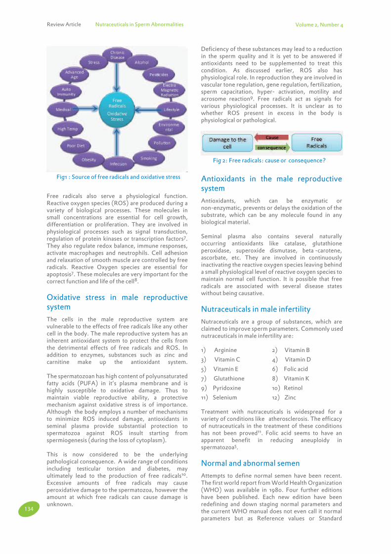
Free radicals also serve a physiological function. Reactive oxygen species (ROS) are produced during a variety of biological processes. These molecules in small concentrations are essential for cell growth, differentiation or proliferation. They are involved in physiological processes such as signal transduction, regulation of protein kinases or transcription factors7. They also regulate redox balance, immune responses, activate macrophages and neutrophils. Cell adhesion and relaxation of smooth muscle are controlled by free radicals. Reactive Oxygen species are essential for apoptosis7. These molecules are very important for the correct function and life of the cell8.
Oxidative stress in male reproductive system
The cells in the male reproductive system are vulnerable to the effects of free radicals like any other cell in the body. The male reproductive system has an inherent antioxidant system to protect the cells from the detrimental effects of free radicals and ROS. In addition to enzymes, substances such as zinc and carnitine make up the antioxidant system.
The spermatozoan has high content of polyunsaturated fatty acids (PUFA) in it’s plasma membrane and is highly susceptible to oxidative damage. Thus to maintain viable reproductive ability, a protective mechanism against oxidative stress is of importance. Although the body employs a number of mechanisms to minimize ROS induced damage, antioxidants in seminal plasma provide substantial protection to spermatozoa against ROS insult starting from spermiogenesis (during the loss of cytoplasm).
This is now considered to be the underlying pathological consequence. A wide range of conditions including testicular torsion and diabetes, may ultimately lead to the production of free radicals10. Excessive amounts of free radicals may cause peroxidative damage to the spermatozoa, however the amount at which free radicals can cause damage is unknown.
Review Article Nutraceuticals in Sperm Abnormalities Volume 2, Number 4
Deficiency of these substances may lead to a reduction in the sperm quality and it is yet to be answered if antioxidants need to be supplemented to treat this condition. As discussed earlier, ROS also has physiological role. In reproduction they are involved in vascular tone regulation, gene regulation, fertilization, sperm capacitation, hyper- activation, motility and acrosome reaction9. Free radicals act as signals for various physiological processes. It is unclear as to whether ROS present in excess in the body is physiological or pathological.
Antioxidants in the male reproductive system
Antioxidants, which can be enzymatic or non-enzymatic, prevents or delays the oxidation of the substrate, which can be any molecule found in any biological material.
Seminal plasma also contains several naturally occurring antioxidants like catalase, glutathione peroxidase, superoxide dismutase, beta�-carotene, ascorbate, etc. They are involved in continuously inactivating the reactive oxygen species leaving behind a small physiological level of reactive oxygen species to maintain normal cell function. It is possible that free radicals are associated with several disease states without being causative.
Nutraceuticals in male infertility
Nutraceuticals are a group of substances, which are claimed to improve sperm parameters. Commonly used nutraceuticals in male infertility are:
1) Arginine 2) Vitamin B
3) Vitamin C 4) Vitamin D
5) Vitamin E 6) Folic acid
7) Glutathione 8) Vitamin K
9) Pyridoxine 10) Retinol
11) Selenium 12) Zinc
Treatment with nutraceuticals is widespread for a variety of conditions like atherosclerosis. The efficacy of nutraceuticals in the treatment of these conditions has not been proved11. Folic acid seems to have an apparent benefit in reducing aneuploidy in spermatozoa5.
Normal and abnormal semen
Attempts to define normal semen have been recent. The first world report from World Health Organization (WHO) was available in 1980. Four further editions have been published. Each new edition have been redefining and down staging normal parameters and the current WHO manual does not even call it normal parameters but as Reference values or Standard
Fig1 : Source of free radicals and oxidative stress
Fig 2: Free radicals: cause or consequence?
134

parameters12. There are too many parameters listed. We will confine this paper to concentration, motility and morphology.
Azoospermia
Azoospermia defined as absence of spermatozoa in the ejaculate, both in a neat semen sample and in a centrifuged resuspended semen sample. This may be obstructive or non obstructive12. Obstructive azoospermia requires surgical correction or sperm retrieval techniques. Nutraceuticals are of no value.
In non-obstructive Azoospermia - gonadotrophins are of immense value in hypogonadotrophic hypogonadism. Nutraceuticals have been tried with no proven benefit. Anti oxidants are the primary group of drugs used in other non-obstructive azoospermia patients. The results are conflicting. Significant number of men with non-obstructive azoospermia have chromosomal anomalies or Y chromosome micro deletions. It is unlikely that nutraceuticals would be of any value to these patients.
Oligoasthenoteratozoospermia
These conditions are difficult to define except in the total state as in Total asthenozoospermia. The definition of these conditions have been changed 5 times in the last 30 years .For example the lower reference limit for normal morphology is 4% ( WHO 2010 ) which has come down from 80.5% while the motility has come down from 60% to 40% (WHO 1980). It is not clear if the increased DNA fragmentation and increased ROS levels found in some of these men is the cause or consequence of the sperm abnormalities5.
Risks of antioxidants
Treatment with antioxidants is not required in all situations. Antioxidants do not seem to have any short-term risks. The long-term risks are unknown. Some studies found that cancer patients who took antioxidants had worse outcomes13. Fertility treatment studies show that 40% of men seeking fertility treatment are fertile and devoid of sperm oxidative damage. Supplements given to improve fertility may cause harm than cause good which is the actual purpose they are taken for. For example, selenium given alone or in combination with other antioxidants reduces the number of motile spermatozoa. Vitamin-C and Vitamin-E supplementation causes an increase in DNA damage and plasma membrane damage of spermatozoa14.
There are a large number of studies which emphasise on the beneficial effect of antioxidant supplementation and an equal number of studies which say that there is no effect,15 the duration of therapy, dose, and long term side effects needs to be established by larger studies before implementation of nutraceutical supplementation as a routine in male infertility treatment.
Conclusion
The role of nutraceuticals in sperm abnormalities is not
Review Article Nutraceuticals in Sperm Abnormalities Volume 2, Number 4
clear. A recent Cochrane study on Antioxidants in male infertility concluded that a well-designed large randomized placebo controlled trials are needed to confirm the usefulness of antioxidants in male infertility16. It may be prudent not to empirically use nutraceuticals in sperm anomalies except in a double blind randomized clinical trial setting. If clinical exigencies demand the need for a drug, folic acid may be used. Clinically randomized studies based on scientific evidence are required to standardize therapies with antioxidants. Currently, many drugs are used with minimal data showing any beneficial effect. A definitive conclusion cannot be drawn from the existing heterogeneous literature. Further randomized, controlled, clinical trials are needed to be able to fully understand the efficacy and safety of antioxidants and propose proper protocols for their use.
References
1)
2)
3)
4)
5)
6)
7)
8)
9)
10)
11)
Vicki Brower, A nutraceutical a day may keep the doctor away. EMBO Rep, 2005 August; 6(8): 708–711.
Ekta K. Kalra, Nutraceutical-Definition and intro- Iduction, AAPS PharmSci, 2003;5(3):Article 25
Wong, Wai Yee, et al. "Male factor sub fertility: possible causes and the impact of nutritional factors." Fertility and sterility, 2000; 73.3: 435-442.
Ko, E. Y. and Sabanegh, E. S. The Role of Over-the-Counter Supplements for the Treatment of Male Infertility-Fact or Fiction? Journal of Andrology, 2012, 33: 292–308.
Young, S. S., et al. "The association of folate, zinc and antioxidant intake with sperm aneuploidy in healthy non-smoking men." Human reproduction, 2008; 23.5: 1014-1022.
Fang et al. “Free Radicals, Antioxidants, and Nutrition”, Nutrition, 2002; 18:872– 879.
WulfDroge.“Free radicals in regulation of physiological functions”.Physiol Rev, 2002; 82: 47–95.
Valko, Marian, et al. "Free radicals and antioxidants in normal physiological functions and human disease." The international journal of biochemistry & cell biology, 2007; 39.1: 44-84.
Eve de Lamirande, et al. ”Reactive oxygen species and sperm physiology”, Reviews of Reproduction 1997; 2:48–54.
R. John Aitken and Shaun D. Roman. Antioxidant Systems and Oxidative Stress in the Testes molecular mechanism in spermatogenesis, 2008; 09:155-171
Badimon, L., Vilahur, G. and Padro, T. Nutraceuticals and Atherosclerosis: Human Trials. Cardiovascular Therapeutics, 2010; 28: 202–215.
135

Review Article Nutraceuticals in Sperm Abnormalities Volume 2, Number 4
12)
13)
14)
World Health Organization. WHO Laboratory Manual for the Examination and Processing of Human Semen, 5th ed. Geneva: World Health Organization; 2010.
Salganik RI.The benefits and hazards of antioxidants: controlling apoptosis and other protective mechanisms in cancer patients and the human population, J Am Coll Nutr. 2001; 5:464S-472S.
Lewis SE, et al. The impact of sperm DNA damage in assisted conception and beyond: recent advances in diagnosis and treatment, Reprod Biomed Online, 2013 Oct; 27(4): 325-37
Walk a Lot, to Keep Walking!
Walking briskly for about four hours a week apparently preserves your bone health and the muscle tone, and prevents hip fractures. That is the conclusion of a longitudinal study conducted at Brigham and Women's Hospital on 36000 men over a period of 24 years. The study relied on answers to questionnaires that the participants filled up once in every two years. The questions were designed to obtain information about how they spent their time in various activities including sitting, walking, playing, swimming etc. Information was also collected about the serious skeletal injuries suffered by the participants during that period. The results of the study suggest that the more a person walked, and more vigorously he walked, the lower the risk for hip fracture as he aged. To keep your bone healthy, just brisk walking is enough; no need for any strenuous exercise. The study is published online in Feb. 13th issue of the American Journal of Public Health.
- Dr. K. Ramesh Rao136
15)
16)
Armand Zini and Naif Al-Hathal. Antioxidant therapy in male infertility: fact or fiction? Asian J Andrology, 2011 May; 13(3): 374–381.
Showell MG, Brown J, Yazdani A, Stankiewicz MT, Hart RJ. Antioxidants for male sub fertility (Review) The Cochrane Library,2011, Issue 1
- Dr. M.Chokkalingam, Consultant Cardiology, CSSH. Answer in page: 147
Diagnose the condition
50 year old female presented to emergency with palpitation and breathlessness. Her ECG was taken.

Chettinad Health City Medical Journal
An Innovative Combined Three Dimensional Augmentation of Alveolar Ridge using Titanium Mesh, PRF and Autogenous Bone Graft with Implant Placement
Case Report
Dr.Vinayak S Gowda*, Dr.B.Meena Priya**
Dr.Vinayak S Gowda completed his under graduation from VS Dental College and Hospital, Bangalore in 1997 and post graduation from SDM Dental College and Hospital, Dharwad in 2002. He completed his Diplomate in Implantology from ICOI. He did his Fellowship in implants from International Institute of Implantology, Nuremberg under Professor Dr.Manfred Lang in 2006. He has many publications in national and international journals to his credit. He has given guest lectures in many implantology conferences and conducted preconference programmes. His area of interest is periodontal regeneration, implantology and peri implantitis.
Volume 2, Number 4
*Professor, Department of Periodontics, VS Dental College and Hospital, Bangalore, **Senior Lecturer, Department of Periodontics,Chettinad Dental College and Research Institute, Chennai.
Introduction
Alveolar bone augmentation for dental implant rehabilitation is one of the greatest challenges. In order to place the implant in ideal anatomic position, adequate alveolar ridge dimensions should be present. Alveolar ridge defects are most commonly seen after periodontal infection and tooth extraction. Placement of implants in such defective sites could compromise the functional and aesthetic results. However adequate bone quality and quantity is mandatory prior to implant placement1 because patients with loss of alveolar bone height or width may require reconstructive procedures including vertical ridge augmentation which remains a challenge in the reconstruction of the atrophic maxilla and mandible2.
The use of barrier membranes along with bone grafts and bone substitutes has been proposed and tested for the partial and full augmentation of the alveolar process prior to implant surgery3. The use of titanium mesh for reconstruction of the atrophic alveolus was first introduced by Dr. Philip Boyne et al. in 19854,5. The use of titanium mesh along with bone grafts has been shown to be successful in both vertical and horizontal bone defects. The combination of rigid osteoconductive scaffold(titanium mesh) along with autogenous bone grafts would increase the possibility of vertical augmentation and esthetic results and decrease the post operative morbidity.
Platelet Rich Fibrin contains high concentration of platelets and growth factors which is found to stimulate the osteoblasts and periodontal ligament cells which fastens the wound healing and repair of the ridge defects6. The aim of this case report is to present a
combined surgical approach for a three dimensional augmention of alveolar ridge using titanium mesh, Platelet rich fibrin and autogenous graft prior to implant placement.
Case Report
A 23 year old female patient reported to the Department of Periodontics with the chief complaint of loose upper front tooth for the past 3 months. Patient also had a complaint of sensitivity in upper front & lower back teeth for 2 days. Patient underwent scaling 2 years before she reported as she had severe bleeding from gums.
Clinical examination : Extraorally no abnormality was detected .The gingiva was erythematous, soft and edematous in consistency which was generalized and diffuse, with loss of stippling in attached gingiva (Fig 1). Generalised enlargement of marginal gingiva and interdental papilla and bleeding on probing was present. Periodontal examination revealed the presence of generalized periodontal pocket. Grade II mobility was seen in 21 and 11 with pathologic migration and exudation in relation to 21. There was no discolouration of the involved teeth. Bimaxillary protrusion of teeth was present. Blood investigations were normal.
Radiographic Interpretation: Generalised horizontal bone loss extending till the junction of middle third and apical third and vertical bone loss extending till the apical third was observed in 21 and 11(Fig 2 & 3).
Abstract
Alveolar bone augmentation is mandatory in deficient ridges prior to implant placement but ridge defects require regenerative membranes and bone grafts to achieve and particularly vertical bone augmentation which still remains a challenge to be proven. The aim of this case report is to propose a treatment modality for three dimensional augmentation of alveolar ridge using autogenous symphseal graft, PRF membrane, Titanium mesh and bone morselizer.
Key words : Ridge augmentation, PRF, Titanium mesh, autogenous graft.
Chettinad Health City Medical Journal 2013; 2(4): 137 - 141
Corresponding author - Meena Priya. B ([email protected])
137

Autogenous bone graft was harvested from mandibular symphysis using trephines (Fig 5) and the harvested bone was triturated using bone morselizer (Fig 6). The triturated bone was mixed with blood . The donor site was decorticated using round bur to induce bleeding. The titanium mesh of thickness 0.5 mm was contoured around the alveolar ridge so that there was a gap of 2mm above the crest of the alveolar ridge and sharp edges were removed. The mesh was adapted on both the buccal and lingual aspects of the ridge. The titanium mesh was stabilized using titanium screws (Fig 7). The morselized bone blend was placed under the mesh (Fig8).
Case Report An Innovative Combined Three Dimensional Augmentation of Alveolar Ridge sing Titanium Mesh, PRF and Autogenous Bone Graft with Implant Placement.
Volume 2, Number 4
138
Figure - 1
Figure - 2
Figure - 3
Management: Scaling, root planing and curettage was carried out initially to reduce the inflammation. The patient was recalled after a month and periodontal flap surgery was carried out to control the inflammation and to prevent bacterial translocation in the defect area where the ridge augmentation was planned. In the maxillary anterior teeth, sulcular incisions followed by vertical incisions were placed distal to the line angles of 11 and 21. The flap was elevated and extraction of 21 was carried out as there was increase in the grade of mobility and pathologic migration of the teeth. The area was thoroughly debrided. Ridge mapping was at 3mm and 6mm away from the crest of the ridge which revealed a ridge width of 4 mm approximately7. The vertical defect area was measured corresponding to the CEJ of the adjacent tooth using UNC 15 probe (Fig 4).
Figure - 4
Figure - 5
Figure - 6
Volume 2, Number 4
139
Case Report An Innovative Combined Three Dimensional Augmentation of Alveolar Ridge sing Titanium Mesh, PRF and Autogenous Bone Graft with Implant Placement.
Figure - 7
Figure - 8
Figure - 9
Figure - 10
PRF was prepared following the protocol developed by Choukroun et al8. The procedure of PRF preparation consisted of withdrawing 10 ml of intravenous blood from the antecubital fossa. The blood was transferred into 10 ml sterile tube without anticoagulant and immediately centrifuged at 3000 rpm for 10 minutes. Fibrin clot formed in between the acellular plasma on top and the red blood cells at the bottom was separated using sterile tweezers and scissors. The PRF membrane is placed over the titanium mesh and black silk 3-0 sutures were placed. (Fig 9 &10)
The titanium mesh was removed at the end of 4 months as there was a slight exposure of the mesh. A crestal incision was placed and the buccal and lingual flaps were elevated. Then the screws were loosened and the titanium mesh was removed from the bone. There was an increase in a ridge width of 2 mm and height by 4-5 mm(Fig 11&12). The flap was sutured after removal of the mesh. At the end of six months the patient was recalled and subsequently 11 was also extracted and implants of dimension 3.25 X 13mm was placed in relation to 11, 21(Fig 13). PRF was placed subsequently around the implants to enhance the regeneration (Fig 14) and sutures were placed .
Subsequently healing collars were placed after 6 months. After obtaining a sufficient emergence profile, impressions were made and the final prosthesis was given.(Fig 15)
Figure - 11
Figure - 12
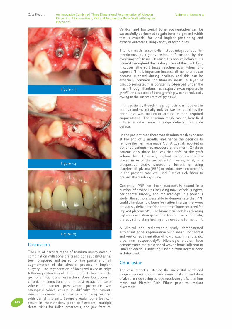
Volume 2, Number 4
Figure - 13
Figure -14
Figure -15
140
Case Report An Innovative Combined Three Dimensional Augmentation of Alveolar Ridge sing Titanium Mesh, PRF and Autogenous Bone Graft with Implant Placement.
Vertical and horizontal bone augmentation can be successfully performed to gain bone height and width that is essential for ideal implant positioning and esthetic outcomes using variety of techniques.
Titanium mesh has some distinct advantages as a barrier membrane. Its rigidity resists deformation by the overlying soft tissue. Because it is non-resorbable it is present throughout the healing phase of the graft. Last, it causes little soft tissue reaction even when it is exposed. This is important because all membranes can become exposed during healing, and this can be especially common for titanium mesh. A layer of pseudo periosteum is constantly observed under the mesh. Though titanium mesh exposure was reported in 51.11%, the success of bone grafting was not reduced , owing to the success rate of 97.72%9.
In this patient , though the prognosis was hopeless in both 21 and 11, initially only 21 was extracted, as the bone loss was maximum around 21 and required augmentation. The titanium mesh can be beneficial only in isolated areas of ridge defects than wide defects.
In the present case there was titanium mesh exposure at the end of 4 months and hence the decision to remove the mesh was made. Von Arx, et al. reported 10 out of 20 patients had exposure of the mesh. Of those patients only three had less than 10% of the graft volume lost. However, implants were successfully placed in 19 of the 20 patients5. Torres, et al, in a prospective study, showed a benefit of using platelet-rich plasma (PRP) to reduce mesh exposure10. In the present case we used Platelet rich fibrin to prevent the mesh exposure.
Currently, PRF has been successfully tested in a number of procedures including maxillofacial surgery, periodontal surgery, and implantology. In a previous study, the authors were able to demonstrate that PRF could stimulate new bone formation in areas that were previously deficient of the amount of bone required for implant placement11. The biomaterial acts by releasing high-concentration growth factors to the wound site, thereby stimulating healing and new bone formation12.
A clinical and radiographic study demonstrated significant bone regeneration with mean horizontal and vertical augmentation of 3.71± 1.24mm and 4.16± 0.59 mm respectively13. Histologic studies have demonstrated the presence of woven bone adjacent to lamellar which is indistinguishable from normal bone architecture3.
Conclusion
The case report illustrated the successful combined surgical approach for three dimensional augmentation of alveolar ridge using autogenous bone graft, titanium mesh and Platelet Rich Fibrin prior to implant placement.
Discussion
The use of barriers made of titanium macro-mesh in combination with bone grafts and bone substitutes has been proposed and tested for the partial and full augmentation of the alveolar process in implant surgery. The regeneration of localized alveolar ridge following extraction of chronic defects has been the goal of clinicians and researchers. Bone loss occurs in chronic inflammation, and in post extraction cases where no socket preservation procedure was attempted which results in difficulty for patients wearing a conventional prosthesis or being restored with dental implants. Severe alveolar bone loss can result in malnutrition, poor self-esteem, multiple dental visits for failed prosthesis, and jaw fracture.
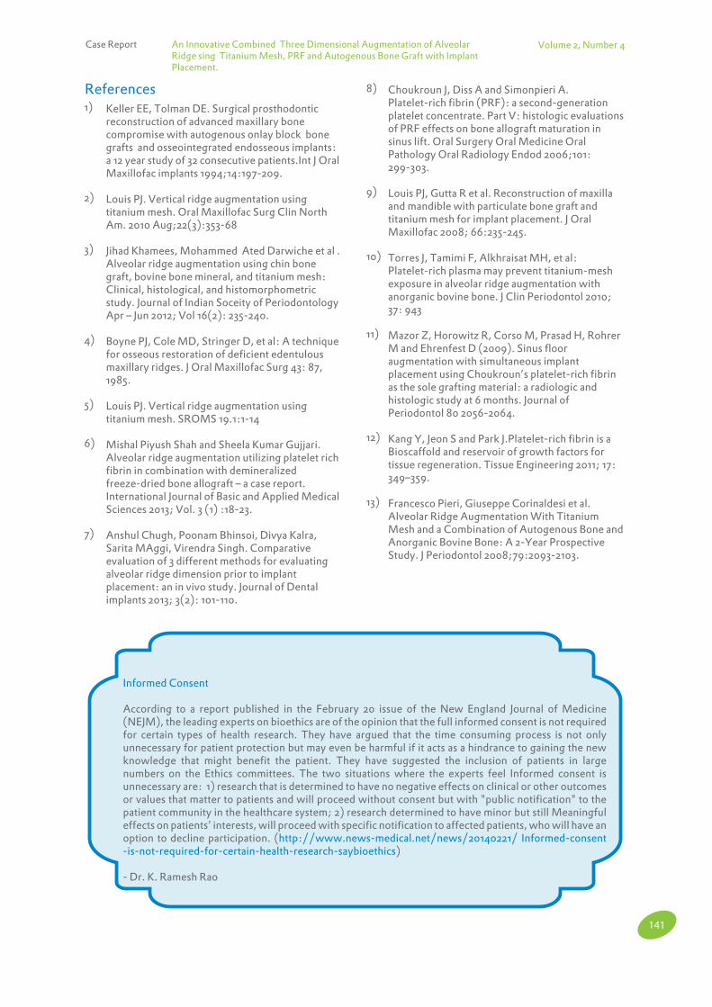
Volume 2, Number 4
References1)
2)
3)
4)
5)
6)
7)
Keller EE, Tolman DE. Surgical prosthodontic reconstruction of advanced maxillary bone compromise with autogenous onlay block bone grafts and osseointegrated endosseous implants: a 12 year study of 32 consecutive patients.Int J Oral Maxillofac implants 1994;14:197-209.
Louis PJ. Vertical ridge augmentation using titanium mesh. Oral Maxillofac Surg Clin North Am. 2010 Aug;22(3):353-68
Jihad Khamees, Mohammed Ated Darwiche et al . Alveolar ridge augmentation using chin bone graft, bovine bone mineral, and titanium mesh: Clinical, histological, and histomorphometric study. Journal of Indian Soceity of Periodontology Apr – Jun 2012; Vol 16(2): 235-240.
Boyne PJ, Cole MD, Stringer D, et al: A technique for osseous restoration of deficient edentulous maxillary ridges. J Oral Maxillofac Surg 43: 87, 1985.
Louis PJ. Vertical ridge augmentation using titanium mesh. SROMS 19.1:1-14
Mishal Piyush Shah and Sheela Kumar Gujjari. Alveolar ridge augmentation utilizing platelet rich fibrin in combination with demineralized freeze-dried bone allograft – a case report. International Journal of Basic and Applied Medical Sciences 2013; Vol. 3 (1) :18-23.
Anshul Chugh, Poonam Bhinsoi, Divya Kalra, Sarita MAggi, Virendra Singh. Comparative evaluation of 3 different methods for evaluating alveolar ridge dimension prior to implant placement: an in vivo study. Journal of Dental implants 2013; 3(2): 101-110.
8)
9)
10)
11)
12)
13)
Choukroun J, Diss A and Simonpieri A. Platelet-rich fibrin (PRF): a second-generation platelet concentrate. Part V: histologic evaluations of PRF effects on bone allograft maturation in sinus lift. Oral Surgery Oral Medicine Oral Pathology Oral Radiology Endod 2006;101: 299-303.
Louis PJ, Gutta R et al. Reconstruction of maxilla and mandible with particulate bone graft and titanium mesh for implant placement. J Oral Maxillofac 2008; 66:235-245.
Torres J, Tamimi F, Alkhraisat MH, et al: Platelet-rich plasma may prevent titanium-mesh exposure in alveolar ridge augmentation with anorganic bovine bone. J Clin Periodontol 2010; 37: 943
Mazor Z, Horowitz R, Corso M, Prasad H, Rohrer M and Ehrenfest D (2009). Sinus floor augmentation with simultaneous implant placement using Choukroun’s platelet-rich fibrin as the sole grafting material: a radiologic and histologic study at 6 months. Journal of Periodontol 80 2056-2064.
Kang Y, Jeon S and Park J.Platelet-rich fibrin is a Bioscaffold and reservoir of growth factors for tissue regeneration. Tissue Engineering 2011; 17: 349–359.
Francesco Pieri, Giuseppe Corinaldesi et al. Alveolar Ridge Augmentation With Titanium Mesh and a Combination of Autogenous Bone and Anorganic Bovine Bone: A 2-Year Prospective Study. J Periodontol 2008;79:2093-2103.
Informed Consent
According to a report published in the February 20 issue of the New England Journal of Medicine (NEJM), the leading experts on bioethics are of the opinion that the full informed consent is not required for certain types of health research. They have argued that the time consuming process is not only unnecessary for patient protection but may even be harmful if it acts as a hindrance to gaining the new knowledge that might benefit the patient. They have suggested the inclusion of patients in large numbers on the Ethics committees. The two situations where the experts feel Informed consent is unnecessary are: 1) research that is determined to have no negative effects on clinical or other outcomes or values that matter to patients and will proceed without consent but with "public notification" to the patient community in the healthcare system; 2) research determined to have minor but still Meaningful effects on patients' interests, will proceed with specific notification to affected patients, who will have an option to decline participation. (http://www.news-medical.net/news/20140221/ Informed-consent -is-not-required-for-certain-health-research-saybioethics)
- Dr. K. Ramesh Rao
141
Case Report An Innovative Combined Three Dimensional Augmentation of Alveolar Ridge sing Titanium Mesh, PRF and Autogenous Bone Graft with Implant Placement.

Volume 2, Number 4 Chettinad Health City Medical Journal
HARVEY CUSHING (1869 – 1939)From the Pages of History
Dr. V.G.Ramesh, Professor and HOD, Dept. of Neuro Surgery, Chettinad Hospital and Research Institute, Chennai, India
Father of Modern Neurosurgery, a great medical scientist, a gifted author – all-in-one – is what Harvey Cushing can be described in a single sentence. He was an extraordinary human being who by his untiring work, established Neurosurgery as a separate specialty, brought new concepts in the practice of medicine in general. He authored enormous amount of literature, in an era when modern medicine was just establishing itself.
Harvey Williams Cushing was born in Cleveland, Ohio, on April 8th, 1869. He came from a family of physicians. Harvey Cushing went to school in Cleveland and then to Yale where he received his A.B. degree in 1891. At the Harvard Medical School he gained his A.M. and M.D. cum laude in 1895. From this time he served for one year in surgery in the Massachusetts General Hospital and then, in 1896, he was appointed to the house-staff in surgery under Dr. Halsted at the Johns Hopkins Hospital, Baltimore where he remained until 1900 as resident in surgery. He learned meticulous surgical technique from Halsted while modeling his intellectual pursuits after William Osler, who at that time was chairman of the Department of Medicine. In 1900, he went to Berne, Switzerland along with Kocher and Kronecker, began his work in experimental neurology. Later he was associated with Sherrington in Liverpool. On his return, at the age of 32, he was made Associate Professor of Surgery at Johns Hopkins Hospital, and at the hospital was placed in full charge of cases of surgery of the central nervous system. In 1911, he was appointed Surgeon-in-Chief at the Peter Bent Brigham Hospital in Boston. He became a Professor of
Chettinad Health City Medical Journal 2013; 2(4): 142
Surgery at the Harvard Medical School starting in 1912. During 1917-1919, he was director of U.S. Base Hospital attached to the British Expeditionary Force in France. In 1918, he was made Senior Consultant in Neurological surgery for the American Expeditionary Forces in Europe during World War I. He served in the U.S. Army Medical Corps, attaining the rank of Colonel. After the war he returned to Boston and continued until 1932. Next year, in 1933, he went to Yale as Sterling Professor of Neurology, a position which he held until 1937. Cushing died on October 7, 1939 in New Haven, Connecticut, from complications of a myocardial infarction. He was interred at Lake View Cemetery in Cleveland. Interestingly, an autopsy performed on Cushing revealed that his brain harbored a colloid cyst of the third ventricle.
Contributions to Medicine in general
As a medical student, he developed the first continuous record for recording pulse and respiration during surgery in 1895, which forms the basis for present day anesthesia records.
Cushing was involved in the introduction of X-ray technology into the clinical realm at Massachusetts General Hospital and Johns Hopkins Hospital after its discovery by Roentgen.
He did the first human experimentation with nerve block anesthesia for operation using cocaine.
He was the first to use blood pressure monitoring during surgery.
He studied typhoid disease and developed a pioneering approach to surgical management of perforations of the esophagus due to typhoid infection.
He showed that the gut can be sterilized by fasting and this has formed the basis for pre-operative bowel preparation.
He predicted the routine use of positive-pressure endotracheal anesthesia twenty years before it was used routinely.
He was the first to describe the tolerance of the heart to surgical manipulation, which was accepted only twenty years later.
1)
2)
3)
4)
5)
6)
7)
8)
Contributions to Neurosurgery
He established the safe surgical method of removing intracranial tumors with meticulous surgical technique.
He was the first to establish the basic techniques in Neurosurgery like finger pressure for haemostasis of scalp; waxing the bone edges; haemostatic clips; motor driven suction during surgery, etc. He brought down the surgical mortality after neurosurgical procedure from over 50% to less than 20%.
He invented the electrosurgical cautery along with Bovie.
He was the first to map human cerebral cortex with faradic stimulation in conscious patients.
He did the first surgery for acromegaly (1909).
He described Cushing’s law and Cushing’s triad in relation to raised intracranial pressure.
He described the C.P.Angle syndrome and advocated intracapsular excision of acoustic neurinoma.
He described the Cushing’s syndrome due to excess adrenal corticosteroids.
He described the Cushing’s ulcers – the gastric ulcers due to raised intracranial pressure.
He was the first to classify brain tumours along with Percival Bailey.
1)
2)
3)
4)
5)
6)
7)
8)
9)
10)
142
Contributions to Medical Literature: He has authored several medical books. He won the prestigious Pulitzer prize for his biography book “Life of Sir William Osler” in 1926.
References1. Wikipedia: Harvey Williams Cushing2. Biographical memoir of Harvey Cushing by W. G. Maccallum: National Academy of Sciences, 1940.

Volume 2, Number 4
143
Abu Backer Sulaiman See Srinija P, 65Agnishwar Girigoswami Nanomedicine: Promising Tools in Biomedical Sciences, 99Amritha See Anitha V, 110Anitha V The frequency of medically compromised patients visiting Dental College and Research Institute, Kancheepuram dt., A retrospective study (2011 - 2013), 110
See Shanmugam M, 96Anitha Elango Hand, Foot and Mouth Disease, 7Aneeshya Jose See Sasireka K, 48Annamalai See Venkateswaran S, 52Anoop Sreevalsan See Srinija P, 65
Anantharamakrishnan R See Senthil Kumar K, 11Antony J See Nathan R, 94 ArunThevaraja K See Gopalakrishnan Raman, 123Asha Benziger A Prospective Study On The Impact Of Radiofrequency Electromagnetic Waves (RF - EMWS) Emitted By Cell Phone During Call Attended Mode On The Semen Sample, 70
See Ranjani S, 133Arun Kumar R See Ruckmani A, 92Arun Kumar G Nobel Prize in Physiology? Medicine – 2003, 103Arunprasad G See Ramesh V, 19Bobby Kurian See Sasireka K, 48Dalim Kumar Baidya Pain relief in Neonates: No Doubt it’s Duty, 28Divya See Venkateswaran S, 53Ganavi R Variant of Pierre Robin Sequence Requiring Prolonged Tracheostomy, 63Ganesh N Treadmill testing – Where does it stand today?, 31Ganesan R See Ramanujam S, 17Gayathiri Devi SS The Correlation between endometrial Thickness and outcome of Pregnancy in an Assisted Reproductive Technology Programme – Retrospective Observational Study, 70Gopalakrishnan Raman Management of severe Traumatic Brain
Author Index
Injury in adults, 123Indumathi A Comparative Study of The Outcome of the Individualized GnRH Antagonist Stimulation Protocol (Early Initiation) with the fixed antagonist Protocol In IVF/ICSI – ET cycles, 70Jaishree See Anitha Elango, 7Jeya M Antimicrobial Resistance – an Overview, 80
Jothi Ramalingam S B See Ganavi R, 63
Joseph John See Prabhu S, 74Karthikeyan K V Solitary Giant Neurocysticercosis in a Child with Combined Immunodeficiency, 9
Dumb Bell Skull Base Meningioma, 60
See Gopalakrishnan Raman, 123Karunanithi R See Senthil Kumar K, 11 See Ramanujam S, 17Kurinji Pandiyan Epigenetics - A paradigm shift in Cancer Management, 33Mariano Does serum progesterone level on the day of Human Chorionic Gonadotropin administration affect Clinical Pregnancy rates in fresh Embryo Transfer cycles? : A Retrospective cohort study, 71Meenapriya B See Shanmugam M, 96 See Anitha V, 110 See Vinayak S Gowda, 137Mohanarangam T See Gopalakrishnan Raman, 123Namrata Sanjeevi Antioxidants in health and disease: Review of clinical trials, 113Nathan R Neonatal Cholera, 94Loganathan M See Ramanujam S, 17Padmapriya See Srinivasan, 14Pandiyan N Editorial, 1 Editorial, 27 Editorial, 73 Editorial, 107 See Prathima T, 108 See Ranjani S, 133Prabhu S Impact of Dental Caries and Dental Fluorosis on the Quality of Life of 12- year old Children in Tamil Nadu, India, 74Prathima T Definition of Oligozoospermia- A commentary, 108Priyadarshini G See Anitha V, 110
Chettinad Health City Medical Journal
Volume 2, Number 4Chettinad Health City Medical Journal
144
Rajasekaran D Tackling Antimicrobial Resistance in Tertiary Hospital, Antimicrobial Stewardship Programme (ASP), 88 Rajesh P See Anitha V, 110Raj Kumar V J To do or Not to do! 2Ramanujam S Management of Lingual Thyroid by Suprahyoid Approach, 17Ramesh V G See Karthikeyan K V, 9 A Case of Central Giant Cell Granuloma Involving the Maxillary Sinus, 19
Clinically Masquerading as a Malignant Neoplasm Cerebrospinal Fluid Dynamics Study: Applications in Clinical Practice, 40
See Karthikeyan K V, 60
See Gopalakrishnan Raman, 123
Harvey Cushing (1869 – 1939), 142Ramesh Raja A Pilot study : Immotile Spermatozoa in Semen sample – Resting or Immotile?, 71 Ramesh Rao Hippocrates and his Oath, 23 Ignaz Philipp Semmelweis, 69 Medical Fashion – A Brief Look at its Evolution, 105Ranjani S Leucocytospermia –Does It Mean Anything?, 71
Nutraceuticals in Sperm Abnormalities, 133Rathinasamy M See Anitha Elango, 7 See Ganavi R, 63Ruckmani A Non Microbial Drug Resistance, 85Saravanakumar See Venkateswaran S, 52Saravanan S See Prabhu S, 74Senthil Kumar K T- Cell Lymphoma Arising From Gluteal Muscle –A Rare Presentation, 11
Schwannoma of Intercostal nerve- An Uncommon Localization, 92Shilpa Jha See Anitha Elango, 7Shivakumar V See Sasireka K, 48 See Shanmugam M, 96 See Anitha V, 110Somasundaram See Srinija P, 65Souvik Maitra See Dalim Kumar Baidya, 28Sumi Thomas Chromosomal Aberrations in Couples with Recurrent Miscarriages – An Experience of 479 Couples at a Tertiary Care Centre in India, 72
Author Index
Suresh P See Anitha Elango, 7 See Nathan R, 94Syed Meraj Ahmed An Objective View of Problems in Rural Healthcare Infrastructure in India, 4Thayumanavan S See Srinivasan, 14 See Ganavi R, 63 See Nathan R, 94Thilaka Muthiah See Dalim Kumar Baidya, 28Umadevi See Anitha Elango, 7Vasantha N Subbiah See Srinija P, 65Venkateswaran P See Nathan R, 94Venkateswaran S Diagnosis & Management of Temporomandibular Joint Disorders - What the Medical and Dental practitioners should know, 52 Vinayak S Gowda An innovative combined three dimensional augmentation of alveolar ridge using Titanium Mesh, PRF and autogenous bone graft with implant placement, 137Vinayak Meti See Ruckmani A, 85Yamini See Venkateswaran S, 53Sasireka K Pigmented Lesions Of The Oral avity-Review And Differential Diagnosis, 48Shah Dupesh Khan Immune Mediated Male Infertility, 44Shanmugam M A Rare Combination of Aggressive Periodontitis with Multiple Impacted Supernumerary Teeth, 96
See Anitha V, 110Sriram K See Ramesh V, 19Srinija P Posterior Reversible Encephalopathy Syndrome, 65
Srinivasan K See Ganavi R, 63Srinivasan Aniridia with Sydney Crease, 14Shilpa Jha See Anitha Elango, 7Shivakumar V See Sasireka K, 48 See Shanmugam M, 96 See Anitha V, 110

Volume 2, Number 4Chettinad Health City Medical Journal
145
Addison’s disease, see pigmented lesionsALADIN, see trialsALADIN II, see trialsALADIN III, see trialsAlzheimer’s, 51,112Aniridia, 14Anthelminthics, see resistanceAnti asthmatics, see resistanceAntibodies Antisperm, 45Antibiotics Cycling strategies, 90Gene transfer Horizontal, 82 Vertical, 82 Mechanism, 81 Resistance- 80-83, Anthelminthics, 86 Anti asthmatics, 86 Anti cancer, 85 Anti emetics,86 Anti epileptics, 85,86,130 Anti hypertensive, 86 Antimicrobial 80,81,88 Acquired, 82 ESBL- 81,82 Insulin, 86MRSA- 81,85Multi Drug Resistant (MDR),85Non microbial, 85-87Total Drug Resistant (TDR), 85 Vancomycin Resistant Enterococci(VRE)- 81 Targets, 80Anti cancer, see resistanceAnti emetics, see resistanceAnti epileptics, see resistanceAnti hypertensive, see resistanceAntimicrobial Resistance- see resistance Restriction strategies, 89Antimicrobial Stewardship Programme Clinical Pharmacist, 89 Clinical Microbiologist, 89 Goals, 88 Hospital Administrators, 89 Infectious Disease Physician, 88 Members, 88Antioxidants Cancer, 113,114 Chronic Obstructive Pulmonary Disease (COPD), 115 Diabetes, 115 Male reproductive system, 134 Pre- eclampsia, 114,115ATBC, see trialsAzoospermia, 135Bacteria
Subject Index
Akkermansia muciniphilia, 3 Survival, 81Blueberries,59Bottle feeding, 94Brain Development, 143 Herniation, 125Caries Epidemiology, 74,75Impact- 74-78Onset- 74- 76 Prevention 74Cancer, 33,34,see antioxidantsCardiovascular disease, 110-112CARET, see trialsCataract, 14Cell phone, 70Cerebral Salt Wasting, 131Cerebrospinal fluid Dynamics, 40-42 Outflow resistance, 40,41 Bolus injection method, 41Constant pressure infusion, 41Katzman method, 40,41Phase contrast MRI, 41 Radio isotope dilution, 41CHAOS, see trialsChild Perception Questionnaire, 74,75Chocolates, 84Chromosomal aberrations, 72Chronic Obstructive Pulmonary Disease (COPD), see antioxidantsComputed tomography, see traumatic brain injuryConsent, 141Corticosteroids, 19CRASH, see trialsCushing Harvey, 142DEKAN, see trialsDelusion, 132Diabetes Insipidus, 130Mellitus, 59, 72,110-112, see antioxidantsDisorder Renal, 110-112 Systemic, 110-112DNA Aberrant DNA methylation, 34 Methyltransferase inhibitors, 35Economic strata, 94Enzyme EZH2, 43Epilepsy, 110-112Epigenetics Aberrations, 34 Components, 33 Therapy, 35,36Ethics, 98
Volume 2, Number 4Chettinad Health City Medical Journal
146
Exercise, 24Extended Spectrum Beta lactamases (ESBL), see resistanceFree radicals, 113,114,133,134Fluid resuscitation, see traumatic brain injuryFluorosis 74-76Food, 26Gene transfer, see antibioticsGluteal, 11GnRH Antagonist protocol, 70Graft Autogenous, 137,138Granuloma Giant cell, 19 Jaw bone, 19Hand foot and mouth disease, 7,8Health Mental, 6, 113-119Healthcare Roadblocks, 4,5 Rural- 4-6 Infrastructure, 4-6Hepatitis, 110-112HOPE, see trialsHippocrates Oath, 23Human Chorionic Gonadotropin, 71Ignaz Philipp Semmelweis, 69Immunodeficiency Combined, 9Infection Focal, 110-112 Prevalence, 110-112Infertility Male, 44,109,133 Immune mediated, 44,45Infrastructure, see healthcareIntra Cranial Pressure (ICP),see traumatic brain injuryInvitro Fertilization (IVF), 70Leucocytospermia, 71Linxian, see trialsLymphoma Non Hodgkin, 11 T- cell, 11,12Marijuana, 16Medical fashion, 105Melanoma,see pigmented lesionsMeningioma Dumb bell skull, 60Mesh Titanium, 137Methicillin Resistant Staphylococcus aureus (MRSA), see resistance Mycobacterium tuberculosis 80, Multi Drug Resistant (MDR), see resistanceNanomedicine Challenges, 101
Subject Index
Nanomedicine Challenges, 101Diagnostics, 100Therapeutics, 101Neonatal Cholera, 94,95Examination, 14Pain, 28,29Nerve Intercostal, 92 Peripheral, 92Nevi,see pigmented lesionsNeurocysticercosis Giant, 9NHS, see trialsNobel prize Medicine, 103 Physiology, 103Nutraceuticals, 133,134Obesity Subcellular stress, 16Obstructive Sleep apnoea, 106Oligoasthenozoospermia, 109,135Oligozoospermia Definition, 108 WHO manual, 108Oxidative stress, 113-117,133,134Pain, see neonatesPAP smear, 95Periodontitis, 96PHS, see trialsPPP, see trialsPierre Robin, 63Pigmented lesions Addison’s disease, 49 Drug induced, 49 Focal, 49 Melanoma, 51 Nevi, 51 Oral cavity, 48 Physiological, 48 Smoker’s melanosis, 49 Varix, 50Platelet Rich Fibrin, 137Pollution Noise, 68Posterior Reversible Encephalopathy Syndrome, 65,66Pre- eclampsia, see antioxidantsPregnancy Outcome, 70 Rate, 70,71Prevalence, see infectionProgesterone, 71Prostate Specific Antigen Limitation, 2 Range, 2
Volume 2, Number 4Chettinad Health City Medical Journal
147
Protocol Fixed, 70 Individualised, 70Pulmonary oedema, 130Reality testing, 132Renal disorder, see disorderResistance, see antibioticsRespiratory disease, 110-112Ridge augmentation, 137, 138Schwannoma, 92,93Screening HPV, 95 Prostate cancer, 3SELECT, see trialsSemen, 70,108,109,134Simvastatin, 24Skin disease, 110-112Smoking, 102SPACE, see trialsSperm Abnormalities, 133 Antigen, 44 Count, 108 Immotile, 71Spermatogenesis, 133Sydney Crease, 14 Trial, see trialsSyndrome of Inappropriate ADH secretion (SIADH), 130Temperomandibular joint Disorders Differential diagnosis, 55 Symptoms, 84 Therapies, 57,58Thyroid Disease, 110-112 Lingual, 17,18 Suprahyoid approach, 17Tooth Multiple impacted, 96 Supernumerary, 96Total Drug Resistant (TDR),see antibiotics
Subject Index
Toxins, 143Tracheostomy, 63Traumatic Brain Injury Computed tomography Canadian criteria, 126 Marshall classification, 126 New Orleans criteria, 126 Fluid resuscitation, 126 Intra Cranial Pressure (ICP), 128 Management Anaesthetic management, 126 Emergency management, 125 Pre- hospital management, 124 Pathology, 123Treadmill test, 31,32Trials ALADIN, 117 ALADIN II, 117 ALADIN III, 117 ATBC, 116,117 CARET, 116,117 CHAOS, 117 CRASH, 130 DEKAN, 117 HOPE, 117 Linxian, 116 NHS, 117 PHS, 117 PPP, 117 SELECT, 116 SPACE, 117 SYDNEY, 117 WASC, 116 WHS, 117Vancomycin Resistant Enterococci (VRE), see antibioticsVibrio cholerae, 94Walking, 136WASC, see trialsWine, 32 WHO manual, see oligozoospermia, NutraceuticalWHS, see trials
Answer to : Diagnose the condition
The ECG shows wide QRS tachycardia with varying R-R interval with no definite P waves. There is left axis deviation with poor R wave progression in the anterior chest leads.
Diagnosis: Atrial fibrillation with fast ventricular rate with LAHB . The ECG is showing atrial fibrillation with interventricular conduction defect most probably in the left anterior fascicle. The left axis deviation with poor R wave progression and wide QRS denotes LAHB(left anterior hemi block) pattern. The irregularity and the absence of capture, fusion beats and AV dissociation differentiates this from Ventricular tachycardia.
- Dr. M.Chokkalingam, Consultant Cardiology, CSSH.
Volume 2, Number 4Chettinad Health City Medical Journal
148
Thank You reviewers!
The success of a journal depends on the authors and the quality of the papers they submit. Equally important is the immense contribution from the peer reviewers. They voluntarily, freely provide their time and expertise to critically evaluate the articles and contribute to their improvement with their valuable comments and suggestions. The following scholars have kindly reviewed the articles for our journal. Chettinad Health City Medical journal gratefully acknowledges their help and support for the journal.
1. Dr. Aravind, Reader, Department of Periodontics, Tagore Dental College, Chennai
2. Dr. S. Ahmed, Department of Medicine, Himalayan Institute of Medical Sciences
3. Dr. K. Chandrasekaran, Cardiologist, Prashanth Super Specialty Hospital
4. Dr. Deepika, Department of Periodontics, Meenakshiammal Dental College, Chennai
5. Dr. K. Ganapathy, Apollo Hospitals, Chennai
6. Dr. K. Hassan, Department of Ophthalmology, SRM University, Chennai
7. Dr. Jayanth G. Mehta, BHR University Hospital NHS trust, U.K
8. Dr. Jothi Kumar, Gleneagles Hospital, Singapore
9. Dr. Kanchana Devi, Assistant Professor, CSSH, Chennai
10. Dr. B. Krishnamurthy, Professor of Pediatrics, Mysore Medical College
11. Dr. Kumara Manikavel, Narayana Nethralaya
12. Dr. Latha Ravi, Prof of Pediatrics, Sri Ramachandra Medical College, Chennai
13. Dr. D.C. Mathangi, Professor, CHRI, Chennai
14. Prof. J. Mangayarkarasi, SRM University, Chennai
15. Dr. Meena Priya, Assistant Professor in Department of Periodontics, CDCRI, Chennai
16. Dr. N.S Muthiah, Prof of Pharmacology, Balaji Medical College
17. Dr. N. Muthukumar, Prof and H.O.D of Neurosurgery, Madurai Medical College, Tamil Nadu
18. Prof. P.S Muralidharan, Retired Director, Institute of Social Pediatrics, Stanley Medical College
19. Prof. M. S. Ramachandran, Retired Director, Institute of Medicine, Madras Medical College
20. Ms. Namrata Sanjeevi, Doctoral candidate, University of Texas, USA
21. Prof. Premkumar, Professor of Surgery, Karpaga Vinayaga Medical Institute.
22. Prof. Radha Bhai, Retired Director, I.O.G, Chennai
23. Dr. C. Raja, Assistant Professor, Thoothukudi Government Medical College, Tamil Nadu
24. Prof. V. Raji, Vice Chancellor, CARE, Chennai
25. Dr. Ramachandran, Professor of Pediatrics, Sri Ramachandra Medical College, Chennai
26. Dr. Ranganathan, Urologist, CSSH, Chennai
27. Dr. Shanmugam, Reader, Department of Periodontics, CDCRI, Chennai
28. Dr. Siva Kumar, Professor and H.O.D, Department of Periodontics, CDCRI, Chennai
29. Dr. K. R. Suresh Bapu Kandallu, Neurosurgeon, Apollo Hospitals, Chennai
30. Dr. Suresh Nair, Sri Chitra Institute of Medical Sciences & Technology, Thiruvananthapuram
31. Dr. Thilaka Jeeva, Consultant Anesthetist, CSSH, Chennai

33