Embed Size (px)
Citation preview
April - 2020
April - 2020
044 - 61434250044 - 61434230
INTRODUCTION:
CASE REPORT:
This report describes a case of rare tumour occurring in the right pinna of a 79 year old female patient. The Chondroid Syringoma is a rare and benign, mixed epithelial tumour, more frequently present as a cutaneous lesion in the head and neck region.
They share a resemblance to p l e o m o r p h i c a d e n o m a s o n histopathological examination. In 1961, Hirsch and Helwig introduced the term chondroid syringoma to describe a tumour which is characterized by presence of sweat glands lying within elements of cartilage like stroma. They can be both apocrine or eccrine in origin.
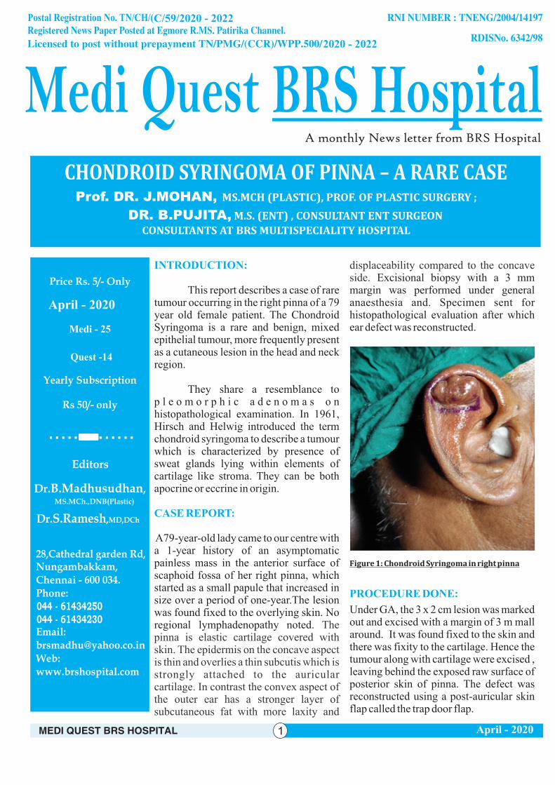
A79-year-old lady came to our centre with a 1-year history of an asymptomatic painless mass in the anterior surface of scaphoid fossa of her right pinna, which started as a small papule that increased in size over a period of one-year.The lesion was found fixed to the overlying skin. No regional lymphadenopathy noted. The pinna is elastic cartilage covered with skin. The epidermis on the concave aspect is thin and overlies a thin subcutis which is strongly attached to the auricular cartilage. In contrast the convex aspect of the outer ear has a stronger layer of subcutaneous fat with more laxity and
displaceability compared to the concave side. Excisional biopsy with a 3 mm margin was performed under general anaesthesia and. Specimen sent for histopathological evaluation after which ear defect was reconstructed.
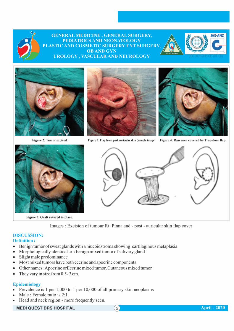
Under GA, the 3 x 2 cm lesion was marked out and excised with a margin of 3 m mall around. It was found fixed to the skin and there was fixity to the cartilage. Hence the tumour along with cartilage were excised , leaving behind the exposed raw surface of posterior skin of pinna. The defect was reconstructed using a post-auricular skin flap called the trap door flap.
Figure 1: Chondroid Syringoma in right pinna
PROCEDURE DONE:
CHONDROID SYRINGOMA OF PINNA – A RARE CASE
2020 - 2022
C/59/2020 - 2022
Prof. DR. J.MOHAN, MS.MCH (PLASTIC), PROF. OF PLASTIC SURGERY ;
DR. B.PUJITA, M.S. (ENT) , CONSULTANT ENT SURGEON
CONSULTANTS AT BRS MULTISPECIALITY HOSPITAL
April - 2020
DISCUSSION:Definition :
Epidemiology
· Benign tumor of sweat glands with a mucoidstroma showing cartilaginous metaplasia· Morphologically identical to / benign mixed tumor of salivary gland· Slight male predominance· Most mixed tumors have both eccrine and apocrine components· Other names :Apocrine orEccrine mixed tumor, Cutaneous mixed tumor· They vary in size from 0.5- 3 cm.
· Prevalence is 1 per 1,000 to 1 per 10,000 of all primary skin neoplasms · Male : Female ratio is 2:1· Head and neck region - more frequently seen.
Images : Excision of tumour Rt. Pinna and - post - auricular skin flap cover
Figure 2: Tumor excised Figure 3: Flap from post auricular skin (sample image)
Figure 5: Graft sutured in place.
Figure 4: Raw area covered by Trap door flap.
April - 2020
Prognostic factors
Clinical features
Radiology description
Treatment
Gross description
Microscopic (histologic) description
· Chondroidsyringoma can turn malignant and hence the pathological confirmation is a must. Surgery with margins of excision and post op irradiation might be required.
· Benign; rarely recurs· Most often seen in middle aged men· Most commonly in nose, followed by cheek
and upper lip
· Ultrasonography shows well encapsulated, hyperechoic mass with radiating, hypoechoicseptae· Color Doppler shows vascularization of intervening
septae· MRI shows peripheral high intensity signal with
central low intensity signal on T1 weighted imaging· T2 weighted imaging shows heterogeneously
mixed signal intensities
· Surgical excision · Recurrence is rare
· Nodular, circumscribed, nonulcerated with marked variation in size
· Cut surfaces are tan-white, often with grossly apparent chondroid component
· Slow growing, painless, firm, deep dermal to subcutaneous nodule
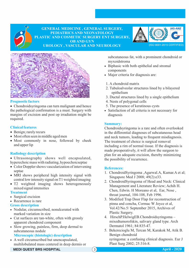
· A well circumscribed but unencapsulated, multilobulated mass centered in deep dermis or
subcutaneous fat, with a prominent chondroid or myxoidstroma
· Biphasic with both epithelial and stromal components
· Major criteria for diagnosis are:
1. A chondroid matrix2. Tubuloalveolar structures lined by a bilayered
epithelium3. Ductal structures lined by a single epithelium4. Nests of polygonal cells5. The presence of keratinous cysts · Satisfaction of all criteria is not necessary for
diagnosis
Chondroidsyringoma is a rare and often overlooked in the differential diagnoses of subcutaneous head and neck tumors, leading to frequent misdiagnosis. The treatment of choice is surgical removal including a rim of normal tissue. If the diagnosis is made preoperatively, it will allow the surgeon to plan for an adequate excision, thereby minimizing the possibility of recurrence.
1. ChondroidSyringoma ,Agarwal.A, Kumar.A et al; Singapore Med J 2008; 49(2):e33.
2. ChondroidSyringoma of Head and Neck: Clinical Management and Literature Review; Achih H. Chen, Edwin. H Moreano et al, Ear, Nose , throat journal, 104-108, Feb 1996.
3. Modified Trap Door Flap for reconstruction of pinna and concha, Cormac W Joyce et al, Vol.42/No.5/ September 2015, Archives of Plastic Surgery.
4. HirschP.HelwigEB.Chondroidsyringoma – mixedtumorofskin, salivary gland type. Arch Dermatol 1961; 84:835-47.
5. Bekerecioglu M, Tercan M, Karakok M, Atik B. Benign chondroid
syringoma: a confusing clinical diagnosis. Eur J Plast Surg 2002; 25:316-8.
Summary:
References:
April - 2020
No.28, Cathedral Garden Road, Nungambakkam, Chennai - 600 034.044 - 6143 4200 / 230 / 250 / 2823 5859
www.hospitalsinchennai.in : [email protected]
SPECIALITIES General MedicineENTPlastic SurgeryUrology
v 24x7 CASUALTY SERVICES v ICUv LAB
Paediatrics & NeonatologyObstetrics & GynecologyCosmetic SurgeryNephrology
General SurgeryAnaesthesiologyPaediatric SurgeryNeurology
v MARC-INFERTILITY CLINICv DENTAL CLINICv PHYSIOTHERAPY
CardiologyRadiologyVascular SurgeryOncology
2020 - 2022
Expertise Meets Care
2020 - 2022