Embed Size (px)
Citation preview
CILIATED PROTOZOA An illustrated guide to the species used as biological
indicators in freshwater biology
HARTMUT BICK
Professor and Head, Institute of Agricultural Zoology, University of Bonn, Federal Republic of Germany
(9) WORLD HEALTH ORGANIZATION
GENEVA
1972
© World Health Organization 1972
Publications of the World Health Organization enjoy copyright protection in accordance with the provisions of Protocol 2 of the Universal Copyright Conventoin. For rights of reproduction or translation of WHO publications, in part or in toto, application should be made to the Office of Publications and Translation, World Health Organization, Geneva, Switzerland. The World Health Organization welcomes such applications.
The designations employed and the presentation of the material in this publication do not imply the expression of any opinion whatsoever on the part of the DirectorGeneral of the World Health Organization concerning the legal status of any country or territory or of its authorities, or concerning the delimitation of its frontiers.
The mention of specific companies or of certain manufacturers ' products does not imply that they are endorsed or recommended by the World Health Organization in preference to others of a similar nature that are not mentioned. Errors and omissions excepted, the names of proprietary products are distinguished by initial capital letters.
PRINTED IN SWITZERLA ND
~
CONTENTS
Introduction ...........................
Synopsis of the most important taxa of non-parasitic ciliates
Key to families and genera .....................
Species descriptions
Acknowledgements
Selected bibliography
........................
· ..................... .
· ..................... .
Annex 1 : Glossary. . · .......... . · ......... . Annex 2: Key to abbreviations used in Fig. 1-94 · ......... . Index of genera and species . . . . . . . . . . · ......... .
Page
5
7
9
33
188
189
193
195
197

INTRODUCTION
Many species of ciliated protozoa are used as indicators for the ecological monitoring of water quality and they can also be used in ecological studies of aquatic habitats in which mosquitos and other vectors and intermediate hosts of disease organisms are breeding. However, taxonomic difficulties are frequently experienced by investigators and this illustrated guide has therefore been prepared as a service to freshwater biologists in the fields of vector control and sanitary engineering. The guide includes a taxonomic key to the most important families and genera of nonparasitic freshwater ciliates and 84 species descriptions dealing with morphological and ecological features of the ciliated protozoa employed in biological methods for assessing water quality. In order to allow broad comparisons to be made between the various species and genera, 135 species are illustrated.
When ciliated protozoa are to be identified, the following points should be remembered. A general feature of the Class Ciliatea is the presence of hairlike structures called cilia. The cilia, or compound ciliary structures, serve as organelles of locomotion or feeding or both. In contrast to the other classes of the Phylum Protozoa, two kinds of nuclei, the macronucleus and the micronucleus, are always present.
Species identifications of ciliates are based on the size and shape of the body, and on the structure and arrangement of certain organelles, such as the ciliation, macronucleus, contractile vacuole, and pellicle. Shape and size are best determined in living animals since specimens killed and stored in preserving fluids such as formol and ethanol cannot in general be identified at all. Most ciliates being rather small (size range: 10 p.m-l mm; average size about 20-200 p.m), it is always necessary to use a high-power microscope (magnification range: X 100-1000) for identifications. The use of phase-contrast microscopy is frequently helpful. In view of the rapid movements of many ciliates, it is useful to slow down the organisms by adding a small drop of methyl cellulose 1 to the biological material.
A quick method of determining the shape of nuclei is to kill and stain simultaneously with methyl-green-acetic acid,2 the stain being allowed to run under the cover glass. In order to determine the ciliation of spirotrich ciliates, a drop of a saturated aqueous solution of mercuric chloride may be added to the sample.
Material to be examined for ciliated protozoa may be bottom sediments, sludge, scrapings from stones or plants, and plankton samples (taken by means of a plankton net with a mesh smaller than 25 p.m). The sample may be concentrated by centrifugation. A very promising method for the evaluation of water quality is the investigation of the periphyton community (Aufwuchs) living on an artificial substrate such as a microscope slide. Slides are exposed in bodies of water for 4-8 weeks and then may be used directly for microscopic analysis (Sladeckova, 1960; Wilbert, 1969). The number of species of ciliated protozoa occurring in the periphyton community
1 Prepared by dissolving 109 of methyl cellulose in 90 ml of hot water . • Prepared by dissolving 0.1 g of methyl green dye in 99 ml of water and adding 1 ml of glacial
acetic acid.
-5-
6 INTRODUCTION
is very high; Wilbert (1969) and Nusch (1969) listed about 140 species of freeswimming and creeping ciliate and about 30 species of sessile ciliate, in the periphyton
of ponds and reservoirs. Ciliates should always be identified in a small drop of material placed on a
microscope slide and covered with a cover glass. In order to make individual counts of ciliates, a Sedgewick-Rafter cell or a Kolkwitz chamber (a plankton-counting chamber containing 0.5 ml offiuid) may be used; low-power objectives only (magnification approx. X 125) can be used with these chambers. When ciliates are counted in a chamber, their movements must be slowed down by adding methyl cellulose to the medium or stopped by adding a drop of aqueous Lugol's iodine solution.! When periphyton communities growing on artificially exposed microscope slides are to be counted the counts may be made directly from the slide, calculating the number
of individuals per cm2•
1 To prepare this reagent, dissolve 6 g of potassium iodide in 40 ml of water, dissolve 4 g of iodine crystals in the solution, and add water up to 100 ml.
SYNOPSIS OF THE MOST IMPORTANT TAXA OF NON-PARASITIC CILIATES
Nomenclature and classification as recommended by the Committee on Taxonomy and Taxonomic Problems of the Society of Protozoologists 1
Phylum PROTOZOA
Class CIL/ATEA
Subclass HOLOTRICHIA
Order GYMNOSTOMATIDA
Suborder RHABDOPHORINA (= PROSTOMATINA + PLEUROSTOMATINA)
Family COLEPIDAE Family ENCHELYIDAE Family AMPHILEPTIDAE Family ACTINOBOLINIDAE 2
Family DIDINIDAE Family TRACHELIIDAE Family LOXODIDAE 2
Family SPATHIDIIDAE 2
Family METACYSTIDAE 2
Suborder CYRTOPHORINA (= HYPOSTOMATINA)
Family DYSTERIIDAE 2
Family CHLAMYDODONTIDAE (= CHILODONELLIDAE)
Family NASSULIDAE 2
Order TRICHOSTOMATIDA
Family COLPODIDAE Family MICROTHORACIDAE 2
Family PLAGIOPYLIDAE Family TRIMYEMIDAE Family MARYNIDAE 2
Order HYMENOSTOMATIDA
Suborder TETRAHYMENINA
Unassigned tetrahymenine hymenostomes sensu Corliss (1961)
Family OPHRYOGLENIDAE 2
Family COHNILEMBIDAE (= LEMBIDAE) Family TETRAHYMENIDAE
Suborder PENICULINA
Family PARAMECIIDAE Family CINETOCHILIDAE Family UROCENTRIDAE Family FRONTONIIDAE
Suborder PLEURONEMATINA
Family PLEURONEMATIDAE (= CYCLIDIIDAE)
Subclass PERITRICHIA
Order PERITRICHIDA
Suborder SESSILINA
Family OPHRYDIIDAE Family VORTICELLIDAE Family EPISTYLIDAE Family V AGINICOLIDAE Family LAGENOPHRYIDAE 2
Family SCYPHIIDAE 2
Family ASTYLOZOIDAE 2
Suborder MOBILINA
Family URCEOLARIIDAE 2
1 Honigberg et aI. (1964); see also Corliss (1961). , Family is included in the key (p. 10) but no species descriptions are given.
-7-
8 NON-PARASITIC CILIATES
Subclass SUCTORIA
Family PODOPHRYIDAE Family DENDROSOMATIDAE Family DISCOPHRYIDAE
Subclass SPIROTRICHIA
Order HETEROTRICHIDA
Family BURSARIIDAE 1
Family STENTORIDAE Family GYROCORYTHIDAE (= METOPIDAE
= CAENOMORPHIDAE) Family SPIROSTOMATIDAE Family CONDYLOSTOMATIDAE 1
Family FOLLICULINIDAE 1
Order OLIGOTRICHIDA
Family HALTERIIDAE Family STROBILIDIIDAE 1
Order TINTINNIDA
Family TINTINNIDIIDAE
Order ODONTOSTOMATIDA (= CTENOSTOMATIDA)
Family DISCOMORPHELLIDAE (= DISCOMORPHIDAE)
Family EPALXELLIDAE (= EPALXIDAE)
Order HYPOTRICHIDA
Family ASPIDISCIDAE Family EUPLOTIDAE Family OXYTRICHIDAE
1 Family is included in the key (p. 10) but no species descriptions are given.
----------------------............
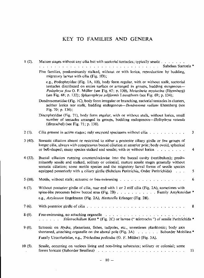
KEY TO FAMILIES AND GENERA
The key offered below is basically dichotomous and consists of a question and a counterquestion; the questions are numbered 1, 2, 3, etc., and the appropriate counter-questions are placed in parentheses: (2), 2 (1), 3 (40), etc. Each question leads either to a new pair of contrasting features or to the taxon sought. Each terminal point of the key is indicated by an asterisk (*).
This key is designed for use with live non-parasitic ciliates. Nearly all species mentioned are illustrated either in the key itself or in the individual species characterizations. In these illustrations, simple somatic ciliation is frequently represented by dots only. The scale line on the figures is equivalent to 10 /Lm unless otherwise indicated. The terms right and left refer to the organism itself and not to the drawing.
Although the key was designed primarily for the identification of ciliated protozoa used in ecological monitoring of water quality, it was necessary to include additional species in order to allow broad comparisons to be made between the various taxa. Those using the key should remember that about 5 000 species of ciliated protozoa have been described and where the identification of any specimen is in doubt, more complete monographs (e.g., Kahl, 1930-35) should be consulted.
Useful keys to the genera of ciliated protozoa are found in Noland (1959) and Matthes & Wenzel (1966). Full descriptions of many genera and species are included in Kudo (1966).
-9-
KEY TO FAMILIES AND GENERA
1 (2). Mature stages without any cilia but with suctorial tentacles; typically sessile. . . . . . . . . . . . . . . . . . . . . . . . . . . . . . . . . . . . . .. Subclass Suctoria *
2 (1).
Five families, predominantly stalked, without or with lorica, reproduction by budding, migratory larvae with cilia (Fig. lD);
e.g., Podophryidae (Fig. lA, lB), body form regular, with or without stalk, suctorial tentacles distributed on entire surface or arranged in groups, budding exogenousPodophrya fixa O. F. MUller (see Fig. 67; p. 130), Metacineta mystacina (Ehrenberg) (see Fig. 68; p. 132); Sphaerophrya solijormis Lauterborn (see Fig. 69; p. 134);
Dendrosomatidae (Fig. 1 C), body form irregular or branching, suctorial tentacles in clusters, neither lorica nor stalk, budding endogenous-Dendrosoma radians Ehrenberg (see Fig. 70; p. 136);
Discophryidae (Fig. 71), body form regular, with or without stalk, without lorica, small number of tentacles arranged in groups, budding endogenous-Heliophrya rotunda (Hentschel) (see Fig. 71; p. 138).
Cilia present in active stages; only encysted specimens without cilia
3 (40). Somatic ciliation absent or restricted to either a posterior ciliary girdle or few groups of longer cilia, always with conspicuous buccal ciliation at anterior pole; body ovoid, spherical
3
or bell-shaped; many species stalked and sessile; with or without lorica . . . . . . .. 4
4 (33). Buccal ciliation running counterclockwise into the buccal cavity (vestibulum); predominantly sessile and stalked, solitary or colonial, mature sessile stages generally without somatic ciliation; some motile species and the migratory larval forms of sessile species equipped posteriorly with a ciliary girdle (Subclass Peritrichia, Order Peritrichida) 5
5 (10). Motile, without stalk; ectozoic or free-swimming . . . . . . . . . . . . . . . 6
6 (7). Without posterior girdle of cilia, rear end with 1 or 2 stiff cilia (Fig. 2A), sometimes with spine-like processes below buccal area (Fig. 2B). . . . . . . . . . . Family Astylozoidae * e.g., Astylozoon Engelmann (Fig. 2A), Hastatella Erlanger (Fig. 2B).
7 (6). With posterior girdle of cilia. . . . . 8
8 (9). Free-swimming, no attaching organelle . . . . .. Telotrochidium Kent * (Fig. 2C) or larvae (" telotrochs ") of sessile Peritrichida *
9 (8). Ectozoic on Hydra, planarians, fishes, tadpoles, etc., sometimes planktonic; body axis shortened, attaching organelle on the aboral pole (Fig. 3A) . . . . . . Suborder Mobilina * Family Urceolariidae, e.g., Trichodina pediculus (0. F. Muller) (Fig. 3A).
10 (5). Sessile, occurring on various living and non-living substrates; solitary or colonial; some forms loricate (Suborder Sessilina) . . . . . . . . . . . . . . . . . .. 11
10 -
KEY TO FAMILIES AND GENERA
Fig. 1. Suctoria, diagrammatic representations;
A, Podophrya fixa O. F. Muller; B, Metacineta Butschli;
B
o
C, Dendrosoma Ehrenberg;
D, migratory larva of Trichophrya Claparede & Lachmann (Dendrosomatidae).
c
1\\\\\\\\1111/11 JIII!!1 I
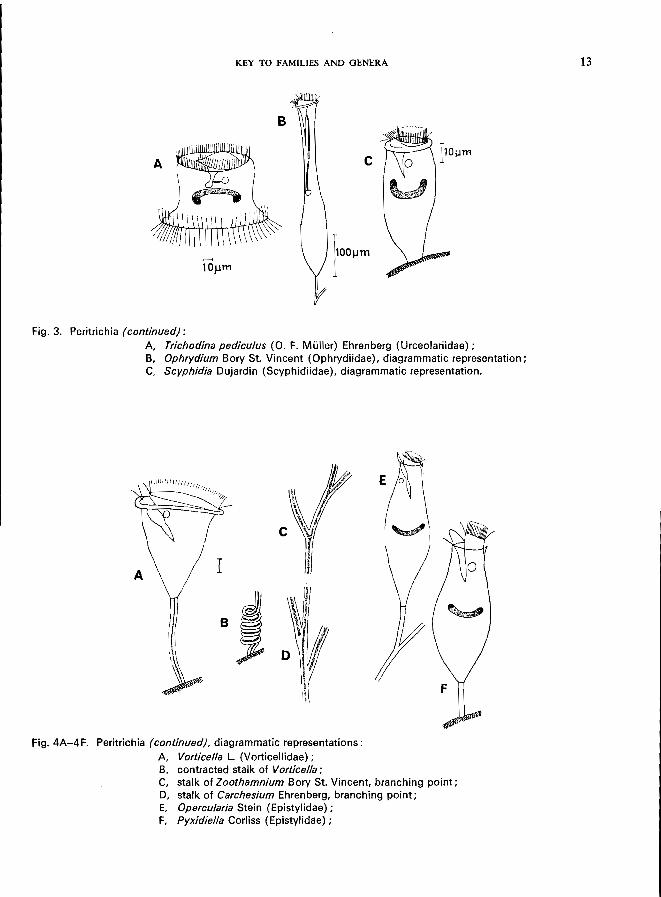
Fig. 2. Peritrichia, diagrammatic representations:
A, Astylozoon Engelmann (Astylozoidae); B, Hastatella Erlanger (Astylozoidae) ; C, Telotrochidium Kent (Epistylidae).
11
12 KEY TO FAMILIES AND GENERA
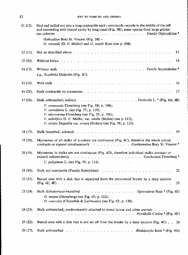
11 (12). Oral end pulled out into a long contractile neck; contractile vacuole in the middle of the cell and connecting with buccal cavity by long canal (Fig. 3B); some species form large gelatin-ous colonies . . . . . . . . . . . . . . . . . . . . . . . Family Ophrydiidae *
Ophrydium Bory St. Vincent (Fig. 3B)-O. versatile (0. F. MUller) and O. sessile Kent (see p. 104).
12 (11). Not as described above ........................... 13
14 13 (30). Without lorica ..................
Without stalk . . . . . . . . .
e.g., Scyphidia Dujardin (Fig. 3C).
. . . . . . . . . . . . . . . Family Scyphidiidae * 14 (15).
15 (14). With stalk. . . . . . . . . ........................ 16
16 (21). Stalk contractile by myonemes ........... 17
17 (18). Stalk unbranched; solitary. . . Vorticella L. * (Fig. 4A, 4B)
18 (17).
V. campanula Ehrenberg (see Fig. 5B; p. 108); V. convallaria L. (see Fig. 57; p. 110); V. microstoma Ehrenberg (see Fig. 55; p. 106); V. nebulifera O. F. MUller, var. similis (Stokes) (see p. 112); V. striata Dujardin var. octava (Stokes) (see Fig. 58; p. 113).
Stalk branched; colonial. . . . . . . . . . . . . . . . . . ........... 19
19 (20). Myonemes of all stalks of a colony are continuous (Fig. 4C), therefore the whole colony contracts or expand simultaneously ........... Zoothamnium Bory St. Vincent *
20 (19). Myonemes in stalks are not continuous (Fig. 4D), therefore individual stalks contract or expand independently . . . . . . . . . . . . . . . . . Carchesium Ehrenberg *
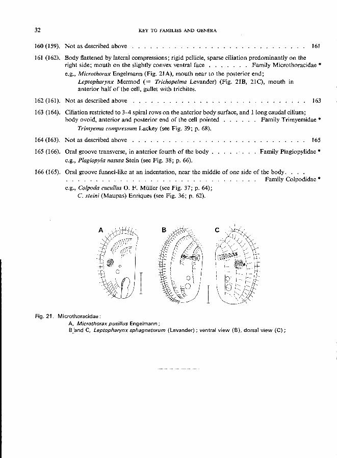
C. polypinum L. (see Fig. 59; p. 114).
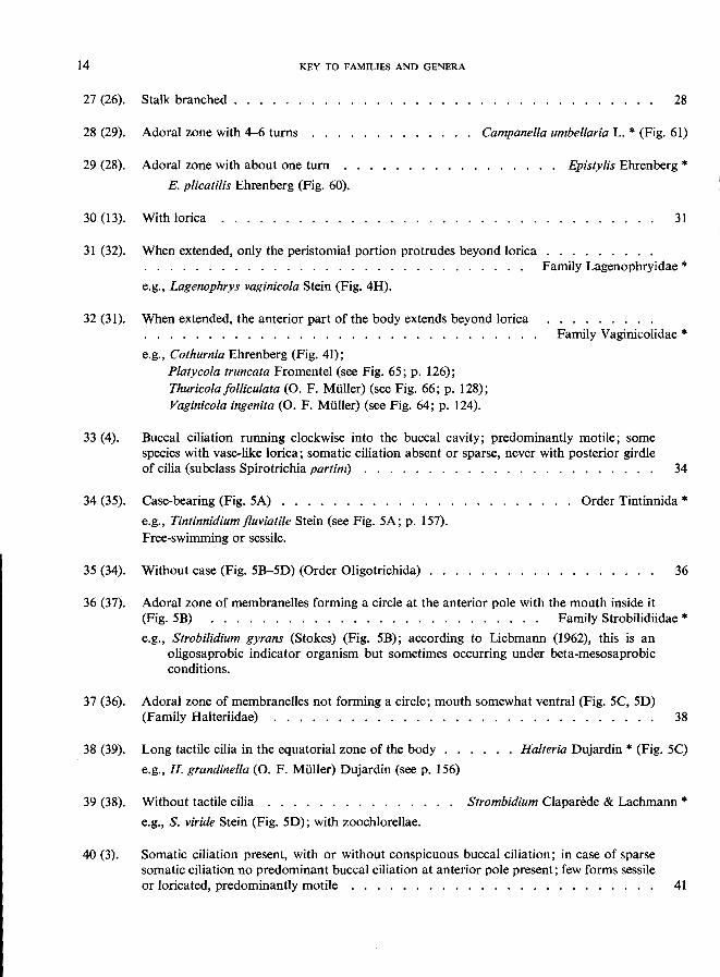
21 (16). Stalk not contractile (Family Epistylidae) 22 .....................
22 (25). Buccal area with a disk that is separated from the peristomial border by a deep incision (Fig. 4E, 4F). . . . . . . . . . . . . . . . .. 23
23 (24). Stalk dichotomous-branched Opercularia Stein * (Fig. 4E)
O. nutans (Ehrenberg) (see Fig. 63; p. 122); O. coarctata (Clapari!de & Lachmann) (see Fig. 62; p. 120).
24 (23). Stalk unbranched; predominantly attached to insect larvae and other animals . . ... . . . . . . . . . . . . . . . . . . . . . . . . . . . . . pyxidiella Corliss * (Fig. 4F)
25 (22). Buccal area with a disk that is not set off from the border by a deep incision (Fig. 4G). . 26
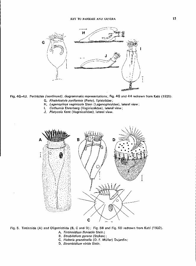
26 (27). Stalk unbranched . . . . . . . . . . . . . . . . . . . . Rhabdostyla Kent * (Fig. 4G)
KEY TO FAMILIES AND GENERA
B
c
Fig. 3. Peritrichia (continued):
A, Trichodina pediculus (0. F. Muller) Ehrenberg (Urceolariidae); B, Ophrydium Bory St. Vincent (Ophrydiidae), diagrammatic representation; C, Scyphidia Dujardin (Scyphidiidae), diagrammatic representation.
Fig.4A-4F. Peritrichia (continued), diagrammatic representations:
A, Vorticella L. (Vorticellidae); B, contracted stalk of Vorticella; C, stalk of Zoothamnium Bory St. Vincent, branching point; D, stalk of Carchesium Ehrenberg, branching point; E, Opercularia Stein (Epistylidae); F, Pyxidiella Corliss (Epistylidae);
13
14 KEY TO FAMILIES AND GENERA
28 27 (26). Stalk branched . . . . . .
..............
28 (29). Adoral zone with 4-6 turns Campanella umbellaria L. '" (Fig. 61)
29 (28). Adoral zone with about one turn . . . . . . . . . . . . . . . . Epistylis Ehrenberg '"
E. plicatilis Ehrenberg (Fig. 60).
31 30 (13). With lorica .........................
31 (32). When extended, only the peristomial portion protrudes beyond lorica . . . . . . . . . . . . . . . . . . . . . . . . . .... , Family Lagenophryidae '"
e.g., Lagenophrys vaginicola Stein (Fig. 4H).
32 (31). When extended, the anterior part of the body extends beyond lorica ........ Family Vaginicolidae '"
33 (4).
34 (3S).
3S (34).
............................... e.g., Cothurnia Ehrenberg (Fig. 41);
Platycola truncata Fromentel (see Fig. 6S; p. 126); Thuricolafolliculata (0. F. Muller) (see Fig. 66; p. 128); Vaginicola ingenita (0. F. MUller) (see Fig. 64; p. 124).
Buccal ciliation running clockwise into the buccal cavity; predominantly motile; some species with vase-like lorica; somatic ciliation absent or sparse, never with posterior girdle of cilia (subclass Spirotrichia partim) . . . . . . . . . . . . . . . . . . . ., 34
Case-bearing (Fig. SA) . . . . . . e.g., Tintinnidium fluviatile Stein (see Fig. SA; p. IS7).
Free-swimming or sessile.
. ........ Order Tintinnida '"
Without case (Fig. SB-SO) (Order Oligotrichida) .................. 36
36 (37). Adoral zone of membranelles forming a circle at the anterior pole with the mouth inside it (Fig. SB) .......................... Family Strobilidiidae '"
e.g., Strobilidium gyrans (Stokes) (Fig. SB); according to Liebmann (1962), this is an oligosaprobic indicator organism but sometimes occurring under beta-mesosaprobic
conditions.
37 (36). Adoral zone of membranelles not forming a circle; mouth somewhat ventral (Fig. SC, SO) (Family Halteriidae) . . . . . . . . . . . . . . . . . . . . . . ., 38
38 (39). Long tactile cilia in the equatorial zone of the body Halteria Oujardin '" (Fig. SC)
e.g., H. grandinella (0. F. MUller) Oujardin (see p. IS6)
39 (38). Without tactile cilia . . . . . . . . . . . . . Strombidium Claparede & Lachmann '"
40 (3).
e.g., S. viride Stein (Fig. SO); with zoochlorellae.
Somatic ciliation present, with or without conspicuous buccal ciliation; in case of sparse somatic ciliation no predominant buccal ciliation at anterior pole present; few forms sessile or loricated, predominantly motile ....................... , 41
KEY TO FAMILIES AND GENERA
G
I
"I I I X
Fig. 4G-4J. Peritrichia (continued), diagrammatic representations; Fig. 4G and 4H redrawn from Kahl (1935). G, Rhabdostyla pyriformis (Perty), Epistylidae; H, Lagenophrys vaginicola Stein (Lagenophryidae), lateral view; I. Cothurnia Ehrenberg (Vaginicolidae), lateral view; J, Platycola Kent (Vaginicolidae), lateral view.
Fig. 5. Tintinnida (A) and Oligotrichida (8, C and 0); Fig. 58 and Fig. 50 redrawn from Kahl (1932). A. Tintinnidium fluviatile Stein; 8, Strobilidium gyrans (Stokes); C, Halteria grandinel/a (0. F. Muller) Oujardin; 0, Strombidium viride Stein.
15
16 KEY TO FAMILIES AND GENERA
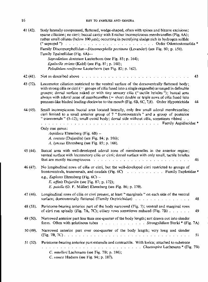
41 (42). Body laterally compressed, flattened, wedge-shaped, often with spines and bizarre excisions; sparse ciliation; no cirri; buccal cavity with 8 rather inconspicuous membranelles (Fig. 6A); rather small ciliates (below 100 /Lm); occurring in putrefying sludge rich in hydrogen sulfide
42 (41).
43 (72).
(" sapropel") . . . . . . . . . . . . . . . . . . . . . . . . Order Odontostomatida * Family Discomorphellidae-Discomorphella pectinata (Levander) (see Fig. 80; p. 158). Family Epalxellidae (Fig. 6A)-
Saprodinium den tatum Lauterborn (see Fig. 83; p. 164); Epalxella striata (Kahl) (see Fig. 81; p. 160); Pelodinium remforme Lauterborn (see Fig. 82; p. 162).
Not as described above . . . . . . . . . . . . . . .
Locomotor ciliation restricted to the ventral surface of the dorsoventrally flattened body;
43
with strong cilia or cirri (= groups of cilia fused into a single organelle) arranged in definable' groups; dorsal surface naked or with tiny sensory cilia (" tactile bristles "); buccal area always with adoral zone of membranelles (= short double or triple rows of cilia fused into pennant-like blades) leading clockwise to the mouth (Fig. 6B, 6C, 7,8). Order Hypotrichida 44
44 (45). Small inconspicuous buccal area located laterally, only few small adoral membranelles; cirri limited to a small anterior group of 7 "frontoventrals " and a group of posterior " transversals" (5-12); small ovoid body; dorsal side without cilia, sometimes ribbed .. . . . . . . . . . . . . . . . . . . . . . . . . . . . . . . . . Family Aspidiscidae * Only one genus:
Aspidisca Ehrenberg (Fig. 6B)-A. costata (Dujardin) (see Fig. 84; p. 166); A. lynceus Ehrenberg (see Fig. 85; p. 168).
45 (44). Buccal area with well-developed adoral zone of membranelles in the anterior region; ventral surface with locomotory cilia or cirri; dorsal surface with only small, tactile bristles that are mostly inconspicuous . . . . . . . . . . . . . . . . . . . . . . . . .. 46
46 (47). No longitudinal rows of cilia or cirri, but the well-developed cirri restricted to groups of frontoventrals, transversals, and caudals (Fig. 6C) . . . . . . . . . . Family Euplotidae * e.g., Euplotes Ehrenberg (Fig. 6C)-
E. affinis Dujardin (see Fig. 87; p. 172); E. patella (0. F. Muller) Ehrenberg (see Fig. 86; p. 170).
47 (46). Longitudinal rows of cilia or cirri present, at least" marginals " on each side of the ventral surface; dorsoventrally flattened (Family Oxytrichidae) . . . . . . . . . . . . . .. 48
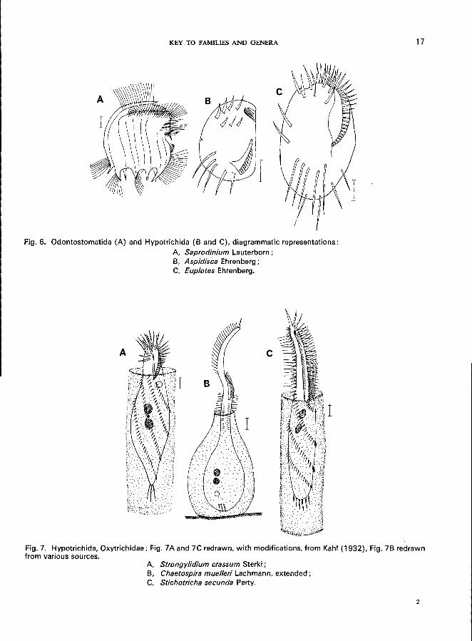
48 (53). Peristome-bearing anterior part of the body narrowed (Fig. 7); ventral and marginal rows of cirri run spirally (Fig. 7A, 7C); ciliary rows sometimes reduced (Fig. 7B) . . . . .. 49
49 (50). Narrowed anterior part less than one-quarter of the body length; not drawn out into slender form. Often with gelatinous tubes . . . .. . .... Strongylidium Sterki * (Fig. 7A)
50 (49). Narrowed anterior part over one-quarter of the body length; very long and slender (Fig. 7B, 7C). . . . . . . . . . . . . . . . . . . . . . . . . . . . . . . . .. 51
51 (52). Peristome-bearing anterior part extensile and contractile. With lorica; attached to substrate . . . . . . . . . . . . . . . . . . . . . . Chaetospira Lachmann * (Fig. 7B) C. muelleri Lachmann (see Fig. 7B; p. 186); C. remex Hudson (see Fig. 94; p. 187).
KEY TO FAMILIES AND GENERA
Fig. 6. Odontostomatida (A) and Hypotrichida (B and C), diagrammatic representations:
A, Saprodinium Lauterborn; B, Aspidisca Ehrenberg; C, Euplotes Ehrenberg.
c
I
Fig. 7. Hypotrichida, Oxytrichidae; Fig. 7A and 7C redrawn, with modifications, from Kahl (1932), Fig. 7B redrawn from various sources.
A, Strongylidium crassum Sterki; B, Chaetospira muelleri Lachmann, extended; C, Stichotricha secunda Perty.
2
17
18 KEY TO FAMILIES AND GENERA
52 (51). Peristome-bearing part not contractile; sometimes in mucilaginous tubes, which may be branched. Posterior part of the body with or without cirri . . . Stichotricha Perty * (Fig. 7C)
53 (48). Peristome-bearing zone not narrowed; ventral and marginal rows of cirri not running spirally . . . . . . . . . . . . . . . . . . . 54
54 (57). Elongated body drawn out into a tail-like portion 55
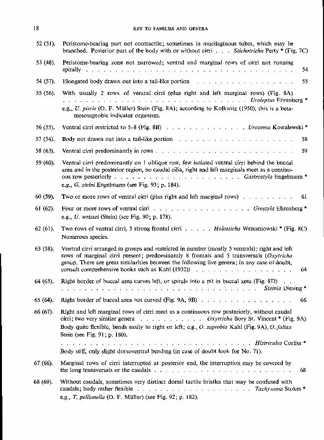
55 (56). With usually 2 rows of ventral cirri (plus right and left marginal rows) (Fig. 8A) . . . . . . . . . . . . . . . . . . . . . . . . . . . . . . . . Uroleptus Ehrenberg * e.g., U. piscis (0. F. Muller) Stein (Fig. 8A); according to Kolkwitz (1950), this is a beta
mesosaprobic indicator organism.
56 (55). Ventral cirri restricted to 5-8 (Fig. 8B) Urosoma Kowalewski * 57 (54). Body not drawn out into a tail-like portion
58 (63). Ventral cirri predominantly in rows . . . .
59 (60). Ventral cirri predominantly on 1 oblique row, few isolated ventral cirri behind the buccal area and in the posterior region, no caudal cilia, right and left marginals meet as a continu-
58
59
ous row posteriorly . . . . . . . . . . . . . . . . . . . . . . Gastrostyla Engelmann * e.g., G. steini Engelmann (see Fig. 93; p. 184).
60 (59).
61 (62).
Two or more rows of ventral cirri (plus right and left marginal rows)
Four or more rows of ventral cirri . . . . e.g., U. weissei (Stein) (see Fig. 90; p. 178).
61
Urostyla Ehrenberg *
62 (61). Two rows of ventral cirri, 3 strong frontal cirri . . . . . Holosticha Wrzesniowski * (Fig. 8e) Numerous species.
63 (58). Ventral cirri arranged in groups and restricted in number (mostly 5 ventrals); right and left rows of marginal cirri present; predominantly 8 frontals and 5 transversals (Oxytricha group. There are great similarities between the following five genera; in any case of doubt, consult comprehensive books such as Kahl (1932» . . . . . . . . . . . . . . . 64
64 (65). Right border of buccal area curves left, or spirals into a pit in buccal area (Fig. 8D) . . . . . . . . . . . . . . . . . . . . . . . . Steinia Diesing *
65 (64). Right border of buccal area not curved (Fig. 9A, 9B) 66
66 (67). Right and left marginal rows of cirri meet as a continuous row posteriorly, without caudal cirri; two very similar genera . . . . . . . . . . . Oxytricha Bory St. Vincent * (Fig. 9A) Body quite flexible, bends easily to right or left; e.g., O. saprobia Kahl (Fig. 9A), O./allax Stein (see Fig. 91; p. 180). . . . . . . . . . . . . . . . . . . . . . . . . . . . . . . . . . Histriculus Corliss * Body stiff, only slight dorsoventral bending (in case of doubt look for No. 71).
67 (66). Marginal rows of cirri interrupted at posterior end, the interruption may be covered by the long transversals or the caudals . . . . . . . . . . . . . . . . . . . . . . .. 68
68 (69). Without caudals, sometimes very distinct dorsal tactile bristles that may be confused with caudals; body rather flexible . . . . . . . . . . . . . . . . . . . . Tachysoma Stokes * e.g., T. pellionella (0. F. Muller) (see Fig. 92; p. 182).
KEY TO FAMILIES AND GENERA 19
Fig. S. Hypotrichida, Oxytrichidae (continued); Fig. SC and SD redrawn, with some modifications, from Kahl (1932). A Uro/eptus piscis (0. F. Muller) Stein; B, Urosoma cienkowski Kowalewski; C, H%sticha a/givora Kahl; D, Steinia p/atystoma (Ehrenberg) Stein.
69 (68). With caudals ................................. 70
70 (71). Body not flexible; 3 conspicuous long caudals that show very often fringes at the end; body ovoid to reniform . . . . . . . . . . . . . . . . . . . . " Sty/onychia Ehrenberg * e.g., S. mytilus Ehrenberg (see Fig. 88; p. 174); S. putrina (Stokes) (see Fig. 89; p. 176).
71 (70). Body flexible, bends easily to right and left; 3 (or 4) rather smooth and sometimes inconspicuous caudals; body mostly long ellipsoid . . . . . . . . Opisthotricha Kent * (Fig. 9B)
72 (43). Body not dorsoventrally flattened, or in case of flattened body no cirri present, and buccal area without adoral zone of membranelles; body surface wholly or partly ciliated; with or without buccal ciliation . . . . . . . . . . . . . . . . . . . . . . . . . . . " 73
73 (74). Body medusoid with a long posterior spine; ciliation limited to the spiral peristome region, and to 1 or 2 dorsal rows of thick flexible cirri ................. .
.................. Caenomorpha Perty * (Order Heterotrichida)
C. medusula Perty (see Fig. 77; p. 150).
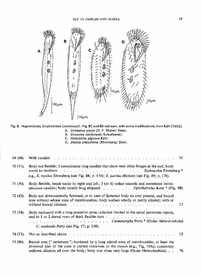
74 (73). Not as described above . . . . . . . . . .................... 75 75 (90). Buccal area (" peristome ") bordered by a long adoral zone of membranelles, at least the
proximal part of the zone is curved clockwise to the mouth (e.g., Fig. lOA); commonly uniform ciliation all over the body; body size often very large (Order Heterotrichida) " 76
20 KEY TO FAMILIES AND GENERA
76 (77). Anterior part of the body twisted; adoral zone of membranelles oblique or spiral, starting anterior left and turning to posterior right ..... Family Gyrocorythidae * (= Metopidae)
Brachonella spiralis (Smith) Jankowski (see Fig. 76; p. 148); Metopus es (0. F. Muller) Kahl (see Fig. 75; p. 146).
77 (76). Not as described above . . . . . . . . . . . . . . . 78
78 (79). Peristome deeply sunken into the anterior part of the body forming a funnel-like cavity (Fig. 9C) . . . . . . . . . . . . . . . . . . . . . . . .. . Family Bursariidae * e.g., Bursaridium pseudobursaria (Faure-Fremiet) Kahl (Fig. 9C).
79 (78). Peristome on surface level . . . . . . . . . . . . . . . . . 80
80 (83). Peristome linear, narrow, adoral zone of membranelles very long, though sometimes in-conspicuous; large, often elongated forms (Spirostomatidae) . . . . . . . . . . . .. 81
81 (82). Conspicuous undulating membrane on right side of peristome; body spindle-form or ellipsoid, always somewhat narrowed anteriorly; dense ciliation; several species rose-coloured ........................ Blepharisma Perty * B. lateritium (Ehrenberg) (Fig. lOA); according to Kolkwitz (1950), this is a beta-
mesosaprobic indicator organism.
82 (81). No undulating membrane present; body elongated, cylindrical, somewhat compressed, highly contractile; contractile vacuole terminal, large, with long dorsal canal . . . . . .
. . . . . . . . . . . . . . . . . . . . . . . . . .. Spirostomum Ehrenberg * S. ambiguum (0. F. Miiller) Ehrenberg (see Fig. 78; p. 152); S. teres Claparede & Lachmann (see Fig. 79; p. 154).
83 (80). Peristome broad, often circular in outline . . . . . . . 84
84 (85). Peristome large triangular, wide at anterior end and V-shaped; peristomial field not ciliated but with a large membrane on right edge and adoral zone of membranelles on left
85 (84).
. . . . . . . . . . . . . . . . . . . . . . . . . . . . . Family Condylostomatidae * e.g., Condylostoma Bory St. Vincent (Fig. lOB).
Peristome not as described above; peristomial field ciliated, without membrane 86
86 (87). Anterior end of the body extended into 2 wings bearing the adoral zone of membranelles (Fig. 10C); with flask-like pseudochitinous lorica attached to plants or other substrate . . . . . . . . . . . . . . . . . . . . . . . . . . . . . . . . Family Folliculinidae * Mostly marine, only one freshwater species-namely, Folliculina bolteni Kent (Fig. lOC)
87 (86). Anterior part without wing-like processes; free-swimming or attached; often housed in mucilaginous tubes (Stentoridae) . . . . . . . . . . . . . . . . . . . . . . . .. 88
88 (89). Body trumpet-shaped (when extended, on account of its high degree of contractibility, the body may change its shape very greatly); adoral zone of membranelles encircles peristome on spiral form; contractile vacuole located in the anterior region . . . . . . Stentor Oken *
S. coeruleus Ehrenberg (see Fig. 72; p. 140); S. muelleri (Bory St. Vincent) (see p. 144); S. polymorphus (0. F. Miiller) (see Fig. 74; p. 144); S. roeseli Ehrenberg (see Fig. 73; p. 142).
KEY TO FAMILIES AND GENERA
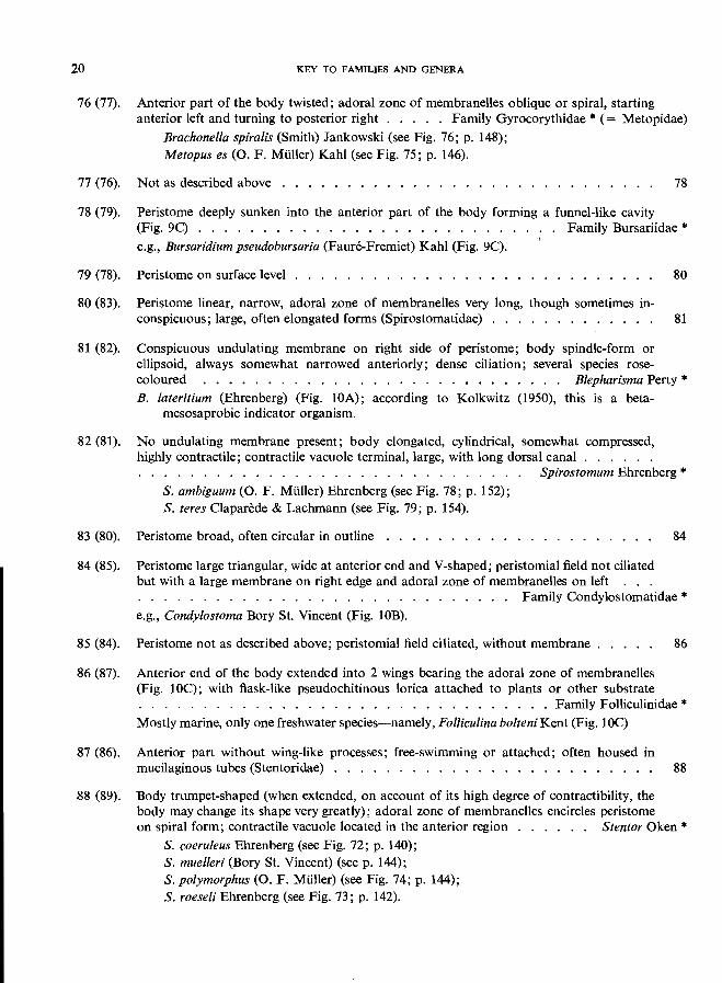
Fig. 9. Hypotrichida, Oxytrichidae (continued) (A and B), and Heterotrichida (C) : A Oxytricha saprobia Kahl; B, Opisthotricha simi/is Engelmann; C, Bursaridium pseudobursaria (Faure-Fremiet) Kahl, Bursariidae.
100)..lm
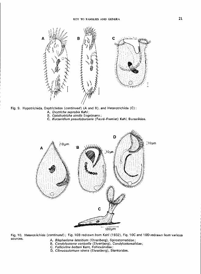
sources. Fig. 10. Heterotrichida (continued); Fig. 10B redrawn from Kahl (1932), Fig. 1 OC and 10D redrawn from various
A Blepharisma lateritium (Ehrenberg), Spirostomatidae; B, Condylostoma vorticella (Ehrenberg), Condylostomatidae; C, Folliculina bolteni Kent, Folliculinidae; D, Climacostomum virens (Ehrenberg), Stentoridae.
21
22 KEY TO FAMILIES AND GENERA
89 (88). Body ovoid, flattened. Noncontractile. Peristome ventral, near to the anterior pole (Fig. 10D). Contractile vacuole with 2 long canals . . . . Climacostomum Stein * (Fig. 10D)
90 (75). Buccal area without a long adoral zone of membranelles; oral ciliation lacking or consisting of simple cilia or composed of few predominantly inconspicuous membranous organelles 91
91 (124). Cytostome near or at surface; no oral ciliation; sometimes mouth a nearly invisible lateral slit opening only when feeding; margins of the slit are provided with trichocysts (cf., line 109) (Order Gymnostomatida) . . . . . . . . . . . . . . . . . . . . . . .. 92
92 (97). Circular mouth located mid-ventrally; gullet wall with fused armature of rods (" trichites "); body often dorsoventrally flattened and ciliation restricted to ventral surface (Suborder Cyrtophorina) . . . . . . . . . . . . . . . . . . . . . . . . . . . . . . . .. 93
93 (94). Ciliation all over the body but dorsal ciliation usually less dense than that of the ventral surface . . . . . . . . . . . . . . . . . . . . . . . . . . . . . Family Nassulidae *
94 (93).
e.g., Nassula gracilis Kahl (Fig. IIA); according to Liebmann (1962), this is an oligosaprobic indicator organism.
Ciliation only on the ventral surface . ..................... . 95
95 (96). With movable posterior stylus (Fig. llB) . . . . . . . . . . . . . . Family Dysteriidae * e.g., Trochilia minuta (Roux) (Fig. lIB); according to Kolkwitz (1950), this is a beta
mesosaprobic indicator organism.
96 (95). Without stylus . . . . . . . . . . . . . . . . . . . . . . Family Chlamydodontidae * e.g., Chilodonella cucullulus (0. F. Muller) (see Fig. 34; p. 58); Chilodonella uncinata
Ehrenberg (see Fig. 35; p. 60); Phascolodon vorticella Stein (Fig. 11 C).
97 (92). The circular or slit-like mouth at or near anterior pole or lateral (Suborder Rhabdophorina) 98
98 (99). With pseudochitinous lorica, sedentary; cytostome terminal; uniform ciliation, sometimes one or more caudal cilia; with peculiar ectoplasmic alveolar zone . . . Family Metacystidae * Predominantly in sapropelic habitats, feeding on sulfur-bacteria (especially purple-coloured
bacteria); e.g., Metacystis Cohn (Fig. 12A); Vasicola Tatem (Fig. 12B).
99 (98). Not as described above 100 .....................
100 (101). With pellicular armour consisting of many small plates; barrel-shaped body; mouth at anterior pole . . . . . . . . . . . . . . . . . . . . . . . . . . . Family Colepidae * e.g., Coleps Nitzsch-C. hirtus Nitzsch (see Fig. 22; p. 34).
101 (100). Not as described above . . . . . . . . . . . . . . . . ............. 102
102 (103). With retractable tentacles in addition to uniform covering of somatic ciliation; tentacles extending in all directions when at rest, but retracted and inconspicuous when swimming; cytostome located anteriorly . . . . . . . . . . . . . . . . . . Family Actinobolinida * e.g., Actinobolina Strand (Fig. 12C); Enchelyomorpha vermicularis (Smith) Kahl, Fig. 12D).
According to Liebmann (1962), this is a polysaprobic indicator organism; ecology is similar to that of Trimyema compressum (p. 68).
103 (102). Not as described above . . . . . . . . '. . . . . ................
104 (105). Mouth apical, often at the tip of a blunt cone, and surrounded by an unciliated area; cytopharynx with trichites (" toxicysts "), the mouth may be surrounded by retractile
104
KEY TO FAMILIES AND GENERA
J
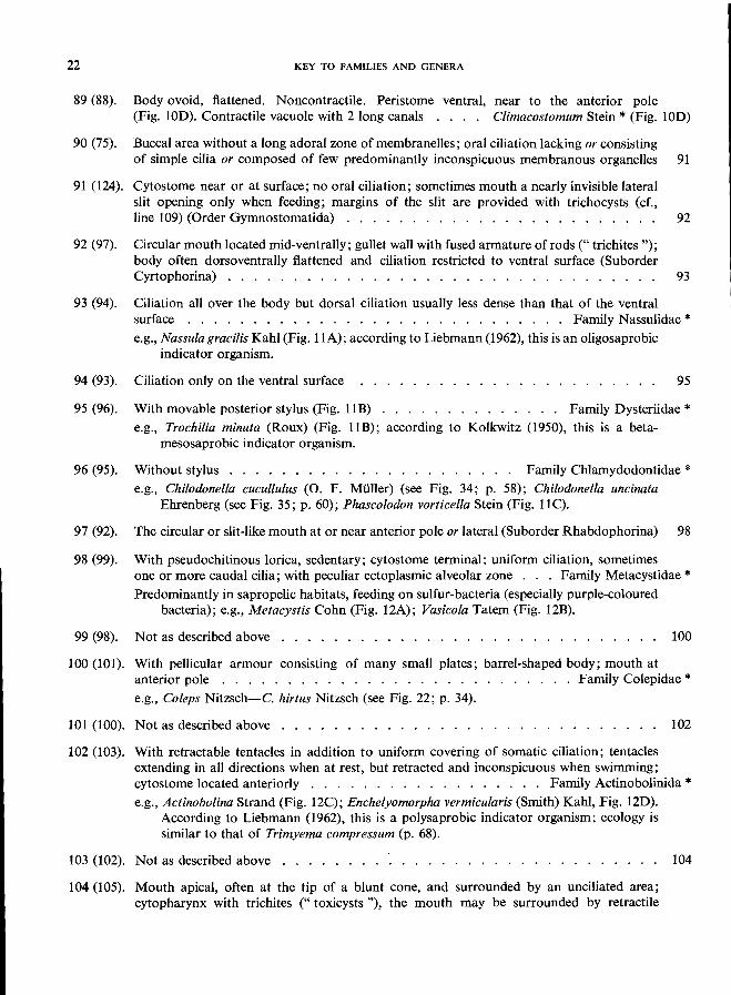
Fig. 11. Gymnostomatida, Cyrtophorina, ventral views:
A, Nassula gracilis Kahl ( = N. elegans Blochmann), Nassulidae; B, Trochilia min uta (Roux), Oysteriidae; C, Phascolodon vorticella Stein, Chlamydodontidae.
I~
Fig. 12. Gymnostomatida, Rhabdophorina; Fig. 12B redrawn from Kahl (1930), Fig. 120 redrawn from various sources.
A, Metacystis sp., Metacystidae; B, Vasicola ciliata Tatem, Metacystidae; C, Actinobolina radians Stein, Actinobolinidae; 0, Enchelyomorpha vermicularis (Smith) Kahl, Actinobolinidae.
23
24 KEY TO FAMILIES AND GENERA
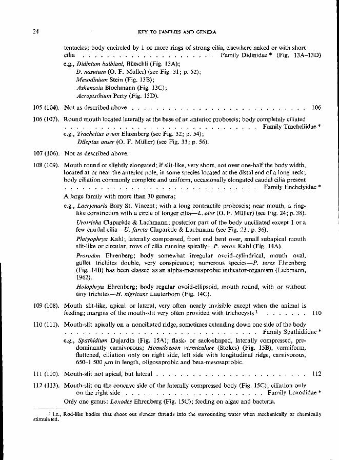
tentacles; body encircled by 1 or more rings of strong cilia, elsewhere naked or with short cilia ...•................ Family Didinidae * (Fig. 13A-13D)
e.g., Didinium balbiani, BUtschli (Fig. 13A); D. nasutum (0. F. MUller) (see Fig. 31; p. 52); Mesodinium Stein (Fig. 13B); Askenasia Blochmann (Fig. 13C); Acropisthium Perty (Fig. 13D).
105 (104). Not as described above . . . . . ........................ 106
106 (107). Round mouth located laterally at the base of an anterior proboscis; body completely ciliated . . . . . . . . . . . . . . . . . . . . . . . . . . . . . . . . Family Tracheliidae * e.g., Trachelius ovum Ehrenberg (see Fig. 32; p. 54);
Dileptus anser (0. F. MUller) (see Fig. 33; p. 56).
107 (106). Not as described above.
108 (109). Mouth round or slightly elongated; if slit-like, very short, not over one-half the body width, located at or near the anterior pole, in some species located at the distal end of a long neck; body ciliation commonly complete and uniform, occasionally elongated caudal cilia present . . . . . . . . . . . . . . . . . . . . . . . . . . . . . . ., Family Enchelyidae * A large family with more than 30 genera; e.g., Lacrymaria Bory St. Vincent; with a long contractile proboscis; near mouth, a ring
like constriction with a circle oflonger cilia-L. olor (0. F. MUller) (see Fig. 24; p. 38).
Urotricha Clapari!de & Lachmann; posterior part of the body unciliated except I or a few caudal cilia-U. farcta Clapari!de & Lachmann (see Fig. 23; p. 36).
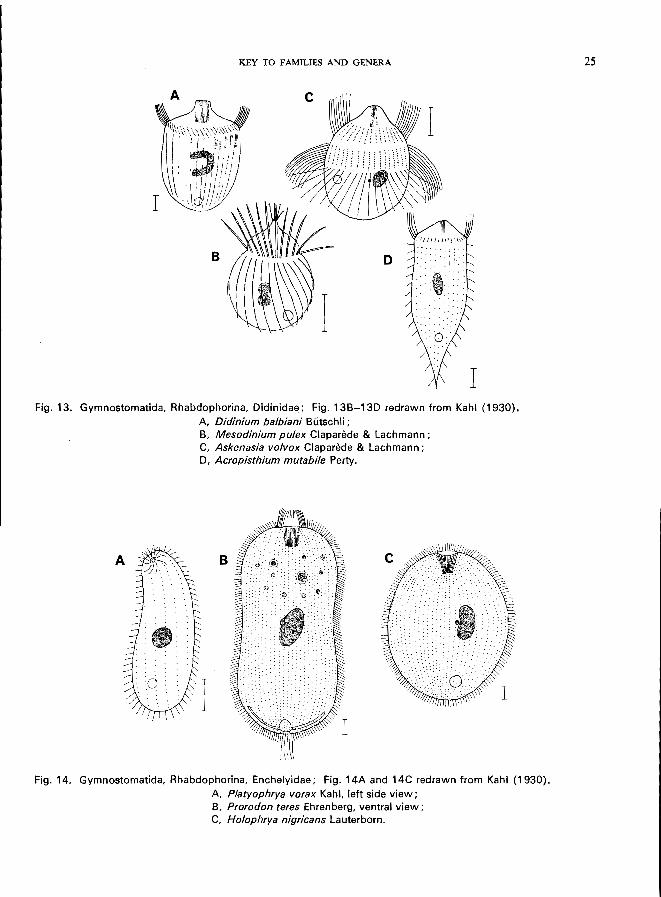
Platyophrya Kahl; laterally compressed, front end bent over, small subapical mouth slit-like or circular, rows of cilia running spirally-Po vorax Kahl (Fig. 14A).
Prorodon Ehrenberg; body somewhat irregular ovoid-cylindrical, mouth oval, gullet trichites double, very conspicuous; numerous species-Po teres Ehrenberg (Fig. 14B) has been classed as an alpha-mesosaprobic indicator-organism (Liebmann,
1962). Holophrya Ehrenberg; body regular ovoid-ellipsoid, mouth round, with or without tiny trichites-H. nigricans Lauterborn (Fig. 14C).
109 (108). Mouth slit-like, apical or lateral, very often nearly invisible except when the animal is feeding; margins of the mouth-slit very often provided with trichocysts 1 •...••. 110
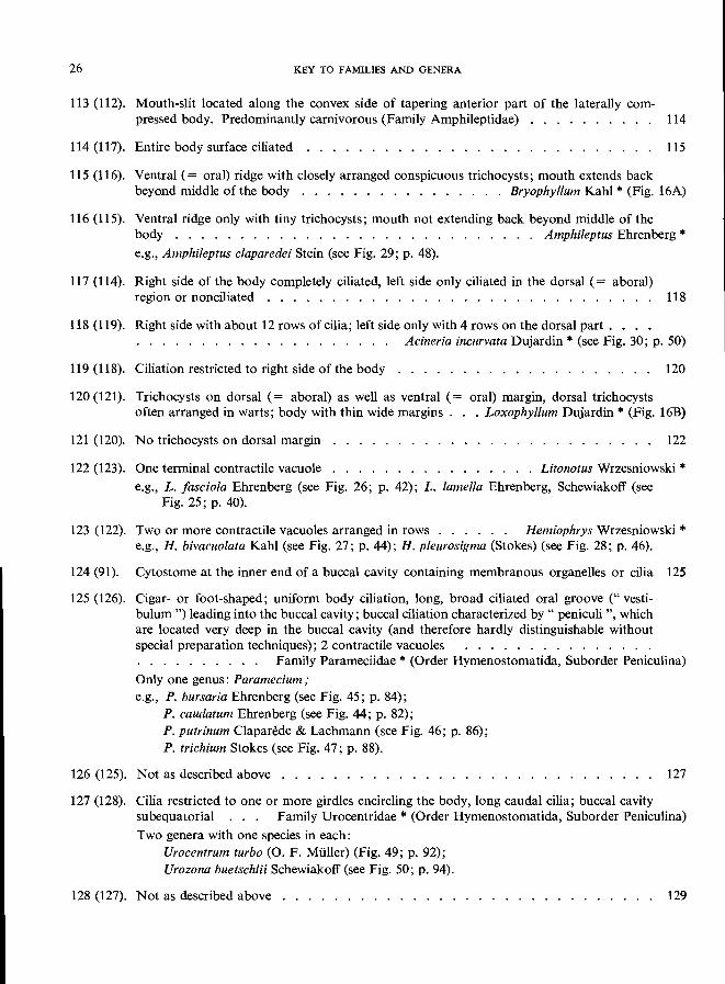
11 0 (111). Mouth-slit apically on a nonciliated ridge, sometimes extending down one side of the body . . . . . . . . . . . . . . . . . . . . . . . . . . . . . . . . Family Spathidiidae * e.g., Spathidium Dujardin (Fig. 15A); flask- or sack-shaped, laterally compressed, pre
dominantly carnivorous; Homalozoon vermiculare (Stokes) (Fig. 15B), vermiform, flattened, ciliation only on right side, left side with longitudinal ridge, carnivorous, 650-1 500 pm in length, oligosaprobic and beta-mesosaprobic.
111 (110). Mouth-slit not apical, but lateral . . . . . . . . . . . . . . . . . . . . . . . .. 112
112 (113). Mouth-slit on the concave side of the laterally compressed body (Fig. 15C); ciliation only on the right side . . . . . . . . . . . . . . . . . . . . . . . Family Loxodidae *
Only one genus: Loxodes Ehrenberg (Fig. 15C); feeding on algae and bacteria.
1 i.e .• Rod-like bodies that shoot out slender threads into the surrounding water when mechanically or chemically
stimulated.
KEY TO FAMILIES AND GENERA
Fig. 13. Gymnostomatida, Rhabdophorina, Oidinidae; Fig. 13B-130 redrawn from Kahl (1930). A, Didinium balbiani Butschli ; B, Mesodinium pulex Claparede & Lachmann; C, Askenasia volvox Claparede & Lachmann; 0, Acropisthium mutabile Perty.
Fig. 14. Gymnostomatida, Rhabdophorina, Enchelyidae; Fig. 14A and 14C redrawn from Kahl (1930). A, Platyophrya vorax Kahl, left side view; B, Prorodon teres Ehrenberg, ventral view; C, Holophrya nigricans Lauterborn.
25
26 KEY TO FAMILIES AND GENERA
113 (112). Mouth-slit located along the convex side of tapering anterior part of the laterally com-pressed body. Predominantly carnivorous (Family Amphileptidae) 114
114 (117). Entire body surface ciliated . . . . . . . . . . . . . . . . . 115
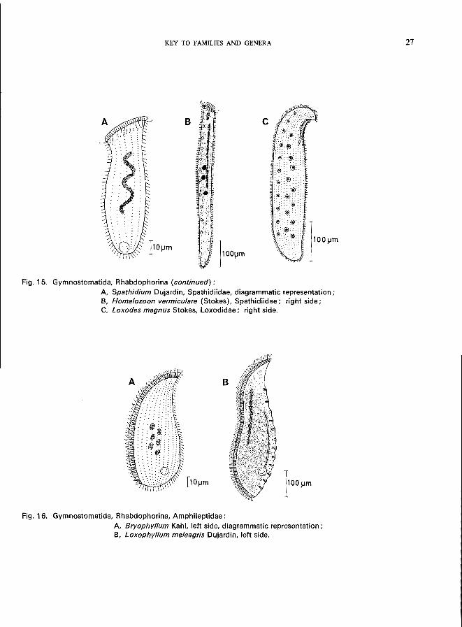
115 (116). Ventral (= oral) ridge with closely arranged conspicuous trichocysts; mouth extends back beyond middle of the body . . . . . . . . . . . . . . . . Bryophyllum Kahl * (Fig. 16A)
116 (115). Ventral ridge only with tiny trichocysts; mouth not extending back beyond middle of the body . . . . . . . . . . . . . . . . . . . . . . . . . . . . Amphileptus Ehrenberg * e.g., Amphileptus claparedei Stein (see Fig. 29; p. 48).
117 (114). Right side of the body completely ciliated, left side only ciliated in the dorsal (= aboral) region or nonciliated . . . . . . . . . . . . . . . . . . . . . . . . . . . 118
118 (119). Right side with about 12 rows of cilia; left side only with 4 rows on the dorsal part. . . . . . . . . . . . . . . . . . . .. Acineria incurvata Dujardin * (see Fig. 30; p. 50)
119 (118). Ciliation restricted to right side of the body 120
120 (121). Trichoeysts on dorsal (= aboral) as well as ventral (= oral) margin, dorsal trichocysts often arranged in warts; body with thin wide margins. . Loxophyllum Dujardin * (Fig. 16B)
121 (120). No trichocysts on dorsal margin 122
122 (123). One terminal contractile vacuole Lilonotus Wrzesniowski * e.g., L. fasciola Ehrenberg (see Fig. 26; p. 42); L. lamella Ehrenberg, Schewiakoff (see
Fig. 25; p. 40).
123 (122). Two or more contractile vacuoles arranged in rows . . . . .. Hemiophrys Wrzesniowski * e.g., H. bivacuolata Kahl (see Fig. 27; p. 44); H. pleurosigma (Stokes) (see Fig. 28; p. 46).
124 (91). Cytostome at the inner end of a buccal cavity containing membranous organelles or cilia 125
125 (126). Cigar- or foot-shaped; uniform body ciliation, long, broad ciliated oral groove (" vestibulum ") leading into the buccal cavity; buccal ciliation characterized by" peniculi ", which are located very deep in the buccal cavity (and therefore hardly distinguishable without special preparation techniques); 2 contractile vacuoles .............. . . . . . . . . . .. Family Parameciidae * (Order Hymenostomatida, Suborder Peniculina)
Only one genus: Paramecium; e.g., P. bursaria Ehrenberg (see Fig. 45; p. 84);
P. caudatum Ehrenberg (see Fig. 44; p. 82); P. putrinum Claparede & Lachmann (see Fig. 46; p. 86); P. trichium Stokes (see Fig. 47; p. 88).
126 (125). Not as described above . . . . . . . . .
127 (128). Cilia restricted to one or more girdles encircling the body, long caudal cilia; buccal cavity
127
subequatorial Family Urocentridae * (Order Hymenostomatida, Suborder Peniculina)
Two genera with one species in each: Urocentrum turbo (0. F. Mi.iller) (Fig. 49; p. 92); Urozona buetschlii Schewiakoff (see Fig. 50; p. 94).
128 (127). Not as described above . . . . . . . . . . . . . . 129
KEY TO FAMILIES AND GENERA
B
Fig. 15. Gymnostomatida, Rhabdophorina (continued):
A Spathidium Dujardin, Spathidiidae, diagrammatic representation; B, Homalozoon vermiculare (Stokes), Spathidiidae; right side; C, Loxodes magnus Stokes, Loxodidae; right side.
Fig. 16. Gymnostomatida, Rhabdophorina, Amphileptidae :
A, Bryophyllum Kahl, left side, diagrammatic representation; B, Loxophyllum meleagris Dujardin, left side.
27
28 KEY TO FAMILIES AND GENERA
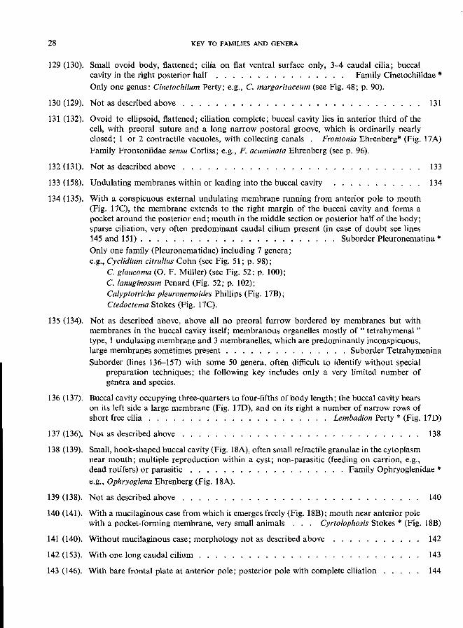
129 (130). Small ovoid body, flattened; cilia on flat ventral surface only, 3-4 caudal cilia; buccal cavity in the right posterior half . . . . . . . . . . . . . . .. Family Cinetochilidae * Only one genus: Cinetochilum Perty; e.g., C. margaritaceum (see Fig. 48; p. 90).
130 (129). Not as described above . . . . . . . . . . . . . . . . • . . . . . . . . 131
131 (132). Ovoid to ellipsoid, flattened; ciliation complete; buccal cavity lies in anterior third of the cell, with preoral suture and a long narrow postoral groove, which is ordinarily nearly closed; 1 or 2 contractile vacuoles, with collecting canals . Frontonia Ehrenberg* (Fig. 17A) Family Frontoniidae sensu Corliss; e.g., F. acuminata Ehrenberg (see p. 96).
132 (131). Not as described above . . . . . . . . . . . . . . . . ..
133 (158). Undulating membranes within or leading into the buccal cavity
134 (135). With a conspicuous external undulating membrane running from anterior pole to mouth (Fig. 17C), the membrane extends to the right margin of the buccal cavity and forms a pocket around the posterior end; mouth in the middle section or posterior half of the body; sparse ciliation, very often predominant caudal cilium present (in case of doubt see lines
133
134
145 and 151). . . . . . . . . . . . . . . . . . . . . . . . Suborder Pleuronematina * Only one family (Pleuronematidae) including 7 genera; e.g., Cyclidium citrul/us Cohn (see Fig. 51; p. 98);
c. glaucoma (0. F. MUller) (see Fig. 52; p. 100); C. lanuginosum Penard (Fig. 52; p. 102); Calyptotricha pleuronemoides Phillips (Fig. 17B); Ctedoctema Stokes (Fig. 17C).
135 (134). Not as described above, above all no preoral furrow bordered by membranes but with membranes in the buccal cavity itself; membranous organelles mostly of " tetrahymenal " type, 1 undulating membrane and 3 membranelles, which are predominantly inconspicuous, large membranes sometimes present . . . . . . . . . . . . . . . Suborder Tetrahymenina Suborder (lines 136-157) with some 50 genera, often difficult to identify without special
preparation techniques; the following key includes only a very limited number of genera and species.
136 (137). Buccal cavity occupying three-quarters to four-fifths of body length; the buccal cavity bears on its left side a large membrane (Fig. 17D), and on its right a number of narrow rows of short free cilia . . . . Lembadion Perty * (Fig. 17D)
137 (136). Not as described above . . . . . . . . . . . . . . . . . . . . . . . . . . . .. 138
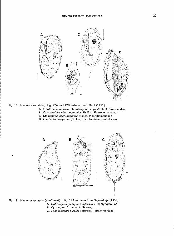
138 (139). Small, hook-shaped buccal cavity (Fig. 18A), often small refractile granulae in the cytoplasm near mouth; multiple reproduction within a cyst; non-parasitic (feeding on carrion, e.g., dead rotifers) or parasitic . . . . . . . . . . . . . . . . . . . Family Ophryoglenidae * e.g., Ophryoglena Ehrenberg (Fig. 18A).
139 (138). Not as described above . . . . . . . 140
140 (141). With a mucilaginous case from which it emerges freely (Fig. 18B); mouth near anterior pole with a pocket-forming membrane, very small animals . . . Cyrtolophosis Stokes * (Fig. 18B)
141 (140). Without mucilaginous case; morphology not as described above
142 (153). With one long caudal cilium ............... .
143 (146). With bare frontal plate at anterior pole; posterior pole with complete ciliation
142
143
144
T 1
KEY TO FAMILIES AND GENERA
Fig. 17. Hymenostomatida; Fig. 17 A and 17D redrawn from Kahl (1931).
A, Frontonia acuminata Ehrenberg var. angusta Kahl, Frontoniidae; B, Calyptotricha pleuronemoides Phillips, Pleuronematidae; C, Ctedoctema acanthocrypta Stokes, Pleuronematidae; D, Lembadion magnum (Stokes), Frontoniidae, ventral view.
Fig. 18. Hymenostomatida (continued); Fig. 18A redrawn from Gajewskaja (1933).
A, Ophryoglena pelagica Gajewskaja, Ophryoglenidae; B, Cyrtolophosis mucicola Stokes; C, Loxocephalus plagius (Stokes), Tetrahymenidae.
I
29
30 KEY TO FAMILIES AND GENERA
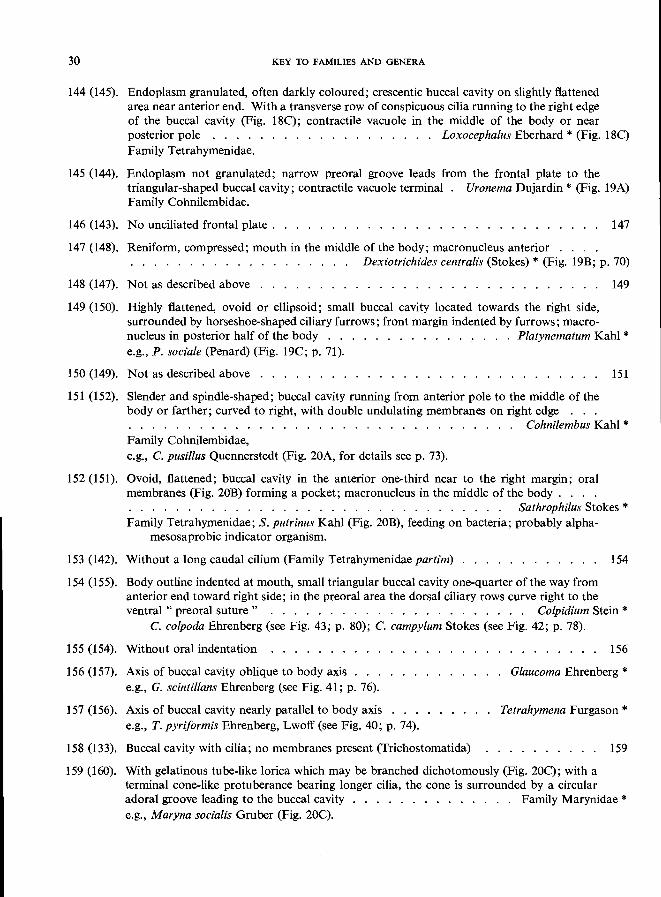
144 (145). Endoplasm granulated, often darkly coloured; crescentic buccal cavity on slightly flattened area near anterior end. With a transverse row of conspicuous cilia running to the right edge of the buccal cavity (Fig. 18C); contractile vacuole in the middle of the body or near posterior pole . . . . . . . . . . . . . . . . . . . Loxocephalus Eberhard * (Fig. 18C) Family Tetrahymenidae.
145 (144). Endoplasm not granulated; narrow preoral groove leads from the frontal plate to the triangular-shaped buccal cavity; contractile vacuole terminal Uronema Dujardin * (Fig. 19A) Family Cohnilembidae.
146 (143). No unciliated frontal plate. . . . . . . . . . . . . . . 147
147 (148). Reniform, compressed; mouth in the middle of the body; macronucleus anterior . . . . . . . . . . . . . . . Dexiotrichides centralis (Stokes) * (Fig. 19B; p. 70)
148 (147). Not as described above . . . . . . . . . . . . . . . . . . . . . . . . . . . . . 149
149 (150). Highly flattened, ovoid or ellipsoid; small buccal cavity located towards the right side, surrounded by horseshoe-shaped ciliary furrows; front margin indented by furrows; macronucleus in posterior half of the body . . . . . . . . . . . . . . . . Platynematum Kahl * e.g., P. sociale (Penard) (Fig. 19C; p. 71).
150 (149). Not as described above . . . . . . . .
151 (152). Slender and spindle-shaped; buccal cavity running from anterior pole to the middle of the body or farther; curved to right, with double undulating membranes on right edge . . .
151
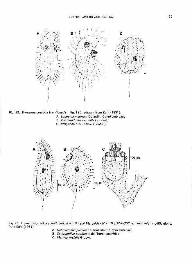
. . . . . . . . . . . . . . . . . . . . . . . . . . . . . . . . . Cohnilembus Kahl * Family Cohnilembidae, e.g., C. pusillus Quennerstedt (Fig. 20A, for details see p. 73).
152 (151). Ovoid, flattened; buccal cavity in the anterior one-third near to the right margin; oral membranes (Fig. 20B) forming a pocket; macronucleus in the middle of the body. . . . . . . . . . . . . . . . . . . . . . . . . . . . . . . . . . .. Sathrophilus Stokes * Family Tetrahymenidae; S. putrinus Kahl (Fig. 20B), feeding on bacteria; probably alpha-
mesosaprobic indicator organism.
153 (142). Without a long caudal cilium (Family Tetrahymenidae partim) . . . . . . . . . . .. 154
154 (155). Body outline indented at mouth, small triangular buccal cavity one-quarter of the way from anterior end toward right side; in the preoral area the dorsal ciliary rows curve right to the ventral " preoral suture" . . . . . . . . . . . . . . . . . . . . . . Colpidium Stein *
C. colpoda Ehrenberg (see Fig. 43; p. 80); c. campylum Stokes (see Fig. 42; p. 78).
155 (154). Without oral indentation 156
156 (157). Axis of buccal cavity oblique to body axis Glaucoma Ehrenberg * e.g., G. scintillans Ehrenberg (see Fig. 41; p. 76).
157 (156). Axis of buccal cavity nearly parallel to body axis Tetrahymena Furgason * e.g., T. pyriformis Ehrenberg, Lwoff (see Fig. 40; p. 74).
158 (133). Buccal cavity with cilia; no membranes present (Trichostomatida) 159
159 (160). With gelatinous tube-like lorica which may be branched dichotomously (Fig. 20C); with a terminal cone-like protuberance bearing longer cilia, the cone is surrounded by a circular adoral groove leading to the buccal cavity . . . . . . . . . . . . . . Family Marynidae * e.g., Maryna socialis Gruber (Fig. 20C).
KEY TO FAMILIES AND GENERA
c
Fig. 19. Hymenostomatida (continued); Fig. 19B redrawn from Kahl (1931).
A, Uronema marinum Dujardin, Cohnilembidae; B, Dexiotrichides centra/is (Stokes) ; C, P/atynematum socia/e (Penard).
Fig. 20. Hymenostomatida (continued: A and B) and Marynidae (C); Fig. 20A-20C redrawn, with modifications, from Kahl (1931).
A, Cohnilembus pusil/us Quennerstedt, Cohnilembidae; B, Sathrophilus putrinus Kahl, Tetrahymenidae; C, Maryna sociafis Gruber.
31
32 KEY TO FAMILIES AND GENERA
.............. 161 160 (159). Not as described above . . . . . . . . . . . . . . .
161 (162), Body flattened by later~l compressions; rigid pellicle, sparse ciliation predominantly on the right side; mouth on the slightly convex ventral face . . . . . . . Family Microthoracidae * e.g., Microthorax Engelmann (Fig. 21A), mouth near to the posterior end;
Leptopharynx Mermod (= Trichopelma Levander) (Fig. 2IB, 21C), mouth in anterior half of the cell, gullet with trichites.
162 (161). Not as described above . . . . . . . . . . . . . . . . . . . . . . . . . . . 163
163 (164). Ciliation restricted to 3-4 spiral rows on the anterior body surface, and 1 long caudal cilium; body ovoid, anterior and posterior end of the cell pointed . . . . . . Family Trimyemidae *
Trimyema compressum Lackey (see Fig. 39; p. 68).
164 (163). Not as described above . . . . . . . . . . . . . .
165 (166). Oral groove transverse, in anterior fourth of the body. e.g., Plagiopyla nasuta Stein (see Fig. 38; p. 66).
........ 165
Family Plagiopylidae *
166 (165). Oral groove funnel-like at an indentation, near the middle of one side of the body. . . . . . . . . . . . . . . . . . . . . . . . . . . . . . . ., Family Colpodidae * e.g., Colpoda cucullus O. F. Miiller (see Fig. 37; p. 64);
C. steini (Maupas) Enriques (see Fig. 36; p. 62).
I I Fig. 21. Microthoracidae:
A Microthorax pusillus Engelmann; BJand C, Leptopharynx sphagnetorum (Levander) ; ventral view (B). dorsal view (C);
SPECIES DESCRIPTIONS
The descriptions are arranged in taxonomic order following that of the key in the previous section. The family and genus are shown for each species described. Where no species descriptions are given, families are mentioned in the same order as in the Synopsis (p. 7) with examples of genera and references to figures in the key.
Family Colepidae Genus Coieps
Subclass Order Suborder
HOLOTRICHIA GYMNOSTOMATIDA RHABDOPHORINA
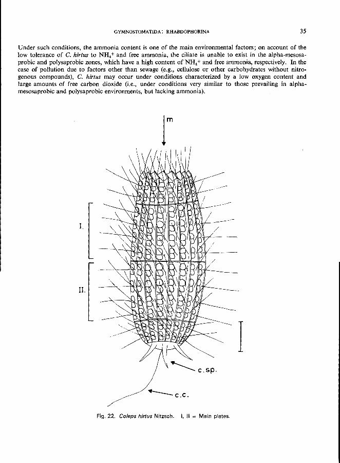
COLEPS H1RTUS Nitzsch (Fig. 22)
Morphology
Length 55-65 /Lm; barrel-shaped, constant body form; regularly arranged ectoplasmatic plates: two main groups of plates (I and II in Fig. 22) with 4 meridional rows of" windows" each; 15-20 longitudinal rows of platelets; mouth at the anterior pole, surrounded by special platelets; 3 spinous processes at the posterior end; uniform ciliation over the whole body except for 1 long caudal cilium; 1 spherical macronucleus; contractile vacuole near the posterior end.
Other species of the genus Coieps may show very similar features; in doubtful cases, make sure of the construction of the main plates and caudal spines. For further information see Kahl (1930).
Food
Feeds saprophytically upon other protozoons or rotifers, etc., and on algae, flagellates, and small ciliates, which are captured alive.
Occurrence and ecology
Cosmopolitan in distribution, occurring in all types of water containing organic detritus. Its occurrence in large numbers indicates beta-mesosaprobic conditions (Liebmann, 1962); single specimens may be found in more-or-Iess polluted waters. Sometimes planktonic in reservoirs, lakes, and ponds.
Ecological characteristics (Bick, 1968)
The following tabulation shows the main ecological characteristics of this species:
Saprobiology
temperature Cc) . pH ...... . dissolved O. (mgfl) free CO. (mg/l). NH. + (mg/l). . free NH. (mg/l) NO.- (mg/l) .. H.S (mgfl) brackish water (seawater type) .
bacteria (plate counts on peptone agar) ........... .
Extreme tolerances
2-30 4.7-9.4 0.1-18.0
0-140 0-26 0-0.2 0-34 0-1.0
up to 3.5 g of total salt content per litre
Optimal ranges
6.5-7.5 0.1-0.5 10-15
0.1-0.5 0-0.05
o o
below 0.25 g of total salt content per litre
300-20000000 ml
From these ecological parameters, C. hirtus only indicates beta-mesosaprobic conditions in selfpurification zones following the discharge of domestic sewage containing large amounts of faecal material.
-34-
GYMNOSTOMATIDA: RHABDOPHORINA 35
Under such conditions, the ammonia content is one of the main environmental factors; on account of the low tolerance of C. hirtus to NH4 + and free ammonia, the ciliate is unable to exist in the alpha-mesosaprobic and polysaprobic zones, which have a high content of NH4 + and free ammonia, respectively. In the case of pollution due to factors other than sewage (e.g., cellulose or other carbohydrates without nitrogenous compounds), C. hirtus may occur under conditions characterized by a low oxygen content and large amounts of free carbon dioxide (i.e., under conditions very similar to those prevailing in alphamesosaprobic and polysaprobic environments, but lacking ammonia).
r
1.
II.
I ~ c.c.
Fig. 22. Co/eps hir/us Nitzsch. I, II = Main plates.
36
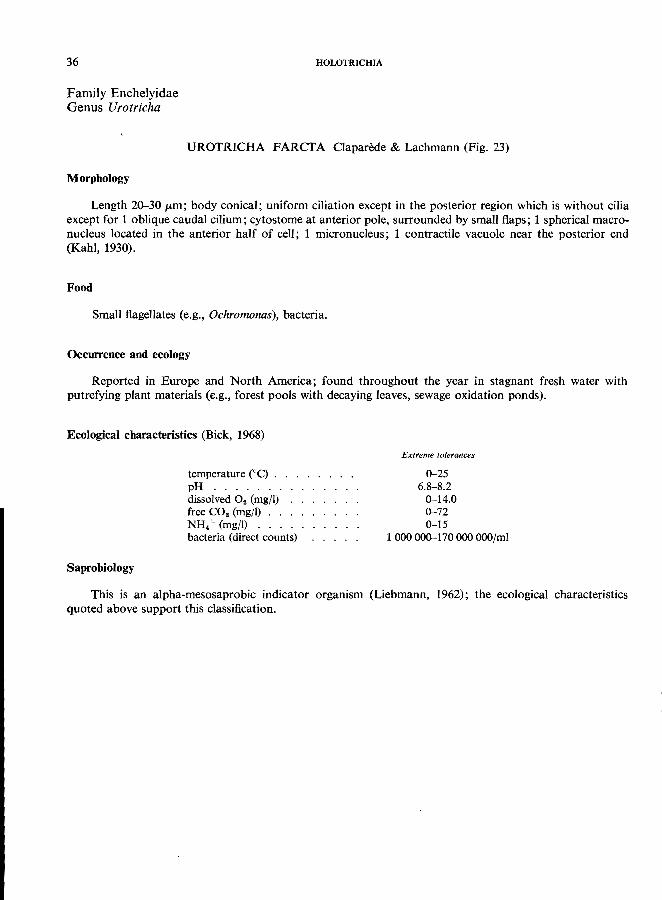
Family Enchelyidae Genus Urotricha
HOLOTRICHIA
UROTRICHA FARCTA Claparede & Lachmann (Fig. 23)
Morphology
Length 20-30 pm; body conical; uniform ciliation except in the posterior region which is without cilia except for 1 oblique caudal cilium; cytostome at anterior pole, surrounded by small flaps; 1 spherical macronucleus located in the anterior half of cell; 1 micronucleus; 1 contractile vacuole near the posterior end
(Kahl, 1930).
Food
Small flagellates (e.g., Ochromonas), bacteria.
Occurrence and ecology
Reported in Europe and North America; found throughout the year in stagnant fresh water with putrefying plant materials (e.g., forest pools with decaying leaves, sewage oxidation ponds).
Ecological characteristics (Bick, 1968)
temperature ceq . . . . . . . . pH ...... . dissolved O. (mg/l) free CO. (mg/l) . . NH/ (mg/l) ... bacteria (direct counts)
Extreme tolerances
0-25 6.8-8.2
0-14.0 0-72 0-15
1000000-170000000/ml
Saprobiology
This is an alpha-mesosaprobic indicator organism (Liebmann, 1962); the ecological characteristics
quoted above support this classification.
GYMNOSTOMATIDA: RHABDOPHORINA 31
c.v.
~ C.C.
Fig. 23. Urotrieha lareta Claparede & Lachmann. Arrows indicate the main features used for identification.
38 HOLOTRICHIA
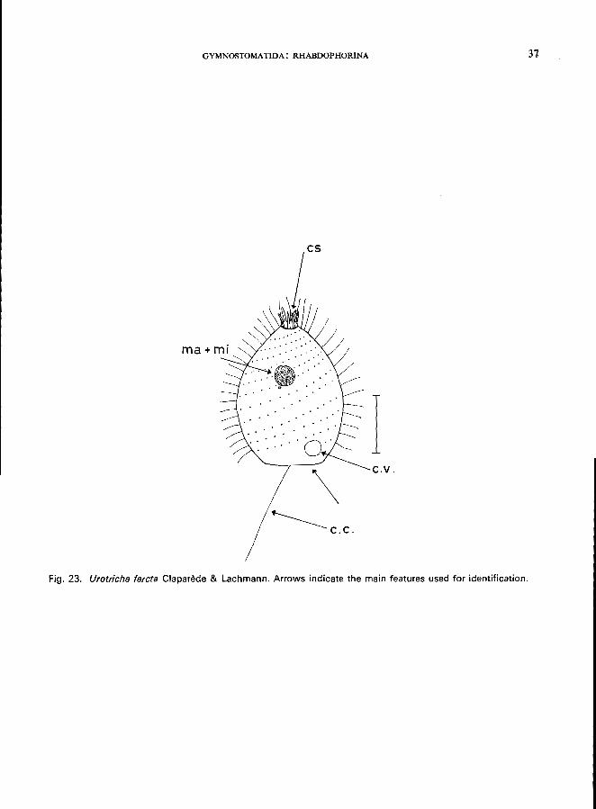
Family Enchelyidae Genus Lacrymaria
LACRYMARIA OLOR (0. F. MUller) (Fig. 24)
Morphology
Length 400 pm (when fully extended, up to 1.2 mm); body elongated, flask-shaped with a long and highly contractile proboscis anteriorly, very polymorphic, body ciliation rather short and dense; a circle of longer cilia is present at the head-like anterior part of the proboscis, which is marked by a ring-like constriction; cytostome at anterior pole, supported by trichites, 2 spherical macronuclei and a single micronucleus
between them; 2 contractile vacuoles (Kahl, 1930; Kudo, 1966).
Food
Carnivorous, feeding on ciliates; when about to feed the body is hidden among detritus while the fully
extended proboscis is searching for prey.
Occurrence and ecology
Widely distributed in stagnant and slowly flowing water; according to Kahl (1930), occurs in saline,
brackish, and fresh water.
Ecological characteristics (Bick, unpublished data)
Saprobiology
temperature eC) . . . pH ........ . dissolved oxygen (mg/I) free CO. (mg/I) NH/ (mg/I) H.S (mg/\) ..
Extreme tolerances
0-25 6.4-7.5 0.2-12
0-56 0-10
up to 0.5
According to Kolkwitz (1950), this is an oligosaprobic indicator organism that may occur also in the beta-mesosaprobic zone. The ecological characteristics given above do not support Kolkwitz's opinion. L. olor is able to withstand comparatively high concentrations of NH4 + and low levels of dissolved oxygen. It is assumed that the optimum for the species are beta-mesosaprobic conditions; single specimens may occur
in the alpha-mesosaprobic zone as well as in the oligosaprobic zone.
Saprobiological classification 1
bos aos 2
bms 6
ams 2
ps i (= indicator value) 3
1 Le., the 10-points method of Zelinka & Marvan (1961); for an explanation refer to the glossary (p. 193).
GYMNOSTOMATIDA: RHABDOPHORINA 39
~--- "head"
~--pr
··~~--c.v.l
ma + --4-~
mi
100~m
Fig. 24. Lacrymaria alar (0. F. Muller). Arrows indicate the main features used for identification.
40
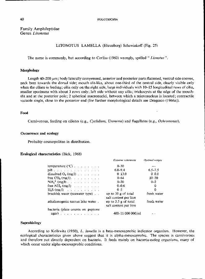
Family Amphileptidae Genus Litonotus
HOLOTRICHIA
LITONOTUS LAMELLA (Ehrenberg) Schewiakoff (Fig. 25)
The name is commonly, but according to Corliss (1960) wrongly, spelled" Lionotus".
Morphology
Length 40-200 pm; body laterally compressed, anterior and posterior parts flattened, ventral side convex, neck bent towards the dorsal side; mouth slit-like, about one-third of the central side, clearly visible only when the ciliate is feeding; cilia only on the right side, large individuals with 10-15 longitudinal rows of cilia, smaller specimens with about 3 rows only; left side without any cilia; trichocysts at the edge of the mouthslit and at the posterior pole; 2 spherical macronuclei, between which a micronucleus is located; contractile vacuole single, close to the posterior end (for further morphological details see Dragesco (1966a».
Food
Carnivorous, feeding on ciliates (e.g., Cyclidium, Uronema) and flagellates (e.g., Ochromonas).
Occurrence and ecology
Probably cosmopolitan in distribution.
Ecological characteristics (Bick, 1968)
Saprobiology
temperature (0C) . . pH ....... . dissolved O. (mgjl) free CO. (mg/I). NH4+ (mg/I). . free NH3 (mg/I) H.S (mg/I) brackish water (seawater type)
athalassogenic natron lake water
bacteria (plate counts on peptone agar) . . . . . . . .
Extreme tolerances
0-30 6.0-9.4
0-13.0 0-64 0-30 0-0.6 0-5
up to 35 g of total salt content per litre up to 3.5 g of total salt content per litre
400-11 OOOOOO/mi
Optimal ranges
6.5-7.5 0-0.1
10-20 0-2
o o
fresh water
fresh water
According to Kolkwitz (1950), L. lamella is a beta-mesosaprobic indicator organism. However, the ecological characteristics given above suggest that it is alpha-mesosaprobic. The species is carnivorous and therefore not directly dependent on bacteria. It feeds mainly on bacteria-eating organisms, many of
which occur under alpha-mesosaprobic conditions.
GYMNOSTOMATIDA: RHABDOPHORINA 41
mj-
Fig. 25. Litonotus lamella (Ehrenberg), right lateral view.
42 HOLOTRICHIA
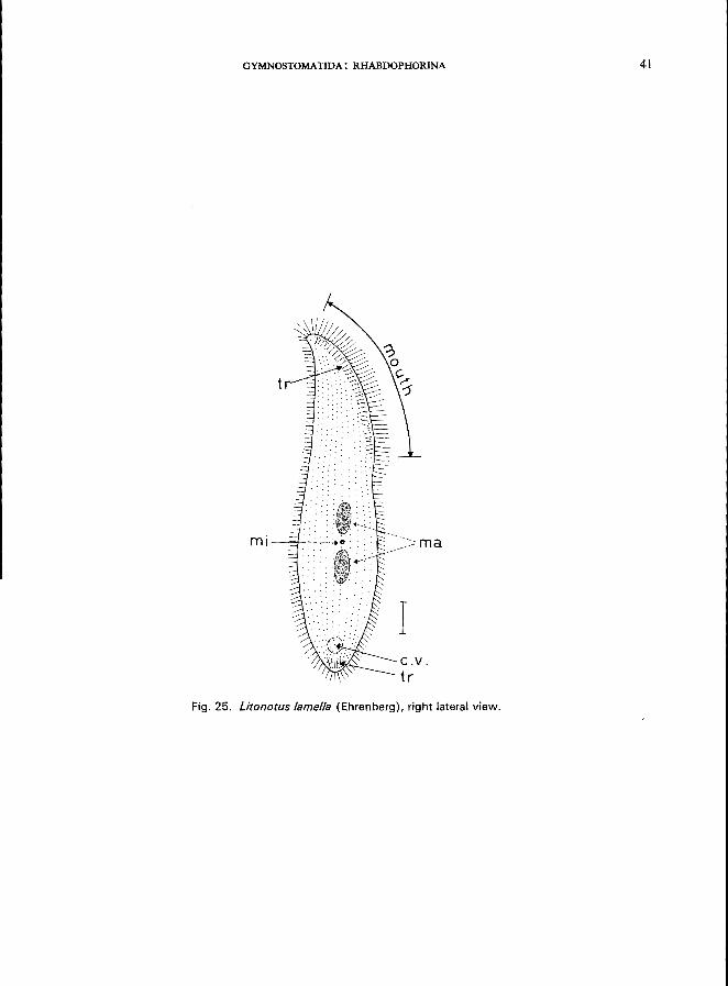
Family Amphileptidae Genus Litonotus
LITONOTUS FASCIOLA (Ehrenberg) (Fig. 26)
Morphology
Length 100 (tm; body elongated, laterally compressed; right side flat, left side more or less convex; anterior and posterior parts flattened and hyaline; neck bent towards the dorsal side; mouth (cytostome) a long slit, about one-third to one-half the total length of the body; cilia on right side only; a single contractile vacuole near the posterior end; 2 spherical macronuclei, between which a single micronucleus is situated.
For further details see Kahl (1931).
Food
Carnivorous, feeding on ciliates and flagellates.
Occurrence and ecology
Probably cosmopolitan in distribution; according to Liebmann (1962), the species occurs in all types of
water polluted with putrescible organic material.
Ecological characteristics (Noland, 1925; Bick, unpublished data)
temperature ("C) . pH ...... . dissolved O. (mg/l) H.S ...... . The species occurs in fresh water
and in seawater. . . . . . .
Saprobiological classification
Extreme tolerances
0-21 7.1-8.0 0.3-6.0
o
. with a total salt content of about 30 gIl (see Bock, 1952).
According to Zelinka & Marvan (1961) (1) and Shideeek (1964) (2) this is an alpha-mesosaprobic indi
cator organism (Liebmann, 1962).
(1) (2)
bos aos bms
1
ams
3 8
ps
7 1
3 4
GYMNOSTOMATIDA: RHABDOPHORINA
th
ma +
mj
'-..L£:>:.-___ c. v .
Fig. 26. Litonotus fasciola (Ehrenberg). right lateral view. Arrows indicate the main features used for identification.
43
44
Family Amphileptidae Genus Hemiophrys
HOLOTRICHIA
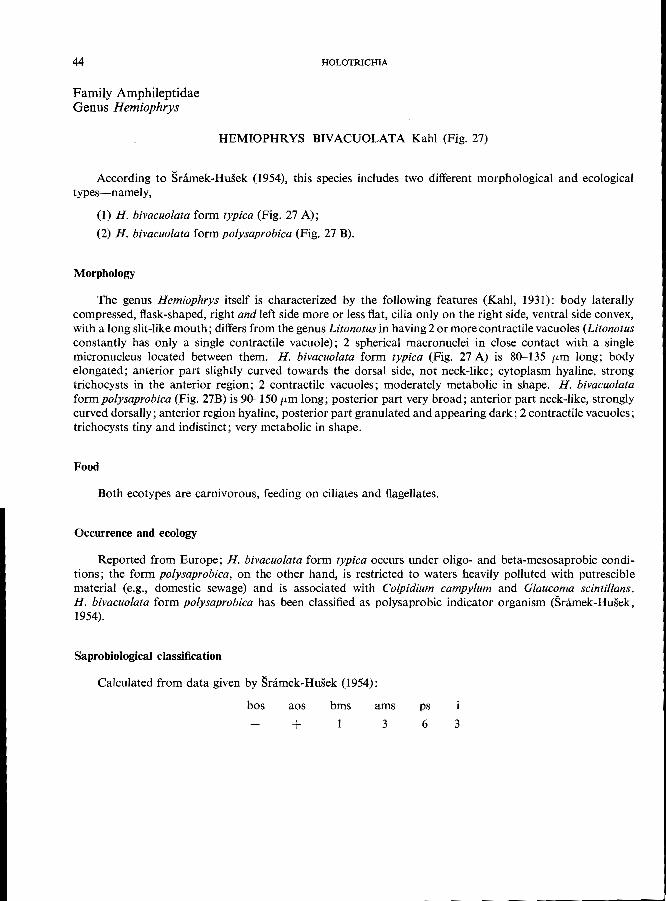
HEMIOPHRYS BIVACUOLATA Kahl (Fig. 27)
According to Snimek-Husek (1954), this species includes two different morphological and ecological
types-namely,
(1) H. bivacuolata form typica (Fig. 27 A);
(2) H. bivacuolata form polysaprobica (Fig. 27 B).
Morphology
The genus Hemiophrys itself is characterized by the following features (Kahl, 1931): body laterally compressed, flask-shaped, right and left side more or less flat, cilia only on the right side, ventral side convex, with a long slit-like mouth; differs from the genus Litonotus in having 2 or more contractile vacuoles (Litonotus constantly has only a single contractile vacuole); 2 spherical macronuclei in close contact with a single micronucleus located between them. H. bivacuolata form typica (Fig. 27 A) is 80-135 fim long; body elongated; anterior part slightly curved towards the dorsal side, not neck-like; cytoplasm hyaline, strong trichocysts in the anterior region; 2 contractile vacuoles; moderately metabolic in shape. H. bivacuolata formpolysaprobica (Fig. 27B) is 90-150 fim long; posterior part very broad; anterior part neck-like, strongly curved dorsally; anterior region hyaline, posterior part granulated and appearing dark; 2 contractile vacuoles;
trichocysts tiny and indistinct; very metabolic in shape.
Food
Both ecotypes are carnivorous, feeding on ciliates and flagellates.
Occurrence and ecology
Reported from Europe; H. bivacuolata form typ;ca occurs under oligo- and beta-mesosaprobic conditions; the form polysaprobica, on the other hand, is restricted to waters heavily polluted with putrescible material (e.g., domestic sewage) and is associated with Colpidium campylum and Glaucoma scintillans. H. bivacuolata form polysaprobica has been classified as polysaprobic indicator organism (Sramek-Husek,
1954).
Saprobiological classification
Calculated from data given by Sramek-Husek (1954):
bos aos bms ams
+ 3
ps
6 3
A
GYMNOSTOMATIDA: RHABDOPHORINA
tr
I
c.v.
ma +
mi
c.v.
B
Fig. 27. A. Hemiophrys bivacuolata form typica, left lateral view. B, Hemiophrys form polysaprobica, left lateral view; redrawn after Sramek-Husek (1954).
45
46 HOLOTRICHIA
Family Amphileptidae Genus Hemiophrys
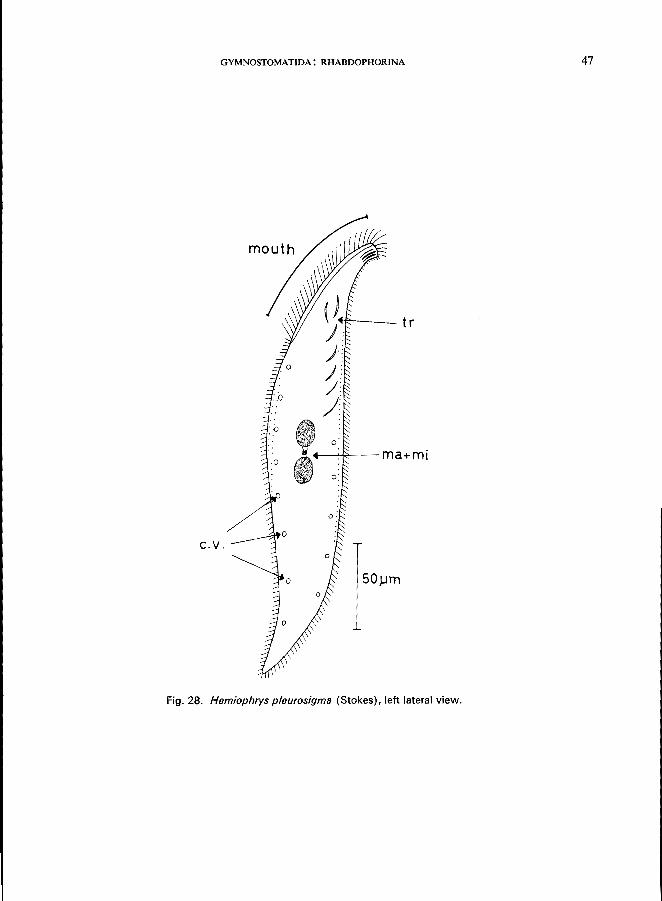
HEMIOPHRYS PLEUROSIGMA (Stokes) (Fig. 28)
Morphology (Kahl, 1931)
Length 300 fLm; for generic features see Hemiophrys bivacuolata (p. 44); body sigmoid; left side without cilia except for single rows near the ventral and dorsal margins; right side with about 20 rows of cilia; a group of strong trichocysts at the anterior pole and individual trichocysts throughout the anterior region; a single micronucleus located in a tiny tunnel connecting the 2 macronuclei; numerous contractile vacuoles
arranged in a ventral and a dorsal row.
Food
Carnivorous, feeding on ciliates and flagellates.
Occurrence and ecology
Reported from Europe in pools, ponds, reservoirs, slowly running waters, associated with Coleps hirtus, Colpidium campy/um, Halteria grandinella, Paramecium caudatum, Stentor roeseli, Urocentrum turbo.
Ecological characteristics (Bick, unpublished data)
Saprobiology
temperature eC) . pH ...... . dissolved O. (mg/I) free CO. (mg/I) . . NH/ (mg/I) ... brackish water (seawater type) bacteria (plate counts on peptone
agar) .......... .
Extreme tolerances
3-25 6.2-7.5 0.2-10
0--52 0-18
up to 3.5 g of total salt content per litre
up to 18000 OOO/mi
According to Snimek-Husek (1956), H. pleurosigma occurs most frequently in beta-mesosaprobic and oligosaprobic environments. However, the ecological data published above do not support this statement.
Saprobiological classification
bos aos bms
4
ams ps
5 2
GYMNOSTOMATIDA: RHABDOPHORINA 47
~---·tr
-~;;?>----ma+mi
c.v.
50 JJl11
Fig. 28. Hemiophrys pleurosigma (Stokes), left lateral view.
48
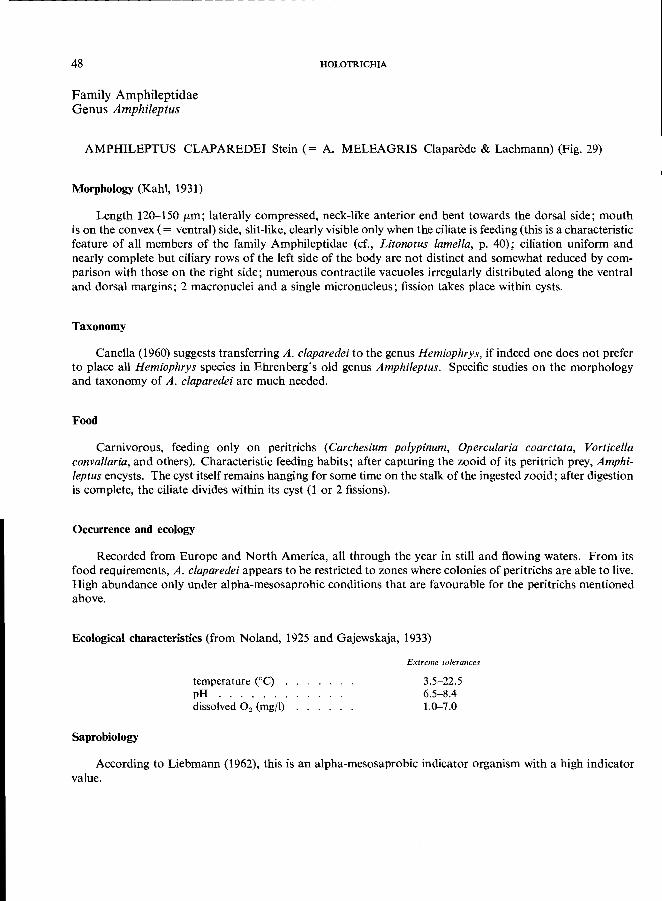
Family Amphileptidae Genus Amphileptus
HOLOTRICHIA
AMPHILEPTUS CLAPAREDEI Stein (= A. MELEAGRIS Claparede & Lachmann) (Fig. 29)
Morphology (Kahl, 1931)
Length 120-150 (Lm; laterally compressed, neck-like anterior end bent towards the dorsal side; mouth is on the convex (= ventral) side, slit-like, clearly visible only when the ciliate is feeding (this is a characteristic feature of all members of the family Amphileptidae (cf., Litonotus lamella, p. 40); ciliation uniform and nearly complete but ciliary rows of the left side of the body are not distinct and somewhat reduced by comparison with those on the right side; numerous contractile vacuoles irregularly distributed along the ventral and dorsal margins; 2 macronuclei and a single micronucleus; fission takes place within cysts.
Taxonomy
Canella (1960) suggests transferring A. claparedei to the genus Hemiophrys, if indeed one does not prefer to place all Hemiophrys species in Ehrenberg's old genus Amphileptus. Specific studies on the morphology and taxonomy of A. claparedei are much needed.
Food
Carnivorous, feeding only on peritrichs (Carchesium polypinum, Opercularia coarctata, Vorticella convallaria, and others). Characteristic feeding habits; after capturing the zooid of its peritrich prey, Amphileptus encysts. The cyst itself remains hanging for some time on the stalk of the ingested zooid; after digestion is complete, the ciliate divides within its cyst (lor 2 fissions).
Occurrence and ecology
Recorded from Europe and North America, all through the year in still and flowing waters. From its food requirements, A. claparedei appears to be restricted to zones where colonies of peritrichs are able to live. High abundance only under alpha-mesosaprobic conditions that are favourable for the peritrichs mentioned above.
Ecological characteristics (from Noland, 1925 and Gajewskaja, 1933)
temperature (0C) . pH ...... . dissolved O 2 (mg/I)
Saprobiology
Extreme tolerances
3.5-22.5 6.5-8.4 1.0-7.0
According to Liebmann (1962), this is an alpha-mesosaprobic indicator organism with a high indicator value.
GYMNOSTOMATIDA: RHABDOPHORINA 49
tr-----"::~"?
c.v.
ma+ml
I Fig. 29. Amphileptus claparedei Stein, right lateral view; redrawn from Canella (1960).
4
50
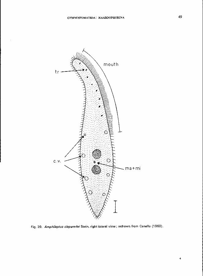
Family Amphileptidae Genus Acineria
HOLOTRICHIA
ACINERIA INCURVATA Dujardin (Fig. 30)
Morphology
Length 50-150 pm; body laterally compressed, outline more or less long and ellipsoid; rather polymorphic; right side with about 12 rows of cilia; left side with only 4 rows in the dorsal part, ventral part of the left side without cilia; mouth slit-like in the ventral part of the anterior region, trichocysts at the edge of the mouth slit; the left ventral region just behind the mouth is somewhat concave and hyaline; 2 macronuclei, and a single micronucleus between them; 1 contractile vacuole at the posterior pole.
Food
Carnivorous, feeding on small ciliates like Cyclidium, Colpidium, and Glaucoma (Kahl, 1931).
Occurrence and ecology
Widely distributed in fresh and brackish water; all types of stagnant and slowly-flowing water. A. incurvata is a true member of the" Colpidietum colpodae" association as described by Snimek-Husek (1958), that is, an association composed of Colpidium colpoda, C. campylum, Glaucoma scintillans, Paramecium caudatum, P. trichium, and Hemiophrys bivacuolata form polysaprobica.
Saprobiology
According to Snimek-Husek (1956), this is a polysaprobic indicator organism, having a high tolerance
to lack of oxygen and high concentrations of NH4 +.
Saprobiological classification
bos aos
Not described:
bms ams
3
ps
7
Family Actinobolinidae e.g., Actinobolina and Enchelyomorpha (p. 22; Fig. 12C, 12D).
4
GYMNOSTOMATIDA: RHABDOPHORINA
mouth
ma +
mi
I c.v .
Fig. 30. Acineria incurvata Dujardin, left lateral view; redrawn from Kahl (1931). Arrows indicate the main features used for identification.
51
52
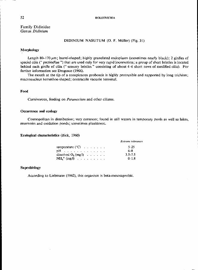
Family Didinidae Genus Didinium
HOLOTRICHIA
DIDINIUM NASUTUM (0. F. Milller) (Fig. 31)
Morphology
Length 80-170 f1-m; barrel-shaped; highly granulated endoplasm (sometimes nearly black); 2 girdles of special cilia (" pectinellae ") that are used only for very rapid locomotion; a group of short bristles is located behind each girdle of cilia (" sensory bristles" consisting of about 4-6 short rows of modified cilia). For
further information see Dragesco (1966). The mouth at the tip of a conspicuous proboscis is highly protrusible and supported by long trichites;
macronucleus horseshoe-shaped; contractile vacuole terminal.
Food
Carnivorous, feeding on Paramecium and other ciliates.
Occurrence and ecology
Cosmopolitan in distribution; very common; found in still waters in temporary pools as well as lakes,
reservoirs and oxidation ponds; sometimes planktonic.
Ecological characteristics (Bick, 1960)
Saprobiology
temperature ceC) . pH ...... . dissolved O. (mg/I) NH/ (mg/I) ...
Extreme tolerances
5-25 6-8
3.5-7.5 0-1.8
According to Liebmann (1962), this organism is beta-mesosaprobic.
GYMNOSTOMATIDA: RHABDOPHORINA 53
/
o I c.v.
Fig. 31. Didinium nasutum (0. F. Muller). Arrows indicate the main features used for identification.
54
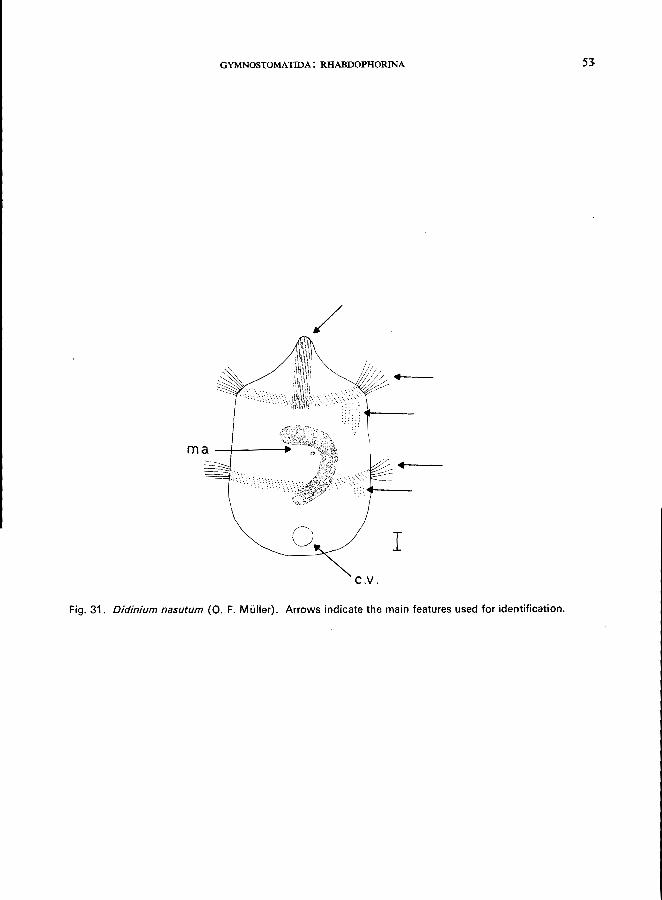
Family Tracheliidae Genus Trachelius
HOLOTRICHIA
TRACHELlUS OVUM Ehrenberg (Fig. 32)
Morphology (Kahl, 1931; Kudo, 1966)
Length 200-400 pm; more or less spheroidal with a distinct short proboscis; right side flattened, often somewhat concave; left side strongly convex; body ciliation uniform, rows of slightly longer cilia on the ventral face of the proboscis, the circular cytostome is located at the base of the proboscis; cytopharynx exhibiting long trichites; numerous contractile vacuoles; macronucleus sausage-shaped; a single micronucleus; endoplasm vacuolated, brown granules often concentrated in the posterior region.
Food
Carnivorous, feeding on flagellates and ciliates.
Occurrence and ecology
Reported from Europe and America, widely distributed in all types of standing and flowing waters; sometimes occurring among the Aufwuchs (periphyton) of artificial substrates (Hentschel, 1916).
Saprobiology
According to Kolkwitz (1950), this is a beta-mesosaprobic indicator organism; single specimens may
occur under alpha-mesosaprobic conditions.
ma +
mi
GYMNOSTOMATIDA: RHABDOPHORINA
pr---~
c.v.
Fig. 32. Trache/ius ovum Ehrenberg, right lateral view. Arrows indicate further features used for identification.
55
56
Family Tracheliidae Genus Dileptus
HOLOTRICHIA
DILEPTUS ANSER (0. F. Muller) (Fig. 33)
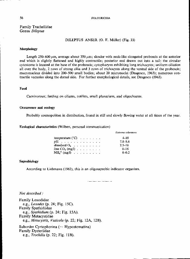
Morphology
Length 250-600 fLm , average about 350 fLm; slender with neck-like elongated proboscis at the anterior end which is slightly flattened and highly contractile; posterior end drawn out into a tail; the circular cytostome is located at the base of the proboscis; cytopharynx exhibiting long trichocysts; uniform ciliation all over the body, 2 rows of strong cilia and 3 rows of trichocysts along the ventral side of the proboscis; macronucleus divided into 200-500 small bodies; about 20 micronuclei (Dragesco, 1963); numerous contractile vacuoles along the dorsal side. For further morphological details, see Dragesco (1963).
Food
Carnivorous; feeding on ciliates, rotifers, small planarians, and oligochaetes.
Occurrence and ecology
Probably cosmopolitan in distribution, found in still and slowly flowing water at all times of the year.
Ecological characteristics (Wilbert, personal communication)
Saprobiology
temperature COc) pH ..... dissolved O. . free CO. (mgfl) NH/ (mgfl) .
Extreme tolerances
4-18 7.8-8.6 2.5-18
0-16 0-0.2
According to Liebmann (1962), this is an oligosaprobic indicator organism.
Not described:
Family Loxodidae e.g., Loxodes (p. 24; Fig. l5C).
Family Spathidiidae e.g., Spathidium (p. 24; Fig. 15A).
Family Metacystidae e.g., Metacystis, Vasicola (p. 22; Fig. 12A, 12B).
Suborder Cyrtophorina (= Hypostomatina) Family Dysteriidae
e.g., Trochilia (p. 22; Fig. lIB).
GYMNOSTOMATIDA: RHABDOPHORINA 57
tr
pr--....
ma
100 J.ltll
Fig. 33. Dileptus anser (0. F. Muller), right lateral view. Arrows indicate the main features used for identification.
58 HOLOTRICHlA
Suborder CYRTOPHORINA (=HYPOSTOMATINA)
Family Chlamydodontidae (= Chilodonellidae) Genus Chilodonella
CHILODONELLA CUCULLULUS (0. F. Milller) (Fig. 34)
Morphology
Length 75-300 p.m, usually about 150 p'm; body dorsoventrally flattened; ventral surface flat with about 20 ciliary rows, the anterior part of the dorsal surface flattened with only 1 transverse row of cilia (" dorsal bristles "), the posterior part more or less convex and lacking cilia; mouth opening round, about 12 cytopharyngeal trichites forming a tube, macronucleus oval with a characteristic concentric structure; 1 small micronucleus, about 6-8 contractile vacuoles. For details on morphology, division, etc., see Radzi-
kowski (1966).
Food
Diatoms, blue-green algae, bacteria.
Occurrence and ecology
Cosmopolitan in distribution, found throughout the year in still and flowing waters (e.g., activated
sludge, trickling filters, sewage drains, oxidation ponds) (Liebmann, 1962).
Ecological characteristics (Bick, 1968)
Saprobiology
temperature COC) . pH ...... . dissolved O2 (mg/i) free CO2 (mg/l) . NH. + (mg/l) . . free NH3 (mgjl) . N02- (mg/l) . . H 2S ..... . brackish water (seawater type) athalassogenic natron lake water bacteria (plate counts on peptone
agar) . . . . . . . . . . .
Extreme tolerances
0-30 6.3-8.5
0-12 0-72
0.1-100 0-20 0-22
o up to 7 g of total salt content per litre up to 7 g of total salt content per litre
140 000-10 000 OOO/ml
This is an alpha-mesosaprobic indicator organism (Liebmann, 1962). C. cucullulus is able to tolerate a wide range of environmental conditions; single specimens may occur in all zones of self-purification. Only its occurrence in great numbers or its mass-development are significant to the monitoring of water quality.
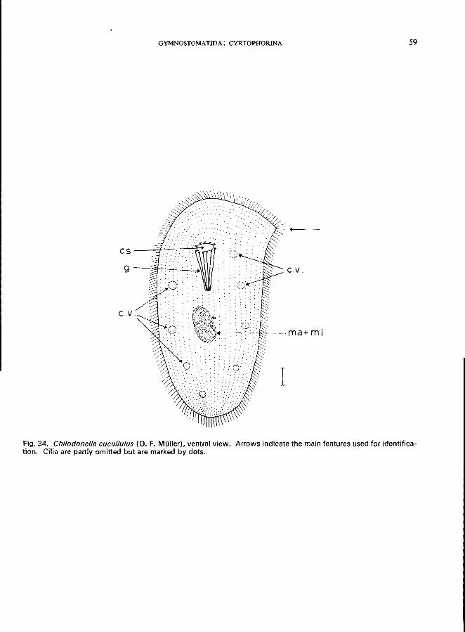
GYMNOSTOMATIDA: CYRTOPHORINA
Fig. 34. Chilodonella cucullulus (0. F. Muller), ventral view. Arrows indicate the main features used for identification. Cilia are partly omitted but are marked by dots.
59
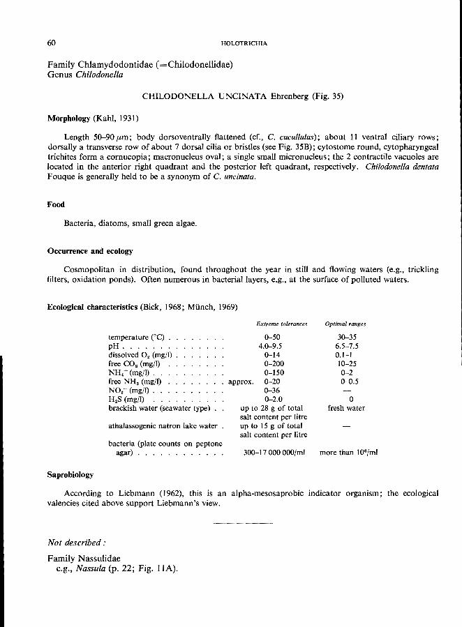
60 HOLOTRICHIA
Family Chlamydodontidae (=Chilodonellidae) Genus Chilodonella
CHILODONELLA UNCINATA Ehrenberg (Fig. 35)
Morphology (Kahl, 1931)
Length 50-90 (Lm; body dorsoventrally flattened (cr., c. cucullulus); about 11 ventral ciliary rows; dorsally a transverse row of about 7 dorsal cilia or bristles (see Fig. 35B); cytostome round, cytopharyngeal trichites form a cornucopia; macronucleus oval; a single small micronucleus; the 2 contractile vacuoles are located in the anterior right quadrant and the posterior left quadrant, respectively. Chilodonella dentata
Fouque is generally held to be a synonym of C. uncinata.
Food
Bacteria, diatoms, small green algae.
Occurrence and ecology
Cosmopolitan in distribution, found throughout the year in still and flowing waters (e.g., trickling filters, oxidation ponds). Often numerous in bacterial layers, e.g., at the surface of polluted waters.
Ecological characteristics (Bick, 1968; MUnch, 1969)
Saprobiology
temperature eC) . pH ...... . dissolved O. (mgfl) free CO. (mg/I) NH.+ (mg/I) .. free NHa (mgfl) NO.- (mg/l) .. H.S (mg/I) brackish water (seawater type) .
athalassogenic natron lake water
bacteria (plate counts on peptone agar) ........... .
Extreme tolerances
0-50 4.0-9.5
0-14 0-200 0-150
approx. 0-20 0-36 0-2.0
up to 28 g of total salt content per litre up to 15 g of total salt content per litre
300-17000000/ml
Optimal ranges
30-35 6.5-7.5 0.1-1 10-25 0-2 0-0.5
o fresh water
more than 106/ml
According to Liebmann (1962), this is an alpha-mesosaprobic indicator organism; the ecological
valencies cited above support Liebmann's view.
Not described:
Family Nassulidae e.g., Nassula (p. 22; Fig. IIA).
GYMNOSTOMATIDA: CYRTOPHORINA
A c de. -'---+-10 B
c. V .---=-'l~ 9
'-"'£=.-- c. v .
I ventral view dorsal view
Fig. 35. Chilodonella uncinata Ehrenberg. A, Ventral view; B, Dorsal view. Arrows indicate the main features used for identification. The posterior part (marked by an arrow) is free from cilia.
61
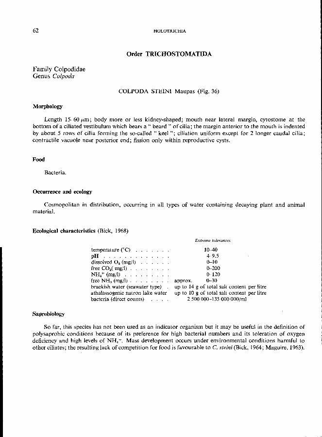
62
Family Colpodidae Genus Colpoda
Morphology
HOLOTRICHIA
Order TRICHOSTOMATIDA
COLPODA STEINI Maupas (Fig. 36)
Length 15-60 pm; body more or less kidney-shaped; mouth near lateral margin, cytostome at the bottom of a ciliated vestibulum which bears a " beard" of cilia; the margin anterior to the mouth is indented by about 5 rows of cilia forming the so-called" keel"; ciliation uniform except for 2 longer caudal cilia; contractile vacuole near posterior end; fission only within reproductive cysts.
Food
Bacteria.
Occurrence and ecology
Cosmopolitan in distribution, occurring in all types of water containing decaying plant and animal
material.
Ecological characteristics (Bick, 1968)
Saprobiology
temperature eq . pH ...... . dissolved O. (mg/I) free CO.( mg/I) . NH4+ (mg/I) . . . free NH3 (mg/I). . brackish water (seawater type) athalassogenic natron lake water bacteria (direct counts)
Extreme tolerances
10-40 4-9.5 0-10 0-200 0-120
approx. 0-30 up to 14 g of total salt content per litre up to 10 g of total salt content per litre
2500000-135000000/m1
So far, this species has not been used as an indicator organism but it may be useful in the definition of polysaprobic conditions because of its preference for high bacterial numbers and its toleration of oxygen deficiency and high levels of NH4 +. Mass development occurs under environmental conditions harmful to other ciliates; the resulting lack of competition for food is favourable to C. steini (Bick, 1964; Maguire, 1963).
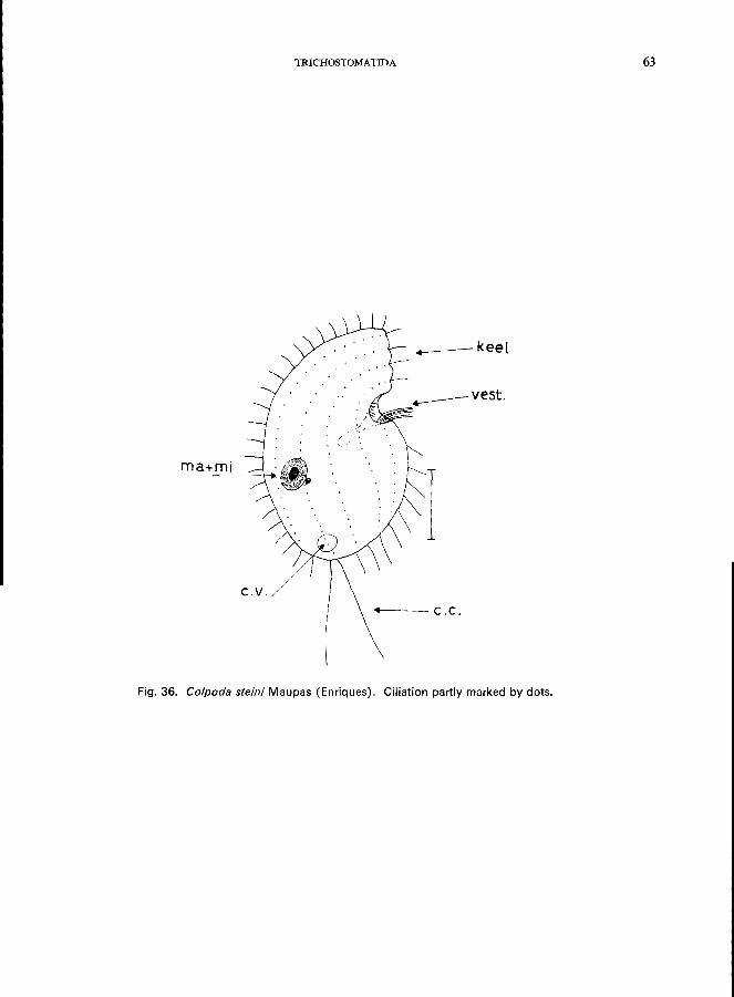
TRICHOSTOMATIDA 63
• ---keel
c.v.
.. C.C.
Fig. 36. Co/poda steini Maupas (Enriques). Ciliation partly marked by dots.
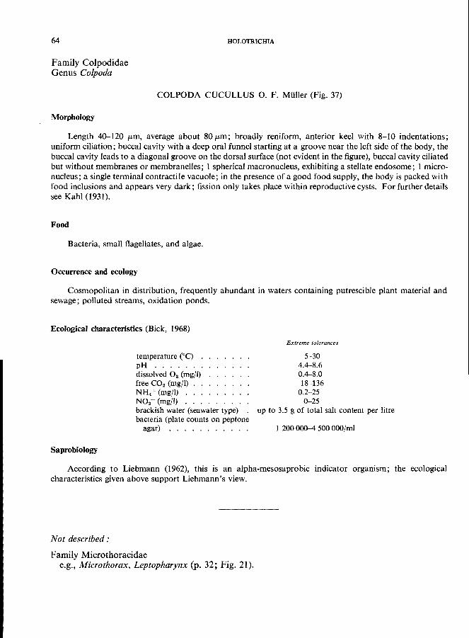
64
Family Colpodidae Genus Co/poda
HOLOTRICHIA
COLPODA CUCULLUS o. F. Mtiller (Fig. 37)
Morphology
Length 40-120 (Lm, average about 80 (Lm; broadly reniform, anterior keel with 8-10 indentations; uniform ciliation; buccal cavity with a deep oral funnel starting at a groove near the left side of the body, the buccal cavity leads to a diagonal groove on the dorsal surface (not evident in the figure), buccal cavity ciliated but without membranes or membranelles; 1 spherical macronucleus, exhibiting a stellate endosome; 1 micronucleus; a single terminal contractile vacuole; in the presence of a good food supply, the body is packed with food inclusions and appears very dark; fission only takes place within reproductive cysts. For further details
see Kahl (1931).
Food
Bacteria, small flagellates, and algae.
Occurrence and ecology
Cosmopolitan in distribution, frequently abundant in waters containing putrescible plant material and
sewage; polluted streams, oxidation ponds.
Ecological characteristics (Bick, 1968)
temperature cae) . pH ...... . dissolved O. (mg/I) free CO2 (mg/I) NH.+ (mg/I) .. . N02- (mg/I) .. . brackish water (seawater type) bacteria (plate counts on peptone
agar) . . . . . . . . . . .
Extreme tolerances
5-30 4.4-8.6 0.4-8.0 18-136
0.2-25 0-25
up to 3.5 g of total salt content per litre
1 200 000-4 500 OOO/mi
Saprobiology
According to Liebmann (1962), this is an alpha-mesosaprobic indicator organism; the ecological
characteristics given above support Liebmann's view.
Not described:
Family Microthoracidae e.g., Microthorax. Leptopharynx (p. 32; Fig. 21).
ma +.
ml
c.v.
TRICHOSTOMATIDA
.-----b.c.
Fig. 37. Co/poda cucullus D. F. Muller, ventral view. Food vacuoles omitted, ciliation partly omitted and marked by dots.
5
65
66
Family Plagiopylidae Genus Plagiopyla
HOLOTRICHIA
PLAGIOPYLA NASUTA Stein (Fig. 38)
Morphology
Length 80--180 fLm; dorsoventrally flattened; uniform ciliation, sometimes yellowish or beige in colour, oral groove transverse, cytostome on the left end; a so-called" striated structure" (of unknown function) is present on the dorsal side starting from the edge of the mouth groove and turning backwards (see Fig. 38B); trichocysts in large numbers all over the body; macronucleus near the right margin in the middle of the body; contractile vacuole at the posterior end; P. nasuta exhibits great morphological variability. For further
details see Kahl (1931) and Liebmann (1962).
Food
Bacteria, algae (Liebmann, 1962).
Occurrence and ecology
Reported in Europe and North America, sapropelobiontic; saprobiological classification according to Liebmann (1962): polysaprobic, indicator of hydrogen sulfide, does not occur in raw sewage; frequently associated with Discomorphella, Metopus, and Caenomorpha.
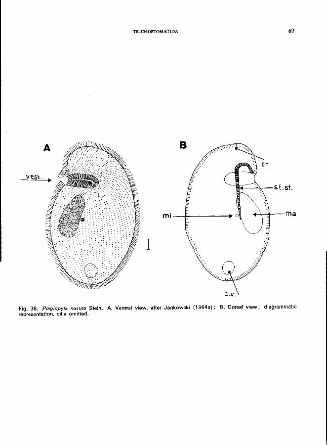
TRICHOSTOMATIDA 67
B
ves~.
I!!I+-~-st.st.
m;--~--______ -+ +-'''''''t----m a
I
c.v. Fig. 38. Plagiopyla nasuta Stein. A, Ventral view, after Jankowski (1964a); 8, Dorsal view; diagrammatic representation, cilia omitted.
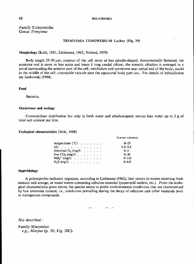
68
Family Trimyemidae Genus Trimyema
HOLOTRICHIA
TRIMYEMA COMPRESSUM Lackey (Fig. 39)
Morphology (Kahl, 1931; Liebmann, 1962; Noland, 1959)
Body length 25-50 pm; contour of the cell more or less spindle-shaped, dorsoventrally flattened, the posterior end is more or less acute and bears 1 long caudal cilium; the somatic ciliation is arranged in a spiral surrounding the anterior part of the cell; vestibulum and cytostome near apical end of the body; nuclei in the middle of the cell; contractile vacuole near the equatorial body part too. For details of infraciliation
see Jankowski (1964).
Food
Bacteria.
Occurrence and ecology
Cosmopolitan distribution but only in fresh water and athalassogenic natron lake water up to 2 g of
total salt content per litre.
Ecological characteristics (Bick, 1968)
Saprobiology
temperature Ce) . pH ...... . dissolved O. (mgfl) free CO. (mg/I) NH.+ (mg/I) H.S (mg/I) ..
Extreme tolerances
0-25 6.8-8.5
0-3 0-30 0-3.0 0-6.0
A polysaprobic indicator organism, according to Liebmann (1962), that occurs in waters receiving fresh manure and sewage, or waste waters containing cellulose material (paper-mill outlets, etc.). From the ecological characteristics given above, the species seems to prefer environmental conditions that are characterized by low ammonia content, i.e., conditions prevailing during the decay of cellulose and other materials poor
in nitrogenous compounds.
Not described:
Family Marynidae e.g., Maryna (p. 30; Fig. 20C).
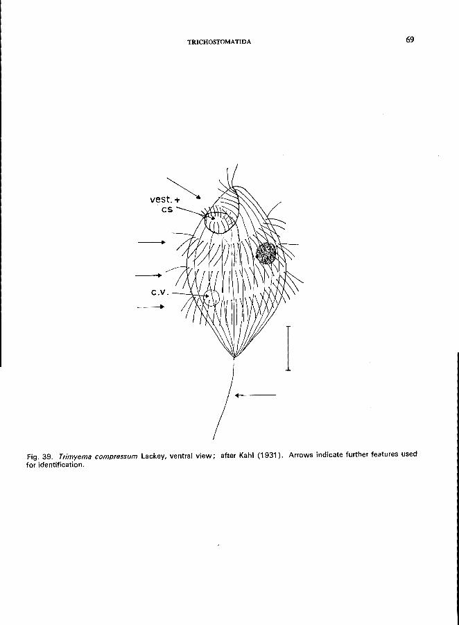
TRICHOSTOMA TIDA
-c . v . --'-LJ. /;
Fig. 39. Trimyema compressum Lackey, ventral view; after Kahl (1931). Arrows indicate further features used for identification.
69
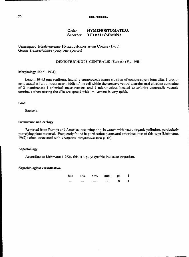
70 HOLOTRICHIA
Order HYMENOSTOMATIDA Suborder TETRAHYMENINA
Unassigned tetrahymenine Hymenostomes sensu Corliss (1961) Genus Dexiotrichides (only one species)
DEXIOTRICHIDES CENTRALIS (Stokes) (Fig. 19B)
Morphology (Kahl, 1931)
Length 30-45 pm; reniform, laterally compressed; sparse ciliation of comparatively long cilia, 1 prominent caudal cilium; mouth near middle of the cell within the concave ventral margin; oral ciliation consisting of 2 membranes; 1 spherical macronucleus and 1 micronucleus located anteriorly; contractile vacuole terminal; when resting the cilia are spread wide; movement is very quick.
Food
Bacteria.
Occurrence and ecology
Reported from Europe and America, occurring only in waters with heavy organic pollution, particularly putrefying plant material. Frequently found in purification plants and other localities of this type (Liebmann, 1962); often associated with Trimyema compressum (see p. 68).
Saprobiology
According to Liebmann (1962), this is a polysaprobic indicator organism.
Saprobiological classification
bos aos bms ams
2
ps
8 4
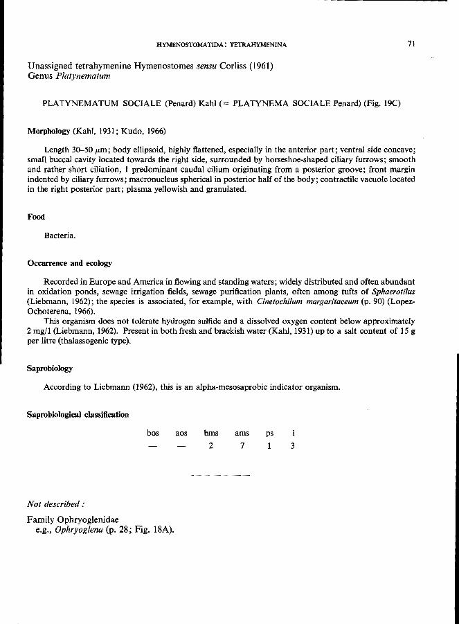
HYMENOSTOMATIDA: TETRAHYMENINA
Unassigned tetrahymenine Hymenostomes sensu Corliss (1961) Genus P/atynematum
PLATYNEMATUM SOCIALE (Penard) Kahl (= PLATYNEMA SOCIALE Penard) (Fig. 19C)
Morphology (Kahl, 1931; Kudo, 1966)
71
Length 30-50 pm; body ellipsoid, highly flattened, especially in the anterior part; ventral side concave; small buccal cavity located towards the right side, surrounded by horseshoe-shaped ciliary furrows; smooth and rather short ciliation, 1 predominant caudal cilium originating from a posterior groove; front margin indented by ciliary furrows; macronucleus spherical in posterior half of the body; contractile vacuole located in the right posterior part; plasma yellowish and granulated.
Food
Bacteria.
Occurrence and ecology
Recorded in Europe and America in flowing and standing waters; widely distributed and often abundant in oxidation ponds, sewage irrigation fields, sewage purification plants, often among tufts of Sphaerotilus (Liebmann, 1962); the species is associated, for example, with Cinetochilum margaritaceum (p. 90) (LopezOchoterena, 1966).
This organism does not tolerate hydrogen sulfide and a dissolved oxygen content below approximately 2 mg/l (Liebmann, 1962). Present in both fresh and brackish water (Kahl, 1931) up to a salt content of 15 g per litre (thalassogenic type).
Saprobiology
According to Liebmann (1962), this is an alpha-mesosaprobic indicator organism.
Saprobiological classification
bos aos
Not described:
Family Ophryoglenidae e.g., Ophryog/ena (p. 28; Fig. 18A).
bms
2 ams
7
ps
1 3
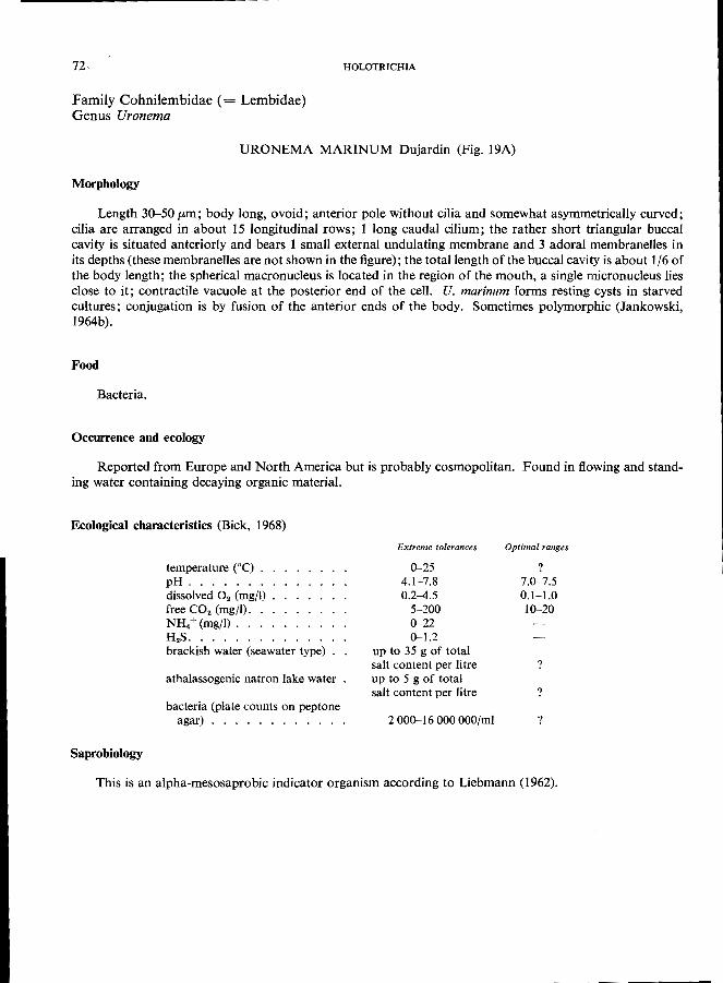
72
Family Cohnilembidae (= Lembidae) Genus Uronema
HOLOTRICHIA
URONEMA MARINUM Dujardin (Fig. 19A)
Morphology
Length 30-50 pm; body long, ovoid; anterior pole without cilia and somewhat asymmetrically curved; cilia are arranged in about 15 longitudinal rows; 1 long caudal cilium; the rather short triangular buccal cavity is situated anteriorly and bears 1 small external undulating membrane and 3 adoral membranelles in its depths (these membranelles are not shown in the figure); the total length of the buccal cavity is about 1/6 of the body length; the spherical macronucleus is located in the region of the mouth, a single micronucleus lies close to it; contractile vacuole at the posterior end of the cell. U. marinum forms resting cysts in starved cultures; conjugation is by fusion of the anterior ends of the body. Sometimes polymorphic (Jankowski,
1964b).
Food
Bacteria.
Occurrence and ecology
Reported from Europe and North America but is probably cosmopolitan. Found in flowing and stand
ing water containing decaying organic material.
Ecological characteristics (Bick, 1968)
Saprobiology
temperature CC) . pH ...... . dissolved O. (mg/I) free CO. (mg/I). NH.+ (mgfl) ... . H.S ....... . brackish water (seawater type) .
athalassogenic natron lake water
bacteria (plate counts on peptone agar) ........... .
Extreme tolerances
0-25 4.1-7.8 0.2--4.5
5-200 0-22 0-1.2
up to 35 g of total salt content per litre up to 5 g of total salt content per litre
2 000--16 000 OOO/mi
Optimal ranges
? 7.0-7.5 0.1-1.0 10--20
?
?
?
This is an alpha-mesosaprobic indicator organism according to Liebmann (1962).
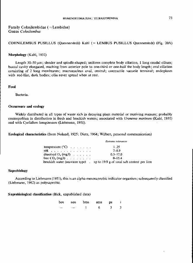
HYMENOSTOMATlDA: TETRAHYMENINA
Family Cohnilembidae (= Lembidae) Genus Cohnilembus
73
COHNILEMBUS PUSILLUS (Quennerstedt) Kahl (= LEMBUS PUSILLUS Quennerstedt) (Fig. 20A)
Morphology (Kahl, 1931)
Length 30-50 /Lm ; slender and spindle-shaped; uniform complete body ciliation, 1 long caudal cilium; buccal cavity elongated, reaching from anterior pole to one-third or one-half the body length; oral ciliation consisting of 2 long membranes; macronucleus oval, central; contractile vacuole terminal; endoplasm with rod-like, dark bodies; cilia never spread when at rest.
Food
Bacteria.
Occurrence and ecology
Widely distributed in all types of water rich in decaying plant material or receiving manure; probably cosmopolitan in distribution in fresh and brackish waters; associated with Uronema marinum (Kahl, 1931) and with Cyclidium lanuginosum (Liebmann, 1951).
Ecological characteristics (from Noland, 1925; Dietz, 1964; Wilbert, personal communication)
Saprobiology
temperature (0C) . pH ...... . dissolved O2 (mg/l) free CO2 (mg/I) . . brackish water (seawater type)
Extreme tolerances
1-25 7-8.9
0.3-17.0 0-15.4
up to 19.9 g of total salt content per litre
According to Liebmann (1951), this is an alpha-mesosaprobic indicator organism; subsequently classified (Liebmann, 1962) as polysaprobic.
SaprobiologicaI classification (Bick, unpublished data)
bos aos bms ams
6
ps
3 3
74
Family Tetrahymenidae Genus Tetrahymena
HOLOTRICHIA
TETRAHYMENA PYRIFORMIS (Ehrenberg) Lwoff (= GLAUCOMA PYRIFORMIS (Ehrenberg) Schewiakoff (Fig. 40)
Morphology
Length 25-90 pm; body ovoid and uniformly ciliated; mouth roughly triangular, longitudinal axis of the buccal cavity parallel to that of the cell itself; buccal cavity containing an undulating membrane on the right side and an adoral zone of 3 membranelles on the left (" tetrahymenal complex "); the spherical macronucleus is situated medially and is usually accompanied by 1 micronucleus (amicronucleate strains occur);
1 contractile vacuole near the posterior end.
Food
Bacteria.
Occurrence and ecology
Cosmopolitan distribution in fresh water, frequently occurring in waters containing plant or animal materials in which bacterial decomposition has commenced, particularly in waters polluted with manure;
recorded in trickling filters, oxidation ponds, sewage drains.
Ecological characteristics (Bick, unpublished data)
Saprobiology
temperature eC) . pH ...... . dissolved O. (mg/I) free CO2 (mg/I) . NH.+ (mg/I) .. free NH3 (mgfl) . H.S (mg/I) ...
Extreme tolerances
1-40 7.0-8.9
0-10 0-200 0-250 0-25 0-2
A polysaprobic indicator organism according to Liebmann (1962).
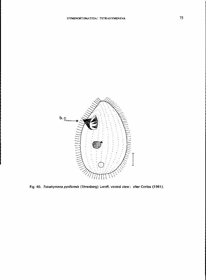
HYMENOSTOMATIDA: TETRAHYMENINA 75
b.c. ----.~
I Fig. 40. Tetrahymena pyriformis (Ehrenberg) Lwoff, ventral view; after Corliss (1961).
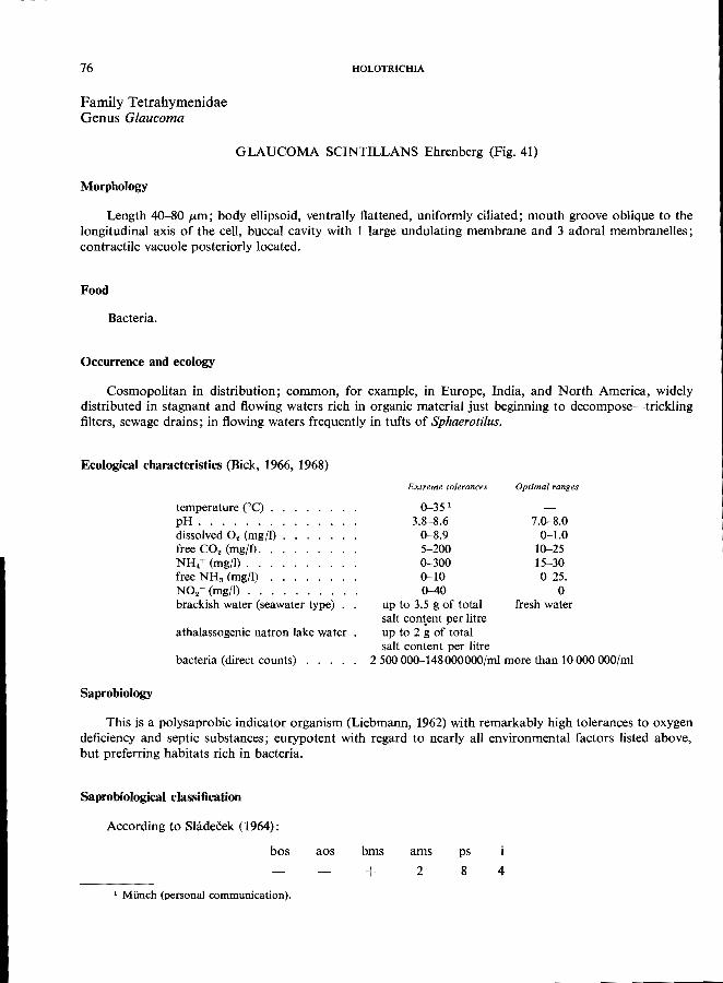
76 HOLOTRICHIA
Family Tetrahymenidae Genus Glaucoma
GLAUCOMA SCINTILLANS Ehrenberg (Fig. 41)
Morphology
Length 40-80 /Lm ; body ellipsoid, ventrally flattened, uniformly ciliated; mouth groove oblique to the longitudinal axis of the cell, buccal cavity with 1 large undulating membrane and 3 adoral membranelles;
contractile vacuole posteriorly located.
Food
Bacteria.
Occurrence and ecology
Cosmopolitan in distribution; common, for example, in Europe, India, and North America, widely distributed in stagnant and flowing waters rich in organic material just beginning to decompose-trickling filters, sewage drains; in flowing waters frequently in tufts of Sphaerotilus.
Ecological characteristics (Bick, 1966, 1968)
Saprobiology
temperature (0e) . pH ...... . dissolved O. (mgfl) free CO. (mg/I). NH4+ (mg/I) .. free NH3 (mg/l) NO.- (mg/l) . . brackish water (seawater type)
athalassogenic natron lake water
bacteria (direct counts) . . . .
Extreme tolerances
0-35 1
3.8-8.6 0-8.9 5-200 0-300 0-10 0-40
Optimal ranges
7.0-8.0 0-1.0
10-25 15-30 0-25.
o up to 3.5 g of total fresh water salt content per litre up to 2 g of total salt content per litre
2500 000-148000000/ml more than 10000 OOO/ml
This is a polysaprobic indicator organism (Liebmann, 1962) with remarkably high tolerances to oxygen deficiency and septic substances; eurypotent with regard to nearly all environmental factors listed above,
but preferring habitats rich in bacteria.
Saprobiological classification
According to Sladecek (1964):
bos
1 MUnch (personal communication).
aos bms ams
+ 2
ps
8 4
HYMENOSTOMATIDA: TETRAHYMENINA 77
Fig. 41. Glaucoma scintil/ans Ehrenberg, ventral view.
78 HOLOTRICHIA
Family Tetrahymenidae Genus Colpidium
COLPIDIUM CAMPYLUM Stokes (Fig. 42)
Morphology
Length 50-120 fLm; elongated, reniform, sometimes ovoid; uniform ciliation except for a group of longer cilia at the posterior pole; preoral suture slightly displaced towards the ventral side; suture curved to right; small triangular buccal cavity near the right side of the body at about one-quarter of the body length from the anterior pole; buccal ciliation tetrahymenal; 1 undulating membrane on the right side and 3 adoral membranelles on the left; 1 spherical macronucleus and 1 micronucleus in the central part of the body; 1 contractile vacuole located near the right margin. For special morphological features (silver line system), see Jankowski (1967). It should be mentioned that Jankowski (1967) proposed to put C. campy/um into a
genus of its own (Dexiostoma n.g.).
Food
Bacteria, small flagellates.
Occurrence and ecology
Cosmopolitan, occurring in waters rich in bacterial decomposition of organic material, e.g., in tufts
of Sphaerotilus.
Ecological characteristics (Bick, 1968)
temperature COC) . pH ...... . dissolved O. (mg/l) free CO. (mgfl) . NH. + (mgfl) . . free NH. (mg/I) . NO.- (mg/l) . . brackish water (seawater type)
Extreme tolerances
4-30 4.0-8.9
0-11.0 0-200
0.2-160 0-15 0-15
up to 3.5 g of total salt content per litre
In laboratory experiments, no development occurred in athalassogenic natron lake water.
Bacteria:
Saprobiology
(1) plate counts on peptone agar
(2) direct counts . (3) optimal ranges . . . . . .
9 000-4 OOOOOO/ml 20000000-150 000 OOO/ml above 10"/ml for (1) or 100 x 10G/ml for (2)
c. campy/um was not used as an indicator organism in the Saprobiensystem established by Liebmann. Snimek-Husek (1956) proposed to use C. campy/um as a beta-polysaprobic indicator organism (betapolysaprobic sensu Snimek-Husek corresponds approximately to the polysaprobic zone of Kolkwitz). Zelinka & Marvan (1961) also classified C. campy/um as a polysaprobic indicator organism.
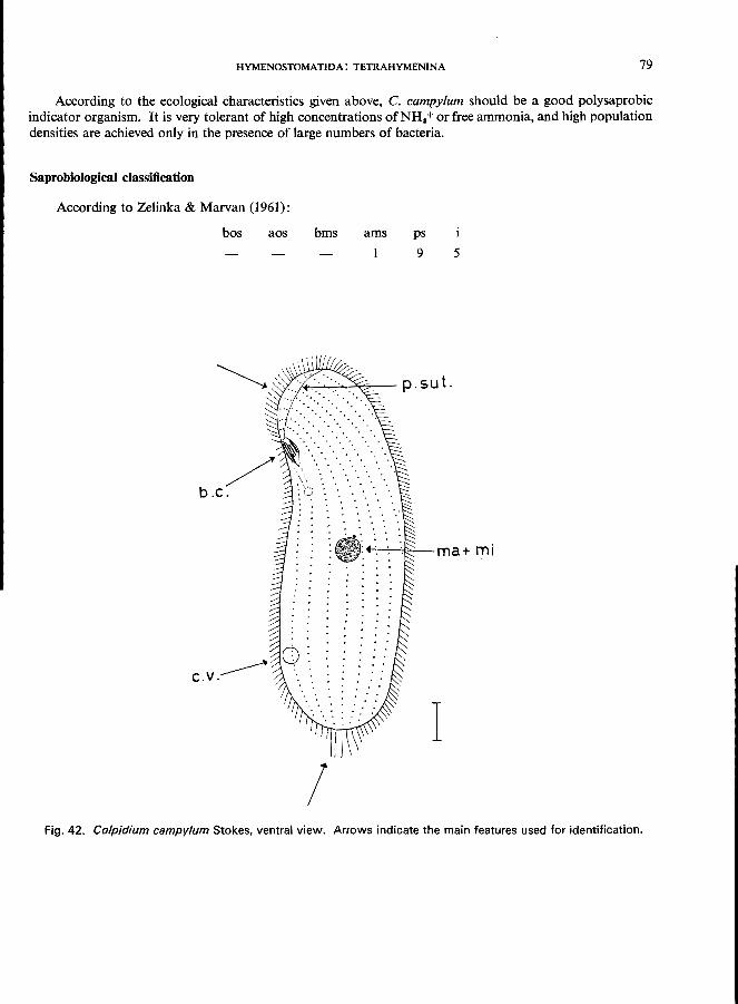
HYMENOSTOMATIDA: TETRAHYMENINA 79
According to the ecological characteristics given above, C. campy/urn should be a good polysaprobic indicator organism. It is very tolerant of high concentrations of NH4 + or free ammonia, and high population densities are achieved only in the presence of large numbers of bacteria.
Saprobiological classification
According to Zelinka & Marvan (1961):
bos aos bms ams ps
9 5
I ..... ~:...,.....:.&-- p.sut.
~~~-ma+mi
I I
Fig. 42. Co/pidium campy/urn Stokes, ventral view. Arrows indicate the main features used for identification.

80 HOLOTRICHIA
Family Tetrahymenidae Genus Colpidium
COLPIDIUM COLPODA (Ehrenberg) (Fig. 43)
Morphology (Kahl, 1931)
Length 100-150 /Lm; ovoid or reniform, elongated; except for a few long caudal cilia, uniform ciliation; about 55 ciliary meridians; preoral suture curved to the left and widely displaced to the ventral side; buccal cavity near the right side of the body at about one-quarter of the body length from the anterior pole; buccal ciliation similar to that of C. campylum; 1 spherical macronucleus, a single micronucleus; the contractile vacuole is located in the central part of the body near the dorsal face. For silver line system, see Jankowski
(1967).
Food
Bacteria, small flagellates.
Occurrence and ecology
Cosmopolitan in distribution, present throughout the year in waters rich in putrefying organic material; very often occurs on excrement just starting to decompose. Frequently associated with Cinetochilum margaritaceum, Colpidium campylum, Glaucoma scintillans, Paramecium trichium, Tetrahymena pyriformis, and other forms; this association was called" Colpidietum colpodae" by Snimek-Husek (1958), the name being
derived from the most characteristic species of the association.
Ecological characteristics (Bick, 1968)
temperature eC) . pH ...... . dissolved O. (mg/l) free CO. (mg/l) . . NH. + (mg/l) . . . free NHa (according to Stammer,
1953) . . . . . . . . . . . brackish water (seawater type) . bacteria (plate counts on peptone
agar) .......... .
Extreme tolerances
4-30 6.2-8.3
0-7.2 10-56
0.4-25
up to 25 mg/l up to 3.5 g of total salt content ptr litre
30 000-17 OOOOOO/ml
Saprobiology C. colpoda was classified as a polysaprobic indicator organism by Liebmann (1962) and as a beta
polysaprobic organism by Snlmek-Husek (1956). The ecological characteristics given above support these statements. Only mass development should be held to indicate polysaprobic conditions, since single examples
may also occur in the alpha-mesosaprobic zone.
Saprobiological classification
According to Zelinka & Marvan (1961) after Siadeeek (1964):
bos aos bms ams
3
ps
7 4
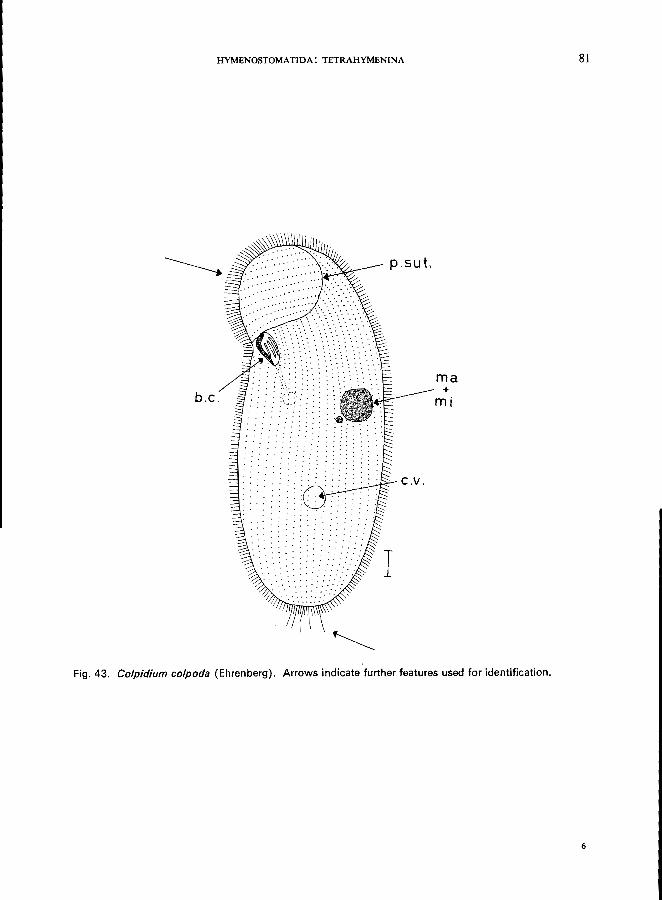
HYMENOSTOMATIDA: TETRAHYMENINA
b.c. ma +
mi
Fig. 43. Colpidium colpoda (Ehrenberg). Arrows indicate further features used for identification.
81
6
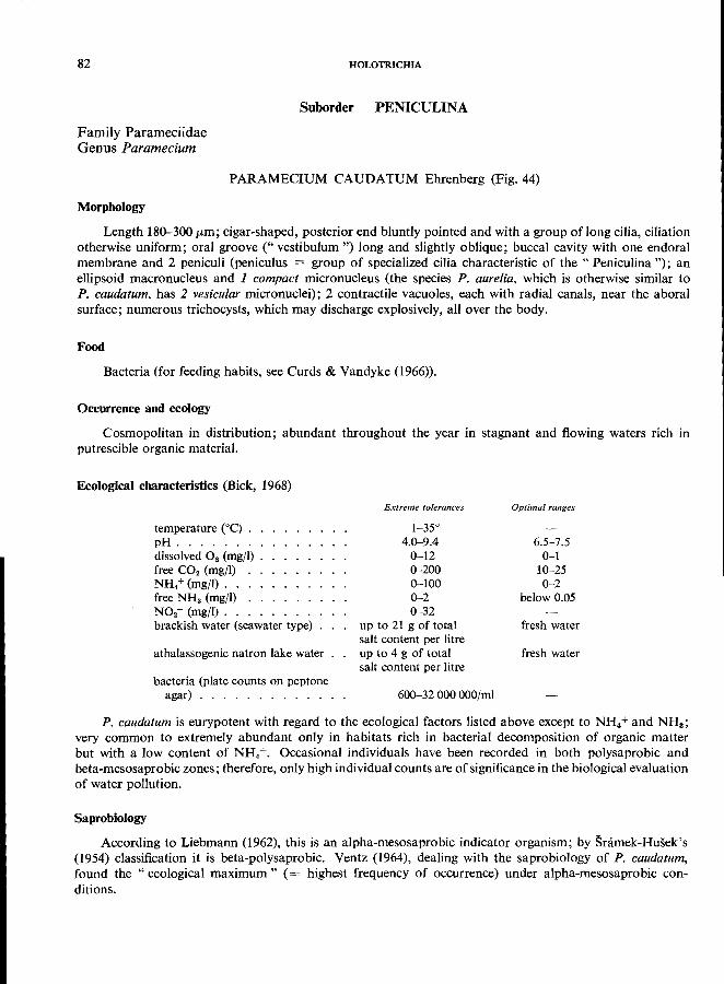
82 HOLOTRICHIA
Suborder PENlCULINA
Family Parameciidae Genus Paramecium
PARAMECIUM CAUDATUM Ehrenberg (Fig. 44)
Morphology Length 180-300 (Lm; cigar-shaped, posterior end bluntly pointed and with a group oflong cilia, ciliation
otherwise uniform; oral groove (" vestibulum ") long and slightly oblique; buccal cavity with one endoral membrane and 2 peniculi (peniculus = group of specialized cilia characteristic of the " Peniculina "); an ellipsoid macronucleus and 1 compact micronucleus (the species P. aurelia, which is otherwise similar to P. caudatum, has 2 vesicular micronuclei); 2 contractile vacuoles, each with radial canals, near the aboral surface; numerous trichocysts, which may discharge explosively, all over the body.
Food
Bacteria (for feeding habits, see Curds & Vandyke (1966)).
Occurrence and ecology Cosmopolitan in distribution; abundant throughout the year in stagnant and flowing waters rich in
putrescible organic material.
Ecological characteristics (Bick, 1968)
temperature eC) . . pH ....... . dissolved O. (mg/I) . free CO. (mg/I) NH/(mg/i) .. free NHa (mg/I) NO.- (mg/I). . brackish water (seawater type)
athalassogenic natron lake water
bacteria (plate counts on peptone agar) ........... .
Extreme tolerances
1-35° 4.0-9.4
0-12 0-200 0-100 0-2 0-32
up to 21 g of total salt content per litre up to 4 g of total salt content per litre
600-32 000 OOO/mi
Optimal ranges
6.5-7.5 0-1
10-25 0-2
below 0.05
fresh water
fresh water
P. cauda tum is eurypotent with regard to the ecological factors listed above except to N H4+ and NHa; very common to extremely abundant only in habitats rich in bacterial decomposition of organic matter but with a low content of NH4+. Occasional individuals have been recorded in both polysaprobic and beta-mesosaprobic zones; therefore, only high individual counts are of significance in the biological evaluation
of water pollution.
Saprobiology According to Liebmann (1962), this is an alpha-mesosaprobic indicator organism; by Snimek-Husek's
(1954) classification it is beta-polysaprobic. Ventz (1964), dealing with the saprobiology of P. caudatum, found the" ecological maximum" (= highest frequency of occurrence) under alpha-mesosaprobic con-
ditions.
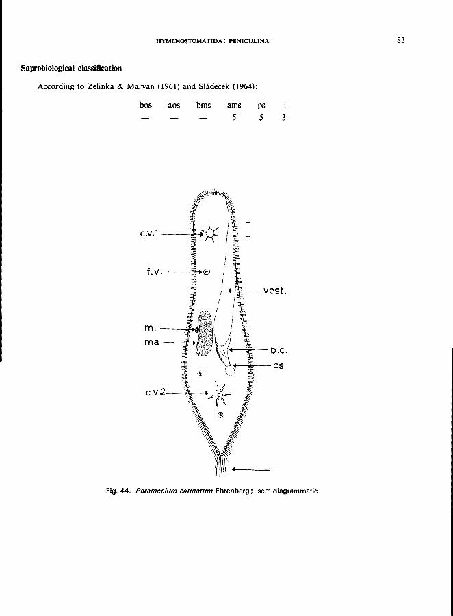
HYMENOSTOMATIDA: PENICULINA
SaprobiolOgical classification
According to Zelinka & Marvan (1961) and Sladeeek (1964):
bos aos bms
C.v.l------"I41
m i ---=100:.,
ma--~
C.v .L. ------"'f,---.
ams
5 pS
5
4-+.lo;-- V es t .
---;~- b.c.
-~;---Cs
3
Fig. 44. Paramecium caudatum Ehrenberg; semidiagrammatic.
83
84
Family Parameciidae Genus Paramecium
HOLOTRICHIA
PARAMECIUM BURSARIA Ehrenberg (Fig. 45)
Morphology
Length 90-150 fLm; foot-shaped, more or less flattened; uniform ciliation except for a group of long caudal cilia; green with symbiotic zoochlorellae; a long broad oral groove (vestibule) leads to the buccal cavity, the buccal ciliary apparatus is characterized by 2 " peniculi "; 1 1 macronucleus, 1 compact micronucleus; 2 contractile vacuoles; numerous prominent trichocysts.
Food
Bacteria (sometimes purple-coloured sulfur bacteria), flagellates; starving individuals digest their symbionts.
Occurrence and ecology
Cosmopolitan in distribution, e.g., Europe, North America, India, in still waters rich in higher plants and algae, such as ponds or littoral zones of lakes, forest pools rich in decaying leaves, oxidation ponds, polluted streams. Under laboratory conditions this ciliate occurs in cultures rich in peptone, under which conditions it can withstand high ammonium and low oxygen levels.
Ecological characteristics (Bick, 1968)
temperature COc) . pH ...... . dissolved O. (mg/I) free CO. (mg/I) . NH.+ (mg/I) . . free NHa (mg/I) . H.S (mg/\) ... bacteria (direct counts) .
Extreme tolerances
0-25 6.4-8.2 0.1-11.0
0-48 0-60 0-3 0-1.1
3 000 000-140 000 OOO/mi
Under laboratory conditions no development occurs in thalassogenic brackish waters. The species develops in athalassogenic natron lake water with a total salt content of up to 2 g per litre.
Saprobiology
According to Liebmann (1962), this is a beta-mesosaprobic indicator organism. My results do not support this opinion; I found P. bursaria able to tolerate high levels of NH. + and a very low oxygen content; under these conditions the populations increased to about 500 individuals per ml, and were accompanied by P. trichium, Colpidium campylum, Glaucoma scintillans, and other alpha-mesosaprobic or even polysaprobic indicator organisms. These results (cf., Krishnamoorthi & Bick) 2 support the following saprobiological classification of Zelinka & Marvan (1961):
bos aos
1 See description of Paramecium caudatum, p. 82.
bms 2
ams 6
ps 2 2
, Krishnamoorthi, K. P. & Bick, H. (1966) Laboratory studies on the succession of ciliates during the decomposition of peptone in relation to certain environmental factors, parts I and II, unpublished WHO working documents WHO/EBL/66/83 and WHO/EBL/66/84.
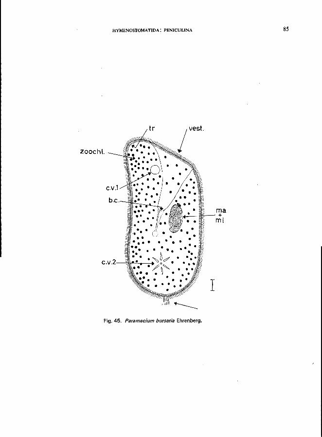
HYMENOSTOMATIDA: PENICULINA
tr
zooeh!.
e.v.1
•• e •
· ~Jc' · e. v .2-~~!--.-_... 0 •
e/ ~ • .. . ~ . • •
Fig. 45. Paramecium bursaria Ehrenberg.
I
ma +
mi
85

86
Family Parameciidae Genus Paramecium
HOLOTRICHIA
PARAMECIUM PUTRINUM Claparede & Lachmann (Fig. 46)
Morphology
About 130 fLm long; similar to P. bursaria but without zoochlorellae; macronucleus elongated, kidneyshaped; 1 micronucleus; only 1 contractile vacuole; no trichocysts (Lep~i, 1926; Kahl, 1931).
Records of P. putrinum are scanty, and it is in fact doubtful whether the species is valid. This problem is of some saprobiological interest since Liebmann (1962) classified P. putrinum as a polysaprobic indicator organism. On the other hand, Snimek-Husek (1954) failed to find any specimens of P. putrinum during his saprobiological studies in Czechoslovakia; all representatives of the genus Paramecium that he found in the more heavily polluted zones were referable to P. trichium. Therefore, Sramek-Husek submitted that the polysaprobic " P. putrinum" of earlier authors is really P. trichium Stokes (Sramek-Husek, 1954, 1956).
In my own studies on ciliates I also failed to find P. putrinum. The figure of P. putrinum given by Liebmann (1962) differs from the original diagnosis: P. putrinum
sensu Liebmann has 2 contractile vacuoles, both shown with radial canals; the specimen illustrated thus resembles individuals of P. caudatum cultivated under polysaprobic conditions (e.g., oxygen lack, high levels of ammonia). P. putrinum sensu Liebmann was recorded from the sludge of polluted reservoirs, dammed rivers, lakes etc., in the upper zone of trickling filters, sewage drains, and in floating tufts of Sphaerotilus (Liebmann, 1962). Taxonomic studies of" P. putrinum" and P. trichium are thus urgently
required.
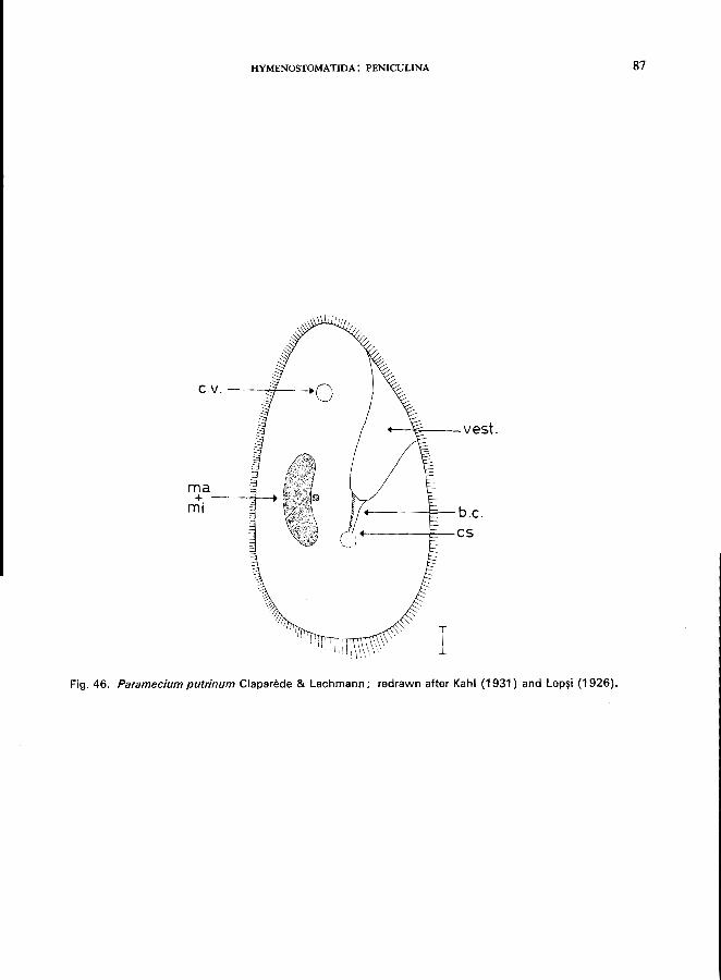
HYMENOSTOMATIDA: PENICULINA
C.v. --_7---.
ma + ---...,,1-....
mi
--'P--vest.
+--------j;"'-- b .c. +-------F-- cs
Fig. 46. Paramecium putrinum Claparede & Lachmann; redrawn after Kahl (1931) and Lep~i (1926).
87
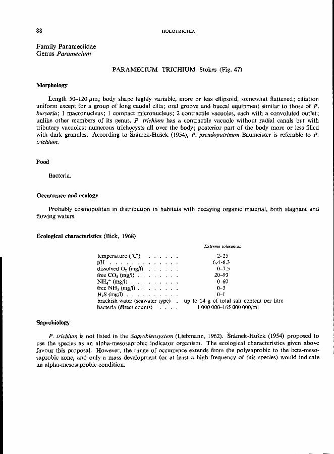
88
Family Parameciidae Genus Paramecium
Morphology
HOLOTRICHIA
PARAMECIUM TRICHIUM Stokes (Fig. 47)
Length 50-120 fLm; body shape highly variable, more or less ellipsoid, somewhat flattened; ciliation uniform except for a group of long caudal cilia; oral groove and buccal equipment similar to those of P. bursaria; 1 macronucleus; 1 compact micronucleus; 2 contractile vacuoles, each with a convoluted outlet; unlike other members of its genus, P. trichium has a contractile vacuole without radial canals but with tributary vacuoles; numerous trichocysts all over the body; posterior part of the body more or less filled with dark granules. According to Snimek-Husek (1954), P. pseudoputrinum Baumeister is referable to P.
trichium.
Food
Bacteria.
Occurrence and ecology
Probably cosmopolitan in distribution in habitats with decaying organic material, both stagnant and
flowing waters.
Ecological characteristics (Bick, 1968)
Saprobiology
temperature eC») pH ..... . dissolved O. (mg/l) free CO. (mgfl) . NH.+ (mg/l) . . free NH3 (mg/l) . H.S (mg/l) ... brackish water (seawater type) bacteria (direct counts)
Extreme tolerances
2-25 6.4-8.3
0-7.5 20-93 0-60 0-3 0-1
up to 14 g of total salt content per litre 1000000-165000000/ml
P. trichium is not listed in the Saprobiensystem (Liebmann, 1962). Snimek-Husek (1954) proposed to use the species as an alpha-mesosaprobic indicator organism. The ecological characteristics given above favour this proposal. However, the range of occurrence extends from the polysaprobic to the beta-mesosaprobic zone, and only a mass development (or at least a high frequency of this species) would indicate
an alpha-mesosaprobic condition.
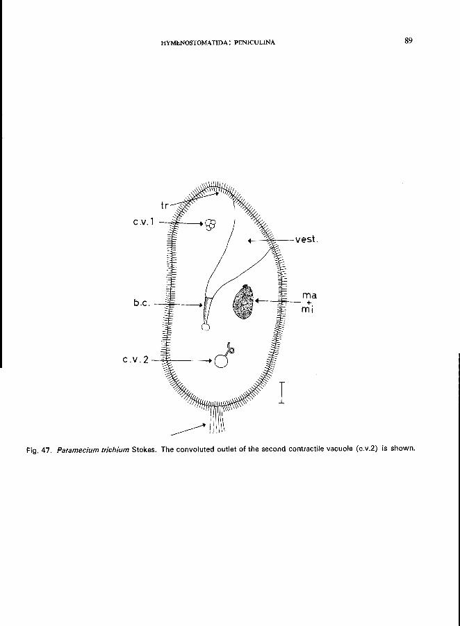
HYMENOSTOMATIDA: PENICULINA
c-v.l
b.c. -:::E----t
\ .. J
c. v. 2 --;;;T----+ J
~~~-vest.
ma .--~fe,-_ +
mi
Fig. 47. Paramecium trichium Stokes. The convoluted outlet of the second contractile vacuole (c.v.2) is shown.
89
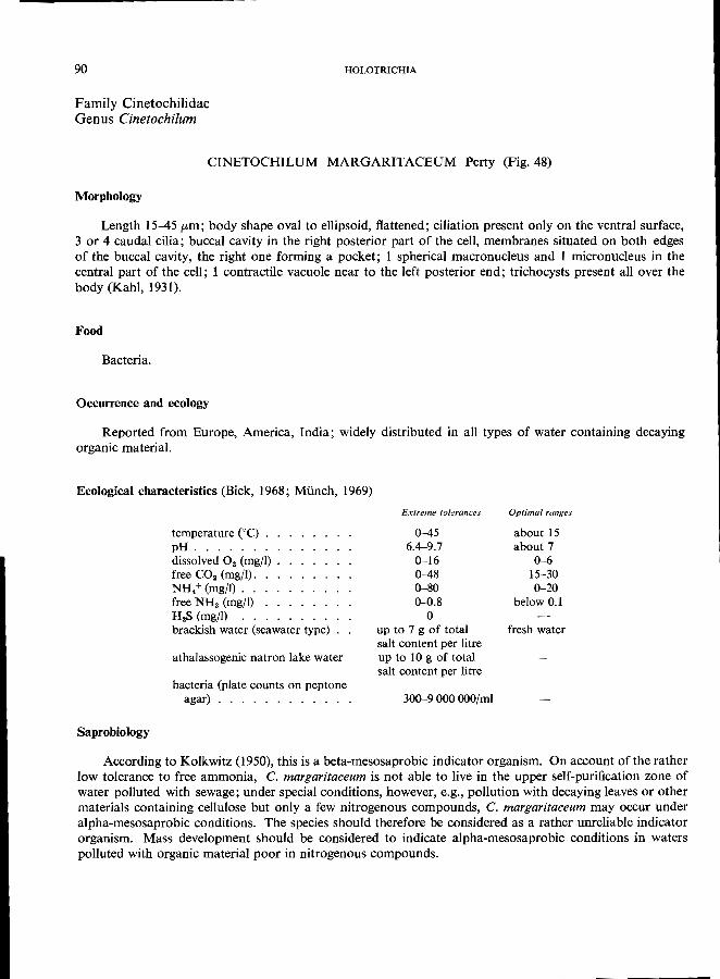
90
Family Cinetochilidae Genus Cinetochilum
HOLOTRICHIA
CINETOCHILUM MARGARITACEUM Perty (Fig. 48)
Morphology
Length 15-45 pm; body shape oval to ellipsoid, flattened; ciliation present only on the ventral surface, 3 or 4 caudal cilia; buccal cavity in the right posterior part of the cell, membranes situated on both edges of the buccal cavity, the right one forming a pocket; 1 spherical macronucleus and 1 micronucleus in the central part of the cell; 1 contractile vacuole near to the left posterior end; trichocysts present all over the
body (Kahl, 1931).
Food
Bacteria.
Occurrence and ecology
Reported from Europe, America, India; widely distributed in all types of water containing decaying
organic material.
Ecological characteristics (Bick, 1968; Munch, 1969)
Saprobiology
temperature CC) . pH ...... . dissolved O2 (mg/I) free CO2 (mg/l). NH.+ (mg/I) .. free NH. (mgfl) H2S (mg/I) brackish water (seawater type)
athalassogenic natron lake water
bacteria (plate counts on peptone agar) ........... .
Extreme tolerances
0-45 6.4-9.7
0--16 0-48 0--80 0-0.8
o up to 7 g of total salt content per litre up to 10 g of total salt content per litre
300--9 000 OOO/mi
Optimal ranges
about 15 about 7
0-6 15-30 0-20
below 0.1
fresh water
According to Kolkwitz (1950), this is a beta-mesosaprobic indicator organism. On account of the rather low tolerance to free ammonia, C. margaritaceum is not able to live in the upper self-purification zone of water polluted with sewage; under special conditions, however, e.g., pollution with decaying leaves or other materials containing cellulose but only a few nitrogenous compounds, C. margaritaceum may occur under alpha-mesosaprobic conditions. The species should therefore be considered as a rather unreliable indicator organism. Mass development should be considered to indicate alpha-mesosaprobic conditions in waters
polluted with organic material poor in nitrogenous compounds.
HYMENOSTOMATIDA: PENICULINA 91
ma+mi
b.c .----l--..a. c.v.
I Fig. 48. Cinetochilum margaritaceum Perty, ventral view.
92
Family Urocentridae Genus Urocentrum
Morphology
HOLOTRICHIA
UROCENTRUM TURBO (0. F. Milller) (Fig. 49)
Length 50-80 [Lm; body cylindrical and ventrally slightly flattened; constricted at the middle; 2 broad girdles of cilia and 1 eccentric posterior tuft; a zone of short cilia in the constricted area; buccal cavity subequatorial with 1 membrane and 2 short undulating membranelles; in the postoral area, a longitudinal zone with small cilia; macronucleus horseshoe-shaped and posteriorly located; a single micronucleus; contractile vacuole terminal, and with 8 collecting canals; large number of trichocysts all over the body
(Kahl, 1931).
Unique movement
Very rapid locomotion with intermittent rotation at one spot fixed by a mucilaginous thread that is
secreted by the ciliate itself.
Food
Bacteria.
Occurrence and ecology
Cosmopolitan in distribution, occurring all through the year in lakes, ponds, pools with natural pollution (e.g., decaying leaves), sewage oxidation ponds with low loadings; in fresh water only.
Ecological characteristics (Bick, 1957; 1968)
Saprobiology
temperature eC) . pH ...... . dissolved O2 (mg/I) free CO2 (mgj1) NH4+ (mg/I) .. . H 2S (mg/l) ... . bacteria (plate counts)
Extreme tolerances
5-25 6.0-7.6
0-12 0-36 0-18 0-0.5
2 000-500 OOO/mi
An alpha-mesosaprobic indicator organism according to Liebmann (1962).
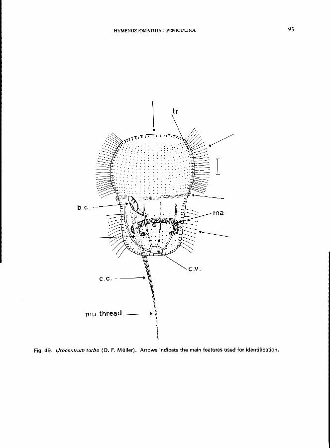
HYMENOSTOMATIDA: PENICULINA 93
b.c.
C.v. c. C. -----+.
mu.thread __ _
Fig. 49. Urocentrum turbo (0. F. Muller). Arrows indicate the main features used for identification.
---...... .... ---------------
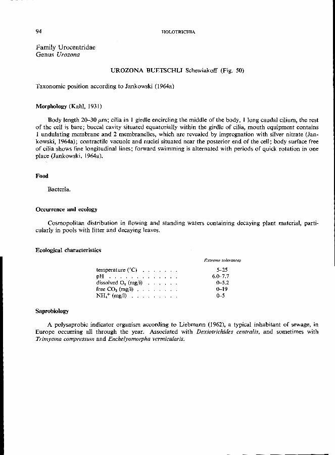
94
Family Urocentridae Genus Urozona
HOLOTRICHIA
UROZONA BUETSCHLJ Schewiakoff (Fig. 50)
Taxonomic position according to Jankowski (1964a)
Morphology (Kahl, 1931)
Body length 20-30 [Lm; cilia in I girdle encircling the middle of the body, I long caudal cilium, the rest of the cell is bare; buccal cavity situated equatorially within the girdle of cilia, mouth equipment contains I undulating membrane and 2 membranelles, which are revealed by impregnation with silver nitrate (Jankowski, 1964a); contractile vacuole and nuclei situated near the posterior end of the cell; body surface free of cilia shows fine longitudinal lines; forward swimming is alternated with periods of quick rotation in one
place (Jankowski, 1964a).
Food
Bacteria.
Occurrence and ecology
Cosmopolitan distribution in flowing and standing waters containing decaying plant material, parti
cularly in pools with litter and decaying leaves.
Ecological characteristics
Saprobiology
temperature COC) • pH ...... . dissolved O. (mg/I) free CO. (mg/I) NH.+ (mg/I) ...
Extreme tolerances
5-25 6.0-7.7
0-5.2 0-19 0-5
A polysaprobic indicator organism according to Liebmann (1962), a typical inhabitant of sewage, in Europe occurring all through the year. Associated with Dexiotrichides centralis, and sometimes with
Trimyema compressum and Enchelyomorpha vermicularis.
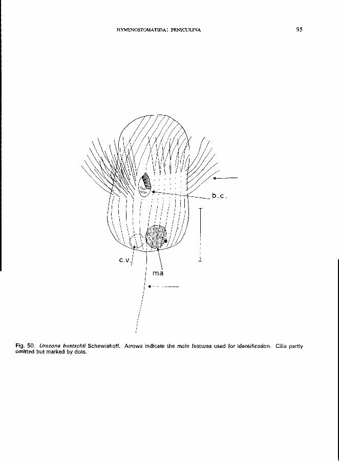
HYMENOSTOMATIDA: PENICULINA
Fig. 50. Urozona buetschli Schewiakoff. Arrows indicate the main features used for identification. Cilia partly omitted but marked by dots.
95
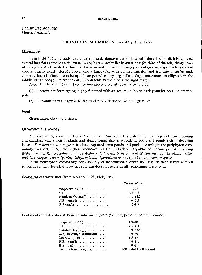
96
Family Frontoniidae Genus Frontonia
HOLOTRICHIA
FRONTONIA ACUMINATA Ehrenberg (Fig. 17A)
Morphology
Length 50-150 pm; body ovoid to ellipsoid, dorsoventrally flattened; dorsal side slightly convex, ventral face flat; complete uniform ciliation; buccal cavity lies in anterior right third of the cell; ciliary rows of the right and left ventral surface meet in a preoral suture and a very postoral groove, respectively; postoral groove usually nearly closed; buccal cavity lancet-like with pointed anterior and truncate posterior end, complex buccal ciliation consisting of compound ciliary organelles; single macronucleus ellipsoid in the middle of the body; 1 micronucleus; 1 contractile vacuole near the right margin.
According to Kahl (1931) there are two morphological types to be found:
(1) F. acuminata form typica; highly flattened with an accumulation of dark granules near the anterior pole.
(2) F. acuminata var. angusta Kahl; moderately flattened, without granules.
Food
Green algae, diatoms, ciliates.
Occurrence and ecology
F. acuminata typica is reported in America and Europe, widely distributed in all types of slowly flowing and standing waters rich in plants and algae; found also in woodland pools and ponds rich in decaying leaves. F. acuminata var. angusta has been reported from ponds and pools occurring in the periphyton community (Wilbert, 1969); the highest abundance in Bonn (Federal Republic of Germany) was in spring (February-April), associated with the diatoms Nitzschia, Synedra, and Tabellaria and the ciliates Cinetochilum margaritaceum (p. 90), Coleps no landi, Opercularia nutans (p. 122), and Stentor igneus.
If the periphyton community consists only of heterotrophic organisms, e.g., in deep layers without sufficient sunlight for algal growth, Frontonia does not occur at all; sometimes planktonic.
Ecological characteristics (from Noland, 1925; Bick, 1957)
temperature (DC) . pH ...... . dissolved O. (mg/I) NH.+ (mg,l) H.S (mg/I) •.•.
Extreme tolerances
1-22 6.5-8.7 0.8-14.3
0-2.2 0-1.1
Ecological characteristics of F. acuminata var. angusta (Wilbert, personal communication)
temperature eC) . pH ......... . dissolved O. (mg/I) . . . O. (percentage saturation) free CO. (mg/I) NH. + (mg/I) . . . . . H.S (mg/I) ..... . bacteria (direct counts)
1.8-20.5 7.4--9.3
0-22.4 0-207
1.5-37 0-3.1 0-1.1
800000-23 OOOOOO/mi
HYMENOSTOMATIDA: PENICULINA 97
Apparently there are no significant differences between the two morphological types of F. acuminata in their ecological tolerance ranges.
Saprobio}ogy
According to Liebmann (1962), the typical F. acuminata is an oligosaprobic indicator organism. The ecological data presented above are in contradiction to this statement. By investigations using the glassslide method, Wilbert (1968) has demonstrated that at least the variety angusta is unable to live under oligosaprobic conditions but grows well in beta-mesosaprobic localities. From the data on the ecological characteristics of the two types of F. acuminata it is evident that both prefer beta-mesosaprobic conditions favouring mass development of green algae and diatoms.
SaprobioJogicaJ classification
bos aos
2
bms
6
ams ps
2 3
7
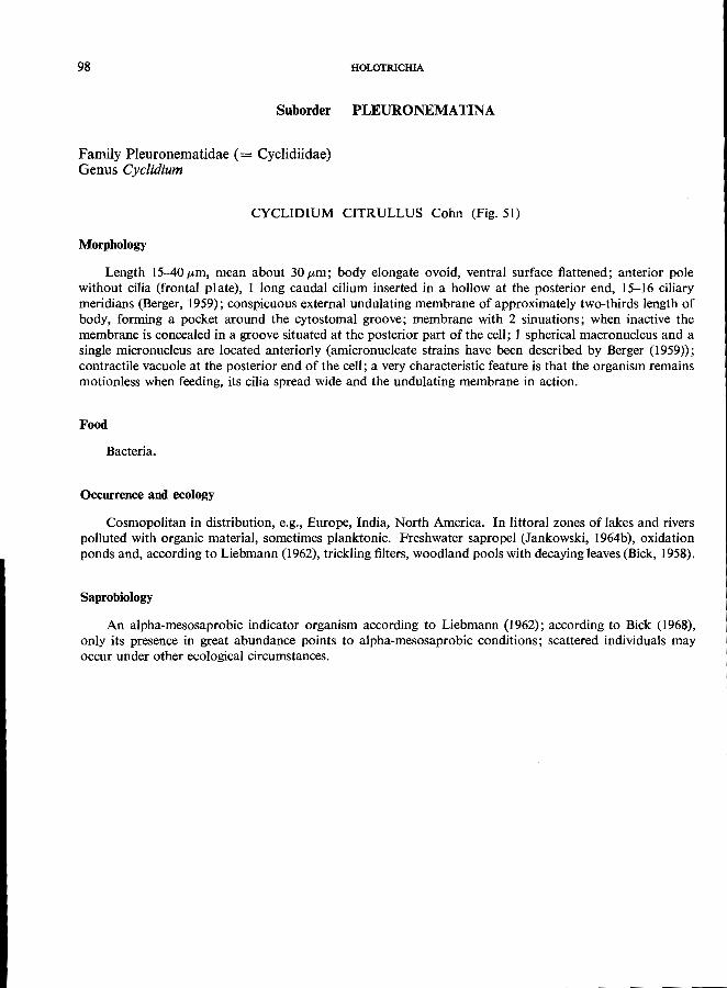
98 HOLOTRICHlA
Suborder PLEURONEMATINA
Family Pleuronematidae (= Cyc1idiidae) Genus Cyclidium
CYCLIDIUM CITRULLUS Cohn (Fig. 51)
Morphology
Length 15-40 pm, mean about 30 pm; body elongate ovoid, ventral surface flattened; anterior pole without cilia (frontal plate), 1 long caudal cilium inserted in a hollow at the posterior end, 15-16 ciliary meridians (Berger, 1959); conspicuous external undulating membrane of approximately two-thirds length of body, forming a pocket around the cytostomal groove; membrane with 2 sinuations; when inactive the membrane is concealed in a groove situated at the posterior part of the cell; 1 spherical macronucleus and a single micronucleus are located anteriorly (amicronucleate strains have been described by Berger (1959)); contractile vacuole at the posterior end of the cell; a very characteristic feature is that the organism remains motionless when feeding, its cilia spread wide and the undulating membrane in action.
Food
Bacteria.
Occurrence and ecology
Cosmopolitan in distribution, e.g., Europe, India, North America. In littoral zones of lakes and rivers polluted with organic material, sometimes planktonic. Freshwater sapropel (Jankowski, 1964b), oxidation ponds and, according to Liebmann (1962), trickling filters, woodland pools with decaying leaves (Bick, 1958).
Saprobiology
An alpha-mesosaprobic indicator organism according to Liebmann (1962); according to Bick (1968), only its presence in great abundance points to alpha-mesosaprobic conditions; scattered individuals may
occur under other ecological circumstances.
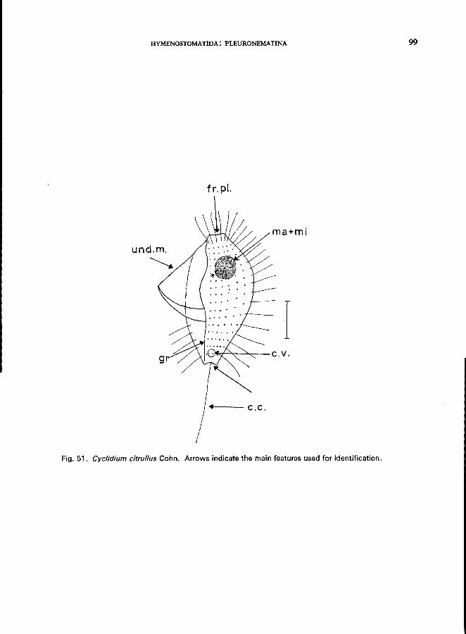
HYMENOSTOMATIDA: PLEURONEMATINA 99
f r. pI.
ma+mi
I .)4.:-1--"-..--..::.... __ C . v .
Fig. 51. Cyclidium citrullus Cohn. Arrows indicate the main features used for identification.
100
Family Pleuronematidae (= Cyclidiidae) Genus Cyclidium
HOLOTRICHIA
CYCLIDIUM GLAUCOMA (0. F. Mi.iller) (Fig. 52)
Morphology
Length 15-30 fLm , mean about 20 fLm; body ovoid with cilia-free anterior end; 1 long caudal cilium; 10 (rarely 11) ciliary meridians (Berger, 1960); the large undulating membrane is free of sinuations and approximately one-half the length of body; a full description of the buccal organelles is given by Berger & Thompson (1960); in addition to the undulating membrane, there are 3 definitive membranelles that are difficult to observe in the living condition; unlike C. citrullus, this species lacks a postoral longitudinal groove; 1 spherical macronucleus and a single micronucleus are located near the anterior end of the body;
the contractile vacuole is posteriorly situated.
Food
Bacteria; during feeding periods the ciliate remains in one position with spread cilia and beating mem
branelles.
Occurrence and ecology
Reported from Europe, North America, New Zealand; widely distributed, inhabiting the polysaprobic zone of freshwater ponds; "one of the most common ciliates recorded from the tens of natural sources"
(Jankowski, 1964b). According to Kolkwitz (1950), it is an alpha-mesosaprobic indicator organism.
Saprobiological classification
According to Sladecek (1969):
bos aos bms ams
9
ps
1 5
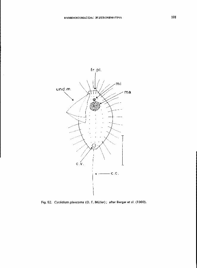
HYMENOSTOMATIDA: PLEURONEMATINA 101
fro pI.
mi und.m.
~ ma
C ..
+----c.c.
Fig. 52. Cyclidium glaucoma (0. F. Miiller); after Berger et al. (1960).
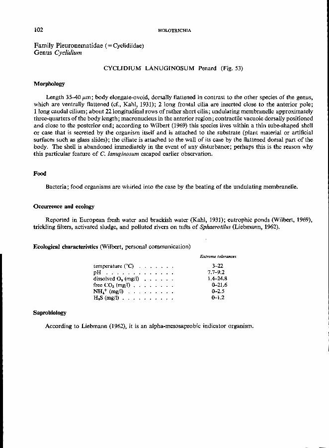
102
Family Pleuronematidae (= Cyclidiidae) Genus Cyclidium
HOLOTRICHIA
CYCLIDIUM LANUGINOSUM Penard (Fig. 53)
Morphology
Length 35-40 pm; body elongate-ovoid, dorsally flattened in contrast to the other species of the genus, which are ventrally flattened (cf., Kahl, 1931); 2 long frontal cilia are inserted close to the anterior pole; 1 long caudal cilium; about 2210ngitudfnal rows of rather short cilia; undulating membranelle approximately three-quarters of the body length; macronucleus in the anterior region; contractile vacuole dorsally positioned and close to the posterior end; according to Wilbert (1969) this species lives within a thin tube-shaped shell or case that is secreted by the organism itsdf and is attached to the substrate (plant material or artificial surfaces such as glass slides); the ciliate is attached to the wall of its case by the flattened dorsal part of the body. The shell is abandoned immediately in the event of any disturbance; perhaps this is the reason why this particular feature of C. lanuginosum escaped earlier observation.
Food
Bacteria; food organisms are whirled into the case by the beating of the undulating membranelle.
Occurrence and ecology
Reported in European fresh water and brackish water (Kahl, 1931); eutrophic ponds (Wilbert, 1969), trickling filters, activated sludge, and polluted rivers on tufts of Sphaerotilus (Liebmann, 1962).
Ecological characteristics (Wilbert, personal communication)
Saprobiology
temperature COe) . pH ...... . dissolved O. (mg/l) free CO. (mgfl) NH/ (mg/l) H.S (mg/l) ..
Extreme tolerances
3-22 7.7-9.2 1.4-24.8
0-21.6 0-2.5 0-1.2
According to Liebmann (1962), it is an alpha-mesosaprobic indicator organism.
HYMENOSTOMATIDA: PLEURONEMATINA 103
Fig. 53. Cyclidium lanuginosum Penard; after Wilbert (1969).
---....... .... ----------------
Family Ophrydiidae Genus Ophrydium
Morphology
Subclass PERITRICHIA Order PERITRICHIDA Suborder SESSILINA
OPHRYDIUM VERSATILE (0. F. MUller) (Fig. 54)
Individuals 300-600 pm long (fully extended), colonies up to 15 cm; body elongated flask-form, the " neck" (see Fig. 54) is extremely contractile; green in colour on account of symbiotic zoochlorellae; long dichotomous stalk; macronucleus long, band-like, extending along the longitudinal axis of the cell; several strains are amicronucleate; contractile vacuole with a long vacuolar canal that empties into the buccal cavity; individuals are embedded in a large mucilaginous mass that is attached to various substrates (e.g., stones, sticks, shells, plants); colonies are green in colour; small colonies are nearly spherical, larger colonies are of an irregular spherical form, sometimes flattened, often with a central cavity; dispersal by telotrochs (= free-swimming larvae), which are equipped with an aboral locomotory girdle of cilia. For a detailed description of morphology and systematics, see Winkler & Corliss (1965).
Food
Bacteria.
Occurrence and ecology
Reported from Europe and America; widely distributed in still and slowly flowing oligotrophic waters near the shore-line; occurs throughout the year, in Michigan, USA, the largest colonies have been recorded in October (Winkler & Corliss, 1965).
Saprobiology
According to Liebmann (1962), it is an oligotrophic indicator organism; according to Kolkwitz (1950), it also occurs in alpha-mesosaprobic habitats.
OPHRYDIUM SESSILE Kent Morphology
Individuals up to 300 /Lm long, colonies 3 mm in diameter; buccal ciliation, contractile vacuole, and macronucleus very similar to O. versatile but zoochlorellae are absent (therefore not green) and there is no stalk; colony-forming; mucilaginous loricae colourless or brownish, colonies comparatively small. It is doubtful whether o. sessile is an independent species, possibly it is a subspecies of O. versatile (cr., Winkler & Corliss, 1965).
Food
Bacteria.
Occurrence and ecology
Reported from European lakes and reservoirs, attached to plants, stones, and artificial substrates (e.g., microscope slides). O. sessile was observed in the Sedlice Reservoir, Czechoslovakia, during a period of heavy pollution by wastes from sugar factories (Shideckova & Siadeeek, 1963).
-104-
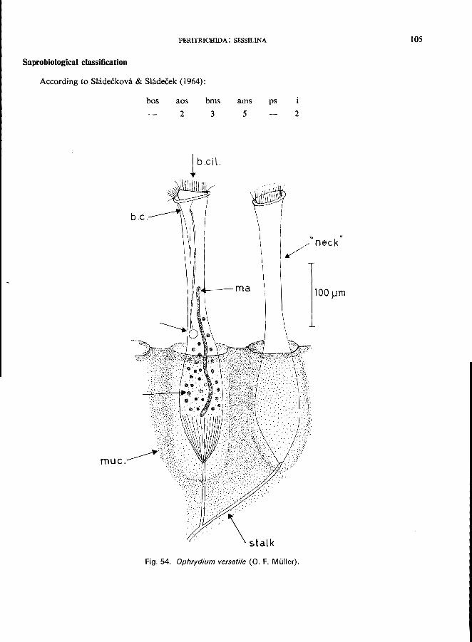
PERITRICHIDA: SESSILINA
Saprobiological classification
According to Shideekova & Sladeeek (1964):
b.c.
muc.
bos aos 2
bms 3
1 b.cil.
ams 5
I>I~---ma
\5talk
ps
2
/
Fig. 54. Ophrydium versatile (0. F. Muller).
105
/" neck·
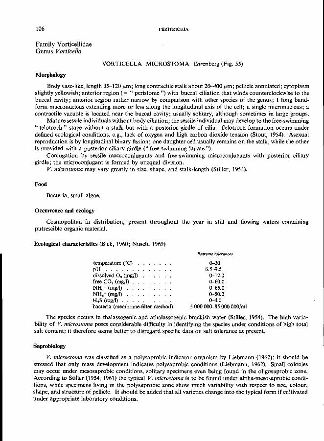
106
Family Vorticellidae Genus Vorticella
PERITRICHIA
VORTICELLA MICROSTOMA Ehrenberg (Fig. 55)
Morphology
Body vase-like, length 35-120 fLm; long contractile stalk about 20-400 fLm; pellicle annulated; cytoplasm slightly yellowish; anterior region (= " peristome ") with buccal ciliation that winds counterclockwise to the buccal cavity; anterior region rather narrow by comparison with other species of the genus; 1 long bandform macronucleus extending more or less along the longitudinal axis of the cell; a single micronucleus; a contractile vacuole is located near the buccal cavity; usually solitary, although sometimes in large groups.
Mature sessile individuals without body ciliation; the sessile individual may develop to the free-swimming " te1otroch " stage without a stalk but with a posterior girdle of cilia. Telotroch formation occurs under defined ecological conditions, e.g., lack of oxygen and high carbon dioxide tension (Stout, 1954). Asexual reproduction is by longitudinal binary fission; one daughter cell usually remains on the stalk, while the other is provided with a posterior ciliary girdle (" free-swimming larvae ").
Conjugation by sessile macroconjugants and free-swimming microconjugants with posterior ciliary girdle; the microconjugant is formed by unequal division.
V. microstoma may vary greatly in size, shape, and stalk-length (Stiller, 1954).
Food
Bacteria, small algae.
Occurrence and ecology
Cosmopolitan in distribution, present throughout the year in still and flowing waters containing putrescible organic material.
Ecological characteristics (Bick, 1960; Nusch, 1969)
temperature eC) . pH ...... . dissolved O2 (mg/I) free CO. (mg/I) NH.+ (mg/I) NH. - (mg/I) . H.S (mg/I) .. bacteria (membrane-filter method)
Extreme tolerances
0-30 6.5-9.5
0-12.0 0-60.0 0-65.0 0-50.0 0-4.0
5000 000-85000 OOO/mi
The species occurs in thalassogenic and athalassogenic brackish water (Stiller, 1954). The high variability of V. microstoma poses considerable difficulty in identifying the species under conditions of high total salt content; it therefore seems better to disregard specific data on salt tolerance at present.
Saprobiology
V. microstoma was classified as a polysaprobic indicator organism by Liebmann (1962); it should be stressed that only mass development indicates polysaprobic conditions (Liebmann, 1962). Small colonies may occur under mesosaprobic conditions, solitary specimens even being found in the oligosaprobic zone. According to Stiller (1954, 1961) the typical V. microstoma is to be found under alpha-mesosaprobic conditions, while specimens living in the polysaprobic zone show much variability with respect to size, colour, shape, and structure of pellicle. It should be added that all varieties change into the typical form if cultivated under appropriate laboratory conditions.
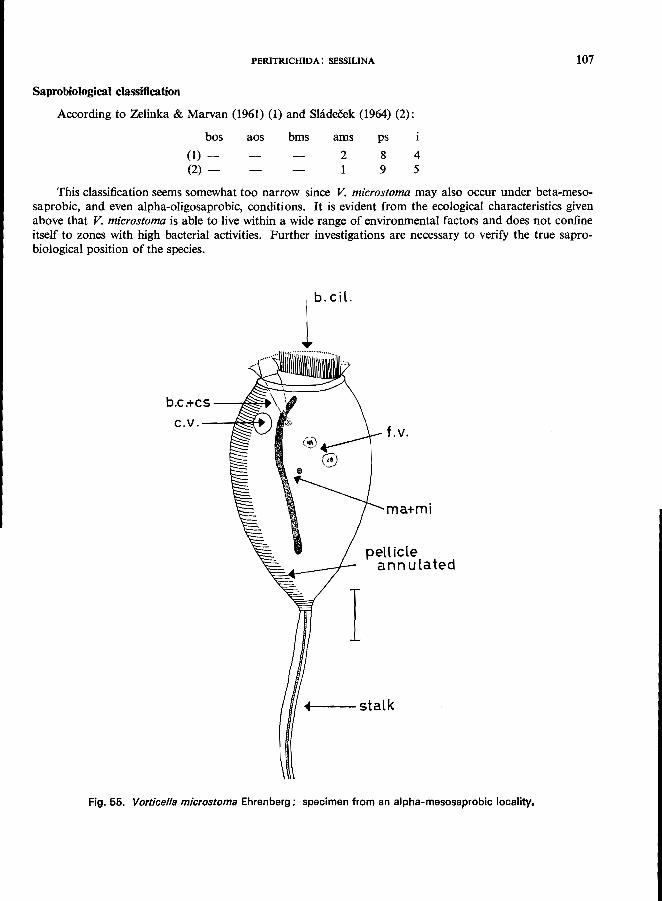
PERITRICHIDA: SESSILINA
Saprobiological classification
According to Zelinka & Marvan (1961) (1) and Shidecek (1964) (2):
bos
(1) -(2) -
aos bms ams
2 1
ps
8 9
4 5
107
This classification seems somewhat too narrow since V. microstoma may also occur under beta-mesosaprobic, and even alpha-oligosaprobic, conditions. It is evident from the ecological characteristics given above that V. microstoma is able to live within a wide range of environmental factors and does not confine itself to zones with high bacterial activities. Further investigations are necessary to verify the true saprobiological position of the species.
b.c.+cs - _____ ~
c.V.--~<i+
lb.Cil.
I
f.v.
ma+mi
pellicle annulated
..---stalk
Fig. 55. Vorticella microstoma Ehrenberg; specimen from an alpha-mesosaprobic locality.
108
Family Vorticellidae Genus Vorticella
PERITRICHIA
VORTICELLA CAMPANULA Ehrenberg (Fig. 56)
Morphology
Body 50-160 /Lm long, 35-100 /Lm wide; stalk about 250-350 /Lm long and 6-12 /Lm wide; bell-shaped, very changeable in outline, sometimes bending back; the central part of the cell is filled with refractile reserve granules, therefore the animals are very conspicuous by their darkish body and they are easy to recognize; the peristome extends considerably outwards; the buccal cavity (" vestibulum ") is very large and equipped with an outer undulating membrane (" pharyngeal membrane "); pellicle faintly annulated; macronucleus extending more or less along the longitudinal axis of the cell; very long, wormlike; a single micronucleus; 1 contractile vacuole near the buccal cavity; the stalk may be somewhat invaginated into the basal portion of the body; sessile (for a full morphological description, see Noland & Finley (1931»; Stiller (1963) described V. campanuia var. monilata, which is characterized by numerous small pellicular tubercles; reproduction and conjugation as described for V. microstoma (p. 106).
Food
Bacteria.
Occurrence and ecology
Cosmopolitan in distribution, frequently occurring throughout the year in all types of flowing and standing waters, avoiding foul water conditions; according to Noland & Finley (1931), it is favoured by a certain amount of current in the water; usually found in groups attached to plants (e.g., Ceratophyl/um, Lemna, Myriophyllum), insect larvae, crustacea, stones, artificial substrates (slides), and detritus; specimens occurring in athalassogenic brackish waters are covered with pellicular tubercles (cf., morphology).
Ecological characteristics (from Noland, 1925; Nusch, 1969)
Saprobiology
temperature (0C) . pH ...... . dissolved O. (mg/I) free CO. (mg/I) . . NH.+(mg/l) ... bacteria (membrane-filter method)
Extreme tolerances
0-23 6.9-9.0 4.5-16
0-12 0-7.5
1 000 000-9 000 OOO/mi
According to Liebmann (1962), this is a beta-mesosaprobic indicator organism but this classification seems too narrow because V. campanu/a is able to tolerate alpha-mesosaprobic conditions also.
Saprobiological classification
According to Shideekova & Sladecek (1966):
bos aos bms
6
ams pa
3 3
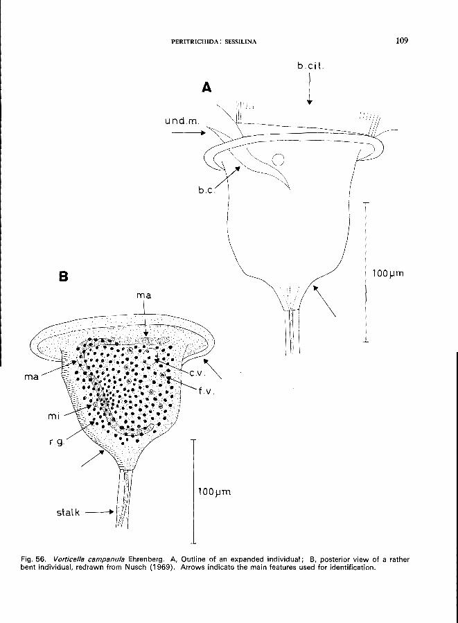
PERITRICHIDA: SESSILINA
b.cil.
A 1 und.m.
B ma
\
Fig. 56. Vorticella campanu/a Ehrenberg. A, Outline of an expanded individual; B, posterior view of a rather bent individual, redrawn from Nusch (1969). Arrows indicate the main features used for identification.
109
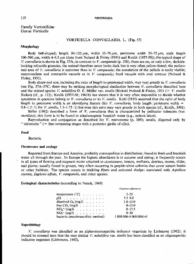
110
Family Vorticellidae Genus Vorticella
PERITRICHIA
VORTICELLA CONY ALLARIA L. (Fig. 57)
Morphology
Body bell-shaped; length 30-120 pm, width 35-70 pm, peristome width 55-75 pm, stalk length 100-500 pm, width 4-6.5 (Lm (data from Noland & Finley (1931) and Kralik (1957-58»; the typical shape of V. convallaria is shown in Fig. 57A; in contrast to V. campanula (p. 108); there are no, or only a few, darkishlooking refractile granules, the animal therefore never looks dark but is very often yellow-tinted; the peristomal area of V. convallaria is narrower than in V. campanula; the annulation of the pellicle is easily visible; macronucleus and contractile vacuole as in V. campanula; food vacuole with oval contour (Noland & Finley, 1931).
Body shape and size, including the ratio oflength to peristomal width, may vary greatly in V. convallaria (see Fig. 57A-57C; there may be striking morphological similarities between V. convallaria described here and the related species V. nebulifera o. F. Muller var. similis (Stokes) Noland & Finley, 1931 (= V. similis Stokes) (cf., p. 112); Kralik (1957-58; 1961b) has shown that it is very often impossible to decide whether specimens in question belong to V. convallaria or to V. similis. Kahl (1935) assumed that the ratio of body length to peristome width is an identifying feature (for V. convallaria, body length: peristome width = 0.8-1.3: 1; for V. similis, 1.5-1.75: 1) but even this ratio may vary greatly in both species (cr., Kralik, 1961).
Stiller (1962) described a form of V. convallaria that is characterized by pellicular tubercles (var. monilata); this form is to be found in athalassogenic brackish water (e.g., natron lakes).
Reproduction and conjugation as described for V. microstoma (p. 106); sessile, dispersal only by " telotrochs " (= free-swimming stages with a posterior girdle of cilia).
Food
Bacteria.
Occurrence and ecology
Reported from Europe and America, probably cosmopolitan in distribution; found in fresh and brackish water all through the year. In Europe the highest abundance is in autumn and spring; it frequently occurs in all types of flowing and stagnant water attached to crustaceans, insects, molluscs, detritus, stones, slides, and plants; usually found in groups, very often occurring in greyish-white colonies that cover stream banks or other habitats. The species occurs in trickling filters and activated sludge; associated with Aspidisca costata, Euplotes ajfinis, V. campanula, and other species.
Ecological characteristics (according to Nusch, 1969)
Saprobiology
temperature eC) . pH ...... . dissolved O2 (mg/l) free CO2 (mgj1) NH. + (mg/l) . . . N02 - (mg/l) . . . bacteria (membrane-filter method)
Extreme tolerances
2-23 5.5-9.0 1.0-13.0
0-15.0 0-17.5 0-30
1 000000-9 000 OOO/mi
V. convallaria was classified as an alpha-mesosaprobic indicator organism by Liebmann (1962); it should be stressed here that the very similar V. nebulifera var. similis has been classified as an oligosaprobic indicator organism (Liebmann, 1962).
PERITRICHIDA: SESSILINA
Saprobiological classification
A
I- B
Fig. 57. Vorticella convallaria L.; redrawn from Nusch (1969). A, Typical specimen; Band C, specimens showing the variety of shapes, C strongly resembles individuals of Vorticella nebulifera var. simi/is. Arrows indicate the main features used for identification.
111
112
Family V orticellidae Genus Vorticella
PERITRICHIA
VORTICELLA NEBULIFERA O. F. Mi.iller var. SJMILIS (Stokes) Noland & Finley (= VORTICELLA SIMILIS Stokes)
Morphology
Body length 40-90 fLm, body width 20-50 fLm, peristome 30-70 fLm, stalk length 50-800 fLm; body shape similar to some varieties of V. convallaria (cf., V. convallaria, Fig. 57C); macronucleus long and band-like, as in V. convallaria; the only features distinguishing this species from V. convallaria may be as follows (Noland & Finley, 1931): body very translucent, pure misty white or slightly greyish in colour, food vacuoles oblong or spindle-shaped; it seems rather doubtful whether these features are valid taxonomic criteria (for extensive discussion of the morphology and ecology, see Kralik (1957-58) and Nusch (1969)); sessile.
Food
Bacteria.
Occurrence and ecology
Reported from Europe and America in flowing and stagnant fresh water (the typical V. nebulifera occurs in marine habitats) singly or in groups attached to plants (including planktonic algae), insect larvae, molluscs, detritus, stones or artificial substrates (e.g., glass slides); according to Noland & Finley (1931), the species" will persist for days in relatively clean laboratory cultures but dies out when bacterial action
becomes intensive".
Ecological characteristics (Noland, 1925)
temperature (0C) . . . . dissolved O2 (mg/l) . . . O2 (percentage saturation) free CO2 (mg/l) . . . . . approx.
Extreme tolerances
4-20 0.5-10 4.5-81.2
0-10
The characteristics given here are very similar to those tabulated for V. convallaria.
Saprobiology
According to Liebmann (1962), this is an oligosaprobic indicator organism but the ecological characteristics given above are not in agreement with Liebmann's opinion. It should be mentioned that Liebmann's indicator organism is explicitly the same as that described by Noland & Finley (1931).
In view of the rather difficult and confusing taxonomic situation, it is assumed that V. nebulifera var. simi/is is a rather bad indicator organism. Probably it is a modification of V. convallaria which occurs only
in clean waters poor in bacteria as a source of food.
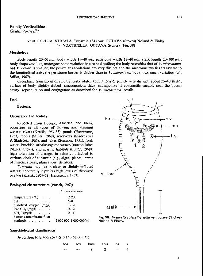
Family Vorticellidae Genus Vorticella
PERITRICHIDA: SESSILINA
VOR TICELLA STRIATA Dujardin 1841 var. OCTA VA (Stokes) Noland & Finley (= VORTICELLA OCTAVA Stokes) (Fig. 58)
Morphology
113
Body length 20-60/Lm, body width 15-40/Lm, peristome width 13-40/Lm, stalk length 20-300/Lm; body shape vase-like, undergoes some variation in size and outline; the body resembles that of V. mierostoma, but V. oetava is smaller, the pellicular annulations are very distinct and the macronucleus lies transverse to the longitudinal axis; the peristome border is thicker than in V. mierostoma but shows much variation (cf., Stiller, 1967).
Cytoplasm translucent or slightly misty white; annulations of pellicle very distinct, about 25-40 striae; surface of body slightly ribbed; macronucleus thick, sausage-like; 1 contractile vacuole near the buccal cavity; reproduction and conjugation as described for V. mierostoma; sessile.
Food
Bacteria.
Occurrence and ecology
Reported from Europe, America, and India, occurring in all types of flowing and stagnant waters: rivers (Kralik, 1957-58), ponds (Hammann, 1953), pools (Stiller, 1968), reservoirs (Shideckova & Sladecek, 1963), and lakes (Sommer, 1951), fresh water, brackish athalassogenic waters (natron lakes (Stiller, 1967», and marine habitats (Stiller, 1968); high toleration of changes in salinity; attached to various kinds of substrate (e.g., algae, plants, larvae of insects, stones, glass slides, detritus).
V. striata may live in clean or slightly polluted waters; apparently it prefers high levels of dissolved oxygen (Kralik, 1957-58; Hammann, 1953).
Ecological characteristics (Nusch, 1969)
temperature COc) pH " .... dissolved oxygen (mg/I) free CO. (mg/I) . . . . NH. + (mg/I) . . . . . bacteria (membrane-filter method) ...... .
Saprobio)ogical classification
Extreme tolerances
2-23 5-8 3-12 0--12 0--15
1000000--9000000/ml
According to Sladeckova & Sladecek (1963);
bos aos bms
8
b . c . ---f'''''-I~\
striae
stalk
-----\-- c.V .
--+---ma
® ® ® @J4--+--f.v. ® ® C!)
® ®
I Fig. 58. Vorticella striata Dujardin var. octava (Stokes) Noland & Finley.
ams
2 ps
4
8
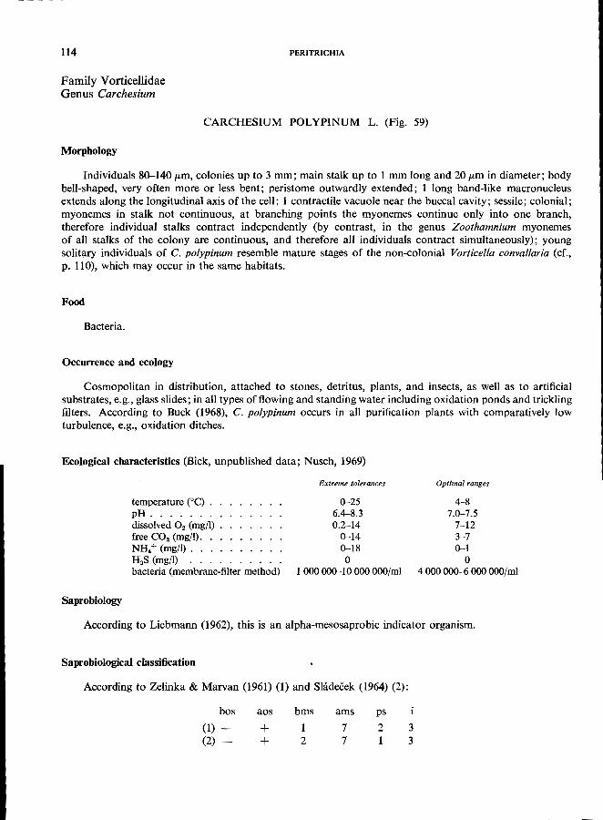
114
Family Vorticellidae Genus Carchesium
Morphology
PERITRICHIA
CARCHESIUM POLYPINUM L. (Fig. 59)
Individuals 80-140 pm, colonies up to 3 mm; main stalk up to 1 mm long and 20 pm in diameter; body bell-shaped, very often more or less bent; peristome outwardly extended; 1 long band-like macronucleus extends along the longitudinal axis of the cell; 1 contractile vacuole near the buccal cavity; sessile; colonial; myonemes in stalk not continuous, at branching points the myonemes continue only into one branch, therefore individual stalks contract independently (by contrast, in the genus Zoothamnium myonemes of all stalks of the colony are continuous, and therefore all individuals contract simultaneously); young solitary individuals of C. po[ypinum resemble mature stages of the non-colonial Vorticella convallaria (cf.,
p. 110), which may occur in the same habitats.
Food
Bacteria.
Occurrence and ecology
Cosmopolitan in distribution, attached to stones, detritus, plants, and insects, as well as to artificial substrates, e.g., glass slides; in all types of flowing and standing water including oxidation ponds and trickling filters. According to Buck (1968), C. poiypinum occurs in all purification plants with comparatively low
turbulence, e.g., oxidation ditches.
Ecological characteristics (Bick, unpublished data; Nusch, 1969)
Saprobiology
temperature ceC) . pH ...... . dissolved O2 (mg/l) free CO2 (mg/l). NH. + (mg/l). . . H 2S (mg/l) bacteria (membrane-filter method)
Extreme tolerances
0-25 6.4-8.3 0.2-14
0-14 0-18 o
1000000-10000000/ml
Optimal ranges
4-8 7.0-7.5
7-12 3-7 0-1
o 4000000-6000000/ml
According to Liebmann (1962), this is an alpha-mesosaprobic indicator organism.
Saprobiological classification
According to Zelinka & Marvan (1961) (1) and Shideeek (1964) (2):
bos
(1) (2) -
aos
+ +
bms
1 2
ams
7 7
ps
2 1
3 3
PERITRICHIDA: SESSILINA
I A
b.c.
\
100~m
1
Fig. 59. Carchesium polypinum L. A. Three individuals of a colony; B, diagram of a colony. Arrows indicate the main features used for identification.
115
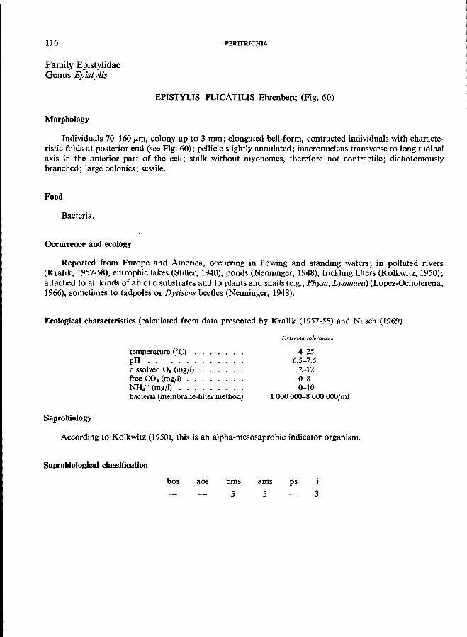
116
Family Epistylidae Genus Epistylis
Morphology
PERITRICHIA
EPISTYLIS PLlCATILlS Ehrenberg (Fig. 60)
Individuals 70-160 p.m, colony up to 3 mm; elongated bell-form, contracted individuals with characteristic folds at posterior end (see Fig. 60); pellicle slightly annulated; macronucleus transverse to longitudinal axis in the anterior part of the cell; stalk without myonemes, therefore not contractile; dichotomously
branched; large colonies; sessile.
Food
Bacteria.
Occurrence and ecology
Reported from Europe and America, occurring in flowing and standing waters; in polluted rivers (Kralik, 1957-58), eutrophic lakes (Stiller, 1940), ponds (Nenninger, 1948), trickling filters (Kolkwitz, 1950); attached to all kinds of abiotic substrates and to plants and snails (e.g., Physa, Lymnaea) (Lopez-Ochoterena, 1966), sometimes to tadpoles or Dytiscus beetles (Nenninger, 1948).
Ecological characteristics (calculated from data presented by Kralik (1957-58) and Nusch (1969)
Saprobiology
temperature eC) . pH ...... . dissolved O 2 (mg/I) free CO2 (mgfl) . . NH/ (mgfl) ... bacteria (membrane-filter method)
Extreme tolerances
4-25 6.5-7.5
2-12 0-8 0-10
1 000000-8 000 OOO/mi
According to Kolkwitz (1950), this is an alpha-mesosaprobic indicator organism.
Saprobiological classification
bos aos bms
5
ams ps
5 3
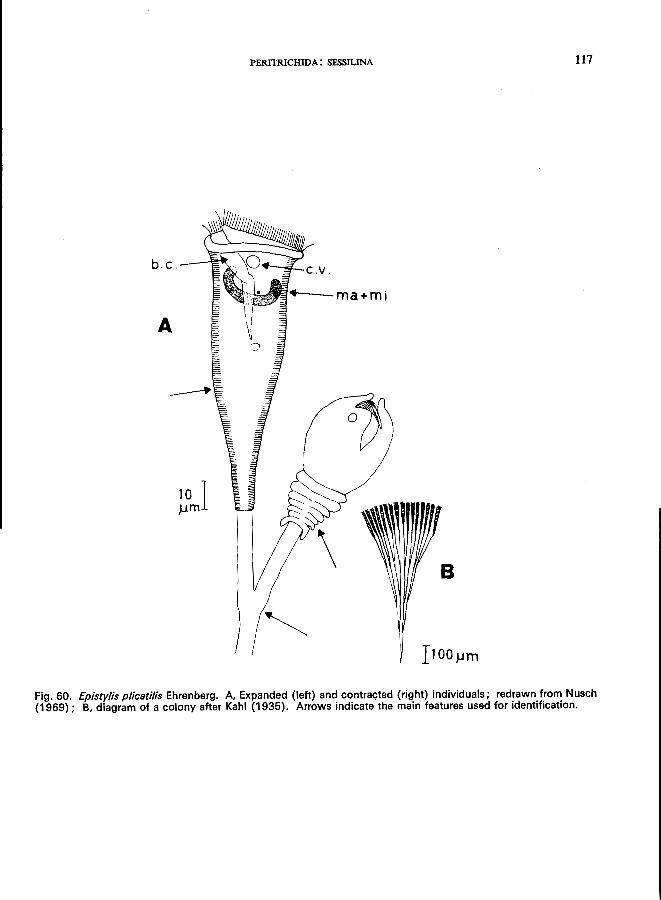
PERITRICHIDA: SESSILINA 117
b.c.--'"'!'='"-..:
A
10 I .urn
Fig. 60. Epistylis plicatilis Ehrenberg. A Expanded (left) and contracted (right) individuals; redrawn from Nusch (1969); B, diagram of a colony after Kahl (1935). Arrows indicate the main features used for identification.
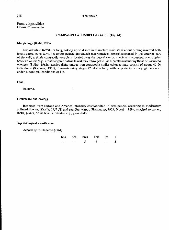
118
Family Epistylidae Genus Campanella
PERITRICHIA
CAMPANELLA UMBELLARIA L. (Fig. 61)
Morphology (Kahl, 1935)
Individuals 200-260/km long, colony up to 4 mm in diameter; main stalk about 3 mm; inverted bellform; adoral zone turns 4-6 times; pellicle annulated; macronucleus horseshoe-shaped in the anterior part of the cell; a single contractile vacuole is located near the buccal cavity; specimens occurring in eutrophic brackish waters (e.g., athalassogenic natron lakes) may show pellicular tubercles resembling those of Vorticella monilata (Stiller, 1962); sessile; dichotomous non-contractile stalk; colonies may consist of about 40-50 individuals (Sommer, 1951); free-swimming stages (" telotrochs") with a posterior ciliary girdle occur
under suboptimal conditions of life.
Food
Bacteria.
Occurrence and ecology
Reported from Europe and America, probably cosmopolitan in distribution, occurring in moderately polluted flowing (Kralik, 1957-58) and standing waters (Hammann, 1953; Nusch, 1969); attached to stones,
shells, plants, or artificial substrates, e.g., glass slides.
Saprobiological classification
According to Shldecek (1964):
bos aos bms
5
ams ps
5 3
PERITRICHIDA: SESSILINA 119
C.V.---t;,.,~~
100J-lm.
Fig. 61. Campanella umbellaria L.; redrawn from Nusch (1969). Arrows indicate the main features used for identification. The individual on the right shows telotroch formation.
120
Family Epistylidae Genus Opercularia
PERITRICHIA
OPERCULARIA COARCTATA (Claparede & Lachmann) (= EPISTYLIS COARCTATA Claparede & Lachmann) (Fig. 62)
Morphology
Individuals 45-65 p,m, stalk about 50-100 p,m; colonies small, consisting of 3-6 individuals; body elongate; peristomal area small and not constricted from the body; buccal area with distinct oblique disk which is set off from the border by a deep incision; obvious undulating membrane; macronucleus horseshoelike, its axis transverse to the longitudinal; stalk without myonemes, dichotomous branching; sessile.
Food
Bacteria.
OccUlTence and ecology
Cosmopolitan in distribution, occurring in all types ofslowly flowing and standing waters rich in bacteria from the decomposition of organic matter, e.g., polluted streams, oxidation ponds, trickling filters, activated sludge; attached to detritus, shells of molluscs, Sphaerotilus; abundant in activated sludge up to 110 OOO/ml (Buck, 1968) with an average of 8 500/mI. O. coarctata, Vorticella putrina, and V. convallaria (p. 110) are characteristic inhabitants of activated sludge in plants using the diffused air system; in this association V. putrina is dominant in " high intensity" plants with comparatively low oxygen content; with decreasing intensity of decomposition and increasing oxygen content the individual counts of Opercu/aria increase first,
and those of V. convallaria later (Buck, 1968).
Saprobiology
According to Liebmann (1962), o. coarctata is an alpha-mesosaprobic indicator organism, Kolkwitz (1950) is of the same opinion but has used the synonym" Epistylis " coarctata.
Saprobioiogical classification
bos aos bms
2
ams
7
ps
1 3
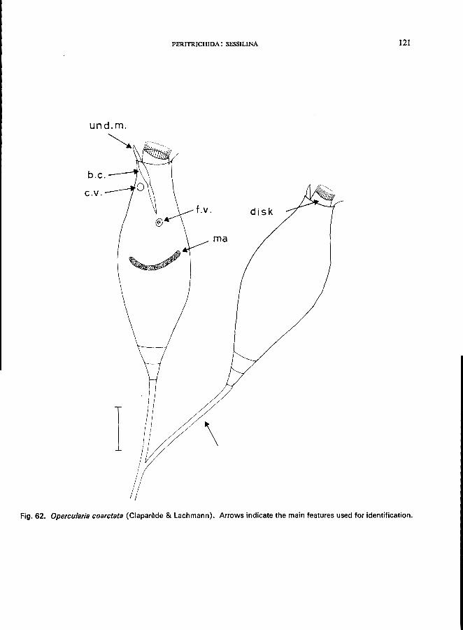
PERITRICHIDA: SESSILINA 121
b.c.
C.V.
f.v. disk
rna
\
Fig. 62. Opercu/aria coarctata (Claparede & Lachmann). Arrows indicate the main features used for identification.
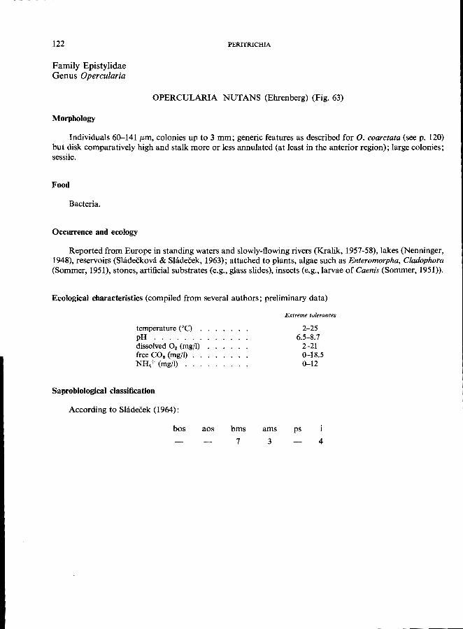
122
Family Epistylidae Genus Opercu/aria
PERITRICHIA
OPERCULARIA NUT ANS (Ehrenberg) (Fig. 63)
Morphology
Individuals 60-141 /Lm, colonies up to 3 mm; generic features as described for O. coarctata (see p. 120) but disk comparatively high and stalk more or less annulated (at least in the anterior region); large colonies;
sessile.
Food
Bacteria.
Occurrence and eoology
Reported from Europe in standing waters and slowly-flowing rivers (Kralik, 1957-58), lakes (Nenninger, 1948), reservoirs (Shideekova & Slidecek, 1963); attached to plants, algae such as Enteromorpha, Cladophora (Sommer, 1951), stones, artificial substrates (e.g., glass slides), insects (e.g., larvae of Caenis (Sommer, 1951)).
Ecological characteristics (compiled from several authors; preliminary data)
temperature COc) . pH ...... . dissolved O2 (mg/I) free CO2 (mgJl) NH,+ (mgjl)
Saprobiological classification
According to Slidecek (1964):
bos aos bms
7
ams
3
Extreme tolerances
2-25 6.5-S.7
2-21 0-1S.5 0-12
ps
4
PERITRICHIDA: SESSILINA 123
und.m-. --..
ma
I j
Fig. 63. Opercu/aria nutans (Ehrenberg). Arrows indicate the main features used for identification.
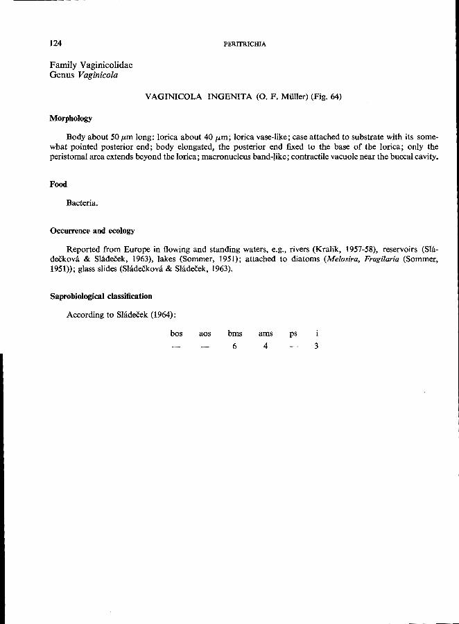
124
Family Vaginicolidae Genus Vaginicola
PERITRICHIA
VAGINICOLA INGENITA (0. F. Mi.iller) (Fig. 64)
Morphology
Body about 50 pm long: lorica about 40 fLm; lorica vase-like; case attached to substrate with its somewhat pointed posterior end; body elongated, the posterior end fixed to the base of the lorica; only the peristomal area extends beyond the lorica; macronucleus band-like; contractile vacuole near the buccal cavity.
Food
Bacteria.
Occurrence and ecology
Reported from Europe in flowing and standing waters, e.g., rivers (Kralik, 1957-58), reservoirs (Shideckova & Siadeeek, 1963), lakes (Sommer, 1951); attached to diatoms (Melosira, Fragilaria (Sommer,
1951»; glass slides (Sladeekova & Shidecek, 1963).
Saprobiological classification
According to Sladecek (1964):
bos aos bms
6
ams ps
4 3
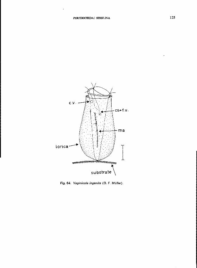
PERITRICHIDA: SESSILINA 125
C.V.
_-I-C--r CS+ f. v .
lorica~ T
1 SUbstrate\
Fig. 64. Vaginico/a ingenita (0. F. Muller).
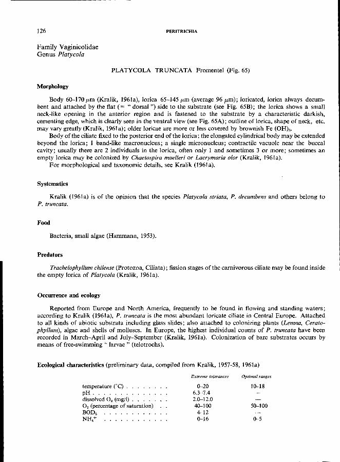
126
Family Vaginicolidae Genus Platycola
Morphology
PERITRICHIA
PLATYCOLA TRUNCATA Fromentel (Fig. 65)
Body 60-170 /Lm (Kralik, 1961a), lorica 65-145/Lm (average 96 /Lm); loricated, lorica always decumbent and attached by the flat (= "dorsal") side to the substrate (see Fig. 65B); the lorica shows a small neck-like opening in the anterior region and is fastened to the substrate by a characteristic darkish, cementing edge, which is clearly seen in the ventral view (see Fig. 65A); outline of lorica, shape of neck, etc. may vary greatly (Kralik, 1961a); older loricae are more or less covered by brownish Fe (OH)3'
Body of the ciliate fixed to the posterior end of the lorica; the elongated cylindrical body may be extended beyond the lorica; 1 band-like macronucleus; a single micronucleus; contractile vacuole near the buccal cavity; usually there are 2 individuals in the lorica, often only 1 and sometimes 3 or more; sometimes an empty lorica may be colonized by Chaetospira muelleri or Lacrymaria olor (Kralik, 1961a).
For morphological and taxonomic details, see Kralik (1961a).
Systematics
Kralik (l961a) is of the opinion that the species Platycola striata, P. decumbens and others belong to
P. truncata.
Food
Bacteria, small algae (Hammann, 1953).
Predators
Trachelophyllum chilense (Protozoa, Ciliata); fission stages of the carnivorous ciliate may be found inside
the empty lorica of Platycola (Kralik, 1961a).
Occurrence and ecology
Reported from Europe and North America, frequently to be found in flowing and standing waters; according to Kralik (1961a), P. truncata is the most abundant loricate ciliate in Central Europe. Attached to all kinds of abiotic substrata including glass slides; also attached to colonizing plants (Lemna, Ceratophyllum), algae and shells of molluscs. In Europe, the highest individual counts of P. truncata have been recorded in March-April and July-September (Kralik, 1961a). Colonization of bare substrates occurs by
means of free-swimming" larvae" (te!otrochs).
Ecological characteristics (preliminary data, compiled from Kralik, 1957-58, 1961a)
temperature (DC) . pH ...... . dissolved O. (mg/I) O. (percentage of saturation) BODs NH/ ......... .
Extreme tolerances
0-20 6.3-7.4 2.0-12.0 40-100 4-12 0-16
Optimal ranges
10-18
50-100
0-5
Saprobiological classification
According to Shideeek (1964):
bos
A
PERITRICHIDA: SESSILINA
aos 2
bms 4
ams ps
4 2
4!---,--'ol'C- f, V ,
.---x
~i--- lor; ca
---+:-_'L- ma
~substrate Fig, 65. Platycola truncata Fromentel. A. View of the convex (" ventral ") face of the lorica; B, diagrammatic longitudinal section through the lorica; redrawn from Kralik (1961 a).
127
128
Family Vaginicolidae Genus Thuricola
PERITRICHIA
THURICOLA FOLLICULATA (0. F. MUller) (Fig. 66)
Morphology (Kahl, 1935; Kudo, 1966)
Length of lorica 130-300 fLm; stalkless, vase-shaped lorica equipped with valve-like apparatus which closes the anterior part of the case obliquely like a trap-door when the cell contracts; body attached to the base of the case by a very short stalk; body elongated, peristomial area extends outwards; pellicle annulated; macronucleus band-like, extending longitudinally nearly the entire length of the cell; contractile vacuole near to the buccal cavity; cytoplasm hyaline, with or without zoochlorellae; the lorica usually contains 2 indi-
viduals.
Food
Algae, bacteria.
Occurrence and ecology
Widely distributed in both standing and flowing water; reported from peat-bogs (Liebmann, 1962), attached to plants, algae, e.g., Enteromorpha sp. (Sommer, 1951), and artificial substrates (glass slides, etc.); occurring throughout the year with highest abundance in summer; associated with Platycola truncata
(Nusch, 1969); very sensitive to low oxygen content (Liebmann, 1962).
Ecological characteristics (preliminary data provided by Nusch, 1969)
Saprobiology
temperature ("C) . pH ...... . dissolved O. (mg/I) free CO. (mgfl) . . NH.+ (mgfl) ... bacteria (membrane-filter
technique) . . . . . .
Extreme tolerances
4-22 7.1-7.9 6.0~12.0
0-12.0 0-2.5
1000000--9000000jml
According to Liebmann (1962), this is an oligosaprobic indicator organism but this characterization
seems too narrow.
Saprobiological classification
bos aos
2
bms
6
ams ps
2 3
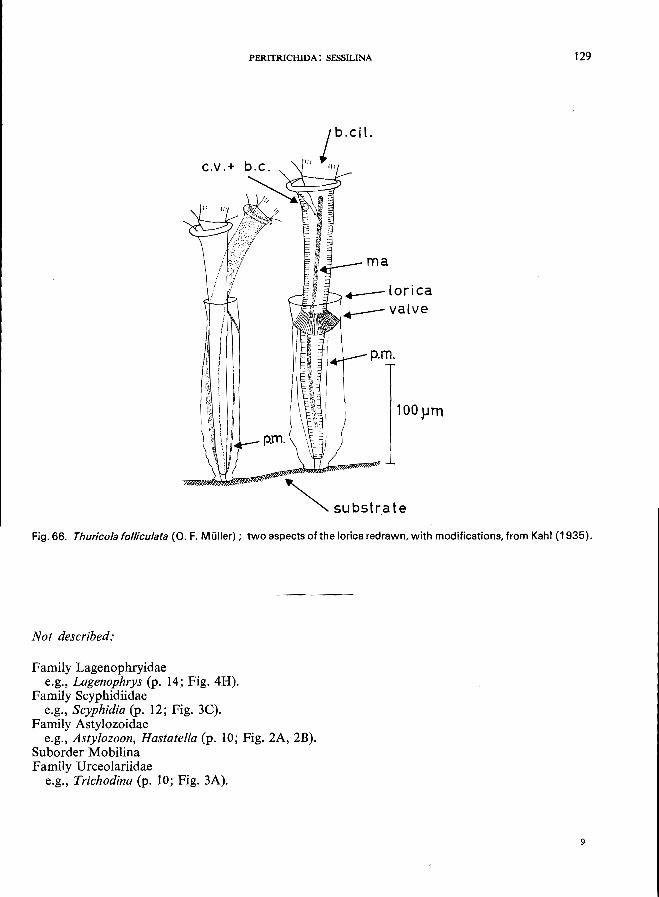
PERITRICHIDA: SESSILINA 129
ma
p.m.
100}JTn
~ substrate
Fig. 66. Thuricola fofliculata (0. F. Muller); two aspects of the lorica redrawn, with modifications, from Kahl (1935).
Not described:
Family Lagenophryidae e.g., Lagenophrys (p. 14; Fig. 4H).
Family Scyphidiidae e.g., Scyphidia (p. 12; Fig. 3C).
Family Astylozoidae e.g., Asty!ozoon, Hastatella (p. 10; Fig, 2A, 2B).
Suborder Mobilina Family Urceolariidae
e.g., Trichodina (p. 10; Fig. 3A).
9
............ ------------------
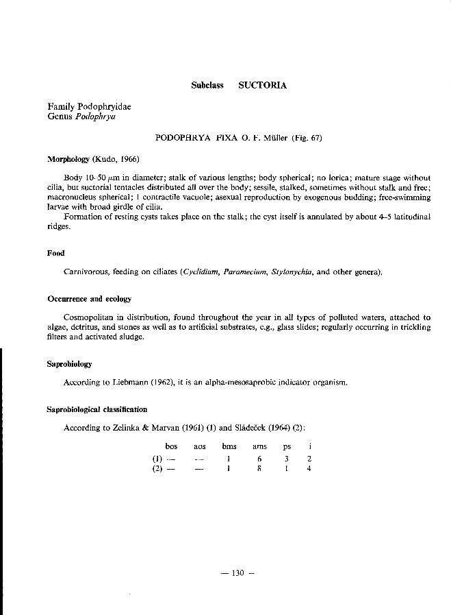
Family Podophryidae Genus Podophrya
Subclass SUCTORIA
PODOPHRYA FIXA O. F. Miiller (Fig. 67)
Morphology (Kudo, 1966)
Body 10-50/Lm in diameter; stalk of various lengths; body spherical; no lorica; mature stage without cilia, but suctorial tentacles distributed all over the body; sessile, stalked, sometimes without stalk and free; macronucleus spherical; 1 contractile vacuole; asexual reproduction by exogenous budding; free-swimming
larvae with broad girdle of cilia. Formation of resting cysts takes place on the stalk; the cyst itself is annulated by about 4-5 latitudinal
ridges.
Food
Carnivorous, feeding on ciliates (Cyclidium, Paramecium, Sty[onychia, and other genera).
Occurrence and ecology
Cosmopolitan in distribution, found throughout the year in all types of polluted waters, attached to algae, detritus, and stones as well as to artificial substrates, e.g., glass slides; regularly occurring in trickling
filters and activated sludge.
Saprobiology
According to Liebmann (1962), it is an alpha-mesosaprobic indicator organism.
Saprobiological classification
According to Zelinka & Marvan (1961) (1) and Sladecek (1964) (2):
bos
(1) -(2) -
aos bms ams
6 8
-130 -
ps
3 2 4
------------------..............
PODOPHRYIDAE 131
4-.--stalk
I
Fig. 67. Podophrya fixa O. F. Muller.
132
Family Podophryidae Genus Metacineta
SUCTORIA
METACINETA MYSTACINA (Ehrenberg) (Fig. 68)
Morphology
Body about 35-100 pm in diameter, tentacles up to 150 p.m, lorica up to 800 p.m; mature stage without any cilia but with suctorial tentacles that are arranged in 6 groups at the anterior end (Fig. 68A); no cytostome; food ingestion by means of the tentacles; the stalked lorica (Fig. 68B) is attached to the substrate (stones, plants, artificial substrates, e.g., glass slides); 1 spherical macronucleus; a single contractile vacuole.
For details see Kudo (1966). Asexual reproduction by exogenous budding; the free-swimming ciliated bud (" larva") undergoes
metamosphosis to the sessile mature stage described above.
Food
Carnivorous, feeding on ciliates (Carchesium, Cyclidium, Halteria, Uronema, Vorticella, and other
genera); the prey is captured by the tentacles.
Occurrence and ecology
Widely distributed in stagnant and flowing waters; in brackish and fresh water (Kahl, 1934), associated with Coleps hirtus, Cyclidium citrullus, Stentor roeseli, Urotricha Jarcta (Bick, unpublished data); in the River Elbe (Hentschel, 1916) a high frequency of M. mystacina on artificial substrates mostly coincides with high individual counts of Epistylis, Vorticella, or Zoothamnium; maximum development in the Elbe occurred in
May-September.
Ecological characteristics (preliminary data)
temperature eC) . pH ...... . dissolved O. (mg/I) NH/ (mg/I) ...
Saprobiological classification
According to Shideckova & Sladecek (1963):
bos aos bms
5
ams
5
Extreme tolerances
5-25 6.5-7.5 0.3-10
0-5
ps
3
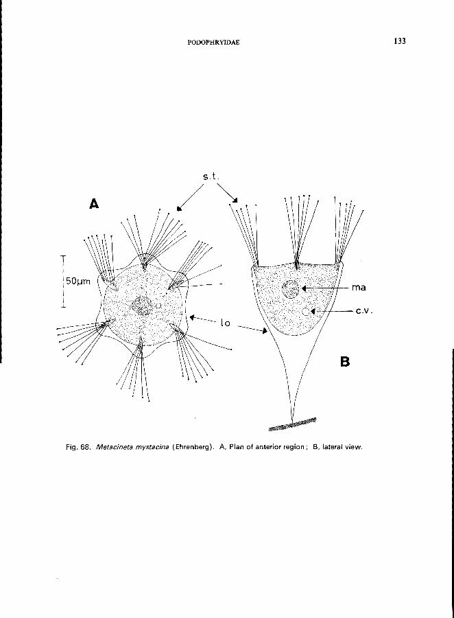
PODOPHRYIDAE 133
S. t.
A /~
~-4-J.'----_ C. v .
B
Fig. 68. Metacineta mystacina (Ehrenberg). A, Plan of anterior region; B, lateral view.
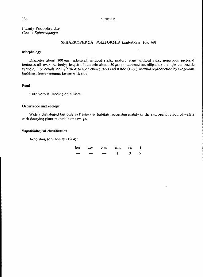
134
Family podophryidae Genus Sphaerophrya
SUCTORIA
SPHAEROPHRYA SOLIFORMIS Lauterborn (Fig. 69)
Morphology
Diameter about 100 fLm; spherical, without stalk; mature stage without cilia; numerous suctorial tentacles all over the body; length of tentacle about 30 fLm; macronucleus ellipsoid; a single contractile vacuole. For details see Eyferth & Schoenichen (1927) and Kudo (1966), asexual reproduction by exogenous
budding; free-swimming larvae with cilia.
Food
Carnivorous; feeding on ciliates.
Occurrence and ecology
Widely distributed but only in freshwater habitats, occurring mainly in the sapropelic region of waters
with decaying plant materials or sewage.
Saprobiological classification
According to Shideeek (1964):
bos aos bms ams ps
9 5
PODOPHRYIDAE 135
ma
50 I ~ml
Fig. 69. Sphaerophrya soliformis Lauterborn.
136
Family Dendrosomatidae Genus Dendrosoma
SUCTORIA
DENDROSOMA RADIANS Ehrenberg (Fig. 70)
Morphology
Body up to 2.5 mm high, branching irregular; adult without cilia but with suctorial tentacles arranged in clusters at the end of the branches; macronucleus long and band-like, branched; numerous contractile vacuoles; sessile; free-swimming larvae with cilia. For morphological details see Gonnert (1935).
Food
Carnivorous, feeding on ciliates.
Occurrence and ecology
Widely distributed, attached to plants, stones, and artificial substrates (e.g., glass slides); in standing and flowing waters, e.g., rivers (Holm, 1928; Hentschel, 1916) and reservoirs (Shideckova & Sladecek, 1963).
Occurring at all times of the year; in the Sedlice Reservoir, Czechoslovakia, conspicuous maxima have been recorded in April, June, and November, partly associated with Campanella umbellaria, Heliophrya rotunda, Ophrydium sessile, and Stentor roeseli (Sladeckova, 1962, 1963). Hentschel (1916) found the highest abundance during May, June, and July (River Elbe, Hamburg, Federal Republic of Germany); associated predominantly with Carchesium poiypinum, Epistylis hentscheli Kahl, Metacineta mystacina, Vorticella
campanuia, and Zoothamnium hentscheli Kahl.
Saprobiology
According to Holm (1928), D. radians occurs in beta- and alpha-mesosaprobic localities.
Saprobiological classification
According to Sladeckova & Sladecek (1966):
bos aos bms
5
ams ps
5 3
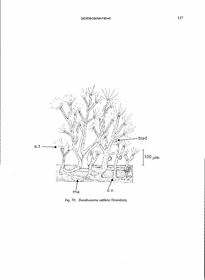
DENDROSOMATIDAE 137
....... ~,--- bud
ma
Fig. 70. Dendrosoma radians Ehrenberg.
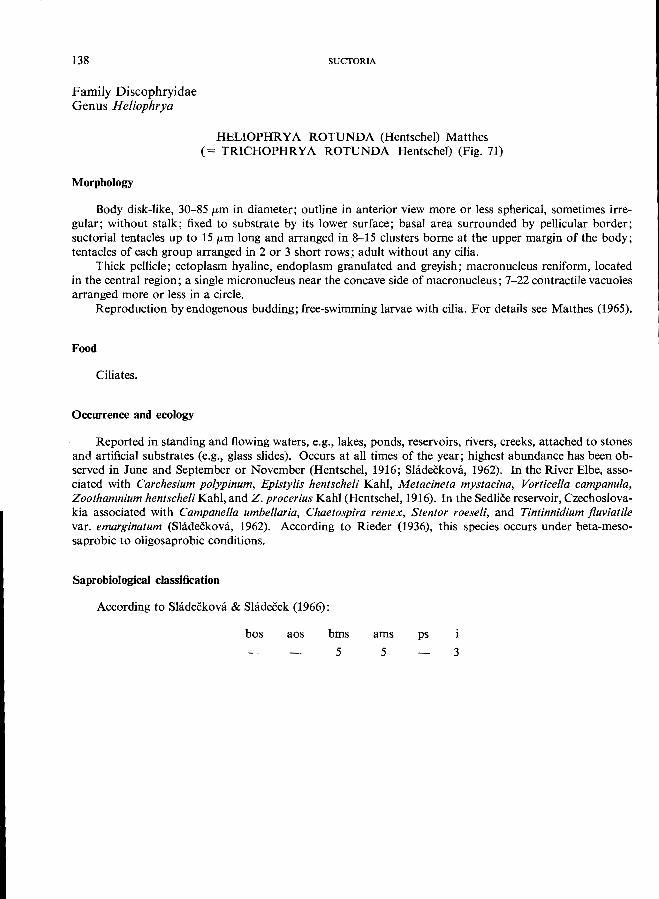
138
Family Discophryidae Genus Heliophrya
SUCTORIA
HELIOPHRYA ROTUNDA (Hentschel) Matthes (= TRICHOPHRYA ROTUNDA Hentschel) (Fig. 71)
Morphology
Body disk-like, 30-85 /km in diameter; outline in anterior view more or less spherical, sometimes irregular; without stalk; fixed to substrate by its lower surface; basal area surrounded by pellicular border; suctorial tentacles up to 15 /km long and arranged in 8-15 clusters borne at the upper margin of the body; tentacles of each group arranged in 2 or 3 short rows; adult without any cilia.
Thick pellicle; ectoplasm hyaline, endoplasm granulated and greyish; macronucleus reniform, located in the central region; a single micronucleus near the concave side of macronucleus; 7-22 contractile vacuoles
arranged more or less in a circle. Reproduction by endogenous budding; free-swimming larvae with cilia. For details see Matthes (1965).
Food
Ciliates.
Occurrence and ecology
Reported in standing and flowing waters, e.g., lakes, ponds, reservoirs, rivers, creeks, attached to stones and artificial substrates (e.g., glass slides). Occurs at all times of the year; highest abundance has been observed in June and September or November (Hentschel, 1916; Shideekova, 1962). In the River Elbe, associated with Carchesium poiypinum, Epistylis hentscheli Kahl, Metacineta mystacina, Vorticella campanuia, Zoothamnium hentscheli Kahl, and Z. procerius Kahl (Hentschel, 1916). In the Sedliee reservoir, Czechoslovakia associated with Campanella umbellaria, Chaetospira remex, Stentor roeseli, and Tintinnidium fluviatile var. emarginatum (Sladeckova, 1962). According to Rieder (1936), this species occurs under beta-meso-
saprobic to oligosaprobic conditions.
Saprobiological classification
According to Sladeckova & Sladecek (1966):
bos aos bms
5
ams ps
5 3
DISCOPHR YIDAE 139
c r' B
..•.. : ::! ma+mi
':',
c.v .
cv.
A I
Fig. 71. He/iophrya rotunda (Hentschel) Matthes. A, Forma typica, anterior view; B, forma sinuosa (Rieder) anterior view, the suctorial tentacles are indicated by dots; C, diagrammatic cross-section .
.............. ------------------
Not described:
Subclass Order
Family Bursariidae e.g., Bursaridium (p. 20; Fig. 9C).
Family Stentoridae Genus Stentor
SPIROTRICIDA HETEROTRICIDDA
STENTOR COERULEUS Ehrenberg (Fig. 72)
Morphology
Length 1-2 mm (fully extended), trumpet-shaped when extended, after contraction more or less spherical; striking blue colour (due to the pigment" stentorin "); uniform ciliation all over the body, a small number of sensory bristles; adoral zone of membranelles extends in a spiral form around the anterior pole of the body; the buccal area itself is equipped with rows of smaller cilia; macronucleus rosary-shaped; contractile vacuole in the anterior part left behind the cytopharynx with long canals directed in posterior and anterior direction.
Food
Flagellates, algae, ciliates.
Occurrence and ecology
s. coeruleus is a sessile organism living attached to stones, roots, or detritus (even on floating material) in a mucilaginous lorica, sometimes free-swimming near the bottom, and often occurring in great colonies. Inhabits still and slowly flowing waters; tolerates free ammonia up to 0.3 mg/l (Stammer, 1953).
Saprobiology
An alpha-mesosaprobic indicator-organism according to Liebmann (1962).
Saprobiological classification
According to Shidecek (1964):
bos aos bms
4
ams
6
-140 -
ps
+ 3
--------------------.............
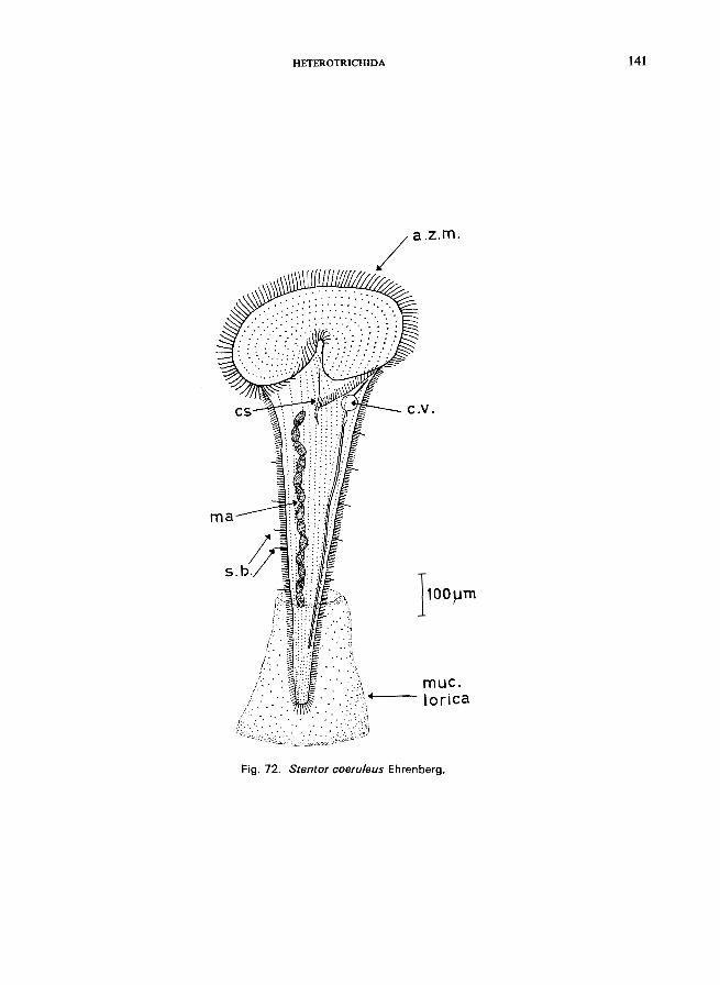
ma
HETEROTRICHIDA
}oo}Jm
muc. lorica
Fig. 72. Stentor coeruleus Ehrenberg.
141
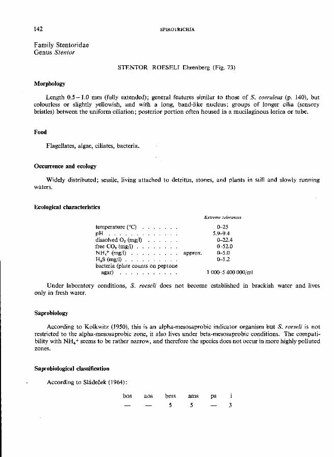
142 SPIROTRICHIA
Family Stentoridae Genus Stentor
STENTOR ROESEL! Ehrenberg (Fig. 73)
Morphology
Length 0.5 -1.0 mm (fully extended); general features similar to those of S. coeruleus (p. 140), but colourless or slightly yellowish, and with a long, band-like nucleus; groups of longer cilia (sensory bristles) between the uniform ciliation; posterior portion often housed in a mucilaginous lorica or tube.
Food
Flagellates, algae, ciliates, bacteria.
Occurrence and ecology
Widely distributed; sessile, living attached to detritus, stones, and plants in still and slowly running
waters.
Ecological characteristics
temperature Ce) . pH ...... . dissolved O2 (mg/I) free CO2 (mg/I) NH/ (mg/I) .. . H 2S (mg/I) ... . bacteria (plate counts on peptone
agar) . . . . . . . .
Extreme tolerances
0-25 5.9-9.4
0-22.4 0-52.0
approx. 0-5.0 0-1.2
1 000-5 400000/ml
Under laboratory conditions, S. roeseli does not become established in brackish water and lives
only in fresh water.
Saprobiology
According to Kolkwitz (1950), this is an alpha-mesosaprobic indicator organism but S. roeseli is not restricted to the alpha-mesosaprobic zone, it also lives under beta-mesosaprobic conditions. The compatibility with NH4 + seems to be rather narrow, and therefore the species does not occur in more highly polluted
zones.
Saprobiological classification
According to Shidecek (1964):
bos aos bms
5
ams
5
ps
3
HETEROTRICHIDA
.41+''--- C . v .
ma--4'-~,~
cs
muc. lorica
Fig. 73. Stentor roeseli Ehrenberg.
143
144
Family Stentoridae Genus Stentor
SPIROTRICHIA
STENTOR POLYMORPHUS (0. F. Milller) (Fig. 74)
Morphology
Length 1-2 mm (fully extended); shape very similar to that of S. coeruleus but colourless and the body is filled with symbiontic zoochlorellae; macronucleus rosary-shaped; usually without a lorica.
Food
Flagellates, algae.
Occurrence and ecology
Distribution and ecology similar to those of S. roeseli; according to Liebmann (1962), it is a betamesosaprobic indicator organism. I believe that it would be more accurate to say beta-mesosaprobic/alphamesosaprobic. S. polymorphus is able to tolerate dissolved oxygen levels down to 0.5 mg/l and may therefore
occur in rather polluted zones.
Ecological characteristics (from various sources)
temperature CC) . pH ...... . dissolved O. (mg/l) free CO. (mg/l) NH.+ (mgfl) H.S (mg/l) ..
Extreme tolerances
4-25 6.8-8.8 0.5-22.5
0-44 0-6 0-1
Saprobiological classification (calculated from data of Snimek-Husek (1956) and unpublished data of Bick)
bos aos bms
4
ams ps
6 3
STENTOR MUELLERI (Bory St. Vincent)
Morphology (Kahl, 1932)
When fully extended, 2-3 mm long., very similar to S. polymorphus but without zoochlorellae (note: sometimes food vacuoles are filled with ingested algae); macronucleus rosary-shaped; often housed in gelatinous tube; posterior end drawn out into stalk (unlike S. polymorphus).
Ecology and saprobiology
Similar to those of S. poly morphus.
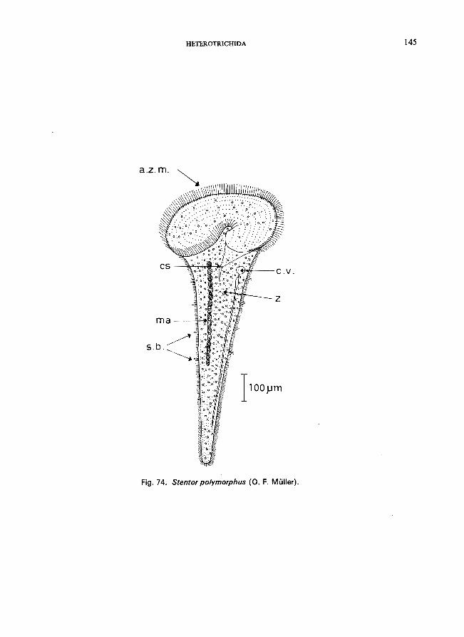
HETEROTRICHIDA 145
a.z.m.
ma --4'-~&
s.b.
Fig. 74. Stentor polymorphus (0. F. Muller).
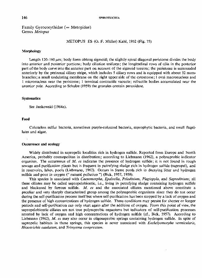
146
Family Gyrocorythidae (= Metopidae) Genus Metopus
SPIROTRICHIA
METOPUS ES (0. F. MUller) Kahl, 1932 (Fig. 75)
Morphology
Length 120-160 /Lm; body form oblong sigmoid; the slightly spiral diagonal peristome divides the body into anterior and posterior portions; body ciliation uniform; the longitudinal rows of cilia in the posterior part ofthe body curve into the anterior part on account of the sigmoid torsion; the peristome is surrounded anteriorly by the perizonal ciliary stripe, which includes 5 ciliary rows and is equipped with about 32 membranelles; a small undulating membrane on the right upper side of the cytostome; 1 oval macronucleus and 1 micronucleus near the peristome; 1 terminal contractile vacuole; refractile bodies accumulated near the anterior pole. According to Schulze (1959) the granules contain peroxidase.
Systematics
See Jankowski (1964a).
Food
Colourless sulfur bacteria, sometimes purple-coloured bacteria, saprophytic bacteria, and small flagellates and algae.
Occurrence and ecology
Widely distributed in sapropelic localities rich in hydrogen sulfide. Reported from Europe and North America, probably cosmopolitan in distribution; according to Liebmann (1962), a polysaprobic indicator organism. The occurrence of M. es indicates the presence of hydrogen sulfide; it is not found in rough sewage and purification plants but is frequent in putrefying sludge rich in hydrogen sulfide (sapropel), and in reservoirs, lakes, pools (Liebmann, 1962). Occurs in forest pools rich in decaying litter and hydrogen sulfide and poor in oxygen (" natural pollution ") (Bick, 1957, 1958).
This species is associated with Caenomorpha, Epaixella, Pelodinium, Plagiopyia, and Saprodinium; all these ciliates may be called sapropelobiontic, i.e., living in putrefying sludge containing hydrogen sulfide and blackened by ferrous sulfide. M. es and the associated ciliates mentioned above constitute a peculiar and very sharply characterized group among the polysaprobic organisms since they do not occur during the self-purification process itself but where self-purification has been stopped by a lack of oxygen and the presence of high concentrations of hydrogen sulfide. These conditions may persist for shorter or longer periods and self-purification can only start again after the addition of oxygen. From this point of view, the sapropelobiontic ciliates are not true polysaprobic organisms but indicators of self-purification processes arrested by lack of oxygen and high concentrations of hydrogen sulfide (cf., Bick, 1957). According to Liebmann (1962), M. es may also occur in oligosaprobic springs containing hydrogen sulfide. In spite of sapropelic habitats in those springs, this species is never associated with Encheiyomorpha vermicuiaris, Hexotrichia caudatum, and Trimyema compressum.
undo rn.
C. v . ---=I..r....:...
HETEROTRICHIDA
pa
+---a.z.rn.
ps
I
Fig. 75. Metopus es (0. F. Muller) Kahl, 1932; ventral view after Jankowski (1964).
147
148
Family Gyrocorythidae (= Metopidae) Genus Brachonella
SPIROTRICHIA
BRACHONELLA SPIRALIS (Smith) Jankowski, 1964a (Fig. 76) (= METOPUS CONTORTUS Levander; sic Liebmann, 1962)
Morphology
Length 80-150 pm; body ovoid, peristome spirally diagonal; cytostome dorsally located, near the posterior end of the cell; body ciliation uniform except for a group of long caudal cilia; brownish in colour; accumulation of refractile granules at the anterior pole; 1 spherical macronucleus and 1 micronucleus located centrally; contractile vacuole near the posterior end; trichocysts present.
Systematics
See Jankowski (1964a).
Food
Purple-coloured and colourless sulfur bacteria; diatoms, flagellates.
Occurrence and ecology
Reported from Europe and North America; sapropelobiontic, associated with Metopus es, Caenomorpha,
Epalxella, and other genera.
Saprobiology
According to Liebmann (1962), this is a polysaprobic indicator organism. However, it seems better to regard it as an indicator of hydrogen sulfide. For a discussion of the ecology of the sapropelobiontic ciliates
see Metopus es.
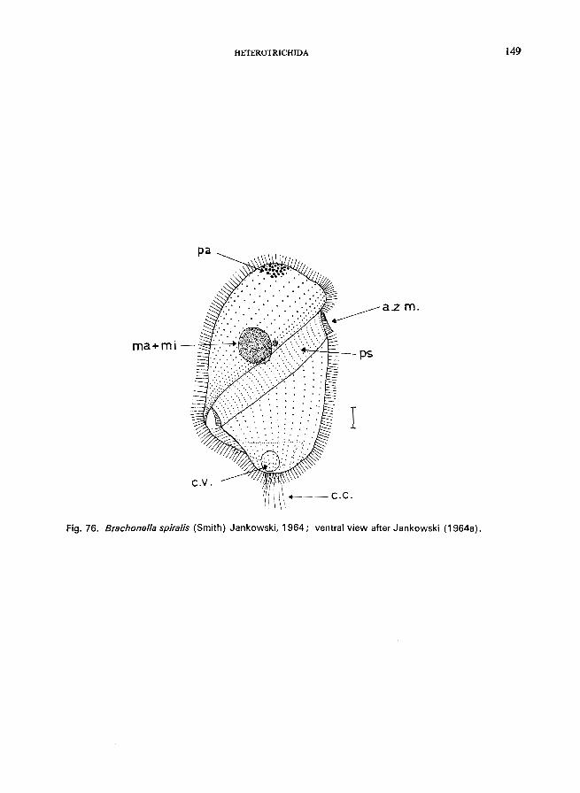
HETEROTRICHIDA 149
Fig. 76. Brachonella spiralis (Smith) Jankowski, 1964; ventral view after Jankowski (1964a).
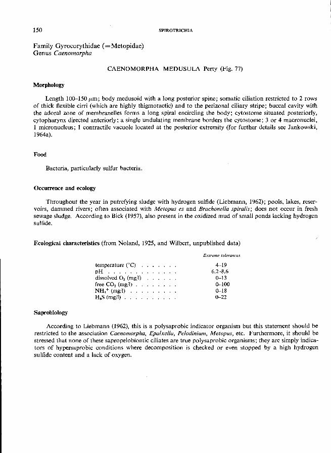
150
Family Gyrocorythidae (=Metopidae) Genus Caenomorpha
SPIROTRICHIA
CAENOMORPHA MEDUSULA Perty (Fig. 77)
Morphology
Length 100-150 (tm; body medusoid with a long posterior spine; somatic ciliation restricted to 2 rows of thick flexible cirri (which are highly thigmotactic) and to the perizonal ciliary stripe; buccal cavity with the adoral zone of membranelles forms a long spiral encircling the body; cytostome situated posteriorly, cytopharynx directed anteriorly; a single undulating membrane borders the cytostome; 3 or 4 macronuclei, 1 micronucleus; 1 contractile vacuole located at the posterior extremity (for further details see Jankowski,
1964a).
Food
Bacteria, particularly sulfur bacteria.
Occurrence and ecology
Throughout the year in putrefying sludge with hydrogen sulfide (Liebmann, 1962); pools, lakes, reservoirs, dammed rivers; often associated with Metopus es and Brachonella spiralis; does not occur in fresh sewage sludge. According to Bick (1957), also present in the oxidized mud of small ponds lacking hydrogen
sulfide.
Ecological characteristics (from Noland, 1925, and Wilbert, unpublished data)
Saprobiology
temperature eC) . pH ...... . dissolved O2 (mg/I) free CO2 (mgfl) NH/ (mg/I) H 2S (mg/I) ..
Extreme tolerances
4-19 6.2-8.6
0-13 0-100 0-18 0-22
According to Liebmann (1962), this is a polysaprobic indicator organism but this statement should be restricted to the association Caenomorpha, Epalxella, Pelodinium, Metopus, etc. Furthermore, it should be stressed that none of these sapropelobiontic ciliates are true polysaprobic organisms; they are simply indicators of hypersaprobic conditions where decomposition is checked or even stopped by a high hydrogen
sulfide content and a lack of oxygen.
HETEROTRICHIDA 151
I ma
a.z.m.
spine
Fig. 77. Caenomorpha medusula Perty, left lateral view; after Jankowski (1964). The small inset diagram illustrates the course of the adoral zone of membranelles and the perizonal ciliary stripe.
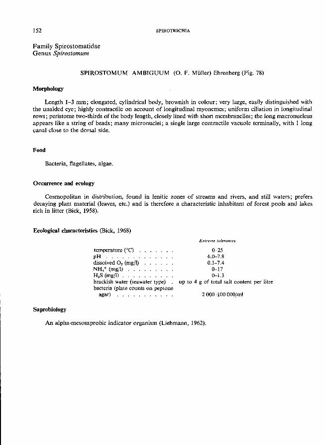
152 SPIROTRICHIA
Family Spirostomatidae Genus Spirostomum
SPJROSTOMUM AMBIGUUM (0. F. Muller) Ehrenberg (Fig. 78)
Morphology
Length 1-3 mm; elongated, cylindrical body, brownish in colour; very large, easily distinguished with the unaided eye; highly contractile on account of longitudinal myonemes; uniform ciliation in longitudinal rows; peristome two-thirds of the body length, closely lined with short membranelles; the long macronucleus appears like a string of beads; many micronuclei; a single large contractile vacuole terminally, with 1 long
canal close to the dorsal side.
Food
Bacteria, flagellates, algae.
Occurrence and ecology
Cosmopolitan in distribution, found in lenitic zones of streams and rivers, and still waters; prefers decaying plant material (leaves, etc.) and is therefore a characteristic inhabitant of forest pools and lakes
rich in litter (Bick, 1958).
Ecological characteristics (Bick, 1968)
Saprobiology
temperature eC) . pH ...... . dissolved O2 (mgfl) NH. + (mg/l) . . . H 2S (mg/l) .... brackish water (seawater type) bacteria (plate counts on peptone
agar) .......... .
Extreme tolerances
0-25 6.0-7.8 0.1-7.4
0-17 0-1.3
up to 4 g of total salt content per litre
2000-100000/ml
An alpha-mesosaprobic indicator organism (Liebmann, 1962).
------------------------............ .
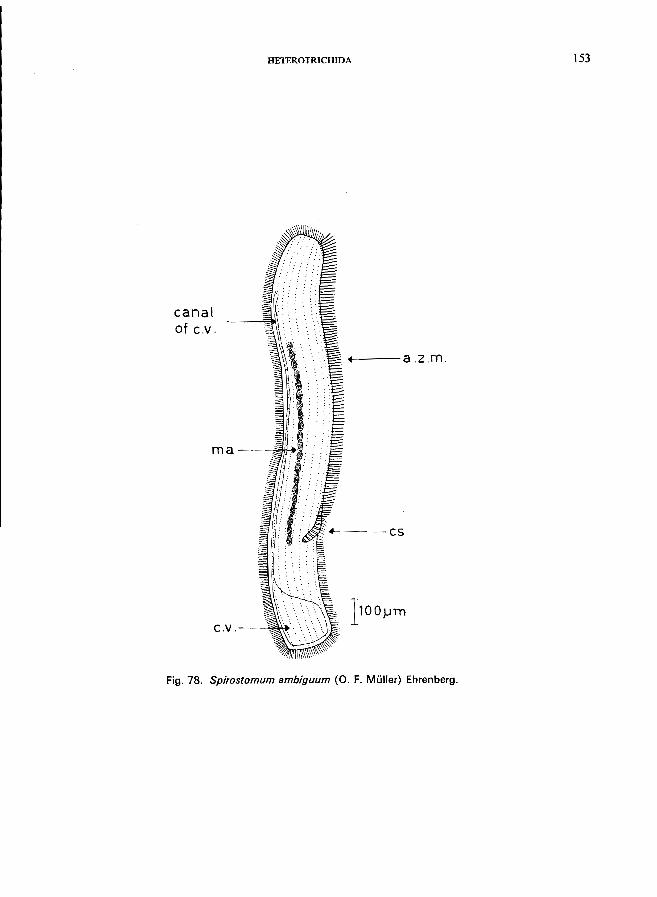
HETEROTRICHIDA
canal of C.v.
ma--~:III-i.'l:i
c.V.--~'"
----a.z.m.
4-----cs
1100}Jln
Fig. 78. Spirostomum ambiguum (0. F. Muller) Ehrenberg.
153
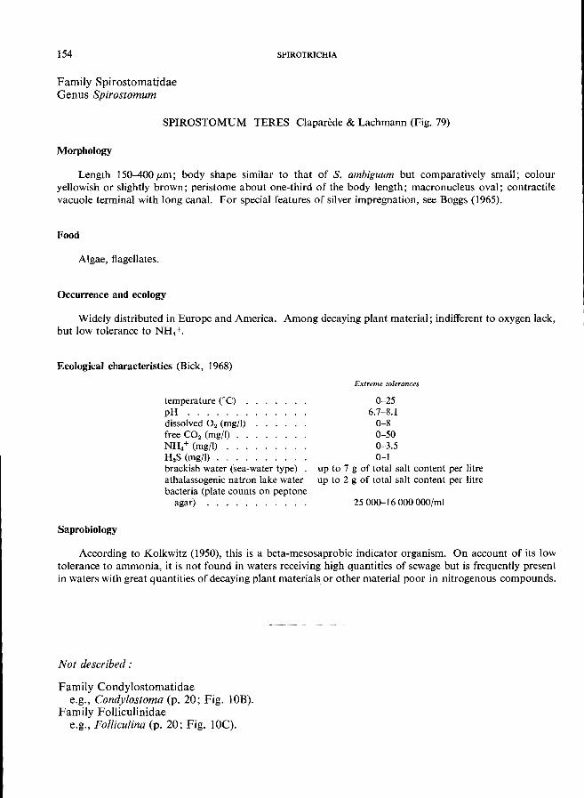
154 SPIROTRICHIA
Family Spirostomatidae Genus SpiNJstomum
SPIROSTOMUM TERES Claparede & Lachmann (Fig. 79)
Morphology
Length 150-400 fLm; body shape similar to that of S. ambiguum but comparatively small; colour yellowish or slightly brown; peristome about one-third of the body length; macronucleus oval; contractile vacuole terminal with long canal. For special features of silver impregnation, see Boggs (1965).
Food
Algae, flagellates.
Occurrence and ecology
Widely distributed in Europe and America. Among decaying plant material; indifferent to oxygen lack,
but low tolerance to NH4+'
Ecological characteristics (Bick, 1968)
Saprobio)ogy
temperature COC) . pH ...... . dissolved O2 (mg/I) free CO2 (mg/I) NH/ (mg/I) .. . H.s (mg/I) ... . brackish water (sea-water type) athalassogenic natron lake water bacteria (plate counts on peptone
agar) . . . . . . . . . . .
Extreme tolerances
0-25 6.7-8.1
0-8 0-50 0-3.5 0-1
up to 7 g of total salt content per litre up to 2 g of total salt content per litre
25000-16000000/ml
According to Kolkwitz (1950), this is a beta-mesosaprobic indicator organism. On account of its low tolerance to ammonia, it is not found in waters receiving high quantities of sewage but is frequently present in waters with great quantities of decaying plant materials or other material poor in nitrogenous compounds.
Not described:
Family Condylostomatidae e.g., Condylostoma (p. 20; Fig. lOB).
Family Folliculinidae e.g., Folliculina (p. 20; Fig. lOC)o
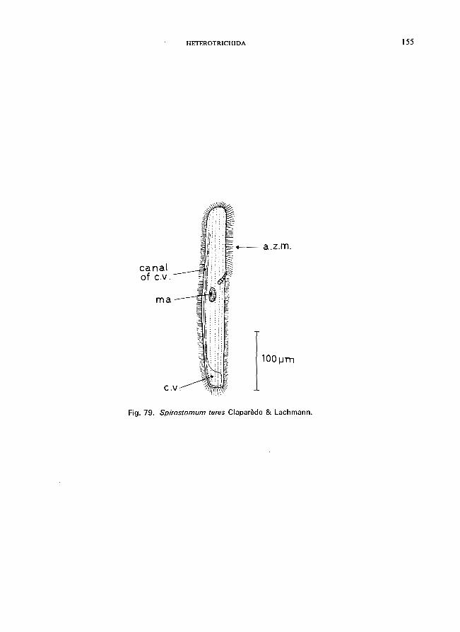
canal of c.v.
rna
HETEROTRICHIDA
-a.z.m.
1 00 ~ . .ITn
Fig. 72. Spirostomum teres Claparede & Lachmann.
155
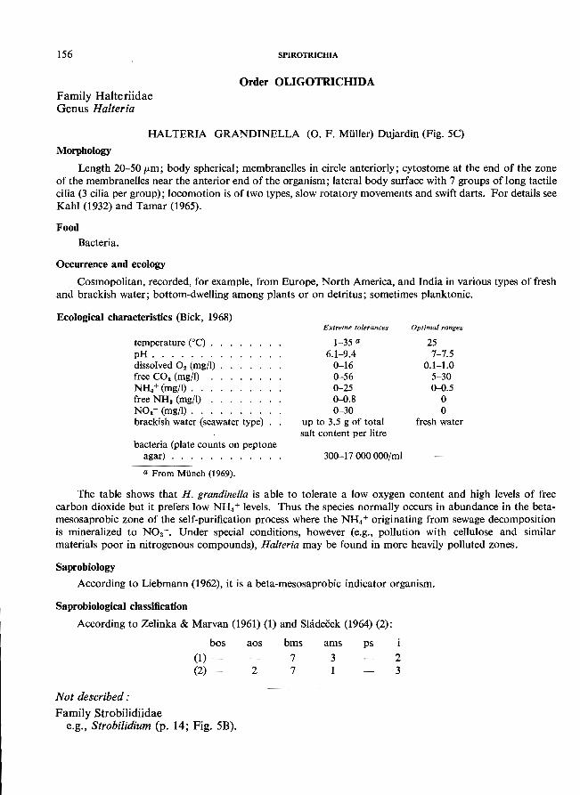
156
Family Halteriidae Genus Halteria
SPIROTRICHIA
Order OLIGOTRICHIDA
HALTERIA GRANDINELLA (0. F. MUller) Dujardin (Fig. 5C)
Morphology Length 20-50 pm; body spherical; membranelles in circle anteriorly; cytostome at the end of the zone
of the membranelles near the anterior end of the organism; lateral body surface with 7 groups of long tactile cilia (3 cilia per group); locomotion is of two types, slow rotatory movements and swift darts. For details see
Kahl (1932) and Tamar (1965).
Food Bacteria.
Occurrence and ecology Cosmopolitan, recorded, for example, from Europe, North America, and India in various types of fresh
and brackish water; bottom-dwelling among plants or on detritus; sometimes planktonic.
Ecological characteristics (Bick, 1968)
temperature (0C) . . pH ....... . dissolved O2 (mgfl) . free CO2 (mg/I) NH4+ (mg/I). . . . free NHa (mg/I) . . NO.- (mg/I) .... brackish water (seawater type)
bacteria (plate counts on peptone agar) . . . . . . . .
a From MUnch (1969).
Extreme tolerances
1-35 a
6.1-9.4 0-16 0-56 0-25 <>-D.S 0-30
up to 3.5 g of total salt content per litre
300-17000000/ml
Optimal ranges
25 7-7.5
0.1-1.0 5-30 <>-D. 5
o o
fresh water
The table shows that H. grandinella is able to tolerate a low oxygen content and high levels of free carbon dioxide but it prefers low NH4 + levels. Thus the species normally occurs in abundance in the betamesosaprobic zone of the self-purification process where the NH4 + originating from sewage decomposition is mineralized to N0
3-. Under special conditions, however (e.g., pollution with cellulose and similar
materials poor in nitrogenous compounds), Halteria may be found in more heavily polluted zones.
Saprobiology According to Liebmann (1962), it is a beta-mesosaprobic indicator organism.
Saprobiological classification
According to Zelinka & Marvan (1961) (1) and Shideeek (1964) (2):
Not described:
bos (1) -(2) -
Family Strobilidiidae e.g., Strobilidium (p. 14; Fig. 5B).
aos
2
bms
7 7
ams
3 1
ps 2 3
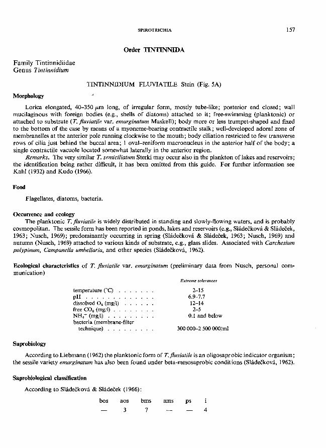
Family Tintinnidiidae Genus Tintinnidium
Morphology
SPIROTRICHIA 157
Order TINTINNIDA
TINTINNIDIUM FLUVIATILE Stein (Fig. 5A)
Lorica elongated, 4O-350/Lm long, of irregular form, mostly tube-like; posterior end closed; wall mucilaginous with foreign bodies (e.g., shells of diatoms) attached to it; free-swimming (planktonic) or attached to substrate (T. flUViatile var. emarginatum Maskell); body more or less trumpet-shaped and fixed to the bottom of the case by means of a myoneme-bearing contractile stalk; well-developed adoral zone of membranelles at the anterior pole running clockwise to the mouth; body ciliation restricted to few transverse rows of cilia just behind the buccal area; 1 oval-reniform macronucleus in the anterior half of the body; a single contractile vacuole located somewhat laterally in the anterior region.
Remarks. The very similar T. semiciliatum Sterki may occur also in the plankton of lakes and reservoirs; the identification being rather difficult, it has been omitted from this guide. For further information see Kahl (1932) and Kudo (1966).
Food
Flagellates, diatoms, bacteria.
Occurrence and ecology The planktonic T. fluviatile is widely distributed in standing and slowly-flowing waters, and is probably
cosmopolitan. The sessile form has been reported in ponds, lakes and reservoirs (e.g., Shideckova & Sladecek, 1963; Nusch, 1969); predominantly occurring in spring (Sladeckova & Sladecek, 1963; Nusch, 1969) and autumn (Nusch, 1969) attached to various kinds of substrate, e.g., glass slides. Associated with Carchesium polypinum, Campanella umbellaria, and other species (Sladeckova, 1962).
Ecological characteristics of T. flUViatile var. emarginatum (preliminary data from Nusch, personal communication)
Saprobiology
temperature COc) . pH ...... . dissolved O. (mg/I) free CO. (mg/I) . . NH. + (mg/l) . . . bacteria (membrane-filter
technique) . . . . . .
Extreme tolerances
2-15 6.9-7.7 12-14 2-5
0.1 and below
300000-2500000/ml
According to Liebmann (1962) the planktonic form of T. fluviatile is an oligosapr 0 bic indicator organism; the sessile variety emarginatum has also been found under beta-mesosaprobic conditions (Sladeckova, 1962).
Saprobiological classification
According to Sladeckova & Sladeeek (1966):
bos aos
3
bms
7
ams ps
4
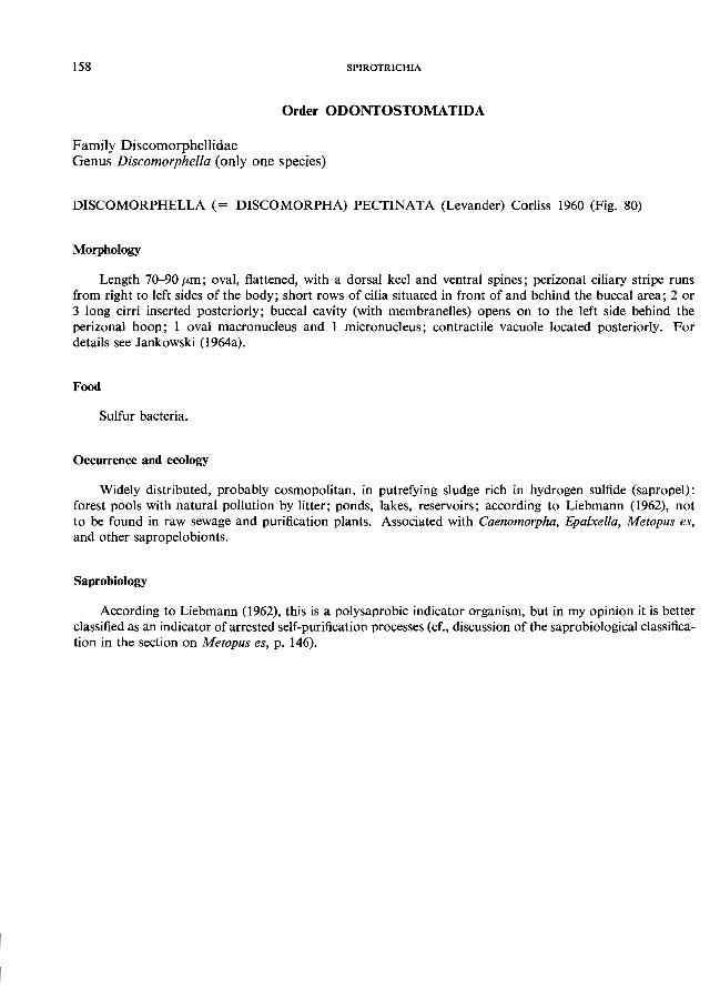
158 SPJROTRICHIA
Order ODONTOSTOMATIDA
Family Discomorphellidae Genus Discomorphella (only one species)
DISCOMORPHELLA (= DISCOMORPHA) PECTINATA (Levander) Corliss 1960 (Fig. 80)
Morphology
Length 70-90 (Lm; oval, flattened, with a dorsal keel and ventral spines; perizonal ciliary stripe runs from right to left sides of the body; short rows of cilia situated in front of and behind the buccal area; 2 or 3 long cirri inserted posteriorly; buccal cavity (with membranelles) opens on to the left side behind the perizonal hoop; 1 oval macronucleus and 1 micronucleus; contractile vacuole located posteriorly. For details see Jankowski (1964a).
Food
Sulfur bacteria.
Occurrence and ecology
Widely distributed, probably cosmopolitan, in putrefying sludge rich in hydrogen sulfide (sapropel): forest pools with natural pollution by litter; ponds, lakes, reservoirs; according to Liebmann (1962), not to be found in raw sewage and purification plants. Associated with Caenomorpha, Epaixella, Metopus es, and other sapropelobionts.
Saprobiology
According to Liebmann (1962), this is a polysaprobic indicator organism, but in my opinion it is better classified as an indicator of arrested self-purification processes (cf., discussion of the saprobiological classification in the section on Metopus es, p. 146).
ODONTOSTOMATIDA
A
sp
e.v ...'--'~>-'-S'J:====: +4 --- p. e. s.
ma+mi--+--....
ei rri
B
p.e.s. -+
I .. cirri
Fig. 80. Discomorphella pectinata (Levander) Corliss; after Jankowski (1964a). A Right lateral view; B, left lateral view.
159
160
Family Epalxellidae Genus Epalxella
Morphology
SPIROTRICHIA
EPALXELLA STRIATA (Kahl) Corliss 1960 (Fig. 81)
Length 20--35 p'm; compressed, pellicle carapaced; right side convex, left side flattened; perizonal ciliary stripe consisting of 7 rows of cilia in the anterior part of the body and 4 short rows of cilia in the posterior part; buccal cavity with 8 membranelles, opening to the left side; 1 macronucleus and 1 micronucleus; the single contractile vacuole posteriorly located, near the buccal cavity; numerous refractile granules give
this ciliate a greyish appearance; for details see Jankowski (l964a).
Food
Sulfur bacteria.
Occurrence and ecology
Reported from Europe and America; sapropelobiontic, occurring in putrefying sludge rich in hydrogen sulfide; associated with Caenomorpha, Metopus, Pelodinium, Plagiopyla (p. 66), and Saprodinium. Like the other sapropelobiontic species, it does not occur in raw sewage.
Saprobiology
According to Liebmann (1962), it is a polysaprobic indicator organism (for discussion of this point,
see the section on Metopus es, p. 146).
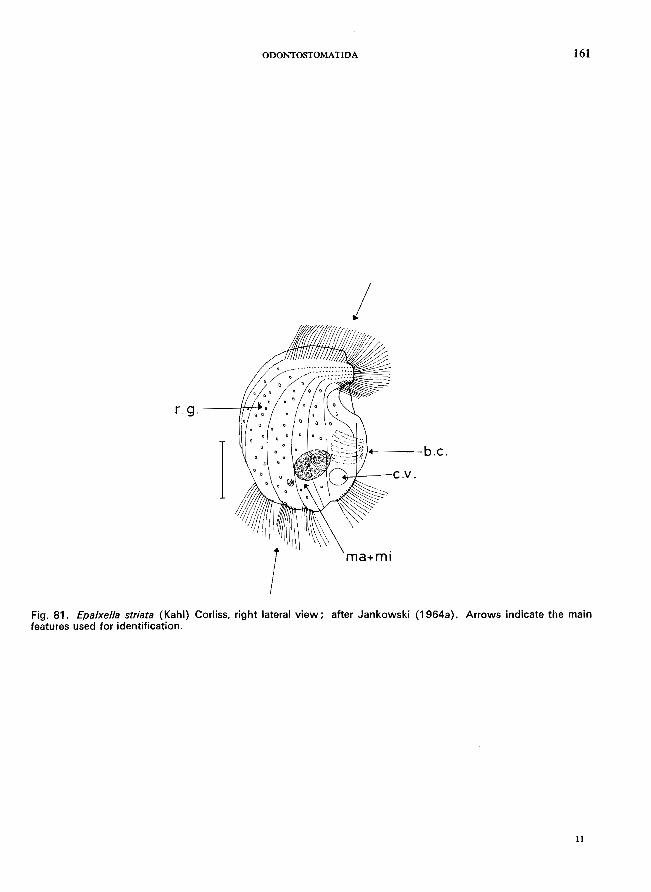
ODONTOSTOMATIDA 161
I
r.g.
I----b.c.
ma+mi
Fig. 81. £pa/xel/a striata (Kahl) Corliss, right lateral view; after Jankowski (1964a). Arrows indicate the main features used for identification.
11
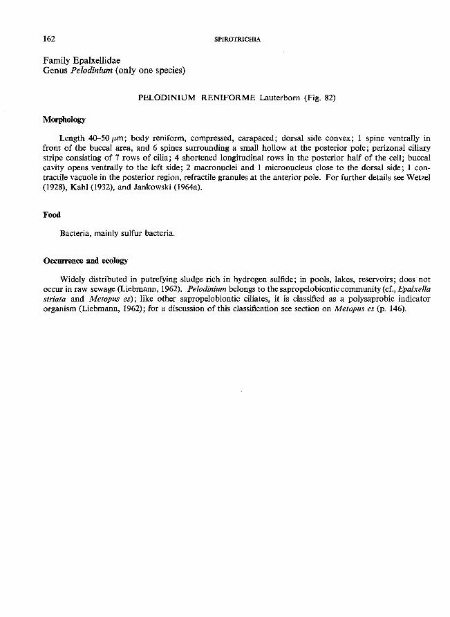
162 SPIROTRICHIA
Family Epalxellidae Genus Pelodinium (only one species)
PELODINIUM RENIFORME Lauterborn (Fig. 82)
Morphology
Length 40-50 fLm; body reniform, compressed, carapaced; dorsal side convex; 1 spine ventrally in front of the buccal area, and 6 spines surrounding a small hollow at the posterior pole; perizonal ciliary stripe consisting of 7 rows of cilia; 4 shortened longitudinal rows in the posterior half of the cell; buccal cavity opens ventrally to the left side; 2 macronuclei and 1 micronucleus close to the dorsal side; 1 contractile vacuole in the posterior region, refractile granules at the anterior pole. For further details see Wetzel (1928), Kahl (1932), and Jankowski (1964a).
Food
Bacteria, mainly sulfur bacteria.
Occurrence and ecology
Widely distributed in putrefying sludge rich in hydrogen sulfide; in pools, lakes, reservoirs; does not occur in raw sewage (Liebmann, 1962). Pelodinium belongs to the sapropelobiontic community (cf., Epalxella striata and Metopus es); like other sapropelobiontic ciliates, it is classified as a polysaprobic indicator organism (Liebmann, 1962); for a discussion of this classification see section on Metopus es (p. 146).
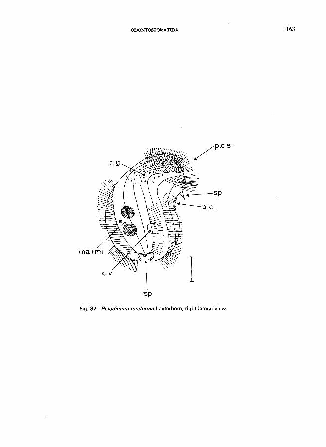
ODONTOSTOMATIDA 163
~--sp
c.v. I 'sp
Fig. 82. Pelodinium reniforme lauterborn, right lateral view.
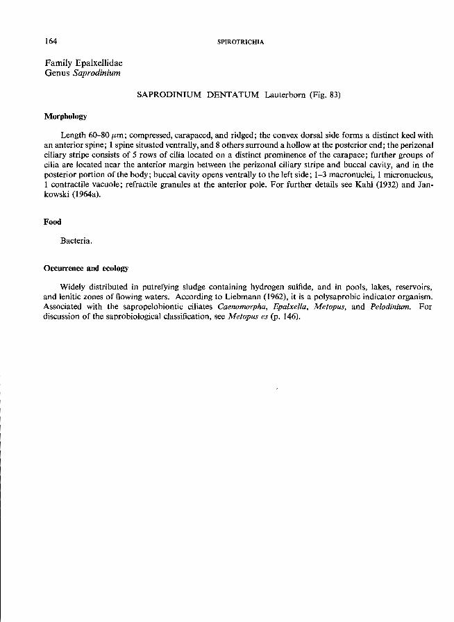
164
Family Epalxellidae Genus Saprodinium
Morphology
SPIROTRICHIA
SAPRODINIUM DENTATUM Lauterbom (Fig. 83)
Length 60--80 (-tm; compressed, carapaced, and ridged; the convex dorsal side forms a distinct keel with an anterior spine; 1 spine situated ventrally, and 8 others surround a hollow at the posterior end; the perizonal ciliary stripe consists of 5 rows of cilia located on a distinct prominence of the carapace; further groups of cilia are located near the anterior margin between the perizonal ciliary stripe and buccal cavity, and in the posterior portion of the body; buccal cavity opens ventrally to the left side; 1-3 macronuclei, 1 micronucleus, 1 contractile vacuole; refractile granules at the anterior pole. For further details see Kahl (1932) and Jan-
kowski (1964a).
Food
Bacteria.
Occurrence and ecology
Widely distributed in putrefying sludge containing hydrogen sulfide, and in pools, lakes, reservoirs, and lenitic zones of flowing waters. According to Liebmann (1962), it is a polysaprobic indicator organism. Associated with the sapropelobiontic ciliates Caenomorpha, Epalxella, Metopus, and Pelodinium. For discussion of the saprobiological classification, see Metopus es (p. 146).
ODONTOSTOMATIDA 165
ma+mi
Fig. 83. Saprodinium dentatum Lauterborn, right lateral view; after Jankowski (1964a).
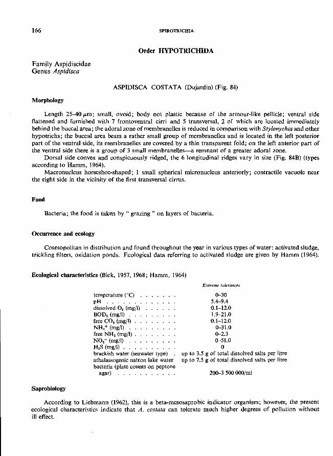
166
Family Aspidiscidae Genus Aspidisca
Morphology
SPIROTRICHIA
Order HYPOTRlCIllDA
ASPIDISCA COSTATA (Dujardin) (Fig. 84)
Length 25-40/Lm; small, ovoid; body not plastic because of the armour-like pellicle; ventral side flattened and furnished with 7 frontoventral cirri and 5 transversal, 2 of which are located immediately behind the buccal area; the adoral zone of membranelles is reduced in comparison with Stylonychia and other hypotrichs; the buccal area bears a rather small group of membranelles and is located in the left posterior part of the ventral side, its membranelles are covered by a thin transparent fold; on the left anterior part of the ventral side there is a group of 3 small membranelles-a remnant of a greater adoral zone.
Dorsal side convex and conspicuously ridged, the 6 longitudinal ridges vary in size (Fig. 84B) (types according to Hamm, 1964).
Macronucleus horseshoe-shaped; 1 small spherical micronucleus anteriorly; contractile vacuole near the right side in the vicinity of the first transversal cirrus.
Food
Bacteria; the food is taken by " grazing" on layers of bacteria.
Occurrence and ecology
Cosmopolitan in distribution and found throughout the year in various types of water: activated sludge, trickling filters, oxidation ponds. Ecological data referring to activated sludge are given by Hamm (1964).
Ecological characteristics (Bick, 1957, 1968; Hamm, 1964)
Saprobiology
temperature eC) . pH ...... . dissolved O. (mg/I) BOD. (mg/I) . free CO. (mg/I) . NH. + (mg/l) . . free NH. (mg/I) . NO.- (mg/l) . . H.S (mg/I) ... brackish water (seawater type) athalassogenic natron lake water bacteria (plate counts on peptone
agar) .......... .
Extreme tolerances
0-30 5.4--9.4 0.1-12.0 1.9-21.0 0.1-12.0
0--31.0 0-2.3 0--51.0
o up to 3.5 g of total dissolved salts per litre up to 7.5 g of total dissolved salts per litre
200--3 500000/ml
According to Liebmann (1962), this is a beta-mesosaprobic indicator organism; however, the present ecological characteristics indicate that A. costata can tolerate much higher degrees of pollution without ill effect.
HYPOTRICHIDA 167
A B
o a
c.v.
b
c
I d
Fig. 84. Aspidisca costata (Dujardin). A Ventral view; the right upper arrow indicates the anterior rudiment of the adoral zone; B, diagrammatic cross-sections of individuals to illustrate the 5 main varieties (according to Hamm (1964». (a) Normal type; (b) overfed form with 6 strong ribs; (c) starved specimen; (d) specimen with extremely high ribs; (e) specimen damaged by adverse environmental conditions .
.............. ------------------
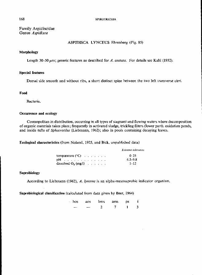
168
Family Aspidiscidae Genus Aspidisca
Morphology
SPIROTRICHIA
ASPIDISCA LYNCEUS Ehrenberg (Fig. 85)
Length 30-50 /Lm; generic features as described for A. costata. For details see Kahl (1932).
Special features
Dorsal side smooth and without ribs, a short distinct spine between the two left transverse cirri.
Food
Bacteria.
Occurrence and ecology
Cosmopolitan in distribution, occurring in all types of stagnant and flowing waters where decomposition of organic materials takes place; frequently in activated sludge, trickling filters (lower part), oxidation ponds, and inside tufts of Sphaerotilus (Liebmann, 1962); also in pools containing decaying leaves.
Ecological characteristics (from Noland, 1925, and Bick, unpublished data)
temperature ("C) . pH ...... . dissolved O. (mgfI)
Saprobiology
Extreme tolerances
0-25 6.5-9.8
1-12
According to Liebmann (1962), A. lynceus is an alpha-mesosaprobic indicator organism.
Saprobiological classification (calculated from data given by Beer, 1964)
bos aos bms
2
ams
7
ps
1 3
HYPOTRICHIDA 169
rna
C.V. I b.a.
Fig. 85. Aspidisca Iynceus Ehrenberg. A Ventral view; B, diagrammatic cross-section. The right upper arrow indicates the anterior rudiment of the adoral zone.
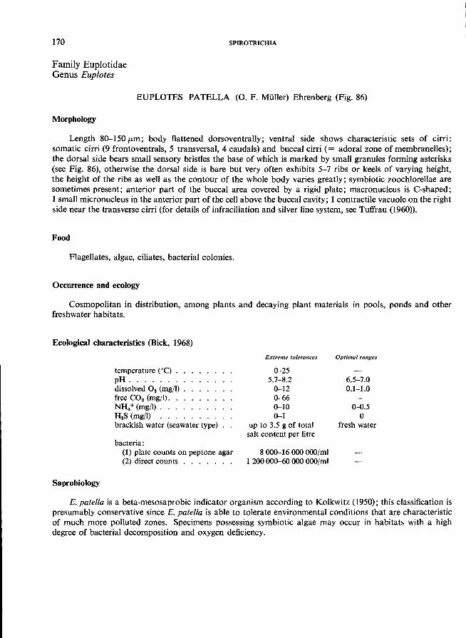
170
Family Euplotidae Genus Euplotes
SPIROTRICHIA
EUPLOTES PATELLA (0. F. Muller) Ehrenberg (Fig. 86)
Morphology
Length 80-150 /km ; body flattened dorsoventrally; ventral side shows characteristic sets of cirri: somatic cirri (9 frontoventrals, 5 transversal, 4 caudals) and buccal cirri (= adoral zone of membranelles); the dorsal side bears small sensory bristles the base of which is marked by small granules forming asterisks (see Fig. 86), otherwise the dorsal side is bare but very often exhibits 5-7 ribs or keels of varying height, the height of the ribs as well as the contour of the whole body varies greatly; symbiotic zoochlorellae are sometimes present; anterior part of the buccal area covered by a rigid plate; macronucleus is C-shaped; 1 small micronucleus in the anterior part of the cell above the buccal cavity; 1 contractile vacuole on the right side near the transverse cirri (for details of infraciliation and silver line system, see Tuffrau (1960)).
Food
Flagellates, algae, ciliates, bacterial colonies.
Occurrence and ecology
Cosmopolitan in distribution, among plants and decaying plant materials in pools, ponds and other
freshwater habitats.
Ecological characteristics (Bick, 1968)
Saprobiology
temperature COC) . . pH ....... . dissolved O. (mgfl) . free CO. (mg/I). NH/(mg/I) .... H.S (mg/I) brackish water (seawater type)
bacteria: (1) plate counts on peptone agar (2) direct counts . . . . . . .
Extreme tolerances
0-25 5.7-8.2
0-12 0-66 0-10 0-1
up to 3.5 g of total salt content per litre
8000-16000000/ml 1200000-60000 OOO/ml
Optimal ranges
6.5-7.0 0.1-1.0
0-0.5 o
fresh water
E. patella is a beta-mesosaprobic indicator organism according to Kolkwitz (1950); this classification is presumably conservative since E. patella is able to tolerate environmental conditions that are characteristic of much more polluted zones. Specimens possessing symbiotic algae may occur in habitats with a high degree of bacterial decomposition and oxygen deficiency.
HYPOTRICHIDA 171
/ f.v.c.
~ r--+---- a
c..Y. a
\: r: c.
I
Fig. 86. Euplotes patella (0. F. Muller) Ehrenberg, ventral view; after Kahl (1932). Arrows indicate the main features used for identification.
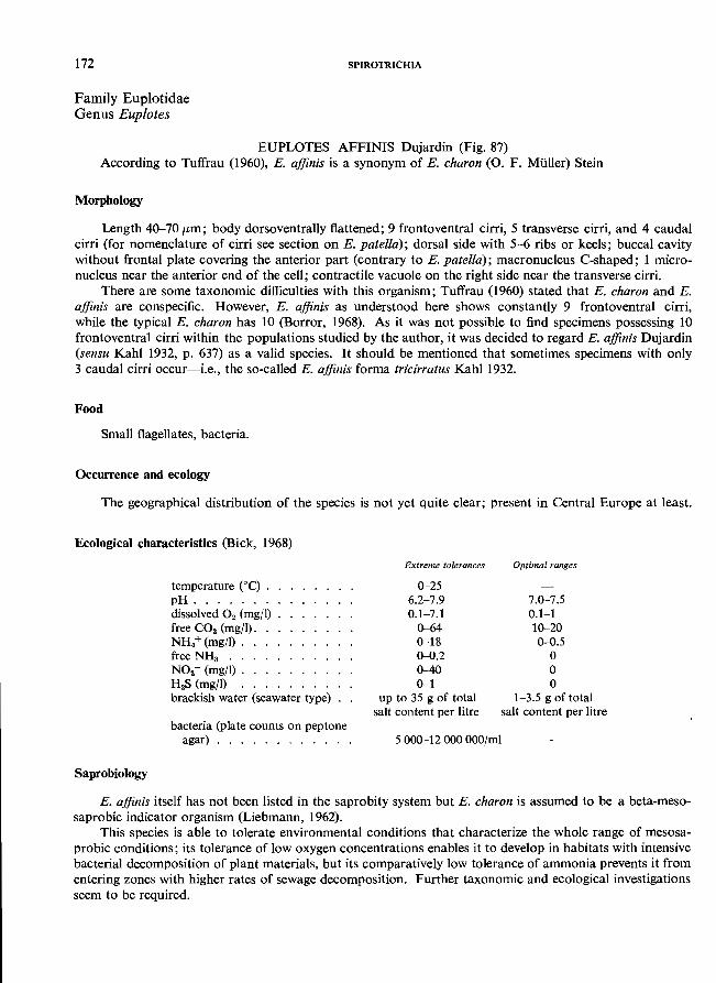
172
Family Euplotidae Genus Euplotes
SPIROTRiCHIA
EUPLOTES AFFINIS Dujardin (Fig. 87) According to Tuffrau (1960), E. affinis is a synonym of E. charon (0. F. MUlier) Stein
Morphology
Length 40-70 fLm; body dorsoventrally flattened; 9 frontoventral cirri, 5 transverse cirri, and 4 caudal cirri (for nomenclature of cirri see section on E. patella); dorsal side with 5-6 ribs or keels; buccal cavity without frontal plate covering the anterior part (contrary to E. patella); macronucleus C-shaped; 1 micronucleus near the anterior end of the cell; contractile vacuole on the right side near the transverse cirri.
There are some taxonomic difficulties with this organism; Tuffrau (1960) stated that E. charon and E. affinis are conspecific. However, E. affinis as understood here shows constantly 9 frontoventral cirri, while the typical E. charon has 10 (Borror, 1968). As it was not possible to find specimens possessing 10 frontoventral cirri within the populations studied by the author, it was decided to regard E. affinis Dujardin (sensu Kahl 1932, p. 637) as a valid species. It should be mentioned that sometimes specimens with only 3 caudal cirri occur-i.e., the so-called E. affinis forma tricirratus Kahl 1932.
Food
Small flagellates, bacteria.
Occurrence and ecology
The geographical distribution of the species is not yet quite clear; present in Central Europe at least.
Ecological characteristics (Bick, 1968)
Saprobiology
temperature Cc) . pH ...... . dissolved O. (mg/I) free CO. (mg/I). NH.+ (mg/I) . free NHa .. NO.- (mg/I) . H.S (mgfl) brackish water (seawater type)
bacteria (plate counts on peptone agar) ........... .
Extreme tolerances
0-25 6.2-7.9 0.1-7.1
0-64 0-18 0-0.2 0-40 0-1
up to 35 g of total salt content per litre
5000-12000000/ml
Optimal ranges
7.0-7.5 0.1-1 10-20 0-0.5
o o o
1-3.5 g of total salt content per litre
E. affinis itself has not been listed in the saprobity system but E. charon is assumed to be a beta-mesosaprobic indicator organism (Liebmann, 1962).
This species is able to tolerate environmental conditions that characterize the whole range of mesosaprobic conditions; its tolerance of low oxygen concentrations enables it to develop in habitats with intensive bacterial decomposition of plant materials, but its comparatively low tolerance of ammonia prevents it from entering zones with higher rates of sewage decomposition. Further taxonomic and ecological investigations seem to be required.
HYPOTRICHIDA 173
_ l----l--I--- b. c.
c.v. i!ii<l'---I-__ rna
Fig. 87. Eup/otes affinis Dujardin, ventral view; after Kahl (1932). Arrows indicate the main features used for identification.
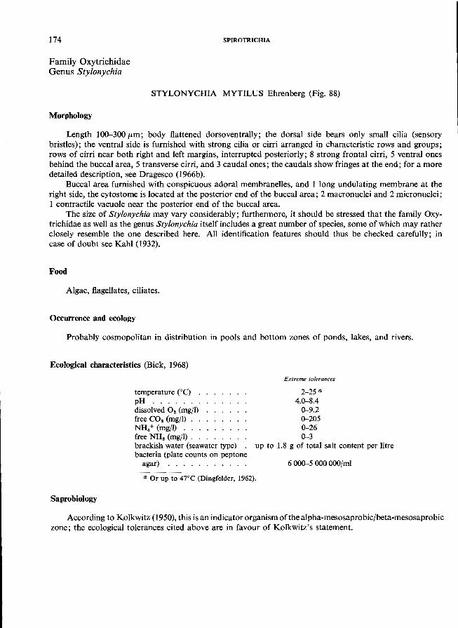
174
Family Oxytrichidae Genus Stylonychia
SPIROTRICHIA
STYLONYCHIA MYTILUS Ehrenberg (Fig. 88)
Morphology
Length 100-300/Lm; body flattened dorsoventrally; the dorsal side bears only small cilia (sensory bristles); the ventral side is furnished with strong cilia or cirri arranged in characteristic rows and groups; rows of cirri near both right and left margins, interrupted posteriorly; 8 strong frontal cirri, 5 ventral ones behind the buccal area, 5 transverse cirri, and 3 caudal ones; the caudals show fringes at the end; for a more detailed description, see Dragesco (1966b).
Buccal area furnished with conspicuous adoral membranelles, and 1 long undulating membrane at the right side, the cytostome is located at the posterior end of the buccal area; 2 macronuclei and 2 micronuclei; 1 contractile vacuole near the posterior end of the buccal area.
The size of Stylonychia may vary considerably; furthermore, it should be stressed that the family Oxytrichidae as well as the genus Stylonychia itself includes a great number of species, some of which may rather closely resemble the one described here. All identification features should thus be checked carefully; in case of doubt see Kahl (1932).
Food
Algae, flagellates, ciliates.
Occurrence and ecology
Probably cosmopolitan in distribution in pools and bottom zones of ponds, lakes, and rivers.
Ecological characteristics (Bick, 1968)
temperature eC) . pH ...... . dissolved O2 (mg/l) free CO2 (mg/l) . NH.+ (mg/I) ... free NH. (mgfl). . brackish water (seawater type) bacteria (plate counts on peptone
agar) .......... .
Extreme tolerances
2-25 a
4.0-8.4 0-9.2 0-205 0-26 0-3
up to 1.8 g of total salt content per litre
6 000-5 OOOOOO/ml
a Or up to 47°C (Dingfelder, 1962).
Saprobiology
According to Kolkwitz (1950), this is an indicator organism ofthe alpha-mesosaprobic/beta-mesosaprobic zone; the ecological tolerances cited above are in favour of Kolkwitz's statement.
HYPOTRICHIDA 175
f r. C .------,-F--f-+
b.a.
C.v. ma---=F-_
ma--_-..
tr.c. I 50)Jm
----c.c.
Fig. 88. Sty/onychia mytilus Ehrenberg. Frontal cirri (fr.c.) shaded black.
176
Family Oxytrichidae Genus Stylonychia
SPIROTRICHIA
STYLONYCHIA PUTRINA (Stokes) (Fig. 89)
Morphology
Length 125-145 pm; similar to S. mytilus but smaller, and right and left sides are more or less parallel; caudal cirri unfringed, 2-4 transverse cirri protruding slightly beyond the posterior end.
Food
Flagellates, bacteria.
Occurrence and ecology
Freshwater habitats with plant material undergoing decomposition; recorded in Europe and North
America.
Ecological characteristics (Bick, 1968)
Saprobiology
temperature COC) . pH ...... . dissolved O. (mg/l) free CO. (mg/l). NH.+ (mg/l). . . . free NHa (mg/l) . . brackish water (seawater type)
athalassogenic natron lake water
bacteria (plate counts on peptone agar) ........... .
Extreme tolerances
2-25 5.8-9.4
0--12.6 0--56 0--22 0--2
up to 14 g of total salt content per litre up to 2 g of total
salt content per litre
1000--20000/ml
Optimal ranges
6.5-7.0 0.1-1.0 10--30
fresh water
According to the saprobiological classification of Kolkwitz; this species inhabits the alpha-meso saprobic to beta-mesosaprobic zones; this statement agrees well with the ecological characteristics given above.
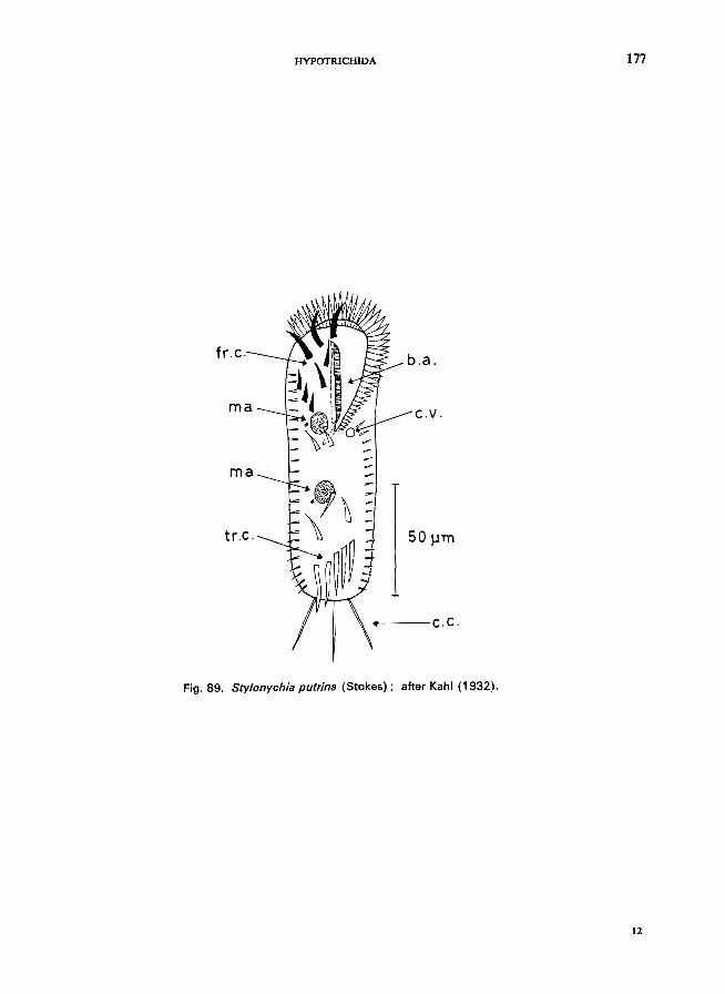
HYPOTRICHIDA 177
fr.c. b.a.
ma c.v.
ma
50 ~m
4----c.c.
Fig. 89. Sty/onychia putrina (Stokes); after Kahl (1932).
12
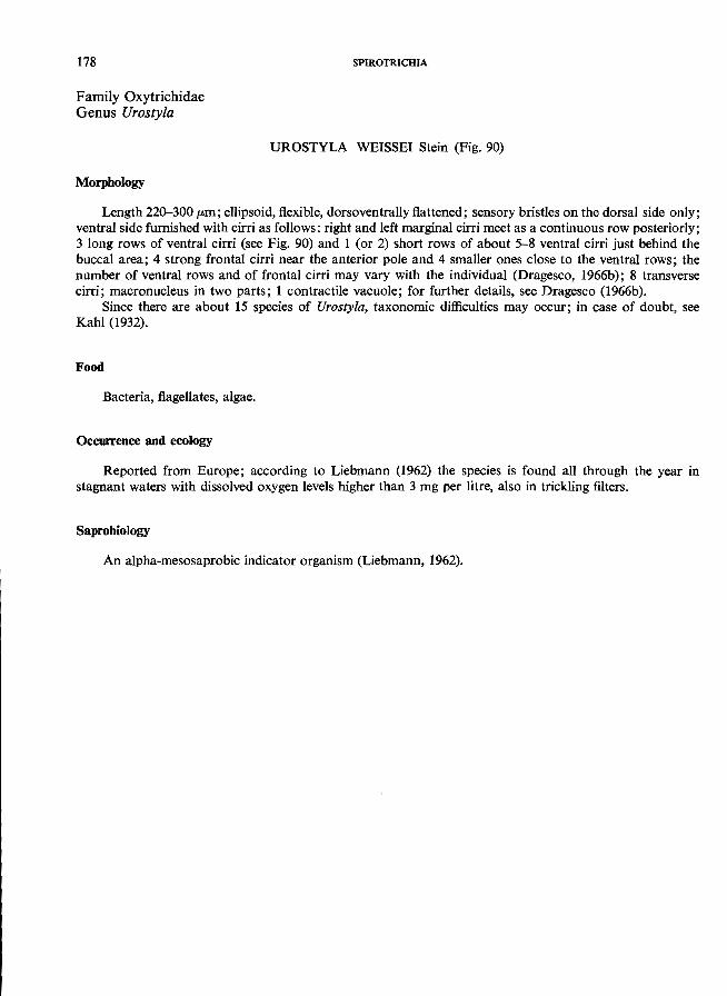
178
Family Oxytrichidae Genus Urostyla
Morphology
SPIROTRICHIA
UROSTYLA WEISSEI Stein (Fig. 90)
Length 220-300 pm; ellipsoid, flexible, dorsoventrally flattened; sensory bristles on the dorsal side only; ventral side furnished with cirri as follows: right and left marginal cirri meet as a continuous row posteriorly; 3 long rows of ventral cirri (see Fig. 90) and 1 (or 2) short rows of about 5-8 ventral cirri just behind the buccal area; 4 strong frontal cirri near the anterior pole and 4 smaller ones close to the ventral rows; the number of ventral rows and of frontal cirri may vary with the individual (Dragesco, 1966b); 8 transverse cirri; macronucleus in two parts; 1 contractile vacuole; for further details, see Dragesco (1966b).
Since there are about 15 species of Urostyla, taxonomic difficulties may occur; in case of doubt, see
Kahl (1932).
Food
Bacteria, flagellates, algae.
Occurrence and ecology
Reported from Europe; according to Liebmann (1962) the species is found all through the year in stagnant waters with dissolved oxygen levels higher than 3 mg per litre, also in trickling filters.
Saprobiology
An alpha-mesosaprobic indicator organism (Liebmann, 1962).
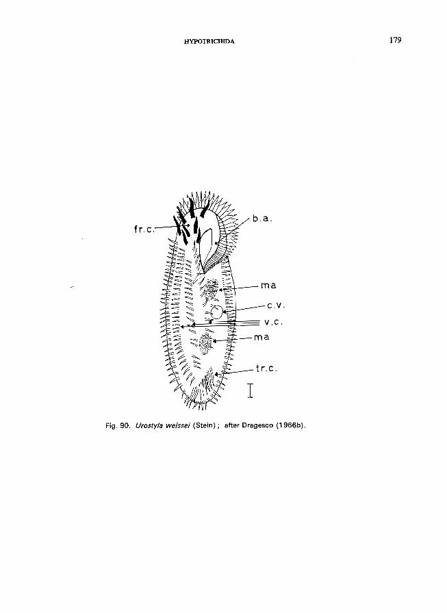
fro C.
HYPOTRICHIDA
b.a.
=.1..--- ma
c.v.
~~~~~=== v.c.
ma
t r.c.
Fig. 90. Urostyla weissei (Stein); after Dragesco (1966b).
179
180
Family Oxytrichidae Genus Oxytricha
Morphology
SPIROTRICHIA
OXYTRICHA FALLAX Stein (Fig. 91)
Length about 150 pm; body outline ellipsoid, posterior region broadly rounded; flexible; dorsal surface convex; ventral side flattened and equipped with cirri: 8 frontals; 5 ventrals, 5 transversals; right and left marginal cirri meet as a continuous row posteriorly (note Stylonychia, with marginal cirri interrupted at posterior end, and with long caudal cirri); buccal area with well-developed adoral zone of membranelles and a long undulating membrane at the right side; cytostome located at the posterior, end of the buccal area; 2 macronuclei and 2 micronuclei; 1 contractile vacuole near to the left margin in the vicinity of the posterior
end of the buccal area.
Food
Bacteria, algae, flagellates.
Occurrence and ecology
Widely distributed in stagnant and flowing waters including trickling filters and activated sludge, not to be found in waters receiving manure. Optimal range of environmental factors about pH 6.8-7.5 and 3-5 mg of dissolved oxygen per ml (Liebmann, 1962).
Saprobiology
Classified as an alpha-mesosaprobic indicator organism by Liebmann (1962).
HYPOTRICHIDA 181
fr.c.
+-f...~-- b. a.
50~m
Fig. 91. Oxytr;cha fal/ax Stein; ventral view. Frontal cirri (fr.c.) shaded black.
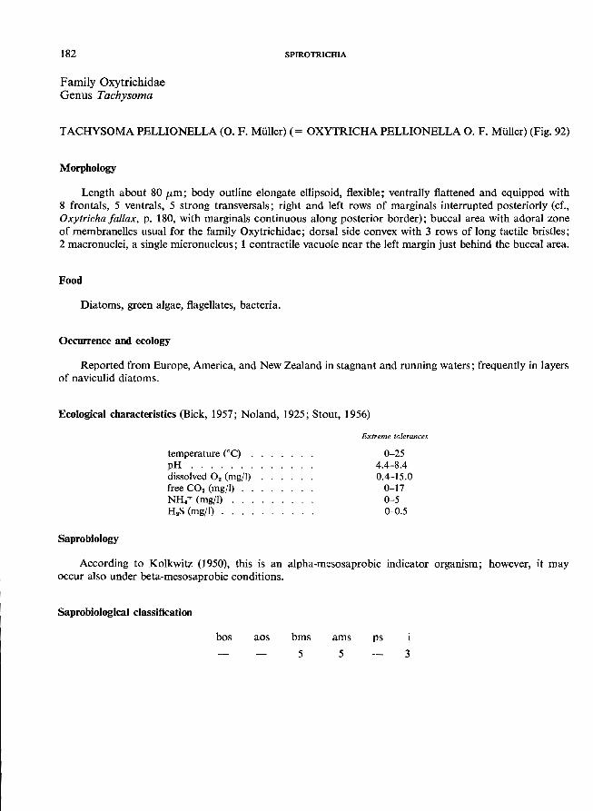
182
Family Oxytrichidae Genus Tachysoma
SPIROTRICHIA
TACHYSOMA PELLIONELLA (0. F. Miiller) (= OXYTRICHA PELLIONELLA o. F. Miiller) (Fig. 92)
Morphology
Length about 80 /Lm ; body outline elongate ellipsoid, flexible; ventrally flattened and equipped with 8 frontals, 5 ventrals, 5 strong transversals; right and left rows of marginals interrupted posteriorly (cf., Oxytricha fal/ax, p. 180, with marginals continuous along posterior border); buccal area with adoral zone of membranelles usual for the family Oxytrichidae; dorsal side convex with 3 rows of long tactile bristles; 2 macronuclei, a single micronucleus; 1 contractile vacuole near the left margin just behind the buccal area.
Food
Diatoms, green algae, flagellates, bacteria.
Occurrence and ecology
Reported from Europe, America, and New Zealand in stagnant and running waters; frequently in layers
of naviculid diatoms.
Ecological characteristics (Bick, 1957; Noland, 1925; Stout, 1956)
Saprobiology
temperature COc) . pH ...... . dissolved O. (mg/I) free CO. (mg/I) NH.+ (mg/I) H.S (mg/I) ..
Extreme tolerances
0-25 4.4-8.4 0.4-15.0
0-17 0-5 0-0.5
According to Kolkwitz (1950), this is an alpha-mesosaprobic indicator organism; however, it may
occur also under beta-mesosaprobic conditions.
Saprobiological classification
bos aos bms
5
ams ps
5 3
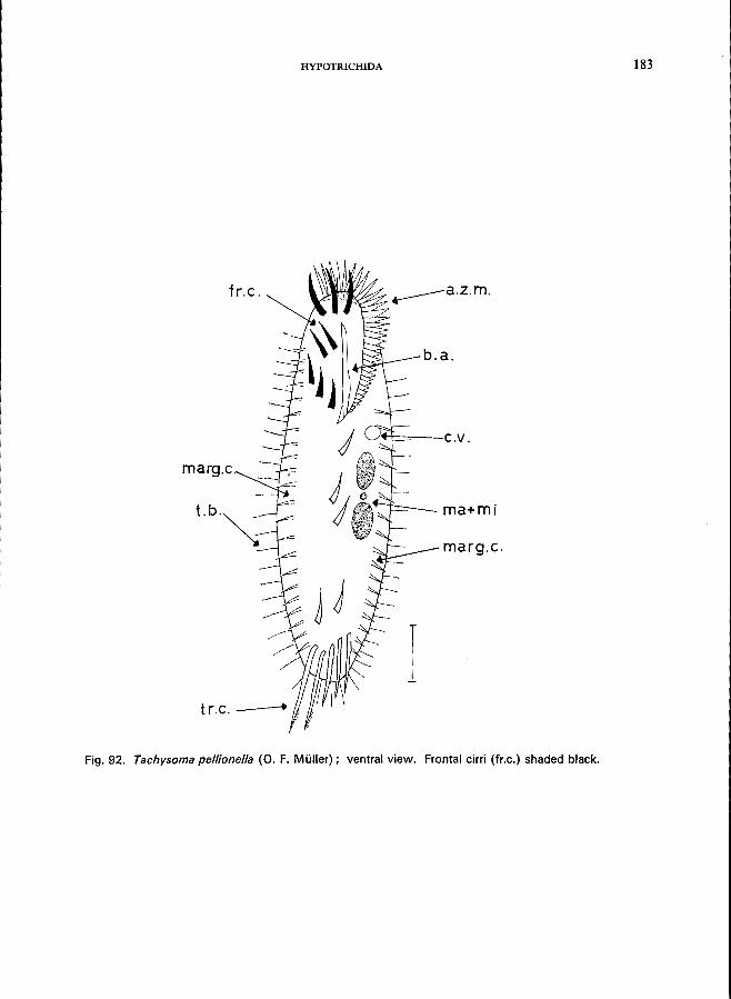
HYPOTRICHIDA 183
fr.c.
b.a .
./~t::::=--- C . v .
marg.c.
t.b.~ I--~- ma+mj
marg.c.
I t r.c. _-----r
Fig. 92. Tachysoma pellionella (0. F. Muller); ventral view. Frontal cirri (fr.c.) shaded black.
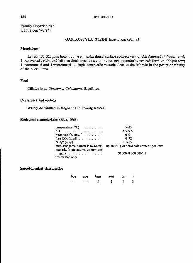
184 SPIROTRICHIA
Family Oxytrichidae Genus Gastrostyla
GASTROSTYLA STEINI Engelmann (Fig. 93)
Morphology
Length 150-320 fLm; body outline ellipsoid; dorsal surface convex; ventral side flattened; 6 frontal cirri, 5 transversals, right and left marginals meet as a continuous row posteriorly, ventrals form an oblique row; 4 macronuclei and 4 micronuclei; a single contractile vacuole close to the left side in the posterior vicinity
of the buccal area.
Food
Ciliates (e.g., Glaucoma, Colpidium), flagellates.
Occurrence and ecology
Widely distributed in stagnant and flowing waters.
Ecological characteristics (Bick, 1968)
temperature eC) pH ..... . dissolved O2 (mg/l) free CO2 (mg/l) . . NH,+ (mg/l) . . . athalassogenic natron lake-water bacteria (plate counts on peptone
agar) .......... . freshwater only
Saprobiological classification
bos aos bms
2
5-25 6.5-9.5
0-9 0-72
0.1-55 up to 10 g of total salt content per litre
ams
7
60 000-8 000 OOO/ml
ps
1 3
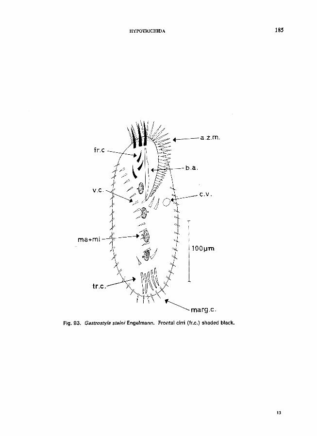
HYPOTRICHIDA
fr.c
~
ma+mi l
.L----a .z.m.
L-/,:::"';;<~- b.a.
c.v.
\.11 100~m
tr.c~~ ~marg.c.
Fig. 93. Gastrostyla steini Engelmann. Frontal cirri (fr.c.) shaded black.
185
13
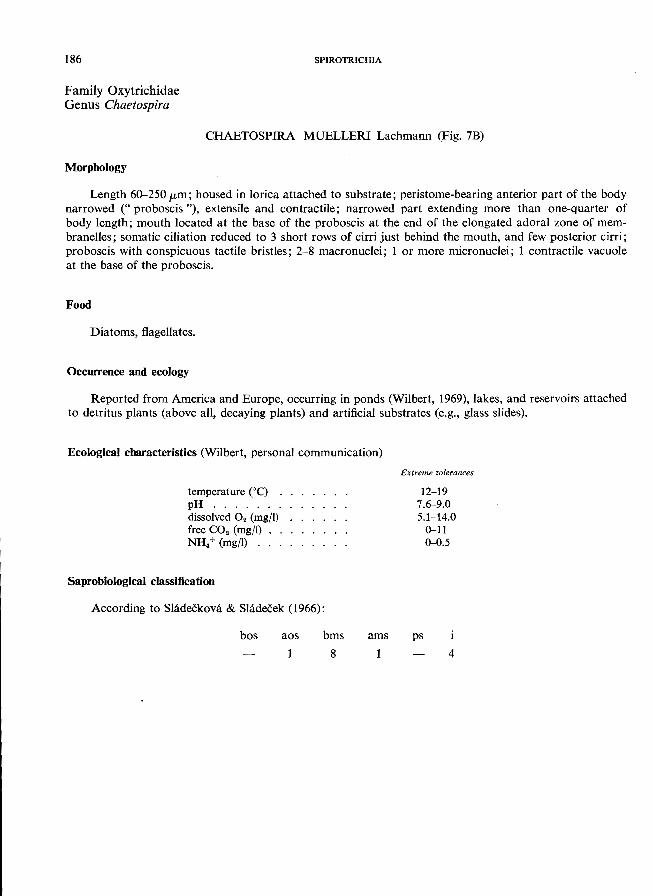
186
Family Oxytrichidae Genus Chaetospira
SPIROTRICHIA
CHAETOSPIRA MUELLERI Lachmann (Fig. 7B)
Morphology
Length 60-250 fLm; housed in lorica attached to substrate; peristome-bearing anterior part of the body narrowed (" proboscis "), extensile and contractile; narrowed part extending more than one-quarter of body length; mouth located at the base of the proboscis at the end of the elongated adoral zone of membranelles; somatic ciliation reduced to 3 short rows of cirri just behind the mouth, and few posterior cirri; proboscis with conspicuous tactile bristles; 2-8 macronuclei; 1 or more micronuclei; 1 contractile vacuole
at the base of the proboscis.
Food
Diatoms, flagellates.
Occurrence and ecology
Reported from America and Europe, occurring in ponds (Wilbert, 1969), lakes, and reservoirs attached to detritus plants (above all, decaying plants) and artificial substrates (e.g., glass slides).
Ecological characteristics (Wilbert, personal communication)
temperature COe) . pH ...... . dissolved O2 (mg/I) free CO2 (mg/I) NH.+ (mg/I) ...
Saprobiological classification
According to Sladeekova & Sladecek (1966):
bos aos bms
8
ams
Extreme tolerances
12-19 7.6-9.0 5.1-14.0
0-11 0-0.5
ps
4
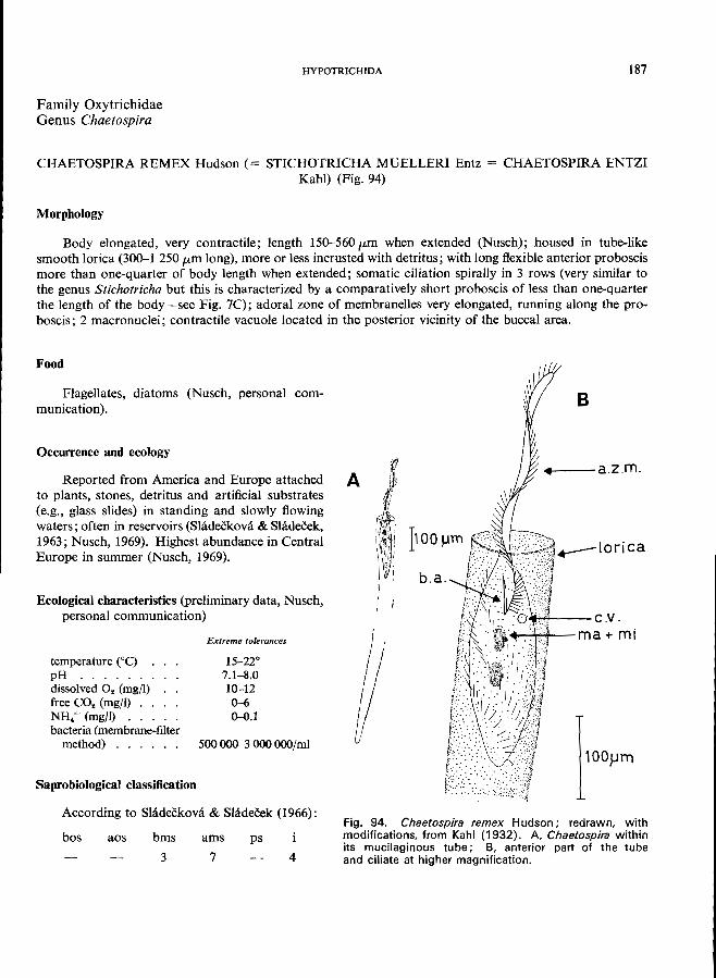
HYPOTRICHIDA 187
Family Oxytrichidae Genus Chaetospira
CHAETOSPIRA REMEX Hudson (= STICHOTRICHA MUELLERI Entz = CHAETOSPIRA ENTZI Kahl) (Fig. 94)
Morphology
Body elongated, very contractile; length 150-560 p,m when extended (Nusch); housed in tube-like smooth lorica (300-1 250 p,m long), more or less incrusted with detritus; with long flexible anterior proboscis more than one-quarter of body length when extended; somatic ciliation spirally in 3 rows (very similar to the genus Stichotricha but this is characterized by a comparatively short proboscis of less than one-quarter the length of the body-see Fig. 7C); adoral zone of membranelles very elongated, running along the proboscis; 2 macronuclei; contractile vacuole located in the posterior vicinity of the buccal area.
Food
Flagellates, diatoms (Nusch, personal communication).
Occurrence and ecology
Reported from America and Europe attached to plants, stones, detritus and artificial substrates (e.g., glass slides) in standing and slowly flowing waters; often in reservoirs (Shideekova & Sladecek, 1963; Nusch, 1969). Highest abundance in Central Europe in summer (Nusch, 1969).
Ecological characteristics (preliminary data, Nusch, personal communication)
temperature (0C) pH ..... . dissolved O. (mg/I) free CO2 (mg/I) . . NH. + (mg/I) . . . bacteria (membrane-filter
method) ..... .
Saprobiological classification
Extreme tolerances
15-22° 7.1-8.0 10-12 0-6 0-0.1
500 000-3000 ooO/ml
According to Sladeckova & Sladeeek (1966):
bos aos bms
3
ams ps
7 4
A
!I f
B
.-.-- a.z .m.
),.,~-- c.v . ~~~-ma+ mi
Fig. 94. Chaetospira remex Hudson; redrawn, with modifications, from Kahl (1932). A, Chaetospira within its mucilaginous tube; B, anterior part of the tube and ciliate at higher magnification.
ACKNOWLEDGEMENTS
The author expresses his gratitude to members of the staff of the Wodd Health Organization for valuable advice in the preparation of this guide and for revising the English text. Special thanks must be expressed to Dr Marshall Laird, Memorial University, Newfoundland, Canada, for his encouragement. The guide was prepared with support from the World Health Organization, and facilities were provided by the Zoological Institute, University of Bonn, Federal Republic of
Germany.
SELECTED BIBLIOGRAPHY
Beer, W. D. (1964) Organismen als Anzeiger der Wassergute und das Saprobiensystem, Wiss. KarlMarx-Univ., Leipzig, 13, Math.-Naturwiss. Reihe, Part 1, pp. 25-30
Berger, J. (1959) The comparative morphology of two species of Cyclidium (Ciliata: Hymenostomatida), J. Protozool., 6, Suppl., p. 13
Berger, J. & Thompson, J. C. (1960) A redescription of Cyclidium glaucoma O. F. M., 1786 (Ciliata: Hymenostomatida), with particular attention to the buccal apparatus, J. Protozool., 7, 256-262
Bick, H. (1957) Beitrage zur Okologie einiger Ciliaten des Saprobiensystems, Vom Wass., 24, 224-246
Bick, H. (1958) Okologische Untersuchungen an Ciliaten fallaubreicher Kleingewasser, Arch. Hydrobiol., 54, 506-542
Bick, H. (1960) Okologische Untersuchungen an Ciliaten und anderen Organismen aus verunreinigten Gewassern, Arch. Hydrobiol., 56, 378-394
Bick, H. (1963) A review of Central European methods for the biological estimation of water pollution levels, Bull. Wid Hlth Org., 29, 401-413
Bick, H. (1964) Die Sukzession der Organismen bei der Selbstreinigung von organisch verunreinigtem Wasser unter verschiedenen Milieubedingungen, Dusseldorf
Bick, H. (1966) Okologische Untersuchungen an Ciliaten des Saprobiensystems I, Int. Rev. ges. Hydrobiol. Hydrogr., 51, 489-520
Bick, H. (1968) Autokologische und saprobiologische Untersuchungen an Siisswasserciliaten, Hydrobiologia, 31, 17-36
Bock, K. J. (1952) Zur Okologie der Ciliaten des marinen Sandgrundes der Kieler Bucht I. Kieler Meeresforsch., 9,77-89
Boggs, N. (1965) Comparative studies on Spirostomum: silver impregnation of three species, J. Protozool., 12, 603-606
Borror, A. C. (1968) Systematics of Euplotes (Ciliophora, Hypotrichida); toward union of the old and the new, J. Protozool., 15, 802-808
Buck, H. (1968) Die Ciliaten des Belebtschlammes in ihrer Abhangigkeit vom Klarverfahren, Miinchner Beitrage zur Abwasser-, Fischerei- und Flussbiologie, 2nd ed., Munich, Oldenbourg, vol. 5
Canella, M. F. (1960) Contributo ad una revisione dei generi Amphileptus, Hemiophrys e Lionotus, Ann. Univ. Ferrara, Sez. III, BioI. animal., 2(2), 47-95
Caspers, H. & Karbe, L. (1966) Trophie und Saprobitat als stoffwechseldynamischer Komplex, Arch. Hydrobioi., 61,453-470
Corliss, J. O. (1959) An illustrated key to the higher groups of the ciliated protozoa, with definition of terms. J. Protozool., 6, 265-281
Corliss, J. O. (1961) The ciliated protozoa, Oxford, Pergamon
Curds, C. R. & Vandyke, J. M. (1966) The feeding habits and growth rates of some fresh-water ciliates found in activated-sludge plants, J. appl. Ecol., 3, 127-137
Dietz, G. (1964) Beitrag zur Kenntnis der Ciliatenfauna einiger Brackwassertiimpel (Etangs) der franzazischen Mittelmeerkliste, Vie Milieu, 15, 47-93
Dingfelder, J. H. (1962). Die Ciliaten vorlibergehender Gewasser, Arch. Protistenk., 105, 509-658
Dragesco, J. (1963) Revision du genre Dileptus, Duj. 1871 (Ciliata Holotricha) (systematique, cytologie, biologie), Bull. bioi. Fr. Be/g., 97, 103-145
Dragesco, J. (1966a) Observations sur quelques cilies libres, Arch. Protistenk., 109, 155-206
Dragesco, J. (1966b) Cilies libres de Thonon et ses environs, Protistologica, 2, 59-95
Eyferth, B. & Schoenichen, W. (1927) Einfachste Lebensformen. In: Schoenichen, W., ed., 5th ed., Berlin, vol. 2
Froud, J. (1949) Observations on hypotrichous ciliates: the genera Stichotricha and Chaetospira, Quart. J. micro Sci., 90, 141-158
Gajewskaja, N. (1933) Zur Okologie, Morphologie und Systematik der Infusorien des Baikalsees, Zoologica, Stuttgart, 83, 1-298
Gannert, R. (1935) Dber Systematik, Morphologie, Entwicklungsgeschichte und Parasiten einiger Dendrosomidae nebst Beschreibung zweier neuer Suctorien, Arch. Protistenk., 86, 113-154
Hamm, A. (1964) Untersuchungen liber die Okologie und Variabilitat von Aspidisca costata (Hypotricha) im Belebtschlamm, Arch. Hydrobiol., 60, 286-339
Hammann, I. (1953) Okologische und biologische Untersuchungen an Siisswasserperitrichen, Arch. Hydrobiol., 47, 177-228
Hentschel, E. (1916) Biologische Untersuchungen liber den tierischen und pflanzlichen Bewuchs im Hamburger Hafen, Mitt. zool. Mus. Hamburg, 33, 1-171
Holm, F. (1928) Dber die Suctorien in der Elbe bei Hamburg und ihre Lebensbedingungen, Arch. Hydrobioi., 4, Suppl., pp. 389-440
Honigberg, B. M. et al. (1964) A revised classification of the phylum Protozoa, J. Protozool., 11, 7-20
Jankowski, A. W. (1964a) Morphology and evolution of Ciliophora: III. Arch. Protistenk., 107, 185-294
Jankowski, A. W. (1964b) Morphology and evolution of Ciliophora: IV. Acta Protozool., 2, 33-58
-189 -
............. --------------------
190 SELECTED BIBLIOGRAPHY
Jankowski, A. W. (1967) [The boundaries and composition of the genera Tetrahymena and Colpidium], Zool. Z., 46, 17-23
Kahl, A. (1930-35) Wimpertiere oder Ciliata (Infusoria). In: Dahl, M. & Peus, F., ed., Tierwelt Deutschlands, Jena, Fischer, parts 18, 21, 25, 30
Kahl, A. (1934) Suctoria. In: Grimpe, G. & Wagler, E., ed., Tierwelt der Nord- und Ostsee, Leipzig, Part 26
Kolkwitz, R. (1950) C>kologie der Saprobien, Schr. Reihe Ver. Wass.- Boden- u. Lufthyg., 4
Kralik, U. (1957-58) Untersuchungen uber den Bewuchs von peritrichen Ciliaten in einigen Fliessgewassern bei Leipzig, Wiss. Karl-Marx-Univ., Leipzig, 7, Math.Naturwiss. Reihe, part 2/3, pp. 309-328
Kralik, U. (1961a) Ein Beitrag zur Biologie von loricaten peritrichen ZiJiaten, insbesondere von Platycola truncata Fromentel 1874, Arch. Protistenk., 105, 201-258
Kralik, U. (1961b) Beitrage zur Systematik von peritrichen Ciliaten, Int. Rev. ges. Hydrobiol. Hydrogr., 46,65-74
Kudo, R. R. (1966) Protozoology, 5th ed. Springfield, Ill., Thomas
Lep~i, J. (1926) Die Infusorien des Siisswassers und Meeres, Berlin.
Liebmann, H. (1951) Handbuch der Frischwasser- und Abwasser-Biologie; Biologie des Trinkwassers, Badewassers, Fischwassers, Vorfluters und Abwassers, 1st ed., Munich, Oldenbourg, vol. 1.
Liebmann, H. (1962) Handbuch der Frischwasser- und Abwasser-Biologie; Biologie des Trinkwassers, Badewassers, Fischwassers, Vorfiuters und Abwassers, 2nd ed., Munich, Oldenbourg, vol. 1
Lopez-Ochoterena, E. (1966) Ciliados mesosaprobicos de Chapultepec (Sistematica, Morfologia, Ecologia), Revta Soc. mex. Hist. nat., 26, 115-247
Maguire, B. (1963) The exclusion of Colpoda (Ciliata) from superficially favourable habitats, Ecology, 44, 781-784
Matthes, D. (1954) Suktorienstudien. VI. Die Gattung Heliophrya Saedeler & Tellier, Arch. Protistenk., 100, 143-152
Matthes, D. & Wenzel, F. (1966) Wimpertiere (Ciliaten), Stuttgart
Munch, F. (1969) Der Einfiuss der Temperatur auf den Abbau organischer Substanz und die damit verkniipfte Organismensukzession unter besonderer Beriicksichtigung der Populationsdynamik der Cilia ten, Dissertation given in the Faculty of Mathematics and Natural Science, University of Bonn
Nenninger, U. (1948) Die Peritrichen der Umgebung von Erlangen mit besonderer Berucksichtigung ihrer Wirtsspezifitat, Zool. Jb. (Systematik), 77, 169-266
Noland, L. E. (1925) Factors influencing the distribution of freshwater ciliates, Ecology, 6, 437-452
Noland, L. E. (1959) Ciliophora. In: Edmondson, W. T., ed., Ward and Whipple's freshwater biology, 2nd ed., New York, London, Wiley
Noland, L. E. & Finley, H. E. (1931) Studies on the taxonomy of the genus Vorticella, Trans. Amer. microsc. Soc., 50, 81-123
Nusch, E. A. (1969) Okologische und systematische Untersuchung der Peritricha (Ciliata, Protozoa) im Aufwuchs von Talsperren und Flussstauen mit verschiedenem Saprobitiitsgrad, Dissertation in the Faculty of Mathematics and Natural Science, University of Bonn
Penard, E. (1922) Etudes sur les infusoires d'eau douce, Geneva
Radzikowski, S. (1966) Study on morphology, division and post-conjugation morphogenesis in (hilodonella cucullulus (0. F. Muller), Acta protozool., 4, 89-95
Rieder, J. (1936) Beitrag zur Kenntnis der SusswasserSuktorien und Revision der Schweizer Suktorienfauna, Rev. suisse Zool., 43, 359-395
Schulze, E. (1959) Morphologische, cytologische und okologisch-physiologische Untersuchungen an Faulschlammciliaten (Metopus sigmoides C. & Lachm. und M. contortus Lev.), Arch. Protistenk., 103, 371-426
Shidecek, V. (1964) Technische Hydrobiologie. II. Tschechoslowakische Beitrage zum Saprobiensystem, Sb. vys. Sk. chemo-technol. Praze, 8, 529-556
Shldecek, V. (1969) Arch. Protistenk., 111, 276-278 Shideckova, A. (1960) Limnological study of the reservoir
Sedlice near Zeliv. XI. Periphyton stratification during the first year-long period (June 1957-July 1958), Sb. vys. Sk. chem.-technol. Praze, 4, 143-261
Sladeckova, A. (1962) Limnological study of the reservoir Sedlice near Zeliv. Xx. Periphyton stratification during the second year-long period (August 1958-June 1959), Sb. vys. Sk. chem.-technol. Praze, 6, 221-291
Shideekova, A. & Sladecek, V. (1963) Periphyton as indicator of the reservoir water quality. I. True periphyton, Sb. vys. Sk. chem.-technol. Praze, 7, 507-561
Sladeckova, A. & Siadeeek, V. (1966) The indicator value of some sessile protozoa, Arch. Protistenk., 109, 223-225
Sommer, G. (1951) Die peritrichen Ciliaten des Grossen Planer Sees, Arch. Hydrobiol., 44, 349-440
Sramek-Husek, R. (1954) Neue und wenig bekannte Ciliaten aus der Tschechoslowakei und ihre Stellung im Saprobiensystem, Arch. Protistenk., 100, 246-267
Sramek-Husek, R. (1956) Zur biologischen Charakteristik der hOheren Saprobitatsstufen, Arch. Hydrobiol., 51, 376-390
Sramek-Husek, R. (1958) Die Rolle der Ciliatenanalyse bei der biologischen Kontrolle von Flussverunreinigungen, Verh. into verein. theor. angew. Limnol., 13, 636-645
Stammer, H. A. (1953) Der Einfluss von Schwefelwasserstoff und Ammoniak auf tierische Leitformen des Saprobiensystems, Vom Wass., 20, 34-71
Stiller, J. (1940) Beitrag zur Peritrichenfauna des Grossen Planer Sees in Holstein, Arch. Hydrobiol., 36, 263-285
Stiller, J. (1954) Vorticella microstoma Ehrenberg (Peritricha, Ciliata) als Bioindikator akologisch verschiedener Gewasser, Ann. hist.-nat. Mus. nat. hung., 5, 191-201
SELECTED BIBLIOGRAPHY 191
Stiller, J. (1961) Durch Einwirkung von Umweltfaktoren entstehende Modifikationen bei peritrichen Cilia ten und ihre Bedeutung bei der Beurteilung des Wassers, Verh. into Verein. theor. angew. Limnol., 14, 1071-1074
Stiller, J. (1962) Die biologische Bedeutung der SchutzhiilIenbildung bei peritrichen Ciliaten und ihre Rolle als Bioindikator bei der Beurteilung des Wassers, Ann. hist.-Nat. Mus. Nat. Hung. (zool.), 54, 231-236
Stiller, J. (1963) Zur Limnologie der Natrongewasser Ungarns. I. Der Natronsee Nagyszek und seine Peritrichenfauna, Int. Rev. ges. Hydrobiol. Hydrogr., 48, 603-612
Stiller, J. (1968) Peritriche Ciliaten okologisch verschiedener Biotope von Rovinj und Umgebung, Acta zool. hung., 14, 185-211
Stout, J. D. (1954) The effect of environmental factors on the life history of the ciliate, Vorticella microstoma, Trans. roy. Soc. N. z., 82, series 3, pp. 705-711
Stout, J. D. (1955) Environmental factors affecting the life history of three soil species of Colpoda (Ciliata), Trans. roy. Soc. N.Z., 82, series 3, pp. 1165-1188
Stout, J. D. (1956) Reactions of ciliates to environmental factors, Ecology, 37, 178-191
Tamar, H. (1965) The culture, structure, and locomotion of Halteria grandinella, Acta protozool., 3, 165-172
Tuffrau, M. (1960) Revision du genre Euplotes, fondee sur la comparaison des structures superficielles, Hydrobiologia, 15, 1-77
Ventz, D. (1964) Beitrag zur Saprobiologie einiger Organismen, Int. Rev. ges. Hydrobiol. Hydrogr., 49, 133-138
Wetzel, A. (1928) Der Faulschlamm und seine ziliaten Leitformen, Z. Morph. Okol. Tiere, 13, 179-328
Wichterman, R. (1953) The biology of Paramecium, Philadelphia, Blakiston
Wilbert, N. (1969) Okologische Untersuchung der Aufwuchs- und Planktonciliaten eines eutrophen Weihers, Arch. Hydrobiol., 35, SuppJ., pp. 411-518
Winkler, R. H. & Corliss, J. O. (1965) Notes on the rarely described, green colonial Protozoon Ophrydium versatile (O.F.M.) (Ciliophora, Peritrichida), Trans. Amer. microsc. Soc., 84, 127-137
Zelinka, M. & Marvan, P. (1961) Zur Prazisierung der biologischen Klassifikation der Reinheit fliessender Gewasser, Arch. Hydrobiol., 57, 389-407

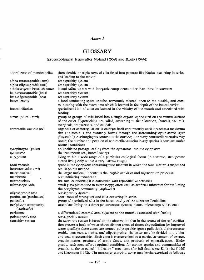
Annex 1
GLOSSARY (protozoological terms after Noland (1959) and Kudo (1966))
adoral zone of membranelles
alpha-mesosaprobic (ams) alpha-oligosaprobic (aos) athalassogenic brackish water beta-mesosaprobic (bms) beta-oligosaprobic (bos) buccal cavity
buccal ciliation
cirrus (plural: cirri)
contractile vacuole (cv)
cytopharynx (gullet) cytostome eurypotent
food vacuole indicator value (= i) macronucleus membrane micronucleus microscope slide
oligosaprobic (os) pectinellae (pectinelles) peniculus periphyton community
(Aufwuchs) peristome polysaprobic (ps) saprobity system
short double or triple rows of cilia fused into pennant-like blades, occurring in series, and leading to the mouth see saprobity system see saprobity system inland saline waters with inorganic components other than those in seawater see saprobity system see saprobity system a food-conducting space or tube, commonly ciliated, open to the outside, and communicating with the cytostome which is located in the depth of the buccal cavity specialized kind of ciIiation located in the vicinity of the mouth and associated with feeding group or groups of cilia fused into a single organelle; the cirri on the ventral surface of the order Hypotrichida are called, according to their location, frontals, ventrals, marginals, transversals, and caudals organelle of osmoregulation; it enlarges itself continuously until it reaches a maximum size (" diastole") and suddenly bursts through the surrounding cytoplasmic layer (" systole "), discharging its content to the outside; 1 or many contractile vacuoles may occur; the number and position of contractile vacuoles in any species is constant under normal conditions an unciliated passage leading from the cytostome into the cytoplasm the true mouth (cf., buccal cavity) living within a wide range of a particular ecological factor (in contrast, stenopotent means living only within a very narrow range) space in the cytoplasm containing fluid medium in which the food matter is suspended see 10-points method the larger nucleus; it controls the trophic activities and regeneration processes see undulating membrane the smaller nucleus; it is concerned with reproductive activities small glass plates used in microscopy; often used as artificial substrates for evaluating the periphyton community (Aufwuchs) see saprobity system short rows of strong unfused cilia occurring in series group of specialized cilia in the buccal cavity of the suborder Peniculina organisms living on submerged substrates (stones, plants, microscope slides, etc.)
a differentiated external area adjacent to the mouth, associated with feeding see saprobity system the saprobity system is based on the observation that in the course of the self-purification process a body of water shows distinct zones of decreasing pollution (or improved water quality); these zones are termed polysaprobic (gross pollution), alpha-mesosaprobic, beta-mesosaprobic, and oligosaprobic; the latter may be divided into alphaand beta-oligosaprobic. Each zone is characterized by a particular content of oxygen, organic matter, products of septic decay, and products of mineralization. Biologically, each zone affords optimal conditions for certain species and communities of organisms, the so-called" indicator" organisms (for full details see Kolkwitz (1950) and Liebmann (I 962). The particular saprobity zones may be characterized as follows:
-193 -
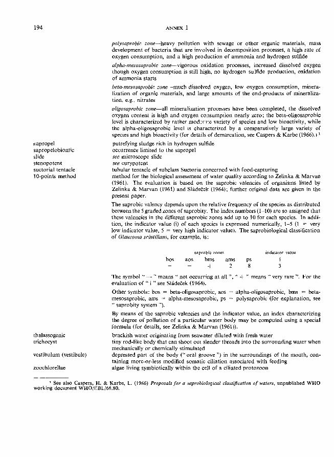
194
sapropel sapropelobiontic slide stenopotent suctorial tentacle 10-points method
thalassogenic trichocyst
vestibulum (vestibule)
zoochlorellae
ANNEX 1
polysaprobic zone-heavy pollution with sewage or other organic materials, mass development of bacteria that are involved in decomposition processes, a high rate of oxygen consumption, and a high production of ammonia and hydrogen sulfide
alpha-mesosaprobic zone-vigorous oxidation processes, increased dissolved oxygen though oxygen consumption is still high, no hydrogen sulfide production, oxidation of ammonia starts
beta-mesosaprobic zone-much dissolved oxygen, low oxygen consumption, mineralization of organic materials, and large amounts of the end-products of mineralization, e.g., nitrates
oligosaprobic zone-all mineralization processes have been completed, the dissolved oxygen content is high and oxygen consumption nearly zero; the beta-oligosaprobic level is characterized by rather mod:nte variety of species and low bioactivity, while the alpha-oJigosaprobic level is characterized by a comparatively large variety of species and high bioactivity (for details of demarcation, see Caspers & Karbe (1966).) 1
putrefying sludge rich in hydrogen sulfide occurrence limited to the sapropel see microscope slide see eurypotent tubular tentacle of subclass Suctoria concerned with food-capturing method for the biological assessment of water quality according to Zelinka & Marvan (1961). The evaluation is based on the saprobic valencies of organisms listed by Zelinka & Marvan (1961) and Sllidecek (1964); further original data are given in the present paper.
The saprobic valency depends upon the relative frequency of the species as distributed between the 5 graded zones of saprobity. The index numbers (1-10) are so assigned that these valencies in the different saprobic zones add up to 10 for each species. In addition, the indicator value (i) of each species is expressed numerically, 1-5 (1 = very low indicator value, 5 = very high indicator value). The saprobiological classification of Glaucoma scintillans, for example, is:
saprobic zones indicator value
bos aos bms
+ ams
2 ps 8
i 3
The symbol" - " means" not occurring at all ", " + " means" very rare". For the evaluation of "i " see Shideeek (1964).
Other symbols: bos = beta-oligosaprobic, aos = alpha-oligosaprobic, bms = betamesosaprobic, ams = alpha-mesosaprobic, ps = polysaprobic (for explanation, see .. saprobity system ").
By means of the saprobic valencies and the indicator value, an index characterizing the degree of pollution of a particular water body may be computed using a special formula (for details, see Zelinka & Marvan (1961)).
brackish water originating from seawater diluted with fresh water tiny rod-like body that can shoot out slender threads into the surrounding water when mechanically or chemically stimulated depressed part of the body (" oral groove ") in the surroundings of the mouth, containing more-or-less modified somatic ciliation associated with feeding algae living symbiotically within the cell of a ciliated protozoon
1 See also Caspers, H. & Karbe, L. (1966) Proposals for a saprobiological classification of waters, unpublished WHO working document WHOjEBLj66.80.
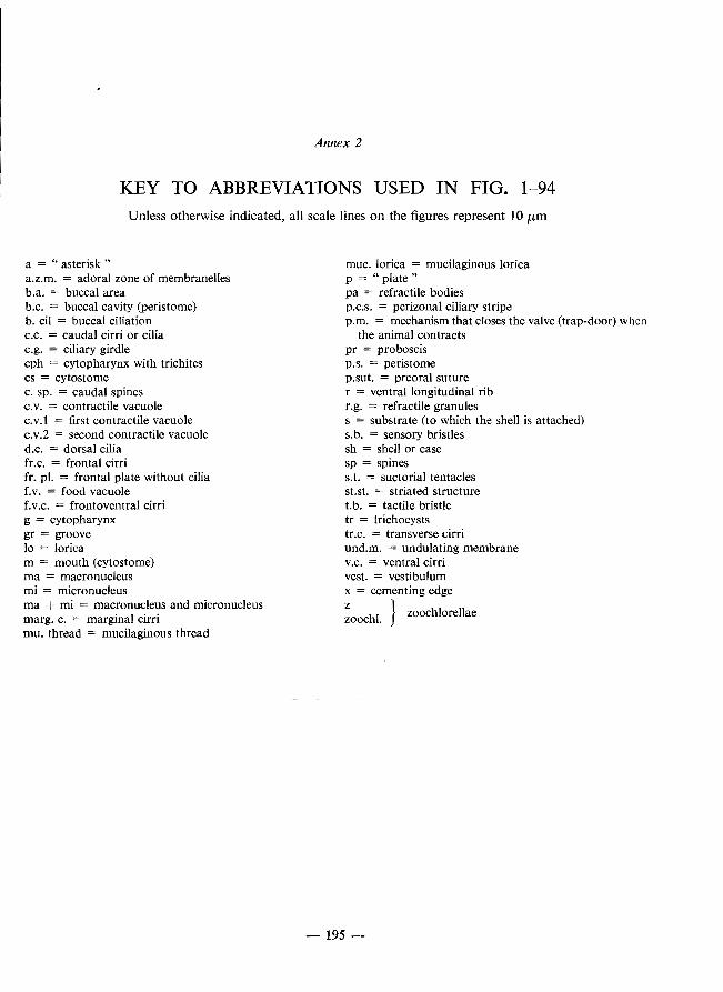
Annex 2
KEY TO ABBREVIATIONS USED IN FIG. 1-94 Unless otherwise indicated, all scale lines on the figures represent lO /kill
a = "asterisk" a.z.m. = adoral zone of membranelles b.a. = buccal area b.c. = buccal cavity (peristome) b. cil = buccal ciliation c.c. = caudal cirri or cilia c.g. = ciliary girdle cph = cytopharynx with trichites cs = cytostome c. sp. = caudal spines c.v. = contractile vacuole c. v.l = first contractile vacuole c.v.2 = second contractile vacuole d.c. = dorsal cilia fr.c. = frontal cirri fro pI. = frontal plate without cilia f.v. = food vacuole f. V.C. = frontoventral cirri g = cytopharynx gr = groove 10 = lorica m = mouth (cytostome) rna = macronucleus mi = micronucleus rna + mi = macronucleus and micronucleus margo C. = marginal cirri mu. thread = mucilaginous thread
muc. lorica = mucilaginous lorica p = "plate" pa = refractile bodies p.C.S. = perizonal ciliary stripe p.m. = mechanism that closes the valve (trap-door) when
the animal contracts pr = proboscis p.S. = peristome p.sut. = preoral suture r = ventral longitudinal rib r.g. = refractile granules s = substrate (to which the shell is attached) S. b. = sensory bristles sh = shell or case sp = spines S. t. = suctorial tentacles st.st. = striated structure t.b. = tactile bristle tr = trichocysts tr.c. = transverse cirri und.m. = undulating membrane V.C. = ventral cirri vest. = vestibulum x = cementing edge
Z hI} zoochlorellae zooc .
-195 -

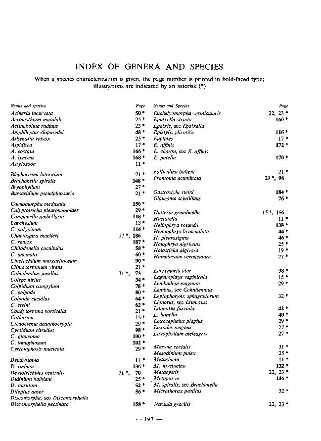
INDEX OF GENERA AND SPECIES When a species characterization is given, the page number is printed in bold-faced type;
illustrations are indicated by an asterisk (*)
Genus and species Page Genus and Species Page Acineria incurvata 50 * Enchelyomorpha vermicularis 22, 23 * Acropisthium mutabile 25 '" Epalxella striata 160 * Actinobolina radians 23 * Epalxis, see Epalxella Amphileptus claparedei 48 * Epistylis plicatilis 116* Askenasia volvox 25 * Euplotes 17 * Aspidisca 17 * E. affinis 172 * A. costata 166 * E. charon, see E. affinis A.lynceus 168 * E.patella 170 * Astylozoon 11*
Blepharisma lateritium 21 * Folliculina bolteni 21 * Brachonella spiralis 148 * Frontonia acuminata 29 *, 96 Bryophyllum 27 * Bursaridium pseudobursaria 21 * Gastrostyla steini 184 *
Glaucoma scintillans 76 * Caenomorpha medusala 150 * Calyptotricha pleuronemoides 29 * Halteria grandinella 15 *, 156 Campanella umbellaria 118 * Hastatella 11* Carchesium 13* Heliophrya rotunda 138 ... C. polypinum 114* Hemiophrys bivacuolata 44* Chaetospira muelleri 17 *, 186 H. pleurosigma 46 * C. remex 187 '" Holophrya nigricans 25 ... Chilodonella cucullulus 58 * Holosticha algivora 19 * C. uncinata 60 '" Homalozoon vermiculare 27 * Cinetochilum margaritaceum 90 '" Climacostomum virens 21 *
Lacrymaria olor 38 ... Cohnilembus pusillus 31 *, 73 Coleps hirtus 34* Lagenophrys vaginicola 15 * Colpidium campylum 78 * Lembadion magnum 29 * C. colpoda 80 '" Lembus, see Cohnilembus Colpoda cucullus 64* Leptopharynx sphagnetorum 32 ... C. steini 62 * Lionotus, see Litonotus Condylostoma vorticella 21 * Litonotus fasciola 42 * Cothurnia 15 ... L. lamella 40 ... Ctedoctema acanthocrypta 29 * Loxocephalus plagius 29 '" Cyclidium citrullus 98 * Loxodes magnus 27 ... C. glaucoma 100 ... Loxophyllum meleagris 27 ... C. lanuginosum 102 * Cyrtolophosis mucicola 29 ... Maryna socialis 31 ...
Mesodinium pulex 25 * Dendrosoma 11* Metacineta 11* D. radians 136 * M. mystacina 132 ... Dexiotrichides centralis 31 *, 70 Metacystis 22, 23'" Didinium balbiani 25 * Metopus es 146 '" D. nasutum 52 * M. spiralis, see Brachonella Dileptus anser 56 ... Microthorax pusillus 32 * Discomorpha, see Discomorphella Discomorphella pectinata 158 * Nassula gracilis 22, 23 *
-197 -
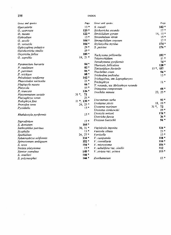
198 INDEX
Genus and species Page Genus and species Page
Opercu/aria 13 * S. roeseli 142 *
O. coarctata 120 * Stichotricha secunda 17 *
O. nutans 122 * Strobilidium gyrans 14, 15 *
Ophrydium 13 * Strombidium viride 15 *
O. sessile 104 * Strongylidium crassum 17*
O. versatile 104 * Stylonychia mytilus 174 *
Ophryoglena pelagica 29 * S. putrina 176 *
Opisthotricha similis 19 * Oxytricha fallax 180 * Tachysoma pellionella 182 *
O. saprobia 18, 21 * Telotrochidium 11*
Paramecium bursaria Tetrahymena pyriformis 74 *
84 * Thuricola folliculata 128 *
P. caudatum 82 * Tintinnidium fluviatile 15 *, 157
P. putrinum 86 * Trachelius ovum 54 * P. trichium 88 * Trichodina pediculus 13 *
Pelodinium reniforme 162 * Trichopelma, see Leptopharynx Phascolodon vorticella 23 * Trichophrya 11*
Plagiopyla nasuta 66 * T. rotunda, see Heliophrya rotunda
Platycola 15 * Trimyema compressum 68 *
P. truncata 126 * Trochilia minuta 22, 23 *
Platynematum sociale 31 *, 71
Platyophrya vorax 25 * Podophrya fixa 11*,130* Urocentrum turbo 92 *
Prorodon teres 24, 25 * Uroleptus piscis 18, 19 *
pyxidiella 13* Uronema marinum 31 *, 72
Urosoma cienkowski 19 *
Rhabdostyla pyriformis 15 * Urostyla weissei 178 *
Urotricha farcta 36 *
Saprodinium 17 * Urozona buetschli 94 *
S. dentatum 164 * Sathrophilus putrinus 30, 31 * Vaginicola ingenita 124 *
Scyphidia 13* Vasicola ciliata 23 *
Spathidium 24, 27 * Vorticella 13*
Sphaerophrya soliformis 134 * V. campanula 108 *
Spirostomum ambiguum 152 * V. convallaria 110 *
S. teres 154 * V. microstoma 106 *
Steinia platystoma 19 * V. nebulifera var. similis 112
Stentor coeruleus 140 * V. striata var. octava 113*
S. muelleri 144 * S. polymorphus 144 * Zoothamnium 13*
WHO publications may be obtained, direct or through booksellers, from:
ALGERIA
ARGENTINA
AUSTRALIA
AUSTRIA
BELGIUM
BURMA
CANADA
COLOMBIA
COSTA RICA
CYPRUS
DENMARK ECUADOR EGYPT FEDERAL
REPUBLIC OF GERMANY
FIn FINLAND
FRANCE
GREECE
HAITI
HUNGARY
ICELAND
INDIA
INDONESIA
IRAN
IRELAND
ISRAEL
ITALY
JAPAN
KENYA
KHMER REPUBLIC
LAOS
LUXEMBOURG
MALAYSIA
MEXICO
MONGOLIA
MOROCCO
Societe Nationale d'Edition et de Diffusion, 3 bd Zirout Youcef, ALGIERS.
Librerla de las Naciones, Cooperativa Ltda. Alsina 500, BUENOS AIREs - Editorial Sudamericana S.A., Humberto 10 545 BUENOS AIRES.
Australian Government Publishing Service, Sales and Distribution, P.O. Box 84, CANBERRA, A.C.T. 2600 (mail orders); AGPS Book Centre, 113-115 London Circuit, CANBERRA CITY; 347 Swanston St., MELBOURNE; Commonwealth Centre, 1-3 St. George's Terrace, PERTH; Bank House, 315 George St., SYDNEy-Hunter Publications, 58A Gipps Street, COLLINGWOOD, Vic. 3066.
Gerold & Co., i. Graben 31, VIENNA 1.
Office international de Librairie, 31 avo Marnix, BRUSSELS.
see India, WHO Regional Office.
Information Canada Bookstore, 171 Slater Street, OTTAWA, Ontario KIA OS9; 1735 Barrington Street, HALIFAX, N.S. ; Edifice Aeterna-Vie, 1182 ouest, rue SteCatherine, MONTREAL, 110 (Que.); 221 Yonge Street, TORONTO 205, Ontario; 657 Granville Street, VANCOUVER 2, B.C. ; 393 Portage Avenue, WINNII'EG, Manitoba.
Distrilibros Ltd, Plo Alfonso Garcia, Carrera 4a, Nos 36-119, CARTAGENA.
Imprenta y Librerla Trejos S.A., Apartado 1313, SAN Jos~.
MAM, P.O. Box 1674, NICOSIA.
Ejnar Munksgaard, Ltd, Nl!Jrregade 6, COPENHAGEN. Librerla Cientlfica S.A., P.O. Box 362, Luque 223, GUAYAQUIL. AI Abram Bookshop, 10 Avenue el Horreya, ALEXANDRIA. Govi-Verlag GmbH, Beethovenplatz 1-3, FRANKFURT A. M. 6 - W. E. Saarbach, Postfach 1510, FoIIerstrasse 2, 5 COLOGNE 1 - Alex. Horn. Spiegelgasse 9, 62 WIESBADEN. The WHO Representative, P.O. Box 113, SUVA.
Akateeminen Kirjakauppa, Keskuskatu 2, HELSINKI 10.
Libraire Arnette, 2, rue Casimir-Delavigne, PARIS 6".
G. C. Eleftheroukadis S.A., Librarie internationale, rue Nikis 4, ATHENS (T. 126).
Max Bouchereau, Librairie" A la CaraveIIe n, Bolte postale ll1-B, PORT-AU-PRINCB.
Kultura, P.O.B. 149, BUDAPEST 62 - Akademiai Konyvesbolt, Vaci utca 22, BUDAPEST V. Snaebjorn Jonsson & CO., P.O. Box 1131, Hafnarstraeti 9, REYKIAVIK.
WHO Regional Office for South-East Asia, World Health House, Indraprastha Estate, Ring Road, NEW DELHI 1 - Oxford Book & Stationery Co., Scindia House, NEW DELHI; 17 Park Street, CALCUTTA 16 (Sub-agent). see India, WHO Regional Office.
Mesrob Grigorian, Naderi Avenue (Arbab-Guiv Building), TEHERAN.
The Stationery Office, DUBLIN.
Heiliger & Co., 3 Nathan Strauss Street, JERUSALEM.
Edizioni Minerva Medica, Corso Bramante 83-85, TURIN; Via Lamarmora 3, MILAN.
Maruzen Co., Ltd, P.O. Box 5050, TOKYO International, 100-31 Japan.
The Caxton Press Ltd, Head Office: Gathani House, Huddersfield Road, P.O. Box 1742, NAIROBI. The WHO Representative, P.O. Box 111, PHNOM-PENH.
The WHO Representative, P.O. Box 343, VIENTIANE.
Librairie Trausch-Schummer, place du Theatre, LUXEMBOURG.
The WHO Representative, P.O. Box 2550, KUALA LUMPUR - Jubilee (Book) Store Ltd, 97 Jalan Tuanku Abdul Raman, P.O. Box 629, KUALA LUMPUR.
La Prensa Medica Mexicana, Ediciones Cientlficas, Paseo de las Facultades, 26 MEXICO CITY 20, D.F. see India, WHO Regional Office.
Editions La Porte, 281 avenue Mohammed V, RABAT. Price: £2.40 $6.00 Sw. fro 24.
Prices are subject to change without notice.
WHO publications may be obtained, direct or through booksellers, from:
NEPAL see India, WHO Regional Office. NETHERLANDS N.V. Martinus Nijhoff's Boekhandel en Uitgevers Maatschappij, Lange Voorhout 9,
THE HAGUE. NEW ZEALAND Government Printing Office, Government Bookshops at: Rutland Street, P.O. Box 5344,
AUCKLAND; 130 Oxford Terrace, P.O. Box 1721, CHRISTCHURCH; Alma Street, P.O. Box 857, HAMILTON; Princes Street, P.O. Box 1104, DUNEDIN; Mulgrave Street, Private Bag, WELLINGTON - R. Hill & Son Ltd, Ideal House, Cnr. Gilles Avenue & Eden St., Newmarket, AUCKLAND S.E. 1.
NIGERIA University Bookshop Nigeria, Ltd, University of Ibadan, IBADAN. NORWAY lohan Grundt Tanum Bokhandel, Karl lohansgt. 43, OSLO 1. PAKISTAN Mirza Book Agency, 65 Shahrah Quaid-E. Azam, P.O. Box 729, LAHORE 3. PARAGUAY Agencia de Librer(as Nizza S.A., Estrella No. 721, ASUNCI6N. PERU Distribuidora Inca S.A., Apartado 3115, Emilio Althaus 470, LIMA. PHILIPPINES World Health Organization, Regional Office for the Western Pacific, P.O. Box 2932,
MANILA - The Modem Book Company Inc., P.O. Box 632, 926 Rizal Avenue, MANILA.
POLAND Skladnica Ksi~garska, ul. Mazowieka 9, WARSAW (except periodicals) - BKWZ Ruch, ul. Wronia 23, WARSAW (periodicals only).
PORTUGAL Livraria Rodrigues, 186 Rua Aurea, LISBON. REPUBLIC OF The WHO Representative, Central P.O. Box 540, SEOUL.
KOREA REPUBLIC OF The WHO Representative, P.O. Box 242, SAIGON.
VIET-NAM SINGAPORE The WHO Representative, 144 Moulmein Road, G.P.O. Box 3457, SINGAPORE 1. SOUTH AFRICA Van Schaik's Bookstore (Pty) Ltd, P.O. Box 724, PRETORIA. SPAIN
SRI LANKA
SWEDEN SWITZERLAND THAILAND TUNISIA TURKEY UGANDA UNITED
KINGDOM
UNITED REP. OF TANZANIA
Comercial Atheneum S.A., Consejo de Ciento 130-136, BARCELONA, 15; General Moscard6 29, MADRID 20 - Librerla Diaz de Santos, Lagasca 95, MADRID 6. see India, WHO Regional Office.
Aktiebolaget C.E. Fritzes Kungl. Hovbokhandel, Fredsgatan 2, STOCKHOLM 16. Medizinischer Verlag Hans Huber, Lllnggass Strasse 76, 3000 BERNE 9. see India, WHO Regional Office. Societe Tunisienne de Diffusion,S avenue de Carthage, TuNIS. Librairie Hachette, 469 avo de l'Independance, ISTANBUL. see address under KENYA. H. M. Stationery Office: 49 High Holborn, LoNDON WCIV 6HB; 13a Castle Street, EDINBURGH EH2 3AR; 109 St. Mary Street, CARDIFF CFI UW; 80 Chichester Street, BELFAST BTl 4JY; Brazennose Street, MANCHESTER M60 8AS; 258 Broad Street, BIRMINGHAM Bl 2HE; 50 Fairfax Street, BRISTOL BSI 3DE. All mail orders should be sent to P.O. Box 569, London SEI9NH. see address under KENYA.
UNITED STATES The American Public Health Association, Inc., lOIS Eighteenth St. N.W., OF AMERICA WASHINGTON D.C. 20036 - United Nations Bookshop, New York, N.Y. 10017
(retail only).
USSR
VENEZUELA
YUGOSLAVIA ZAIRE
For readers in the USSR requiring Russian editions: Komsomo!skij prospekt 18, Medicinskaja Kniga, Moscow - For readers uutside the USSR requiring Russian editions: Kuzneckij most 18, MeMunarodnaja Kniga, Moscow G-200. Editorial Interamericana de Venezuela C.A., Apartado 50785, CARACAS - Librerla del Este, Av. Francisco de Miranda 52, Edificio Galipan, CARACAS. lugoslovenska Knjiga, Terazije 27/11, BELGRADE. Librairie du Zarre, 12 avenue des Aviateurs, KINSHASA.
Orders may also be addressed to : World Health Organization, Distribution and Sales Service, 1211 Geneva 27, Switzerland, but must be paid for
in pounds sterling, US dollars, or Swiss francs.