Embed Size (px)
Citation preview
jf. Cell Set. 15, 379-401 (i974) 379Printed in Great Britain
FEEDING IN CILIATED PROTOZOA
I. PHARYNGEAL DISKS IN EUPLOTES: A SOURCE OFMEMBRANE FOR FOOD VACUOLE FORMATION?
JOHN. A. KLOETZEL
Department of Biological Sciences,University of Maryland Baltimore County,Catonsville, Maryland 21228, U.S.A.
SUMMARY
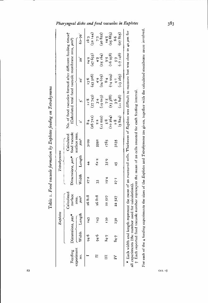
The ciliate Euplotes is able to expend a very large amount of membrane in the formation offood vacuoles. Calculations based on the rate of ingestion of the food organism Tetrahymenaindicate that an amount of food vacuole membrane equivalent to approximately 50—150% ofthe total Euplotes cell surface area can be produced within 5-10 min. An aggregation of osmio-philic, membrane-limited 'pharyngeal disks' is found packed in the cytoplasm just beneath thecell surface membrane in the region of the cell mouth and cytopharynx. These disks, whichcan be seen also in living cells, have average dimensions of 2 /Mn diameter by 100 nm thickness,and contain tightly packed layers of a thin lamellar material. Electron micrographs have revealedthe apparent fusion of the limiting membrane of disks with the cell's plasma membrane at thebase of the gullet. The lamellar disk contents are thereby released to the exterior medium inthe buccal cavity, where they form a loosely packed layer over the surface membrane. It ispostulated that the pharyngeal disks represent a repository of preformed membrane for use infood vacuole formation. The disk contents may also play a role in food ingestion, although thisis not well defined at present. The myeloid content of old food vacuoles is very similar to thatof nearby disks in the cytoplasm, suggesting that the disks may form by pinching from shrinkingfood vacuoles during the digestive cycle. Thus a cycle of membrane flow is envisaged, with thepharyngeal disks (1) coalescing with the surface membrane during food vacuole formation,(2) reforming by pinching from these food vacuoles during digestion, and (3) migrating backto the oral region to serve as a membrane store for subsequent food vacuole formation.
INTRODUCTION
The interrelationship among various types of cell membrane systems in general,and the biosynthesis of the plasma membrane in particular, are areas of considerablecurrent research activity. Protozoan cells offer several advantages for studies on themechanism of formation of new cell membrane (Conner, 1972; McKanna, 1973).For example, when protozoa are feeding rapidly they can produce large amounts ofsurface plasma membrane, which is internalized in the formation of food vacuoles.Marshall & Nachmias (1965) have estimated that an amoeba feeding on Parameciumhas the ability to consume the equivalent of its entire surface area in plasma membranewithin 5 min. Similarly, Wetzel & Korn (1969) have studied the phagocytosis of latexbeads by Acanthamoeba, noting that in 30 min 50—100% of the amoeba's surfacemembrane is converted to phagocytic vacuole membrane surrounding 250-300 in-gested latex particles. Obviously, since these cells do not decrease in size, this means
380 J. A. Kloetzel
that there must be a continuous replenishment of surface membrane during such rapidbursts of feeding.
From the point of view of studying the cellular mechanism of such plasma mem-brane production, the amoebae have the disadvantage that phagocytosis occurs overmuch of their surface area. This is not the case with most ciliated protozoa, whichhave a highly organized cortical surface and a specialized feeding apparatus. Ciliarybeating creates currents in the water that sweep food particles and prey organismsinto a gullet, or buccal cavity. In the region of the cell mouth (cytostome) the foodparticles are wrapped in a piece of membrane from the feeding cell and pinched offinto the cytoplasm for subsequent digestion.
When observing a feeding ciliate, it is immediately obvious that the amount ofmembrane produced in this way must be substantial. It is thus all the more remarkablethat this type of membrane turnover occurs at a very restricted location on the cellsurface. My own calculations (Kloetzel, 1970; see text) indicate that the ciliateEuplotes can ingest sufficient Tetrahymena in 2 min to require the production of2-6 xio4/im2 of food vacuole membrane, or roughly the equivalent of the Euplotes'entire surface area.
The present report provides some data on feeding rate in Euplotes, and describesa set of membranous organ elles, the 'pharyngeal disks', found around the cytopharynxand cytostome of this cell. It is proposed that these organelles represent an internalrepository of preformed cell membrane that is used in the formation of food vacuoles.
METHODS AND MATERIALS
Experimental organisms
Euplotes (probably E. eurystomus; Carolina Biological Supply Co., Burlington, N.C.) weremaintained in wheat infusion containing a mixed population of bacteria. Tetrahymena pyriformiswere grown in 2 % proteose-peptone, washed in amoeba medium (Prescott & Carrier 1964),and fed to Euplotes daily.
Feeding studies
To assess the rate of feeding and to estimate membrane turnovers, Euplotes were maintainedin filtered pond water or old culture media without Tetrahymena for 1-2 days. Large amountsof washed Tetraliymena were then introduced into the fasted Euplotes cultures; cells were re-moved at intervals with a braking pipette (Stone & Cameron, 1964) and fixed in aceto-orcein oracetocarmine solutions to determine the number of food vacuoles formed.
For membrane calculations, a sample of the Tetrahymena to be used in a feeding experimentwas slowed with methyl cellulose, and the length and width of living cells measured micro-scopically with an ocular micrometer. The volume of the average cell was determined using theformula for a prolate ellipsoid. Since food vacuoles are roughly spherical, and Tetrahymenaare ingested individually, the surface area of a sphere of the same volume as the average Tetra-hymena was then calculated, this figure serving as an index of the amount of membrane requiredfor enveloping one Tetrahymena. The dimensions of the Euplotes to be used were obtainedsimilarly. For approximating the surface area of an average Euplotes cell, the formula for thearea of an oblate ellipsoid was employed; a value of (length + width)l\ was used as the majorsemi-axis. Membrane turnover was calculated this way, rather than direcdy from the measureddiameter of freshly formed food vacuoles, because food vacuoles shrink quickly after formation(presumably due to die rapid loss of fluid from the enclosed Tetrahymena).

Pharyngeal disks and food vacuoles in Euplotes 381
Light microscopy
In order to slow living Euplotes for observation and photography, the cells were either placedin methyl cellulose solution or compressed between a petroleum-jelly-ringed coverslip and themicroscope slide. Bright-field, phase-contrast and Zeiss Nomarski interference-contrast opticswere used, and photomicrographs were taken (some with electronic flash illumination) onKodak Panatomic-X film.
Whole mounts of Euplotes fixed briefly with osmium tetroxide vapour were prepared inorder to examine structures in the mouth region. For more intensive staining of osmium-binding structures the procedure of Faur6-Fremiet & Andr6 (1968) was used. Basically, thisinvolves enhancement of the density of bound osmium by reduction with pyrogallol beforemounting the fixed cells in an aqueous mountant.
Electron microscopy
Euplotes were fixed in either of 2 ways: in 3 % OsO4 in 001 M s-collidine buffer, pH 74, with0-5 mg/ml CaCl2; or in a mixture of 0-5 % glutaraldehyde and 2 % OsO4 in 0-05 M phosphatebuffer, pH 76. Fixation (30-60 min), buffer rinses, and ethanol dehydration were carried outat ice-bath temperature, before embedding in Epon-Araldite (Mollenhauer, 1964). Thin sectionswere stained with uranyl acetate in ethanol, methanol, or a mixture, followed by lead citrate,and examined with either a Philips EM 200 or Hitachi HU-12 electron microscope.
OBSERVATIONS
Feeding studies
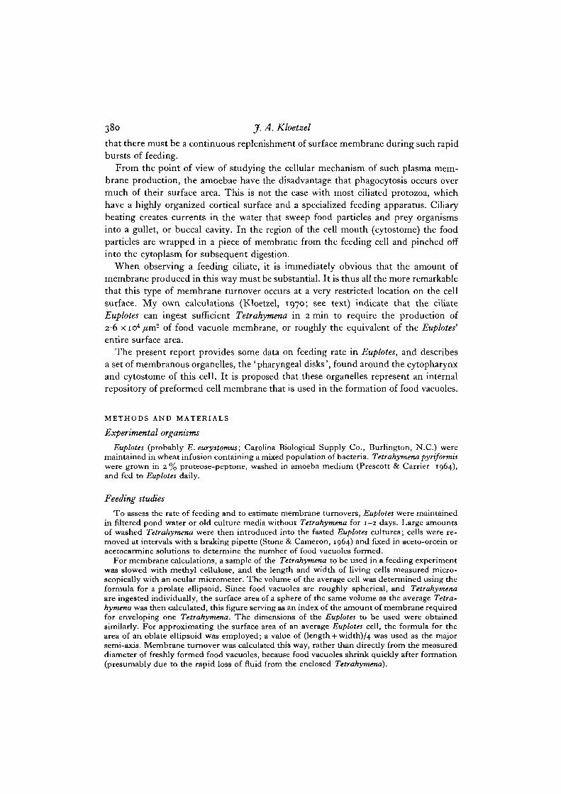
The results of several feeding experiments are presented in Fig. 1. Because ofvariations in size of the Euplotes in different experiments, and especially in size of theTetrahymena (depending on how long they were suspended in washing medium beforebeing used as food), the amount of food vacuole membrane produced is expressed interms of percentage of the total Euplotes surface area. The measured sizes of Euplotesand Tetrahymena for these same experiments, the number of food vacuoles formed,and calculations of the actual membrane areas involved, are presented in Table 1.As an example of the rapidity of feeding, Fig. 3 shows a cell that was exposed toTetrahymena for only 5 min before being fixed and stained. All 17 food vacuoles wereformed within this brief feeding interval.
Light microscopy
Euplotes fixed with osmium tetroxide vapour and prepared as whole mounts reveala variety of osmiophilic structures when viewed with the light microscope. The mostprominent of these structures are food vacuoles, and a collection of structures in theoral region that correspond to the 'epipharyngeal organelle' of Faure"-Fremiet &Andre (1968). Using the pyrogallol reduction technique on osmium-fixed Euplotes, asdescribed by these authors, the epipharyngeal bodies are revealed as very dense,apparently discrete units, wrapped in a layer approximately 30 /im long around theterminus of the gullet (Figs. 4,5). With a 100 x oil-immersion objective and Nomarskioptics, the cytoplasm around the mouth of living, unstained Euplotes can be seen tobe populated with a large number of these oral organelles (Fig. 6). They appear to bearrayed in an ordered layer against the gullet wall, and are in constant motion, movingas individual units in and out of the rows making up this layer.
382 J. A. Kloetzel
200 r-
180 -
10 20 30 40 50
Duration of feeding, mm
60 70
Fig. i. Results of 4 feeding experiments. The amount of food vacuole membraneproduced is expressed in terms of equivalent percentage of the entire calculatedsurface area of the Euplotes used for each experiment, as a function of the time theEuploUs were allowed to feed. Each point represents the mean of 20 cells counted foreach feeding interval. Feeding experiments I, II, III, IV: D, •> O a nd •» respec-tively.
Electron microscopy
A view of the area near the base of Euplotes' buccal cavity is shown in Fig. 7. Inthe cytoplasm, just beneath the non-ciliated dorsal surface of the gullet, one or morerows of oral organelles can be seen, corresponding to those seen with the light micro-scope. In section these organelles appear rod-shaped, generally about 2 fim long ando-i—0-15/im wide. Their long axes are primarily perpendicular to the cell surfacemembrane; rows of microtubules (cytopharyngeal ribbons; Pitelka, 1969) extendbetween and appear to separate these organelles into units or small groups (Fig. 7;Fig. 10, inset).
These oral structures were originally termed 'cytoplasmic rods' (Roth, 1957; seeKloetzel, 1970). The observations of Faure-Fremiet & Andre (1968) on these samestructures led these authors to the opinion that the rods were actually leaves or sheetsmaking up an ensemble that they called the 'foliated epipharyngeal organelle'. It isapparent from light-microscopic observations (see above) that these organelles couldnot be true rods, or they would be below the resolution of the light microscope, andthus not detectable as individual units in living cells. In order to resolve the size and
Tab
le I
. F
ood
vacu
ole f
orm
atio
n by
Eup
lote
s fee
ding
on
Tetr
ahym
ena
Eir
plot
es
Tet
rahy
men
a +u
k
r h
-I -----7
Cal
cula
ted
C
alcu
late
d
No
. of
foo
d va
cuol
es f
orm
ed a
fter
dif
fere
nt
feed
ing
tim
est
3 F
eed
ing
D
imen
sio
ns,
pm
X
surf
ace
Dim
ensi
on
s, p
m*
fo
od
vac
uole
(C
alcu
late
d to
tal
food
vac
uole
mem
bra
ne
area
, p
m2
) 5
exp
erim
ent
rr
A-
,
area
, su
rfac
e ar
ea,
r A
%
n
o.
Wid
th
Len
gth
,u
rn2
Wid
th
Len
gth
p
m2
2'
5'
I o
f 20'
63
-70
' 2
* E
ach
wid
th a
nd
len
gth
rep
rese
nt
the
mea
n o
f 2
0 m
easu
red
cel
ls.
Th
ick
nes
s of
Eup
lote
s w
as d
iffi
cult
to
mea
sure
bu
t w
as c
lose
to
40
pm
fo
r &
al
l ex
per
imen
ts (
the
nu
mb
er u
sed
fo
r ca
lcul
atio
ns).
E
ach
rep
ort
ed f
oo
d v
acuo
le n
um
ber
rep
res$
nts
th
e m
ean
of
20
cel
ls c
ou
nte
d f
or
each
fee
din
g i
nte
rval
. s C
Fo
r ea
ch o
f th
e 4
feed
ing
ex
per
imen
ts t
he
size
s of
th
e E
zrpl
otes
an
d T
etra
hynl
sna
are
give
n, t
og
eth
er w
ith
th
e ca
lcul
ated
mem
bra
ne
area
s in
volv
ed.
384 J.A. Kloetzel
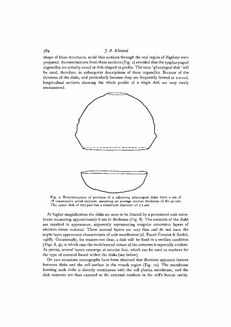
shape of these structures, serial thin sections through the oral region of Euplotes wereprepared. Reconstructions from these sections (Fig. 2) revealed that the epipharyngealorganelles are actually ovoid or disk-shaped in profile. The term ' pharyngeal disk' willbe used, therefore, in subsequent descriptions of these organelles. Because of thethinness of the disks, and particularly because they are frequently bowed or curved,longitudinal sections showing the whole profile of a single disk are very rarelyencountered.
Fig. 2. Reconstruction of portions of 2 adjoining pharyngeal disks from a set of18 consecutive serial sections, assuming an average section thickness of 80-90 nm.The upper disk of this pair has a maximum diameter of 22 fim.
At higher magnification the disks are seen to be limited by a prominent unit mem-brane measuring approximately 6 nm in thickness (Fig. 8). The contents of the disksare myeloid in appearance, apparently representing irregular concentric layers ofelectron-dense material. These internal layers are very thin and do not have thetriple-layer appearance characteristic of unit membranes (cf. Faure-Fremiet & Andre\1968). Occasionally, for reasons not clear, a disk will be fixed in a swollen condition(Figs. 8, 9), in which case the multilayered nature of the contents is especially evident.At points, several layers converge at circular foci, which can be used as markers forthe type of material found within the disks (see below).
On rare occasions micrographs have been obtained that illustrate apparent fusionsbetween disks and the cell surface in the mouth region (Fig. 10). The membranelimiting such disks is directly continuous with the cell plasma membrane, and thedisk contents are thus exposed to the external medium in the cell's buccal cavity.
Pharyngeal disks and food vacuoles in Euplotes 385
Some of this layered material seems to form a coating loosely apposed to the externalsurface of the membrane. This feltwork is extensively developed in some sections(Fig. 11); circular foci can be noted in the layers (Figs. 10, 11), reminiscent of thoseseen within swollen disks in the cytoplasm (Fig. 9).
Upon examination of Euplotes that have recently ingested Tetrahymena, it is ap-parent that some of the layered extracellular material found in the oral region isincorporated into the food vacuole along with the food. Between the food vacuolemembrane and the enclosed Tetrahymena in freshly formed food vacuoles, regions arefound that contain packed arrays of the lamellar material, frequently with cilia fromthe enclosed prey embedded in the layers (Fig. 12).
By far the vast majority of pharyngeal disks found within any single Euplotes isfound congregated around the cytostome. Those disks found elsewhere in the cyto-plasm are frequently near food vacuoles. Fig. 13 represents what is interpreted to bea terminal food vacuole, from which essentially all the digestible material has beenremoved, leaving only residue. Such food vacuoles usually display myeloid contentsthat greatly resemble the material found within the adjacent pharyngeal disks, althoughno sections have been obtained that show clear continuity between food vacuoles anddisks. It may be significant to note that the limiting membranes of food vacuoles,pharyngeal disks, and the surface membrane all exhibit a clear trilaminar appearanceof similar thickness (about 6 nm) in osmium-fixed preparations; all other cytoplasmicmembranes are thinner and not distinctly trilaminar.
DISCUSSION
Feeding and membrane turnover in Euplotes
Simple observation of feeding Euplotes is sufficient to demonstrate that these cells arecapable of very rapid production of food vacuole membrane. An attempt has beenmade in the present study to quantitate this membrane turnover, using the rate ofingestion of Tetrahymena as an index of food vacuole membrane formation. Thereare at least 2 difficulties that affect the accuracy of these calculations. First, theirregular contours of the Euplotes cell (and to a lesser extent those of Tetrahymena)make cell surface areas difficult to measure precisely. Second, newly formed foodvacuoles are not always spherical, as assumed here. Nevertheless, even allowing forthese approximations, the data certainly support the contention that membranerepresenting a large percentage of the Euplotes surface area can be rapidly formed andmoved inside the cell. In extreme cases (with small Tetrahymena, which are eatenmuch more rapidly than large ones) a Euplotes cell can ingest 17 Tetrahymena within5 min, representing an area of food vacuole membrane approximately twice that of theentire Euplotes surface. Certain obvious questions thus arise concerning the source ofsuch an amount of food vacuole membrane.
In theory there are only 2 basic mechanisms for the production of new cell surfacemembrane: molecular or micellar insertion (de novo membrane synthesis); or thefusion of preformed cytoplasmic membrane-limited bodies with the cell surface. Fora cell growing slowly or undergoing only moderate endocytosis, de novo synthesis of
25-2
386 J. A. Kloetzel
new cell surface might suffice. According to classical concepts of membrane flow andphagocytosis, even in rapidly feeding ciliates such molecular addition could conceivablyoccur all over the cell surface, with a concomitant flow of surface membrane into thegullet to accommodate vacuole formation. However, certain considerations would seemto weigh against such molecular surface membrane construction in this case. Firstof all, the observed rate of membrane utilization in food vacuole formation wouldrequire an extremely rapid rate of insertion of new membrane molecules for surfacereplacement, particularly since the new membrane is required at one specializedlocation on the cell surface. However, the pellicle covering most of the surface ofEuplotes (and many other ciliates; see Pitelka, 1969) is a complex structure. The plasmamembrane (where de novo synthesis and associated membrane flow would presumablyhave to occur) is usually separated from the bulk of the cytoplasm by a mosaic offlattened membrane-limited alveoli, layers of fibrous material, and microtubulearrays. This ' isolation' of the plasma membrane would not appear to favour rapidexchange of membrane precursor molecules with the cytoplasm. Even in Acantha-Tnoeba, in which there is neither a localized feeding area nor a multilayered pellicle,Goodall, Lai & Thompson (1972) found that phagocytosis did not stimulate a morerapid incorporation of precursor molecules into the plasma membrane, suggestingthat pre-existing membrane is used to replace the ingested surface membrane.
In the amoebae (Wise & Flickinger, 1970) as well as in other cell systems the Golgiapparatus has been implicated as a source of new surface membrane (see Whaley,Dauwalder & Kephart, 1972). As is true of many ciliates, Euplotes does not possessany Golgi bodies. However, the pharyngeal disks may represent highly modifiedfunctional equivalents of Golgi cisternae, at least in so far as cell surface addition isconcerned.
Role of the pharyngeal disks in the feeding process
The pharyngeal disks in Euplotes have been noted previously, as mentioned above.Originally described by Roth (1957) and termed 'cytoplasmic rods', these organelleswere much more fully characterized by Faur6-Fremiet & Andre (1968). These authorsfirst emphasized the flattened leaf-like arrangement of the disks, their myeloid con-tents, and their preferential localization around the base of the gullet in an array theychristened the 'foliated epipharyngeal organelle'. The term 'pharyngeal disk' is usedhere to designate each of the component organelles making up the array, emphasizingtheir shape and individual nature. None of the authors above speculated on the roleof the disks in the physiology of the cell. However, in the light of the discussionabove, it appears very likely that the pharyngeal disks in Euplotes serve the functionof providing the membrane, in pre-assembled form, necessary for rapid food vacuoleformation. Several factors support this contention. (1) The predominant localizationof the disks around the oral region suggests their involvement with some aspect of thefeeding process. The presence within newly formed food vacuoles of lamellar materialresembling the contents of the disks (Fig. 12) further supports this relationship. (2) Froma theoretical standpoint, the shape of the disks provides a high surface-to-volume ratio.Taking an idealized disk 2 /tm in diameter and o-i /im thick as an example, one finds
Pharyngeal disks and food vacuoles in Euplotes 387
that a sphere occupying the same volume of cytoplasm as this disk (0-21 /tm3) wouldhave only about one-fourth as much surface membrane (1-72/tm2 vs. 6-34/tm2).Conversely a sphere with the same surface area as this disk would occupy approxi-mately seven times as much volume. The shape of the disks also facilitates theirpacking into ordered layers immediately next to their proposed site of fusion with theplasma membrane of the gullet. It is interesting to note that the rows of microtubulesin this region may play a role in the stacking of the disks by serving as a' gate', allowinginsertion of disks into the array in only one preferred orientation. (3) The disks arelimited by a clearly trilaminar unit membrane 6 nm thick, of a type seen elsewhere inthe cell only in the plasma membrane and in the membrane enclosing food vacuoles.This similarity of membrane appearance between disks, food vacuoles and cell surfacesuggests some interrelationship among these membrane systems, as discussed below.(4) Although rare, pictures can be obtained that reveal disks apparently fused with thecell surface in the buccal cavity. Such micrographs could be interpreted as representingthe formation of disks by pinching inward from the surface membrane. However,clumps of layered material that greatly resemble the contents of cytoplasmic disks arefrequently found free in the buccal cavity, making it much more likely that the disks doindeed fuse with the plasma membrane, empty their contents, and supply membranefor cell surface expansion in the cytopharyngeal region of the cell. Certainly the mostplausible use of this expanded cell surface would be to provide for the phagocytosisof food particles.
While this hypothesis of disk function is thus quite attractive, one other possibilityshould be entertained: the disk membranes could conceivably serve to permit atransient stretching of the gullet and cytopharynx while relatively large prey organisms(like Tetrahymena) are being squeezed through the ordinarily narrow cytopharyngealopening, following which the disks could reform as the cytopharynx collapsed to itsnormal dimensions. In this case the disks could play a role similar to that postulatedfor the thick-membraned fusiform vesicles in the transitional epithelial cells of themammalian urinary tract during cycles of stretching and contraction (Hicks, 1966;see Falk, 1969, and Bardele, 1972, for descriptions of similar vesicles in algal cells andpseudoheliozoan protozoa, respectively). This possibility is made less plausible becauseof the myeloid material ejected from the disks following their fusion with the cellmembrane; it again would seem that recapture of this material by disks formingendocytotically from the wall of the buccal cavity might be difficult.
The nature and possible role of the layered contents of the disks require somecomment. In this discussion, primary focus has been placed on the limiting membraneof the disks with respect to membrane turnover during food vacuole formation. Itshould be realized, however, that the disk contents could play a more important rolein the feeding process than the membranes enclosing them. The osmiophilia of thedisks would suggest that their contents are rich in lipids. Indeed, Faure'-Fremiet &Andre (1968) reported staining the disks with Sudan black, although I have not beenable to do so. The lamellar appearance of the disk contents is also suggestive of lipoidallayers, although they are much thinner than the true unit membranes in the Euplotescytoplasm. The volume of extruded disk material in the cytopharynx can be extensive;

388 J. A. Kloetzdthese layers could serve as a lubricant when large prey organisms are squeezed throughthe narrow cytopharyngeal passage, or perhaps aid in immobilizing the cilia of capturedprey. Nilsson (1972) reports that the mucus extruded from the oral mucocysts ofTetrahymena can bind solutes from die medium for subsequent ingestion. Sincebacteria form a staple part of the Euplotes diet, ejected disk layers conceivably couldplay a role in binding and collecting bacteria until sufficient number have been accumu-lated for inclusion within a food vacuole. In Fig. 10 (inset) it can be noted that atpoints over the crests of ridges in the gullet wall, ejected disk lamellae are tightlypacked against the plasma membrane in highly ordered arrays. The significance ofthis ordering is not obvious, but does perhaps permit the speculation that materialfrom extruded lamella layers (possibly lipid) is somehow added to the plasma mem-brane itself during the membrane expansion accompanying phagocytosis of food.One fact is clear: some disk contents are taken into the food vacuole, forming a layerbetween the vacuole membrane and the ingested prey organism(s). It would seem mostefficient if the disks could supply not only the membrane for the envelopment ofthe food particle, but also hydrolytic enzymes to be included within the food vacuolefor initiation of the digestive events. Although preliminary cytochemical investiga-tions indicate that the Euplotes disks do not contain acid phosphatase, Dembitzer(1968) finds that some of the cytopharyngeal vesicles in Blepharisma are acid-phosphatase positive. Further work is needed on this question.
Since many ciliates besides Euplotes are able to undergo rapid bursts of food vacuoleformation, and with the preceding discussion in mind, it is not surprising to find thatcollections of membrane-limited vesicles, vacuoles, disks, or tubules have beenreported frequently near the cytopharynx of a number of ciliates (Randall & Fitton-Jackson, 1958; Faure-Fremiet, 1961; Miller & Stone, 1963; Jenkins, 1973; see Pitelka,1969, for discussion). Such structures are usually found between rows of microtubulesbeneath that portion of the gullet wall limited by a single membrane, as in Euplotes.Although most authors have not speculated on the functions of these oral organelles,Schneider (1964) attempted to relate the disk-shaped structures found near thecytopharynx of Paramecium (cf. Jurand & Selman, 1969) to some secretory processassociated with food vacuole formation. Kennedy (1965) and Elliott & Clemmons(1966) suggested that the oral vesicles of Blepharisma and Tetrahymena, respectively,might contribute structural material to developing food vacuole membranes. Veryrecently, Bradbury (1973) has described elongate 'apostome organelles' underlyingthe cytostome area of Hyalophysa, proposing likewise that these structures join thesurface of the cell to replace the membrane and associated filamentous coat 'used up'in the pinocytotic formation of food vacuoles. In these other ciliates examined to date,the cytopharyngeal vesicles appear to be less prominent and less numerous than are thedisks in Euplotes.
A final point concerns the origin of the pharyngeal disks. Those disks not found inthe array around the gullet are usually seen near food vacuoles in the cytoplasm. Asthe ingested food organisms are digested, the food vacuoles gradually become smaller;at the end of the digestion cycle, only myeloid' residual bodies' are left, whose contentsgreatly resemble those of the disks nearby. Taken together, these observations suggest
Phargyneal disks and food vacuoles in Euplotes 389
that disks may pinch off from shrinking food vacuoles and migrate back to the cyto-stome area; the disks could be looked upon as the intermediate vehicles in a closedcycle of membrane flow, from buccal cavity to food vacuole and back again. Since theEuplotes are continually growing and dividing, a mechanism for the production of newdisks must exist; nevertheless, the thickness and prominent trilaminar nature of themembranes of the cell surface, food vacuoles, and disks suggests some interrelation-ship among these membrane systems, perhaps of the cyclical nature mentioned above.McKanna (1969) has proposed that a similar recycling of membrane occurs duringfeeding in the peritrich ciliate Epistylis, with cup-shaped vesicles serving the role ofintermediate membrane carriers, although the photographs to illustrate this have notyet appeared. Bradbury (1973) finds membranous structures in Hyalophysa that sheinterprets as 'the collapsing walls of former food vacuoles', which are apparentlytransformed into the 'apostome organelles' that fuse with the cell surface near thecytostome during pinocytosis. Clearly, more complete information is required tosubstantiate the concept of membrane cycling in feeding ciliates. This will be soughtby careful ultrastructural observations of food vacuole formation and digestion inEuplotes, with electron-dense tracers included to identify food vacuoles of a given agethroughout the digestive cycle.
The able technical assistance of Patricia M. Willis is gratefully acknowledged. Dr NesslyCraig made valuable suggestions on the manuscript. This investigation was aided by a grantfrom the American Cancer Society, Maryland Division, Inc., and by U.S. Public HealthService Grant No. GM-18825, National Institute of General Medical Sciences.
NOTE ADDED IN PROOF
The full report by McKanna on membrane recycling in feeding peritrichs has now appeared(J. Cell Sci. (1973) 13, 663-686). In a report recently brought to my attention (MonitoreZool. Ital. [N.S.] (1971) 5, 65-80), Nobili & Rosati Raffaelli described 'electron-dense bodies'(disk-type structures) in the oral cytoplasm of 5 species of hypotrichs, including Euplotes.Their micrographs clearly show some of these bodies in continuity with the cytopharyngealsurface membrane, releasing ' extrapellicular fibrils' (extracellular lamellae of my report) intothe buccal cavity. Also, Allen (J. Cell. Biol. (1973) 59, 6 a) has recently provided evidencefor membrane recycling accompanying food vacuole turnover in Paramecium.
REFERENCES
BARDELE, C. F. (1972). Cell cycle, morphogenesis, and ultrastructure in the pseudoheliozoanClathrulina elegant. Z. Zellforsch. mikrosk. Anat. 130, 219—242.
BRADBURY, P. C. (1973). The fine structure of the cytostome of the apostomatous ciliateHyalophysa chattoni. J. Protozool. 20, 405-414.
CONNER, R. L. (1972). The unique value of protozoa in membrane investigation. J. Protozool.19. 225.
DEMBITZER, H. M. (1968). Digestion and the distribution of acid phosphatase in Blepharisma.J. Cell Biol. 37, 329-344-
ELLIOTT, A. M. & CLEMMONS, G. L. (1966). An ultrastructural study of ingestion and digestionin Tetrahymena pyriformis. J. Protozool. 13, 311-323.
FALK, H. (1969). Fusiform vesicles in plant cells. J. Cell Biol. 43, 167-174.FAURE-FREMIET, E. (1961). Le cytoplasme stomo-pharyngien des cilies cytophores. C. r.
hebd. Sianc. Acad. Sci., Paris 253, 357-362.FAURE-FREMIET, E. & ANDRE, J. (1968). Structure fine de YEuplotes eurystomus (WRZ). Archs
Anat. microsc. Morph. exp. 57, 53-78.
39° J-A. KloetzelGOODALL, R., LAI, Y. & THOMPSON, J. E. (1972). Turnover of plasma membrane during phago-
cytosis. J. Cell Sci. 11, 569-580.HICKS, R. M. (1966). The function of the Golgi complex in transitional epithelium. Synthesis
of the thick cell membrane. J. Cell Biol. 30, 623-643.JENKINS, R. A. (1973). Fine structure. In Blepharisma (ed. A. C. Giese), pp. 39—93. Stanford:
Stanford University Press.JURAND, A. & SELMAN, G. G. (1969). The Anatomy of Paramecium aurelia. London and New
York: Macmillan, St Martin's Press.KENNEDY, J. R. (1965). The morphology of Blepharisma undulans Stein. J. Protozool. 12,
542-561.KLOETZEL, J. (1970). The role of 'cytoplasmic rods' in food vacuole membrane formation in
Euplotes. J. Cell Biol. 47, 108 a.MARSHALL, J. M. & NACHMIAS, V. T . (1965). Cell surface and pinocytosis. J. Histochem. Cyto-
chem. 13, 92-104.MCKANNA, J. A. (1969). Transport of membrane by means of vesicles in ciliate protozoans.
J. Cell Biol. 43, 89 a.MCKANNA, J. A. (1973). Membrane recycling: vesiculation of the Amoeba contractile vacuole
at systole. Science, N. Y. 179, 88-90.MILLER, O. L. & STONE, G. E. (1963). Fine structure of the oral area of Tetrahymena patula.
J. Protozool. 10, 280-288.MOLLENHAUER, H. H. (1964). Plastic embedding mixtures for use in electron microscopy.
Stain Technol. 39, 111-114.NILSSON, J. R. (1972). Further studies on vacuole formation in Tetrahymena pyriformis GL.
C. r. Trav. Lab. Carlsberg 39, 83-110.PITELKA, D. (1969). Fibrillar systems in protozoa. In Research in Protozoology, vol. 3 (ed.
T . T. Chen.), pp. 279-388. Oxford: Pergamon.PRESCOTT, D. M. & CARRIER, R. F. (1964). Experimental procedures and culture methods for
Euplotes eurystomus and Amoeba proteus. In Methods in Cell Physiology, vol. 1 (ed. D. M.Prescott), pp. 85—95. New York and London: Academic Press.
RANDALL, J. T. & FITTON-JACKSON, S. (1958). Fine structure and function in Stentor poly-morphus. J. biophys. biochem. Cytol. 4, 807-830.
ROTH, L. E. (1957). An electron microscope study of die cytology of the protozoan Euplotespatella. J. biophys. biochem. Cytol. 3, 985-1000.
SCHNEIDER, L. (1964). Elektronenmikroskopische Untersuchungen an den Ernahrungsorgan-ellen von Paramecium. I. Der Cytopharynx. Z. Zellforsch. mikrosk. Anat. 62, 198-224.
STONE, G. E. & CAMERON, I. L. (1964). Methods for using Tetrahymena in studies of the normalcell cycle. In Methods in Cell Physiology, vol. 1 (ed. D. M. Prescott), pp. 127-140. New Yorkand London: Academic Press.
WETZEL, M. G. & KORN, E. (1969). Phagocytosis of latex beads by Acanthamoeba castellanii(Neff). III. Isolation of the phagocytic vesicles and their membranes. J. Cell Biol. 43, 90-104.
WHALEY, W. G., DAUWALDER, M. & KEPHART, J. (1972). Golgi apparatus: influence on cellsurfaces. Science, N.Y. 175, 596-599.
WISE, G. E. & FLICKINGER, C. (1970). Relation of the Golgi apparatus to the cell coat inamoebae. Expl Cell Res. 61, 13-23.
(Received 9 November 1973)
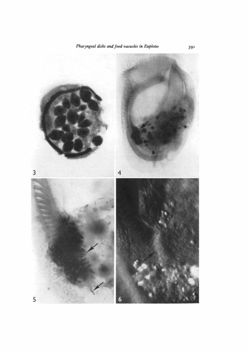
Fig. 3. Acetocarmine preparation of a Euplotes allowed to feed on Tetrahymena for5 min (feeding experiment 1). This cell contains 17 ingested Tetrahymena. x 370.
Fig. 4. Whole-mount of a starved Euplotes prepared by the osmium-pyrogallol pro-cedure. The collection of osmiophilic pharyngeal disks at die base of the gullet isclearly illustrated (arrow). Nomarski interference contrast, x 570.
Fig. 5. Enlarged view of the pharyngeal disks from the same cell as in Fig. 3. Someindividual disks can be seen (arrows). Bright field, x 2100.
Fig. 6. Pharyngeal disks (arrow) as seen in a living Euplotes widi Nomarski interferencecontrast optics. Since this represents an optical section, die actual volume occupiedby the disks is much more extensive, x 2100.
Pharyngeal disks and food vacuoles in Euplotes 391
392 J. A. Kloetzel
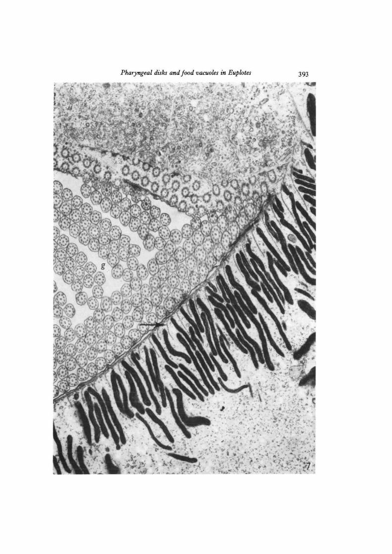
Fig. 7. Electron micrograph showing a number of electron-dense pharyngeal disksarranged in the cytoplasm just beneath the surface membrane near the base of thegullet (g). A number of membranellar cilia are seen in cross-section within the gulletcavity. Note the rows of microtubules (arrow) separating adjacent disks, x 22000.
Pharyngeal disks and food vacuoles in Euplotes 393
-.. v^iv?*v.-
i
394 J-A-- Kloetzel
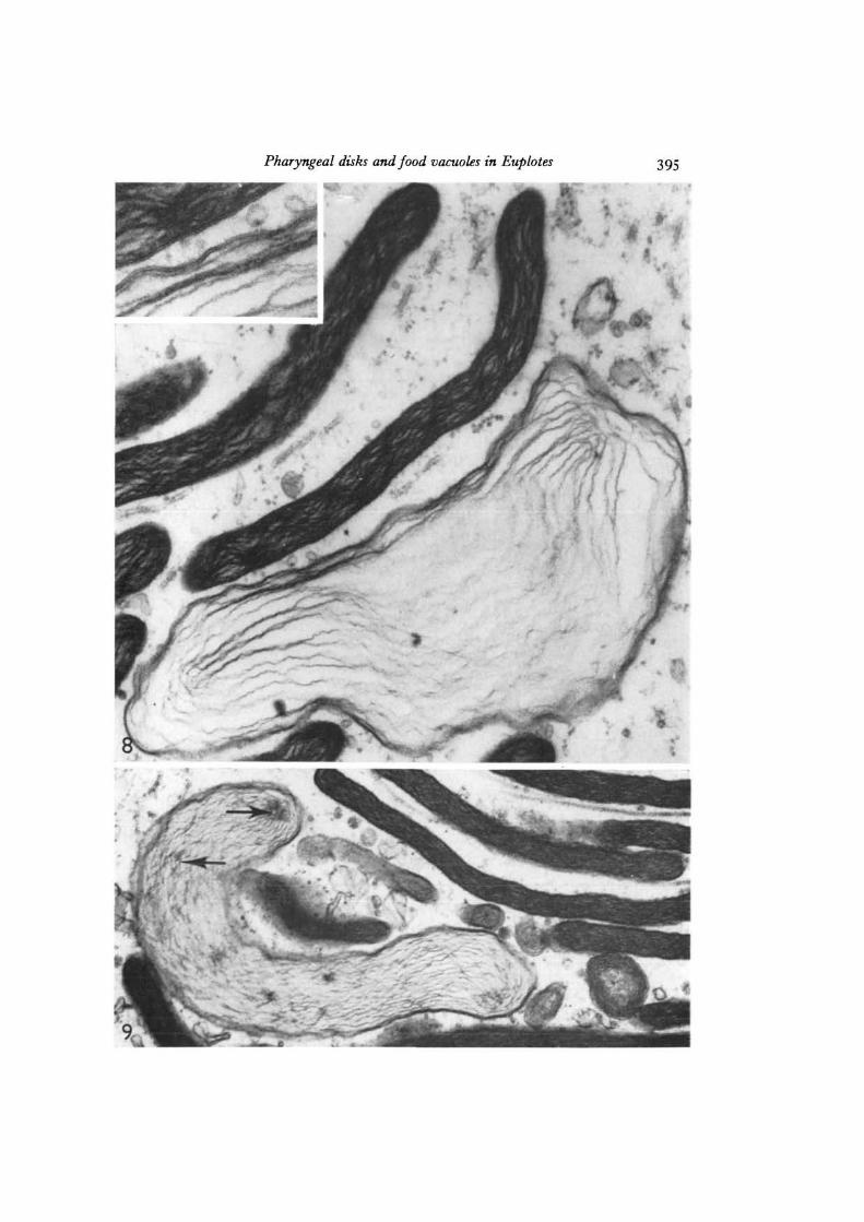
Figs. 8, 9. Pharyngeal disks at higher magnification showing their lamellar contents,which are particularly clear in those disks fixed in a swollen condition. The inset inFig. 8 shows the clear trilaminar unit membrane limiting the disks, and the thinnerprofiles of the internal lamellar layers. In Fig. 9, some of the lamellar layers areaggregated at circular foci (arrows). Fig. 8, x 77500; inset, x 148000; Fig. 9,x 44500.
Pharyngeal disks and food vacuoles in Euplotes 395
396 J.A.Kloetzel
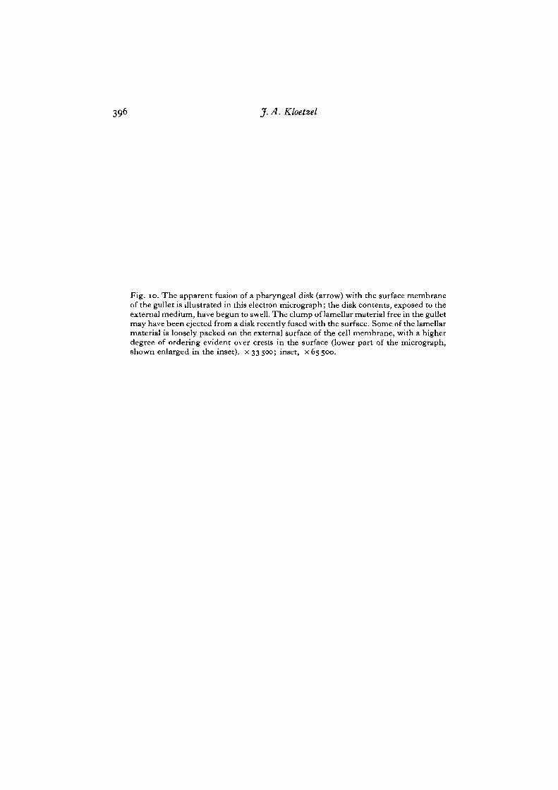
Fig. 10. The apparent fusion of a pharyngeal disk (arrow) with the surface membraneof the gullet is illustrated in this electron micrograph; the disk contents, exposed to theexternal medium, have begun to swell. The clump of lamellar material free in the gulletmay have been ejected from a disk recently fused with the surface. Some of the lamellarmaterial is loosely packed on the external surface of the cell membrane, with a higherdegree of ordering evident over crests in the surface (lower part of the micrograph,shown enlarged in the inset), x 33500; inset, x65500.
Pharyngeal disks and food vacuoles in Euplotes
398 J. A. Kloetzel
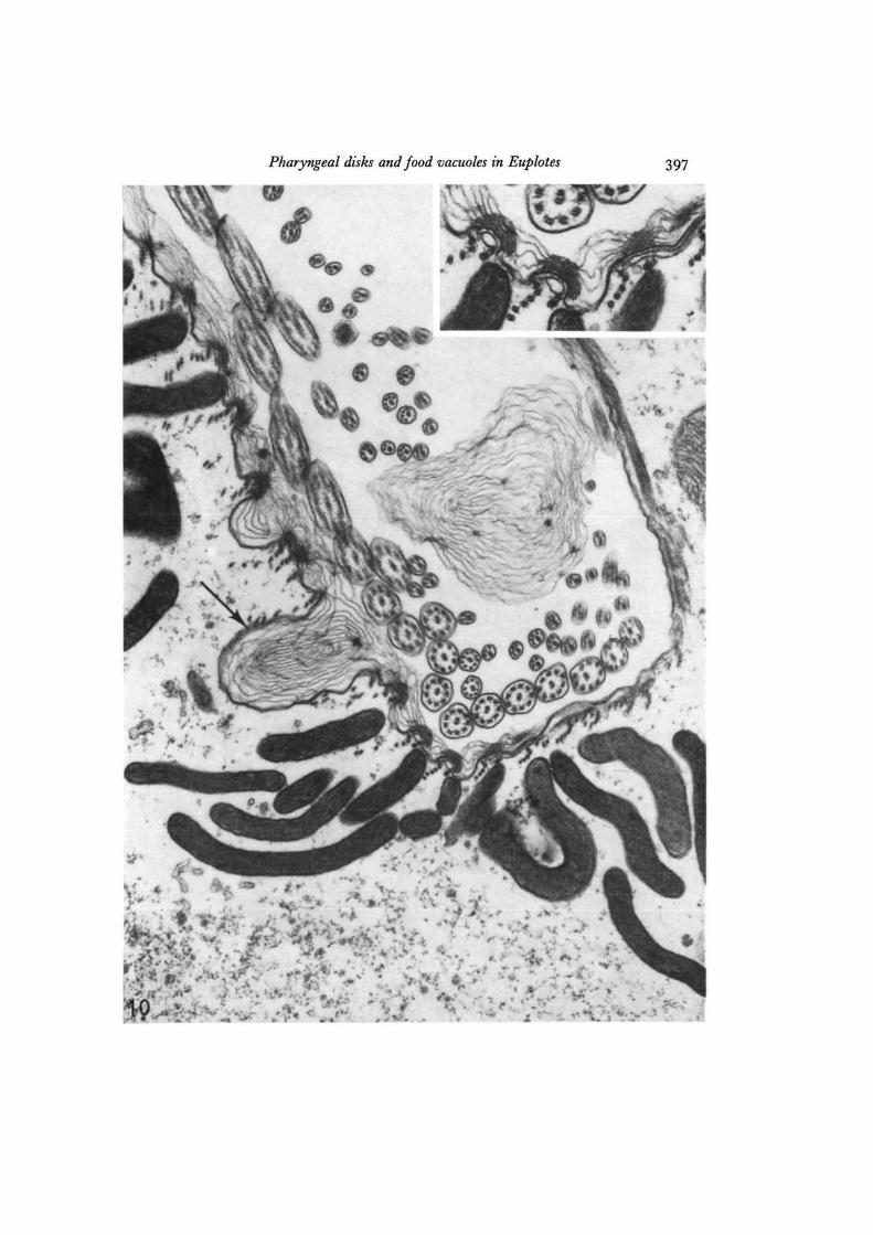
Fig. I I . A cross-section near the terminus of the gullet (g), showing a whorl ofpharyngeal disks and thick layers of lamellar material on the outer surface of theplasma membrane. Numerous circular foci are seen among the loosely packed extra-cellular layers, x 22coo.

Pharyngeal disks and food vacuoles in Euplotes 399

400 J. A. Kloetzel
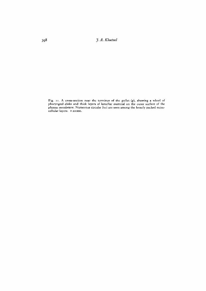
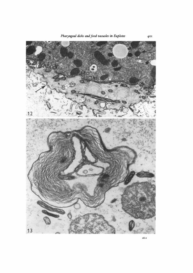
Fig. 12. A portion of a food vacuole in a Euplotes fed with Tetrahymena 8 min beforefixation. Between the food vacuole membrane (arrow) and the enclosed Tetrahymena (t),layers of lamellar material can be seen in which are embedded Tetrahymena cilia.Glutaraldehyde-osmium tetroxide combination fixation, x 13000.Fig. 13. This structure probably represents a terminal food vacuole from which almostall digestible material has gone. Note the similarity between the lamellar contents ofthe food vacuole and those of the nearby pharyngeal disks, x 47000.
Pharyngeal disks and food vacuoles in Euplotes 401
26-2