Embed Size (px)
Citation preview

1
Oxidative stress, imposed by pathophysiological condi-tions such as hypertension, hyperlipidemia, and aging,
triggers inflammatory responses of vascular endothelial cells (ECs). Although not viewed as typical immunogenic cells, ECs are suggested to be sentinel cells that detect danger signals, initiate innate immune responses, produce proinflammatory cytokines and chemokines, and recruit immune cells.1,2 Such an increase in the endothelial innate immunity has emerged
as an important mechanism underlying the interplay among oxidative stress, inflammation, and endothelial dysfunction.
Clinical Perspective on p XXXSterol regulatory element (SRE)–binding proteins
(SREBPs) are master regulators in cholesterol and lipid homeostasis.3 Decreases in the intracellular level of fatty acid or cholesterol activate SREBP1 or SREBP2 through a 2-step proteolytic cleavage, and the resulting mature N-terminal
Background—Oxidative stress activates endothelial innate immunity and disrupts endothelial functions, including endothelial nitric oxide synthase–derived nitric oxide bioavailability. Here, we postulated that oxidative stress induces sterol regulatory element–binding protein 2 (SREBP2) and microRNA-92a (miR-92a), which in turn activate endothelial innate immune response, leading to dysfunctional endothelium.
Methods and Results—Using cultured endothelial cells challenged by diverse oxidative stresses, hypercholesterolemic zebrafish, and angiotensin II–infused or aged mice, we demonstrated that SREBP2 transactivation of microRNA-92a (miR-92a) is oxidative stress inducible. The SREBP2-induced miR-92a targets key molecules in endothelial homeostasis, including sirtuin 1, Krüppel-like factor 2, and Krüppel-like factor 4, leading to NOD-like receptor family pyrin domain-containing 3 inflammasome activation and endothelial nitric oxide synthase inhibition. In endothelial cell–specific SREBP2 transgenic mice, locked nucleic acid–modified antisense miR-92a attenuates inflammasome, improves vasodilation, and ameliorates angiotensin II–induced and aging-related atherogenesis. In patients with coronary artery disease, the level of circulating miR-92a is inversely correlated with endothelial cell–dependent, flow-mediated vasodilation and is positively correlated with serum level of interleukin-1β.
Conclusions—Our findings suggest that SREBP2–miR-92a–inflammasome exacerbates endothelial dysfunction during oxidative stress. Identification of this mechanism may help in the diagnosis or treatment of disorders associated with oxidative stress, innate immune activation, and endothelial dysfunction. (Circulation. 2015;131:00-00. DOI: 10.1161/CIRCULATIONAHA.114.013675.)
Key Words: endothelium ◼ inflammasomes ◼ MIRN92 microRNA, human ◼ oxidative stress ◼ sterol regulatory element binding protein 2 AQ4
© 2015 American Heart Association, Inc.
Circulation is available at http://circ.ahajournals.org DOI: 10.1161/CIRCULATIONAHA.114.013675
Circulation
10.1161/CIRCULATIONAHA.114.013675
2015
24
February
131
XXX
00
00
30July201419December2014
© 2015 American Heart Association, Inc.
2015
Received July 30, 2014; accepted December 19, 2014.From Department of Medicine, School of Medicine (Z.C., L.W., M.M., L.F., T.-Y.L., M.J.C., Y.I.M., J.Y.-J.S.) and Department of Bioengineering (S.C.),
University of California, San Diego; Biochemistry and Molecular Biology Graduate Program (M.M.) and Division of Biomedical Sciences, School of Medicine (D.A.J., J.Y.-J.S.), University of California, Riverside; Division of Cardiology, Department of Medicine, Taipei Veterans General Hospital, Taipei, Taiwan (C.-Y.H., J.-W.C., S.-J.L., P.-H.H.); Cardiovascular Research Center, National Yang-Ming University, Taipei, Taiwan (C.-Y.H., J.-W.C., S.-J.L., P.-H.H.); Institute of Bioinformatics and Systems Biology and Department of Biological Science and Technology, National Chiao Tung University, Hsin-Chu, Taiwan (F.-M.L., H.-D.H.); Department of Kinesiology and Health Sciences, California State University, San Bernardino (G.G.); and Cardiovascular Research Center, Medical School, Xi’an Jiaotong University, Xi’an, China (Y.Z.).
*Drs Chen and Wen contributed equally.The online-only Data Supplement is available with this article at http://circ.ahajournals.org/lookup/suppl/doi:10.1161/CIRCULATIONAHA.
114.013675/-/DC1.Circulation is available at http://circ.ahajournals.orgCorrespondence John Y-J. Shyy, PhD, or Zhen Chen, PhD, Department of Medicine, School of Medicine, University of California, San Diego, 9500
Gilman Dr, La Jolla, CA 92093. E-mail [email protected] or [email protected]
AQ3
Oxidative Stress Activates Endothelial Innate Immunity via Sterol Regulatory Element Binding Protein 2 (SREBP2)
Transactivation of MicroRNA-92aZhen Chen, PhD*; Liang Wen, MD, PhD*; Marcy Martin; Chien-Yi Hsu, MD; Longhou Fang, PhD; Feng-Mao Lin, PhD; Ting-Yang Lin; McKenna J. Geary;
Gregory Geary, PhD; Yongli Zhao, MD; David A. Johnson, PhD; Jaw-Wen Chen, MD; Shing-Jong Lin, MD, PhD; Shu Chien, MD, PhD; Hsien-Da Huang, PhD;
Yury I. Miller, MD, PhD; Po-Hsun Huang, MD, PhD; John Y-J. Shyy, PhD
AQ1
AQ2
LWW
95,138,143
Febr
uary
24,
201
5
Original Article
<zjs;Original Article> • <zjss;95,138,143> • <zdoi;10.1161/CIRCULATIONAHA.114.013675>
LWW 01/9/15 4 Color Fig(s): F2,5-6 13:24 Art:CIR013675

2 Circulation February 24, 2015
SREBPs transactivate genes involved in cholesterol and lipid synthesis. Recently, SREBPs have been implicated in innate immune responses in vascular cells because of their regulation of inflammasomes, the intracellular multiprotein complexes mediating the activation of caspase-1, and sub-sequent maturation of interleukin (IL)-1β/IL-18.4 Im et al5 showed that SREBP1 activates the NOD-like receptor fam-ily pyrin domain-containing (NLRP) 1 inflammasome in lipopolysaccharide-stimulated macrophages, and we reported that SREBP2 activates NLRP3 inflammasome in ECs under disturbed flow.6 Importantly, ectopic expression of SREBP2 in murine endothelium is sufficient to aggravate atheroscle-rosis, partially through the increased caspase-1–dependent IL-1β production, which suggests a primary role of EC innate immunity in atherogenesis.6 In human aortic sections, acti-vated SREBPs are observed in macrophages and ECs in the atherosclerotic lesions.7 Although these studies suggest that SREBPs are critical regulators in vascular innate immunity, the upstream stress stimuli that induce or activate SREBPs and the SREBP downstream targets that disturb EC homeo-stasis are largely unexplored.
Stimuli such as disturbed flow and oxidized lipids that impose oxidative stress in ECs increase SREBP levels.6,8,9 They also induce microRNA-92a (miR-92a), a crucial miRNA that inhibits EC angiogenesis and impairs EC function.10–12 At the molecular level, miR-92a targets Krüppel-like factor (KLF) 2, KLF4, and possibly sirtuin 1 (SIRT1), all of which are tightly associated with redox balance, endothelial nitric oxide (NO) synthase (eNOS)–derived NO bioavailability, and the inflammatory state.10,12–14 In regard to endothelial innate immune response, KLF2, KLF4, and SIRT1 suppress the production or antagonize the effect of IL-1β.15–17 In terms of translational implications, administration of locked nucleic acid (LNA)–modified antisense miR-92a (LNA-92a) pre-vents ischemic injury in pigs and ameliorates hyperlipidemia-induced atherosclerosis in mice.11,18 Despite the involvement of miR-92a in EC dysfunction, the molecular mechanism underlying stress induction of miR-92a in ECs and its link to endothelial innate immunity are unclear.
Given the common stimuli (eg, disturbed flow and oxidized lipids) and convergent functional consequences (ie, endothelial inflammation and dysfunction) of SREBP2 and miR-92a, we postulated that oxidative stress induces SREBP2 and miR-92a, which in turn activate innate immune response, leading to dys-functional endothelium. Here, we demonstrate that the SREBP2 transactivation of miR-92a is a ubiquitous response to oxidative stress. Downstream, this redox-sensitive pathway decreases the expression of anti-inflammatory genes, including SIRT1, KLF2, and KLF4, thus resulting in increased inflammasome and impaired eNOS-NO bioavailability. Furthermore, studies of zebrafish, mouse, and human samples suggest an inverse correlation between miR-92a level and functional endothelium.
MethodsCross-Linking Immunoprecipitation and Chromatin ImmunoprecipitationFor crosslinking immunoprecipitation, human umbilical vein endo-thelial cells (HUVECs) were irradiated with ultraviolet light at 400 mJ/cm2 to cross-link RNA and proteins. Cells were then lysed in a
buffer containing 50 mmol/L Tris-HCl, pH 7.5, 150 mmol/L NaCl, 0.1% NP-40, 1 mmol/L EDTA, and 100 U/μL RNase inhibitor. The lysates were incubated with protein G Dynabeads conjugated with anti–argonaute 2 (clone 4G8; Wako Chemicals) at 4°C overnight. Mouse IgG was used as an isotype control. The immunoprecipitated RNAs were then extracted with Trizol. For chromatin immunopre-cipitation, HUVECs were treated with 0.75% formaldehyde for 20 minutes at room temperature. After sonication, the SREBP2-bound chromatin was immunoprecipitated by rabbit anti-SREBP2(N) (Abcam) conjugated to protein A Dynabeads. Protein and RNA were degraded by proteinase K and RNase A, respectively. The purified chromatin DNA was then used as the template for quantitative poly-merase chain reaction. As an isotype control, rabbit IgG was used in chromatin immunoprecipitation.
Flow Cytometry and NO Bioavailability AssayActive caspase-1 was detected in living ECs with the use of the Fluorescent Labeled Inhibitor of Caspases (FLICA) caspase-1 assay kit (ImmunoChemistry Technologies). FLICA-FAM-YVAD-FMK is a cell-permeable, nontoxic fluorochrome inhibitor of caspase-1 that interacts with the enzyme-reactive center of activated caspase-1 via the YVAD recognition sequence, thus forming a covalent thioether adduct with the enzyme through the FMK moiety. The resulting green fluorescence is a direct measure of caspase-1 activity, which was analyzed by fluorescence-activated cell sorter analysis with 488-nm excitation and 530-nm emission. NO was detected as the accu-mulated nitrite/nitrate, the stable breakdown product of NO, in cell culture media by use of a nitrate/nitrite fluorometric assay (Cayman Chemicals).
In Vivo Angiotensin II Infusion and Administration of LNAAll animal experiments were approved by University of California, San Diego Institutional Animal Care and Use Committee. EC-SREBP2(N)-Tg and ApoE−/−/EC-SREBP2(N)-Tg mice were cre-ated as described.6 LNAs were designed and synthesized by Exiqon Inc with the following sequences: 5′-AGGCCGGGACAAGTGCAAT-3′ (LNA-92a) and 5′-TAACACGTCTATACGCCCA-3′ (LNA-control). Male ApoE−/−/EC-SREBP2(N)-Tg and their age- and sex-matched ApoE−/− littermates were used for angiotensin II (Ang II) infusion and LNA injection. As illustrated in Figure I in the online-only Data Supplement, 1 week before Ang II infusion, mice received tail-vein injections of LNA-control or LNA-92a at 16 mg/kg body weight, a dose that effectively inhibits miR-92a expression.11 Osmotic mini-pumps (model 1004, Alzet) filled with Ang II solution were implanted subcutaneously into the dorsal side of mice. Ang II was released at 1 μg·kg−1·min−1 for 28 days. The second dose of LNA was given 10 days after the minipump implantation. The animals were euthanized at the end of the 28th day after minipump implantation.
IK17-EGFP Tg ZebrafishTg(hsp70l:Hsa.IK17-EGFP) zebrafish with temperature-inducible expression of the enhanced green fluorescent protein (EGFP)–labeled single-chain human monoclonal antibody IK17 (ie, IK17-EGFP) was described.19 IK17-EGFP is driven by heat shock protein (hsp70) and hence can be induced by heat shock at 37°C. Zebrafish larvae at 5 days after fertilization were fed a normal diet or high-cholesterol diet (HCD) containing 4% cholesterol for 4 weeks. One group of high-cholesterol diet–fed fish were subjected to heat shock (1 hour at 37°C) once every 3 days to maintain IK17-EGFP expression during the feeding period. The expression of IK17-EGFP was confirmed by microscopy (excitation at 488 nm).
Clinical Samples, Measurement of Circulating miR-92a, IL-1β, and Flow-Mediated DilationAll clinical samples were obtained at Taipei Veterans General Hospital with informed consent and Institutional Review Board approval (protocol
AQ5
LWW 01/9/15 4 Color Fig(s): F2,5-6 13:24 Art:CIR013675

Chen et al Oxidative Stress Induces MiR-92a and Inflammasome 3
No. 2014-02-002A). The baseline characteristics of patients are summa-rized in Table I in the online-only Data Supplement. Sera were collected after an 8-hour overnight fast. Circulating miR-92a level was measured as described.20 The IL-1β level was assessed by ELISA (Human IL-1β Quantikine ELISA Kit, R&D Systems). Each standard and plasma sam-ple were analyzed twice, and the mean values were used in all subsequent analyses. Endothelium-dependent flow-mediated dilation was assessed by use of a 7.5-MHz linear-array transducer (Hewlett-Packard Sonos 5500) to scan the brachial artery in longitudinal sections, as described previously21 (see Methods in the online-only Data Supplement).
Additional experimental procedures are described in the Methods section in the online-only Data Supplement.
Statistical AnalysisData from in vitro experiments are expressed as mean±SD from at least 3 independent experiments unless otherwise noted. Data from in vivo studies are expressed as mean±SEM. Two groups were com-pared by the Student t test. Differences among multiple groups were evaluated by ANOVA followed by the Bonferroni post hoc test for equal sample sizes or Tukey-Kramer test for unequal sample sizes. Correlational analyses were performed with the Spearman correlation after determining the (non)normal distribution of the data. Values of P<0.05 were considered statistically significant.
ResultsSREBP2 and MiR-92a Are Induced By Oxidative Stress in ECsWe first sought to test whether SREBP2 and miR-92a are induced by H
2O
2, Ang II, and oxidized low-density lipoprotein
(ox-LDL), all of which directly or indirectly increase reactive oxygen species in ECs to result in inflammatory responses. All 3 stimuli induced and activated SREBP2 in HUVECs, as evi-denced by the increased levels of the SREBP2 precursor and the mature form of SREBP2 [ie, SREBP2(N); Figure 1A]. The acti-vation of SREBP2 was associated with the induction of SREBP2 transactivation targets such as LDL receptor and squalene syn-thase (Figure 1B). Notably, H
2O
2, Ang II, and ox-LDL also dose-
dependently increased the level of miR-92a (Figure 1C–1E). Pretreatment with the reactive oxygen species scavenger EUK-134, a catalase/superoxide dismutase mimetic,22 attenuated the H
2O
2-, Ang II–, and ox-LDL–induced SREBP2 and miR-92a
levels (Figure 1F–1H). These results indicate that SREBP2 and miR-92a are induced by oxidative stress in ECs.
SREBP2 Transactivates MiR-92a Under Oxidative StressTo examine whether SREBP2 transactivates miR-92a in response to oxidative stress, we performed bioinformat-ics analysis for putative SREs in the promoter region of the miR-17–92 cluster. We found 8 SREs in the promoter region of the human miR-17–92 cluster, which are conserved in the mouse gene (Figure 2A and Table II in the online-only Data Supplement). To validate this in silico prediction, we overexpressed the active/mature form of SREBP2, that is, SREBP2(N), in ECs, which increased the level of miR-92a (Figure 2B). Conversely, knockdown of SREBP2 with siRNA inhibited H
2O
2-induced miR-92a (Figure 2C). To demonstrate
enhanced transactivation of miR-92a by SREBP2 under oxi-dative stress, we performed chromatin immunoprecipitation assays to assess the direct binding of SREBP2 to SREs in the miR-17–92 promoter. H
2O
2 treatment of ECs or ectopic
expression of SREBP2(N) in ECs, mimicking SREBP2 induc-tion by oxidative stress, substantially enriched SREBP2(N) binding to segments of miR-92a promoter containing the predicted SREs (Figure 2D and 2E). Thus, oxidative stress–activated or –induced SREBP2 transactivates miR-92a.
Oxidative Stress–Induced MiR-92a Increases Endothelial Innate ImmunityRecent findings suggest that SREBP induction or activa-tion increases innate immunity, specifically via NLRP3
AQ6
AQ7
F1
F2
Figure 1. H2O2, angiotensin II (Ang II), and oxidized low-density lipoprotein (ox-LDL) induce sterol regulatory element–binding protein 2 (SREBP2) and microRNA-92a (miR-92a) in endothelial cells (ECs). A and B, Human umbilical vein ECs (HUVECs) were treated with H2O2 (100 μmol/L), Ang II (100 nmol/L), or ox-LDL (100 μg/mL) for 16 hours. A, Cellular proteins were collected for immunoblotting of the SREBP2 precursor and the mature form of SREBP2 [SREBP2(N)]. Bar graphs are densitometry quantifications of the ratios of the SREBP2 precursor or SREBP2(N) to β-actin level. B, RNA was collected for reverse transcription–quantitative polymerase chain reaction (qPCR) analysis of mRNA encoding the low-density lipoprotein receptor (LDLR) and squalene synthase. C through E, Taqman miRNA qPCR analysis of the miR-92a level in HUVECs treated with various concentrations of H2O2, Ang II, and ox-LDL for 16 hours. F through H, HUVECs were pretreated with EUK-134 (1 μmol/L) for 2 hours and then incubated with H2O2, Ang II, or ox-LDL for 16 hours. SREBP2 was detected by immunoblotting and miR-92a level by Taqman miRNA qPCR. Data are mean±SD from at least 3 independent experiments. *P<0.05 vs respective control or between the indicated groups.
AQ8
LWW 01/9/15 4 Color Fig(s): F2,5-6 13:24 Art:CIR013675

4 Circulation February 24, 2015
inflammasome activation in ECs and macrophages.5,6 Because oxidative stress activates SREBP2–miR-92a (Figures 1 and 2), we examined whether oxidative stress activates inflammasome and, if so, the role of miR-92a in this innate immune response in ECs. H
2O
2, Ang II, and ox-LDL all increased the cleaved
form of caspase-1 and IL-1β, hallmarks of inflammasome acti-vation (Figure 3A and 3B). Importantly, pre–miR-92a, mimick-ing miR-92a induction by SREBP2, also increased the cleavage
of caspase-1 and IL-1β (Figure 3C and 3D). Conversely, trans-fecting ECs with anti–miR-92a blocked these inflammasome-related events in H
2O
2-stimulated ECs (Figure 3E and 3F).
These results were further confirmed by the use of flow cytom-etry to detect the active caspase-1 in living cells (Figure 3G and 3H). Therefore, the SREBP2–miR-92a axis contributes to oxidative stress induction of the endothelial innate immune response, as evidenced by inflammasome activation.
F3
Figure 3. Oxidative stress–induced microRNA-92a (miR-92a) increases endothelial innate immunity. Immunoblotting of caspase-1 and interleukin (IL)-1β in human umbilical vein endothelial cells (HUVECs; A) treated with H2O2 (100 μmol/L), angiotensin II (Ang II; 100 nmol/L; B), or oxidized low-density lipoprotein (ox-LDL; 100 μg/mL) for 16 hours, (C) transfected with control RNA or pre–miR-92a (20 nmol/L) for 72 hours, or (E) transfected with control RNA or anti–miR-92a for 48 hours before H2O2 treatment for 24 hours. Quantification in B, D, and F is the relative expression of caspase-1 and IL-1β to that of β-actin. G and H, Flow cytometry quantification of active caspase-1 in HUVECs transfected as in E. Histograms in G are representative results, and data in H are mean±SD from 5 independent experiments, with the percentage of caspase-1+ cells in control RNA group set to 1. FLICA indicates Fluorescent Labeled Inhibitor of Caspases.
AQ10
Figure 2. Sterol regulatory element (SRE)–binding protein 2 (SREBP2) transactivates microRNA-92a (miR-92a) under oxidative stress. A, Bioinformatics analysis of SREs in the promoter region of human miR-17–92 cluster. B, Reverse transcription–quantitative polymerase chain reaction (qPCR) and Taqman miRNA qPCR analyses of SREBP2 and miR-92a levels in human umbilical vein endothelial cells (HUVECs) infected with Ad-null or Ad-SREBP2(N). C, miR-92a level in HUVECs transfected with SREBP2 siRNA (10 nmol/L) or control RNA and then treated with H2O2. D and E, Chromatin immunoprecipitation assays were performed with SREBP2 antibody or a nonspecific IgG in extracts from HUVECs treated with H2O2 (D) or infected with Ad-SREBP2(N) (E). The enrichment of SREBP2(N) binding to the putative SREs in the promoter region of miR-17–92 was quantified by qPCR, with the untreated group (D) or Ad-null group (E) set to 1. MOI indicates multiplicity of infection. *P<0.05 vs respective control or between the indicated groups.
AQ9
LWW 01/9/15 4 Color Fig(s): F2,5-6 13:24 Art:CIR013675
Chen et al Oxidative Stress Induces MiR-92a and Inflammasome 5
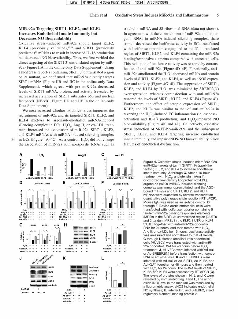
MiR-92a Targeting SIRT1, KLF2, and KLF4 Increases Endothelial Innate Immunity but Decreases NO BioavailabilityOxidative stress–induced miR-92a should target KLF2, KLF4 (previously validated),12,14 and SIRT1 (previously predicted)10 mRNAs to result in increased IL-1β production but decreased NO bioavailability. Thus, we first verified the direct targeting of the SIRT1 3′ untranslated region by miR-92a (Figure IIA in the online-only Data Supplement). Using a luciferase reporter containing SIRT1 3′ untranslated region or its mutant, we confirmed that miR-92a directly targets SIRT1 mRNA (Figure IIB and IIC in the online-only Data Supplement), which agrees with pre–miR-92a–decreased levels of SIRT1 mRNA, protein, and activity (revealed by increased acetylation of SIRT1 substrates p53 and nuclear factor-κB [NF-κB]; Figure IID and IIE in the online-only Data Supplement).
We next assessed whether oxidative stress increases the recruitment of miR-92a and its targeted SIRT1, KLF2, and KLF4 mRNAs to argonaute-mediated miRNA-induced silencing complex in ECs. H
2O
2, Ang II, or ox-LDL treat-
ment increased the association of miR-92a, SIRT1, KLF2, and KLF4 mRNAs with miRNA-induced silencing complex in ECs (Figure 4A–4C). As a control, H
2O
2 did not change
the association of miR-92a with nonspecific RNAs such as
α-tubulin mRNA and 5S ribosomal RNA (data not shown). In agreement with the coenrichment of miR-92a and its tar-get mRNAs in miRNA-induced silencing complex, these stimuli decreased the luciferase activity in ECs transfected with luciferase reporters conjugated to the 3′ untranslated region of SIRT1, KLF2, and KLF4 containing the miR-92a binding/responsive elements compared with untreated cells. This reduction of luciferase activity was restored by cotrans-fection of anti–miR-92a (Figure 4D–4F). Functionally, anti–miR-92a ameliorated the H
2O
2-decreased mRNA and protein
levels of SIRT1, KLF2, and KLF4, as well as eNOS expres-sion and activity (Figure 4G–4I). The suppression of SIRT1, KLF2, and KLF4 by H
2O
2 was mimicked by SREBP2(N)
overexpression, whereas cotransfection with anti–miR-92a restored the levels of SIRT1, KLF2, and KLF4 (Figure 4J). Furthermore, the effect of ectopic expression of SIRT1, KLF2, and KLF4 was similar to that of anti–miR-92a in reversing the H
2O
2-induced EC inflammation (ie, caspase-1
activation and IL-1β production) and H2O
2-impaired NO
bioavailability (Figure 4K and 4L). Collectively, oxidative stress induction of SREBP2–miR-92a and the subsequent SIRT1, KLF2, and KLF4 targeting increase endothelial innate immunity and impair eNOS-NO bioavailability, 2 key features of endothelial dysfunction.
F4
Figure 4. Oxidative stress–induced microRNA-92a (miR-92a) targets sirtuin 1 (SIRT1), Krüppel-like factor (KLF) 2, and KLF4 to increase endothelial innate immunity. A through C, After a 16-hour treatment with H2O2, angiotensin II (Ang II), or oxidized low-density lipoprotein (ox-LDL), argonaute (AGO)–miRNA-induced silencing complex was immunoprecipitated, and the AGO-bound miR-92a and SIRT1, KLF2, and KLF4 mRNAs were quantified by reverse transcription–quantitative polymerase chain reaction (RT-qPCR). Mouse IgG was used as an isotype control. D through F, Bovine aortic endothelial cells were transfected with luciferase reporter containing 5 tandem miR-92a binding/responsive elements (MREs) in the SIRT1 3′ untranslated region (3′UTR) and 2 tandem MREs in the KLF2 3′UTR or KLF4 3′UTR, together with anti–miR-92a or control RNA for 24 hours, and then treated with H2O2, Ang II, or ox-LDL for 16 hours. Luciferase activity was measured and normalized to that of Renilla. G through I, Human umbilical vein endothelial cells (HUVECs) were transfected with anti–miR-92a or control RNA for 48 hours before H2O2 treatment. J, HUVECs were infected with Ad-null or Ad-SREBP2(N) before transfection with control RNA or anti–miR-92a. K and L, HUVECs were infected with Ad-null or Ad-SIRT1, Ad-KLF2, and Ad-KLF4 together for 48 hours and then treated with H2O2 for 24 hours. The mRNA levels of SIRT1, KLF2, and KLF4 were assessed by RT-qPCR (G). The levels of proteins shown in H, J, and K were revealed by immunoblotting. I and L, The nitric oxide (NO) level in the medium was measured by a fluorometric assay. eNOS indicates endothelial NO synthase; IL, interleukin; and SREBP2, sterol regulatory element–binding protein 2.
LWW 01/9/15 4 Color Fig(s): F2,5-6 13:24 Art:CIR013675

6 Circulation February 24, 2015
SREBP2 Induction of miR-92a Leads to Impaired EC Functions in the Vessel WallWe next examined whether miR-92a is oxidative stress inducible in the vessel wall in vivo in mouse and zebraf-ish models. The aortic miR-92a level was higher in the Ang II–infused C57BL6 mice than in normotensive controls (Figure 5A). Consistent with increased oxidative stress in the aging vessel,23,24 the miR-92a level in the aortas was also higher in 12-month-old than in 3-month-old C57BL6 mice (Figure 5B). We also used a hypercholesterolemic zebrafish model with accelerated vascular accumulation of oxidized lipids with enhanced oxidation-specific epitopes.19,25 Four weeks of a high-cholesterol diet significantly increased the miR-92a level in the trunks, where major blood vessels are located. This induction was substantially inhibited in zebraf-ish expressing IK17, a human monoclonal antibody against malondialdehyde-LDL and ox-LDL19,26 (Figure 5C and 5D). Thus, miR-92a was also induced in the zebrafish in an oxida-tive stress–dependent manner. To examine whether SREBP2 increases miR-92a in the endothelium in vivo, we assessed the miR-92a level in intima isolated from EC-SREBP2(N)-Tg mice with the active form of SREBP2 overexpressed only in the endothelium.6 Ectopic expression of SREBP2(N) increased the miR-92a levels and decreased the mRNA levels of SIRT1, KLF2, KLF4, and eNOS in the intima (Figure 5E). Furthermore, an endothelial innate immune response was induced, as evidenced by the increased IL-1β mRNA level in EC-SREBP2(N)-Tg mice (Figure 5E). Consistent with
the reduced eNOS expression, flow-mediated vasodilation, a hallmark of endothelial dysfunction, was attenuated (Figure III in the online-only Data Supplement). To address whether miR-92a mediates the detrimental effects of SREBP2 in the vessel wall, we inhibited miR-92a with LNA in the carotid arteries of EC-SREBP2(N)-Tg mice. Although miR-92a was significantly inhibited by LNA, the mRNA levels of SIRT1, KLF2, KLF4, and eNOS were substantially increased and that of IL-1β was decreased in mice receiving LNA-92a com-pared with control LNA (Figure 5F). Functionally, LNA-92a administration improved the flow-mediated vasoreactivity (Figure 5G).
To evaluate the detrimental effect of the SREBP2–miR-92a pathway at the disease level, we used a model of EC-SREBP2(N)-Tg in an ApoE−/− background treated with Ang II (1 μg·min−1·kg−1). With a regular diet, Ang II infusion accelerates atherosclerosis.27 Thus, the atherosclerosis in this model is due mainly to the oxidative stress–induced endothe-lial dysfunction rather than diet-induced hyperlipidemia. The experimental design is outlined in Figure I in the online-only Data Supplement, and the efficacy of LNA-92a was con-firmed by the decreased miR-92a but increased mRNA levels of miR-92a targets, that is, SIRT1, KLF2, and KLF4, as well as eNOS (Figure IVA in the online-only Data Supplement). Noticeably, atherosclerosis, including overall lesion size and that in the aortic arch, was reduced in mice treated with LNA-92a compared with control LNA. Furthermore, the inhibitory effect of LNA-92a on atherosclerosis was apparent in both
F5
Figure 5. Oxidative stress induction of microRNA-92a (miR-92a) and inflammasome in vivo. A and B, Taqman miRNA quantitative polymerase chain reaction (qPCR) of the miR-92a level in whole aortas isolated from 8-week-old C57BL6 mice with or without angiotensin II (Ang II) infusion (A) and C57BL6 mice at 3 and 12 months of age (B). C and D, hsp70:IK17-EGFP Tg zebrafish were fed a normal diet or a high-cholesterol diet (HCD). Enhanced green fluorescent protein (EGFP)–labeled single-chain human monoclonal antibody IK17 (IK17-EGFP) was heat shock-induced (HS) in 1 group of HCD-fed fish. The trunk regions were collected for miR-92a qPCR (C). IK17 induction was confirmed by visualization of EGFP (D). Bar, 0.1 cm. E and F, Reverse transcription–qPCR analysis of miR-92a and mRNA levels of various genes in intima isolated from EC-SREBP2(N)-Tg and that from wild-type littermates (WT) pooled from 4 animals in each group (E) and carotid arteries of EC-SREBP2(N)-Tg with local delivery of control locked nucleic acid (LNA-Ctrl) or LNA-modified antisense miR-92a (LNA-92a; F). G, Vasodilatory function of carotid arteries from EC-SREBP2(N)-Tg mice treated as in F. n Denotes number of animals used; and SREBP2 indicates sterol regulatory element–binding protein. *P<0.05 vs respective control or between the indicated groups.
AQ11
LWW 01/9/15 4 Color Fig(s): F2,5-6 13:24 Art:CIR013675
Chen et al Oxidative Stress Induces MiR-92a and Inflammasome 7
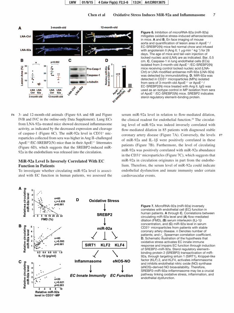
3- and 12-month-old animals (Figure 6A and 6B and Figure IVB and IVC in the online-only Data Supplement). Lung ECs from LNA-92a–treated mice showed decreased inflammasome activity, as indicated by the decreased expression and cleavage of caspase-1 (Figure 6C). The miR-92a level in CD31+ mic-roparticles collected from sera was higher in Ang II–challenged ApoE−/−/EC-SREBP2(N) mice than in their ApoE−/− littermates (Figure 6D), which suggests that the SREBP2-induced miR-92a in the endothelium was released into the circulation.
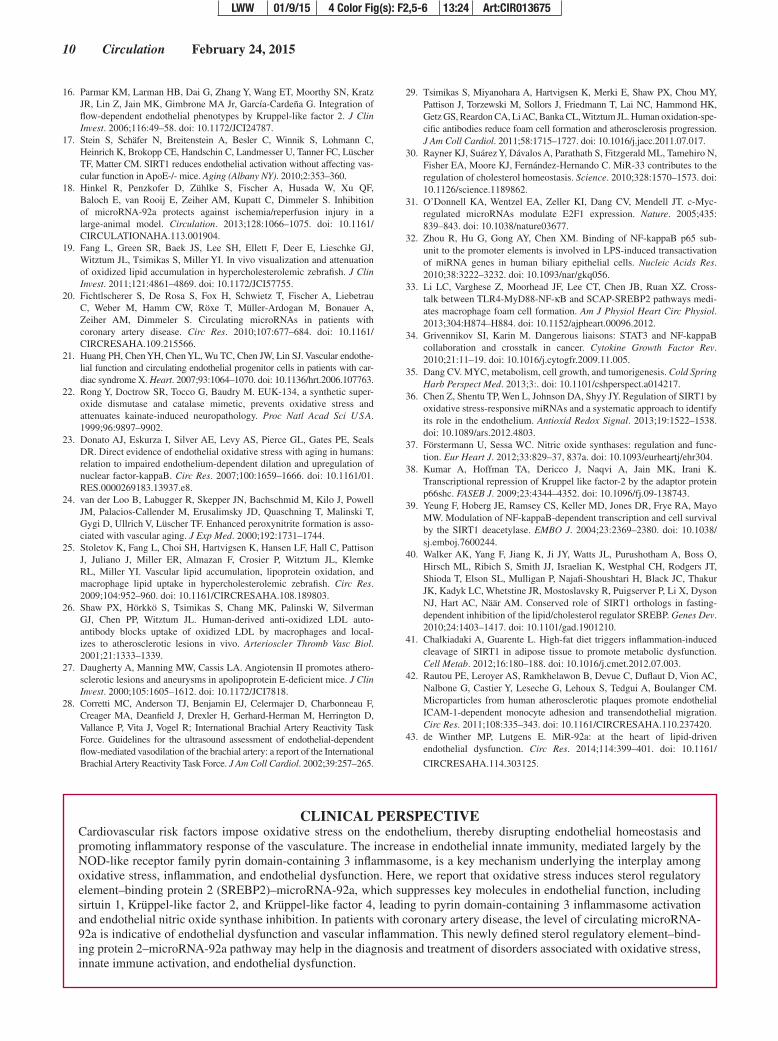
MiR-92a Level Is Inversely Correlated With EC Function in PatientsTo investigate whether circulating miR-92a level is associ-ated with EC function in human patients, we assessed the
serum miR-92a level in relation to flow-mediated dilation, the clinical readout for endothelial function.28 The circulat-ing level of miR-92a was indeed inversely correlated with flow-mediated dilation in 85 patients with diagnosed stable coronary artery disease (Figure 7A). Conversely, the levels of miR-92a and IL-1β were positively correlated in these patients (Figure 7B). Furthermore, the level of circulating miR-92a was positively correlated with miR-92a abundance in the CD31+ microparticles (Figure 7C), which suggests that miR-92a in circulation originates in part from the endothe-lium. Therefore, the serum level of miR-92a could indicate endothelial dysfunction and innate immunity under certain cardiovascular events.
F6
F7
Figure 6. Inhibition of microRNA-92a (miR-92a) mitigates oxidative stress–induced atherosclerosis in mice. A and B, En face imaging of mouse aorta and quantification of lesion area in ApoE−/−/EC-SREBP2(N) mice fed normal chow and infused with angiotensin II (Ang II; 1 μg·min−1·kg−1) for 28 days. The age of mice and tail-vein injection of locked nucleic acid (LNA) are as indicated. Bar, 0.5 cm. C, Caspase-1 in lung endothelial cells (ECs) isolated from 3-month-old ApoE−/−/EC-SREBP2(N) mice receiving control locked nucleic acid (LNA-Ctrl) or LNA-modified antisense miR-92a (LNA-92a) was detected by immunoblotting. D, MiR-92a was detected in CD31+ microparticles (MPs) isolated from sera of 3-month-old ApoE−/− or ApoE−/−/EC-SREBP2(N) mice treated with Ang II. IgG was used as an isotype control in MP isolation from sera of ApoE−/−/EC-SREBP2(N) mice. SREBP2 indicates sterol regulatory element–binding protein.
Figure 7. MicroRNA-92a (miR-92a) inversely correlates with endothelial cell (EC) function in human patients. A through C, Correlations between circulating miR-92a level and (A) flow-mediated dilation (FMD), (B) serum interleukin (IL)-1β concentration, and (C) miR-92a level in serum CD31+ microparticles from patients with stable coronary artery disease. n Denotes number of patients; and rs, Spearman correlation coefficient. D, Schematic illustration of the hypothesis that oxidative stress activates EC innate immune response and impairs EC function through induction of SREBP2–miR-92a. Sterol regulatory element–binding protein 2 (SREBP2) transactivation of miR-92a, through targeting sirtuin 1 (SIRT1), Krüppel-like factor (KLF) 2, and KLF4, activates inflammasome and inhibits endothelial nitric oxide (NO) synthase (eNOS)–derived NO bioavailability. Therefore, SREBP2–miR-92a–inflammasome may be a crucial pathway linking oxidative stress, inflammation, and endothelial dysfunction.
LWW 01/9/15 4 Color Fig(s): F2,5-6 13:24 Art:CIR013675

8 Circulation February 24, 2015
DiscussionHere, we demonstrate that oxidative stress activates the endo-thelial innate immune response by inducing SREBP2–miR-92a. This finding presents a novel function of SREBP2 in addition to its canonical role in cholesterol synthesis and uptake. The induction of this pathway is a common response of ECs to disturbed flow, H
2O
2, Ang II, ox-LDL, and oxidized
palmitoyl-arachidonylphosphatidyl choline, all of which cause oxidative stress6,12 (Figure 1 and Figure V in the online-only Data Supplement). This response is attenuated when ECs are pretreated with the reactive oxygen species scavengers super-oxide dismutase/catalase mimetic EUK-134 or cell-permeable superoxide dismutase (Figure 1 and Figure V in the online-only Data Supplement), which suggests that the SREBP2–miR-92a pathway is redox sensitive. In vivo, miR-92a is induced in the mouse aorta with increased oxidative stress from Ang II infusion or aging (Figure 5A and 5B). Similarly, miR-92a is induced in zebrafish by a high-cholesterol diet (Figure 5C). Because IK17 antagonizes ox-LDL,19,29 the IK17 inhibition of miR-92a further indicates that oxidized lipids and lipo-proteins likely induce miR-92a in zebrafish. Together, these in vitro and in vivo findings strongly suggest that oxidative stress–induced miR-92a is a conserved mechanism among vertebrates. Consistent with this conclusion is the observation that miR-17–92 promoters from human, mouse, and zebraf-ish all contain multiple putative SREs (Tables II and III in the online-only Data Supplement). Intriguingly, when ECs are supplemented with 25-hydroxycholesterol, which blocks the canonical cholesterol-sensing pathway, oxidized palmitoyl-arachidonylphosphatidyl choline and disturbed flow are still able to activate SREBP2 (Figure VC in the online-only Data Supplement).3 Thus, oxidative stress induction of miR-92a via SREBP2 differs from miR-33 induction, which responds to sterol depletion.30 As an SREBP2 intronic miRNA, miR-33a targets the cholesterol transporters ABCA1 and ABCG1 for restoration of cellular cholesterol homeostasis.30 However, miR-92a downregulates KLF2, KLF4, and SIRT1 to affect endothelial innate immunity and function. Thus, the redox-sensitive SREBP2, by transactivating miR-92a, mediates the innate immune response in ECs under oxidative stress (sum-marized in Figure 7D). Because the endothelium does not readily accumulate lipid and cholesterol, these results suggest that the major functional relevance of oxidative stress induc-tion of SREBP2 in ECs is increased innate immunity rather than cholesterol synthesis, uptake, and storage.
The miR-17–92 cluster is upregulated by c-Myc and NF-κB in cancer cells, fibroblasts, and epithelial cells.31,32 Although STAT3 regulates miR-92a in ECs,11 whether STAT3 directly transactivates miR-92a is unknown. Given that NF-κB upreg-ulates SREBP2,33 STAT3 cross-talks with NF-κB,34 and both SREBP2 and c-Myc are basic helix-loop-helix transcription factors that likely share transactivating targets that contain E-box in their promoter regions,35 SREBP2 is likely involved in a network regulating miR-92a. Indeed, the H
2O
2-induced
miR-92a is attenuated in ECs if RelA (ie, NF-κB p65 subunit) or c-Myc has been knocked down (Figure VIA in the online-only Data Supplement). Because toll-like receptors (TLRs) are key mediators in innate immunity and most, if not all, TLRs are expressed in ECs, we have also examined the TLR
pathway involved in the oxidative stress–induced miR-92a. When we inhibited myeloid differentiation factor 88, a com-mon signaling adaptor downstream of TLRs except TLR3, ox-LDL–induced miR-92a was suppressed (Figure VIB in the online-only Data Supplement). In line with this, myeloid differentiation factor 88 inhibition also attenuated the activa-tion and induction of SREBP2 in lipopolysaccharide-stimu-lated macrophages and the consequent foam cell formation.33 Therefore, SREBP2–miR-92a may act as a critical nexus link-ing oxidative stress and endothelial innate immune response and subsequent inflammation.
Most, if not all, oxidative insults suppress SIRT1, KLF2, and KLF4 and uncouple eNOS in ECs.14,36–38 Because of the positive effect of SIRT1, KLF2, and KLF4 on NO bioavail-ability, the finding that SREBP2–miR-92a suppresses the expression of SIRT1, KLF2, and KLF4 (Figures 4 and 5) reveals a posttranscriptional mechanism by which oxidative stress diminishes NO bioavailability, thus resulting in endo-thelial dysfunction. Another new finding is the SREBP2–miR-92a induction of inflammasome in ECs (Figure 3). Given that SIRT1 deacetylates and inhibits NF-κB39 (Figure II in the online-only Data Supplement) and that KLF2 and KLF4 suppress both IL-1β expression and IL-1β–mediated inflammatory responses,15,16 miR-92a–induced innate immune responses (ie, inflammasome and IL-1β induction) likely also involve targeting SIRT1, KLF2, and KLF4. Indeed, ectopic expression of these genes reversed H
2O
2-stimulated inflam-
masome activation (Figure 4K). Notably, SIRT1 can deacety-late and inhibit SREBP2.40 Hence, the miR-92a suppression of SIRT1 would further potentiate SREBP2 and inflamma-some under oxidative stress. Interestingly, activated caspase-1 proteolytically cleaves not only pro–IL-1β but also SIRT1.41 While increasing the production of mature IL-1β and decreas-ing the level of SIRT1, oxidative stress–induced miR-92a may provoke a vicious loop of innate immune responses in ECs via inflammasome-dependent activation of caspase-1.
Within the pathophysiological context, 3 factors would contribute to increased vascular oxidative stress in ApoE−/−/EC-SREBP2(N)-Tg mice receiving Ang II (Figure 6). First, Ang II accelerates atherogenesis in ApoE−/− mice with increased oxidative stress without altering the lipid profile.27 Second, ectopic expression of SREBP2(N) in the endothe-lium increases the miR-92a level (Figure 5C), which enhances the innate immune response in the intima,3 independently of other immuno-sensitive cells. Third, aging is an independent factor contributing to increased oxidative stress in the ves-sel wall.23 Noticeably, in either the LNA-control or LNA-92a group, aging still aggravates atherosclerotic lesion area (Figure 6A and 6B and Figure IVB in the online-only Data Supplement). This observation suggests that part of the ath-erosclerotic process in adulthood is less dependent on miR-92a. Because atherogenesis is a complex and multifactorial process, the aging-induced SREBP2–miR-92a may still be involved in atherosclerosis. Indeed, both SREBP2 and miR-92a levels are increased in the aortas of 12-month-old mice relative to 3-month-old mice (Figure 5B and Figure VII in the online-only Data Supplement). Thus, we could not rule out the possibility that Ang II may induce a higher level of miR-92a in 12-month-old mice, which may require a higher degree
LWW 01/9/15 4 Color Fig(s): F2,5-6 13:24 Art:CIR013675

Chen et al Oxidative Stress Induces MiR-92a and Inflammasome 9
of miR-92a inhibition to ameliorate the lesion development. In terms of the therapeutic potential of targeting miR-92a in treating cardiovascular impairments, Bonauer et al10 demon-strated that antagomiR-92a significantly improves reperfusion in an ischemic hind-limb model, Hinkel et al18 showed that LNA-92a reduces infarct size and preserves cardiac function in a porcine ischemia/reperfusion model, and Loyer et al11 found that LNA-92a attenuates atherogenesis in hypercholes-terolemic LDLR−/− mice. These therapeutic effects involving miR-92a suppression are probably associated with amelio-rated endothelial inflammation and dysfunction. To this end, we observed a pleiotropic effect of LNA–miR-92a in decreas-ing blood pressure and the incidence of aneurysms (data not shown) for which endothelial dysfunction is an independent risk factor.
The increase in miR-92a level in CD31+ microparticles from ApoE−/−/EC-SREBP2(N) mouse sera suggests that miR-92a is released from the endothelium into the circula-tion under SREBP2 activation (Figure 6D). This mechanism is likely common to the human vasculature and is supported by the positive correlation between the miR-92a level in CD31+ microparticles and circulation (Figure 7C). Such an increase could be due in part to the increase in EC-derived microparticles under pathophysiological conditions42 or to the augmentation of miR-92a packaged into microparticles resulting from the increased miR-92a level in ECs. Although the mechanism for EC miR-92a secretion into the circula-tion via microparticles remains to be determined, the trans-lational relevance of this data is demonstrated by the finding that the circulating miR-92a level is inversely correlated with flow-mediated dilation, a clinical readout of EC-dependent NO bioavailability, and positively correlated with IL-1β, the end product of inflammasome activation (Figure 7A and 7B). Because IL-1β can be produced largely by myeloid cells, we propose that oxidative stress–induced miR-92a increases endothelial innate immune response, which predisposes the endothelium to other detrimental factors, including the mac-rophage-mediated inflammatory response. Thus, the positive correlation between circulating miR-92a and serum IL-1β levels in patients with stable coronary artery disease suggests that miR-92a is indicative of vascular inflammation. In line with this, we also observed a significant positive correlation between the circulating miR-92a level and high-sensitivity C-reactive protein, the marker of systemic inflammation in the same group of patients (R=0.502, P<0.001; data not shown). Because of the endothelium-derived nature of miR-92a,11,43 assessment of circulating miR-92a, in conjunction with C-reactive protein, may provide further insight into car-diovascular diseases that are initiated by or highly associated with endothelial dysfunction.
AcknowledgmentsWe thank Dr Yun Fang (Department of Medicine, University of Chicago) for generously sharing the Luc–KLF4–miR-92a binding/responsive elements plasmid, Dr Sotirios Tsimikas (Department of Medicine, University of California, San Diego) for his useful com-ments, Drs Jian Kang and Soo Mun Ngoi (Department of Medicine, University of California, San Diego) for technical assistance, and Dr Traci Marin (Department of Cardiopulmonary Sciences, Loma Linda University) for consultation on statistics.
Sources of Funding This work was supported in part by National Institutes of Health research grants R01HL89940 and R01HL105318 (Dr Shyy), R01HL106579 and R01HL108735 (Drs Shyy and Chien), K99HL122368 (Dr Z. Chen), and R01HL093767 (to Dr Miller) and Taiwan Ministry of Science and Technology I-RiCE Program NSC 102-2911-I-009-101 (Drs H.-D Huang, S.-J. Lin, J.-W. Chen, and P.-H. Huang).
Disclosures None.
References 1. Bell E. Innate immunity endothelial cells as sentinels. Nat Rev Immunol.
2009;9:532–532. 2. Mai J, Virtue A, Shen J, Wang H, Yang XF. An evolving new paradigm:
endothelial cells–conditional innate immune cells. J Hematol Oncol. 2013;6:61. doi: 10.1186/1756-8722-6-61.
3. Horton JD, Goldstein JL, Brown MS. SREBPs: activators of the complete program of cholesterol and fatty acid synthesis in the liver. J Clin Invest. 2002;109:1125–1131. doi: 10.1172/JCI15593.
4. Martinon F, Burns K, Tschopp J. The inflammasome: a molecular plat-form triggering activation of inflammatory caspases and processing of proIL-beta. Mol Cell. 2002;10:417–426.
5. Im SS, Yousef L, Blaschitz C, Liu JZ, Edwards RA, Young SG, Raffatellu M, Osborne TF. Linking lipid metabolism to the innate immune response in macrophages through sterol regulatory element binding protein-1a. Cell Metab. 2011;13:540–549. doi: 10.1016/j.cmet.2011.04.001.
6. Xiao H, Lu M, Lin TY, Chen Z, Chen G, Wang WC, Marin T, Shentu TP, Wen L, Gongol B, Sun W, Liang X, Chen J, Huang HD, Pedra JH, Johnson DA, Shyy JY. Sterol regulatory element binding protein 2 activa-tion of NLRP3 inflammasome in endothelium mediates hemodynamic-induced atherosclerosis susceptibility. Circulation. 2013;128:632–642. doi: 10.1161/CIRCULATIONAHA.113.002714.
7. Li Y, Xu S, Jiang B, Cohen RA, Zang M. Activation of sterol regulatory element binding protein and NLRP3 inflammasome in atherosclerotic lesion development in diabetic pigs. PLoS One. 2013;8:e67532. doi: 10.1371/journal.pone.0067532.
8. Liu Y, Chen BP, Lu M, Zhu Y, Stemerman MB, Chien S, Shyy JY. Shear stress activation of SREBP1 in endothelial cells is mediated by integrins. Arterioscler Thromb Vasc Biol. 2002;22:76–81.
9. Yeh M, Cole AL, Choi J, Liu Y, Tulchinsky D, Qiao JH, Fishbein MC, Dooley AN, Hovnanian T, Mouilleseaux K, Vora DK, Yang WP, Gargalovic P, Kirchgessner T, Shyy JY, Berliner JA. Role for sterol regu-latory element-binding protein in activation of endothelial cells by phos-pholipid oxidation products. Circ Res. 2004;95:780–788. doi: 10.1161/01.RES.0000146030.53089.18.
10. Bonauer A, Carmona G, Iwasaki M, Mione M, Koyanagi M, Fischer A, Burchfield J, Fox H, Doebele C, Ohtani K, Chavakis E, Potente M, Tjwa M, Urbich C, Zeiher AM, Dimmeler S. MicroRNA-92a controls angio-genesis and functional recovery of ischemic tissues in mice. Science. 2009;324:1710–1713. doi: 10.1126/science.1174381.
11. Loyer X, Potteaux S, Vion AC, Guérin CL, Boulkroun S, Rautou PE, Ramkhelawon B, Esposito B, Dalloz M, Paul JL, Julia P, Maccario J, Boulanger CM, Mallat Z, Tedgui A. Inhibition of microRNA-92a pre-vents endothelial dysfunction and atherosclerosis in mice. Circ Res. 2014;114:434–443. doi: 10.1161/CIRCRESAHA.114.302213.
12. Wu W, Xiao H, Laguna-Fernandez A, Villarreal G Jr, Wang KC, Geary GG, Zhang Y, Wang WC, Huang HD, Zhou J, Li YS, Chien S, Garcia-Cardena G, Shyy JY. Flow-dependent regulation of Kruppel-like factor 2 is mediated by microRNA-92a. Circulation. 2011;124:633–641. doi: 10.1161/CIRCULATIONAHA.110.005108.
13. Chen Z, Peng IC, Cui X, Li YS, Chien S, Shyy JY. Shear stress, SIRT1, and vascular homeostasis. Proc Natl Acad Sci U S A. 2010;107: 10268–10273. doi: 10.1073/pnas.1003833107.
14. Fang Y, Davies PF. Site-specific microRNA-92a regulation of Kruppel-like factors 4 and 2 in atherosusceptible endothelium. Arterioscler Thromb Vasc Biol. 2012;32:979–987. doi: 10.1161/ATVBAHA.111.244053.
15. Liu J, Yang T, Liu Y, Zhang H, Wang K, Liu M, Chen G, Xiao X. Krüppel-like factor 4 inhibits the expression of interleukin-1 beta in lipopolysac-charide-induced RAW264.7 macrophages. FEBS Lett. 2012;586:834–840. doi: 10.1016/j.febslet.2012.02.003.
AQ12
AQ13
LWW 01/9/15 4 Color Fig(s): F2,5-6 13:24 Art:CIR013675
10 Circulation February 24, 2015
16. Parmar KM, Larman HB, Dai G, Zhang Y, Wang ET, Moorthy SN, Kratz JR, Lin Z, Jain MK, Gimbrone MA Jr, García-Cardeña G. Integration of flow-dependent endothelial phenotypes by Kruppel-like factor 2. J Clin Invest. 2006;116:49–58. doi: 10.1172/JCI24787.
17. Stein S, Schäfer N, Breitenstein A, Besler C, Winnik S, Lohmann C, Heinrich K, Brokopp CE, Handschin C, Landmesser U, Tanner FC, Lüscher TF, Matter CM. SIRT1 reduces endothelial activation without affecting vas-cular function in ApoE-/- mice. Aging (Albany NY). 2010;2:353–360.
18. Hinkel R, Penzkofer D, Zühlke S, Fischer A, Husada W, Xu QF, Baloch E, van Rooij E, Zeiher AM, Kupatt C, Dimmeler S. Inhibition of microRNA-92a protects against ischemia/reperfusion injury in a large-animal model. Circulation. 2013;128:1066–1075. doi: 10.1161/CIRCULATIONAHA.113.001904.
19. Fang L, Green SR, Baek JS, Lee SH, Ellett F, Deer E, Lieschke GJ, Witztum JL, Tsimikas S, Miller YI. In vivo visualization and attenuation of oxidized lipid accumulation in hypercholesterolemic zebrafish. J Clin Invest. 2011;121:4861–4869. doi: 10.1172/JCI57755.
20. Fichtlscherer S, De Rosa S, Fox H, Schwietz T, Fischer A, Liebetrau C, Weber M, Hamm CW, Röxe T, Müller-Ardogan M, Bonauer A, Zeiher AM, Dimmeler S. Circulating microRNAs in patients with coronary artery disease. Circ Res. 2010;107:677–684. doi: 10.1161/CIRCRESAHA.109.215566.
21. Huang PH, Chen YH, Chen YL, Wu TC, Chen JW, Lin SJ. Vascular endothe-lial function and circulating endothelial progenitor cells in patients with car-diac syndrome X. Heart. 2007;93:1064–1070. doi: 10.1136/hrt.2006.107763.
22. Rong Y, Doctrow SR, Tocco G, Baudry M. EUK-134, a synthetic super-oxide dismutase and catalase mimetic, prevents oxidative stress and attenuates kainate-induced neuropathology. Proc Natl Acad Sci U S A. 1999;96:9897–9902.
23. Donato AJ, Eskurza I, Silver AE, Levy AS, Pierce GL, Gates PE, Seals DR. Direct evidence of endothelial oxidative stress with aging in humans: relation to impaired endothelium-dependent dilation and upregulation of nuclear factor-kappaB. Circ Res. 2007;100:1659–1666. doi: 10.1161/01.RES.0000269183.13937.e8.
24. van der Loo B, Labugger R, Skepper JN, Bachschmid M, Kilo J, Powell JM, Palacios-Callender M, Erusalimsky JD, Quaschning T, Malinski T, Gygi D, Ullrich V, Lüscher TF. Enhanced peroxynitrite formation is asso-ciated with vascular aging. J Exp Med. 2000;192:1731–1744.
25. Stoletov K, Fang L, Choi SH, Hartvigsen K, Hansen LF, Hall C, Pattison J, Juliano J, Miller ER, Almazan F, Crosier P, Witztum JL, Klemke RL, Miller YI. Vascular lipid accumulation, lipoprotein oxidation, and macrophage lipid uptake in hypercholesterolemic zebrafish. Circ Res. 2009;104:952–960. doi: 10.1161/CIRCRESAHA.108.189803.
26. Shaw PX, Hörkkö S, Tsimikas S, Chang MK, Palinski W, Silverman GJ, Chen PP, Witztum JL. Human-derived anti-oxidized LDL auto-antibody blocks uptake of oxidized LDL by macrophages and local-izes to atherosclerotic lesions in vivo. Arterioscler Thromb Vasc Biol. 2001;21:1333–1339.
27. Daugherty A, Manning MW, Cassis LA. Angiotensin II promotes athero-sclerotic lesions and aneurysms in apolipoprotein E-deficient mice. J Clin Invest. 2000;105:1605–1612. doi: 10.1172/JCI7818.
28. Corretti MC, Anderson TJ, Benjamin EJ, Celermajer D, Charbonneau F, Creager MA, Deanfield J, Drexler H, Gerhard-Herman M, Herrington D, Vallance P, Vita J, Vogel R; International Brachial Artery Reactivity Task Force. Guidelines for the ultrasound assessment of endothelial-dependent flow-mediated vasodilation of the brachial artery: a report of the International Brachial Artery Reactivity Task Force. J Am Coll Cardiol. 2002;39:257–265.
29. Tsimikas S, Miyanohara A, Hartvigsen K, Merki E, Shaw PX, Chou MY, Pattison J, Torzewski M, Sollors J, Friedmann T, Lai NC, Hammond HK, Getz GS, Reardon CA, Li AC, Banka CL, Witztum JL. Human oxidation-spe-cific antibodies reduce foam cell formation and atherosclerosis progression. J Am Coll Cardiol. 2011;58:1715–1727. doi: 10.1016/j.jacc.2011.07.017.
30. Rayner KJ, Suárez Y, Dávalos A, Parathath S, Fitzgerald ML, Tamehiro N, Fisher EA, Moore KJ, Fernández-Hernando C. MiR-33 contributes to the regulation of cholesterol homeostasis. Science. 2010;328:1570–1573. doi: 10.1126/science.1189862.
31. O’Donnell KA, Wentzel EA, Zeller KI, Dang CV, Mendell JT. c-Myc-regulated microRNAs modulate E2F1 expression. Nature. 2005;435: 839–843. doi: 10.1038/nature03677.
32. Zhou R, Hu G, Gong AY, Chen XM. Binding of NF-kappaB p65 sub-unit to the promoter elements is involved in LPS-induced transactivation of miRNA genes in human biliary epithelial cells. Nucleic Acids Res. 2010;38:3222–3232. doi: 10.1093/nar/gkq056.
33. Li LC, Varghese Z, Moorhead JF, Lee CT, Chen JB, Ruan XZ. Cross-talk between TLR4-MyD88-NF-κB and SCAP-SREBP2 pathways medi-ates macrophage foam cell formation. Am J Physiol Heart Circ Physiol. 2013;304:H874–H884. doi: 10.1152/ajpheart.00096.2012.
34. Grivennikov SI, Karin M. Dangerous liaisons: STAT3 and NF-kappaB collaboration and crosstalk in cancer. Cytokine Growth Factor Rev. 2010;21:11–19. doi: 10.1016/j.cytogfr.2009.11.005.
35. Dang CV. MYC, metabolism, cell growth, and tumorigenesis. Cold Spring Harb Perspect Med. 2013;3:. doi: 10.1101/cshperspect.a014217.
36. Chen Z, Shentu TP, Wen L, Johnson DA, Shyy JY. Regulation of SIRT1 by oxidative stress-responsive miRNAs and a systematic approach to identify its role in the endothelium. Antioxid Redox Signal. 2013;19:1522–1538. doi: 10.1089/ars.2012.4803.
37. Förstermann U, Sessa WC. Nitric oxide synthases: regulation and func-tion. Eur Heart J. 2012;33:829–37, 837a. doi: 10.1093/eurheartj/ehr304.
38. Kumar A, Hoffman TA, Dericco J, Naqvi A, Jain MK, Irani K. Transcriptional repression of Kruppel like factor-2 by the adaptor protein p66shc. FASEB J. 2009;23:4344–4352. doi: 10.1096/fj.09-138743.
39. Yeung F, Hoberg JE, Ramsey CS, Keller MD, Jones DR, Frye RA, Mayo MW. Modulation of NF-kappaB-dependent transcription and cell survival by the SIRT1 deacetylase. EMBO J. 2004;23:2369–2380. doi: 10.1038/sj.emboj.7600244.
40. Walker AK, Yang F, Jiang K, Ji JY, Watts JL, Purushotham A, Boss O, Hirsch ML, Ribich S, Smith JJ, Israelian K, Westphal CH, Rodgers JT, Shioda T, Elson SL, Mulligan P, Najafi-Shoushtari H, Black JC, Thakur JK, Kadyk LC, Whetstine JR, Mostoslavsky R, Puigserver P, Li X, Dyson NJ, Hart AC, Näär AM. Conserved role of SIRT1 orthologs in fasting-dependent inhibition of the lipid/cholesterol regulator SREBP. Genes Dev. 2010;24:1403–1417. doi: 10.1101/gad.1901210.
41. Chalkiadaki A, Guarente L. High-fat diet triggers inflammation-induced cleavage of SIRT1 in adipose tissue to promote metabolic dysfunction. Cell Metab. 2012;16:180–188. doi: 10.1016/j.cmet.2012.07.003.
42. Rautou PE, Leroyer AS, Ramkhelawon B, Devue C, Duflaut D, Vion AC, Nalbone G, Castier Y, Leseche G, Lehoux S, Tedgui A, Boulanger CM. Microparticles from human atherosclerotic plaques promote endothelial ICAM-1-dependent monocyte adhesion and transendothelial migration. Circ Res. 2011;108:335–343. doi: 10.1161/CIRCRESAHA.110.237420.
43. de Winther MP, Lutgens E. MiR-92a: at the heart of lipid-driven endothelial dysfunction. Circ Res. 2014;114:399–401. doi: 10.1161/
CIRCRESAHA.114.303125.
CLINICAL PERSPECTIVECardiovascular risk factors impose oxidative stress on the endothelium, thereby disrupting endothelial homeostasis and promoting inflammatory response of the vasculature. The increase in endothelial innate immunity, mediated largely by the NOD-like receptor family pyrin domain-containing 3 inflammasome, is a key mechanism underlying the interplay among oxidative stress, inflammation, and endothelial dysfunction. Here, we report that oxidative stress induces sterol regulatory element–binding protein 2 (SREBP2)–microRNA-92a, which suppresses key molecules in endothelial function, including sirtuin 1, Krüppel-like factor 2, and Krüppel-like factor 4, leading to pyrin domain-containing 3 inflammasome activation and endothelial nitric oxide synthase inhibition. In patients with coronary artery disease, the level of circulating microRNA-92a is indicative of endothelial dysfunction and vascular inflammation. This newly defined sterol regulatory element–bind-ing protein 2–microRNA-92a pathway may help in the diagnosis and treatment of disorders associated with oxidative stress, innate immune activation, and endothelial dysfunction.
LWW 01/9/15 4 Color Fig(s): F2,5-6 13:24 Art:CIR013675

AUTHOR QUERIES
Authors please note: Authors are responsible for any page charges as outlined in the acceptance letter or as indicated on the Instructions for Authors (http://circ.ahajournals.org/site/misc/ifora.xhtml). Unless you have selected open access for your article, or it is otherwise noted on the acceptance letter, page charges are as follows: $70 per black and white page (print articles only) or $35 per page (online-only articles only). For each color page (print only), please add $653/page. If there are any concerns regarding these charges, these should be addressed within 48 hours of receiving the s-proof. Author(s) will be invoiced for all color and page charges post publication. If you have selected open access for your article, please refer to details in the queries below.
AUTHOR PLEASE ANSWER ALL QUERIES
AQ1— Please turn to page 3 of your proof and review the running head, which will appear in the upper right-hand margins of odd-numbered pages. Running heads must be 50 or fewer characters in length, including spaces and punctuation. If your original short title was longer than 50 characters, we may have shortened it. Please modify if necessary (but observe our length guidelines).
AQ2— Please confirm that all authors are included in the correct order in the byline and that all names are spelled correctly, including special characters, accents, middle initials, and degrees, if applicable. Note that journal style discourages listing American honorary degrees in the byline; such degrees are deleted during editing.
AQ3— Please confirm that all authors’ institutional affiliations (including city/state/country locations) are correct as shown in the affiliations footnote.
AQ4— Key words may have been edited to match the US National Library of Medicine’s Medical Subject Headings (http://www.nlm.nih.gov/mesh/MBrowser.html). If they need modification, please refer to this site and limit the total number of key words to 7.
AQ5— Please confirm the expansion of EGFP.
AQ6— Please confirm “Human IL-1β Quantikine ELISA Kit.”
AQ7— “Additional experimental procedures are described in the Methods section in the online-only Data Supplement.” was moved closer to the discussion of methods, and the heading Supplemental Methods was deleted. Please confirm.
AQ8— If you have color in your proof, please indicate whether you approve the color charge by returning the color agreement with your corrections. The color agreement can be found at HYPERLINK “http://links.lww.com/circ/A10”. The rate is $653 per 1 printed color page. If you prefer black and white figures, please indicate so on your proof corrections. Please note: There is no online-only color option for Circulation articles. If your article is black and white in print, the online version will also be black and white. If you have any questions, please contact HYPERLINK “mailto:[email protected]”.
AQ9— Does “Ad-“ need to be expanded in the legends?

AQ10— Please confirm the addition of B in the legend for Fig. 3.
AQ11— Please confirm the expansion added to the legend for Fig. 5.
AQ12— Please confirm “R=0.502” (not “r=0.502”?).
AQ13— Please carefully review any Acknowledgments, Sources of Funding, and/or Disclosures listed at the end of the manuscript (before the References) and confirm that they are accurate and complete for all authors.


