Embed Size (px)
Citation preview
Clinical and neuropathologic features of progressivesupranuclear palsy with severe pallido-nigro-luysialdegeneration and axonal dystrophyZeshan Ahmed,1 Keith A. Josephs,2 John Gonzalez,1 Anthony DelleDonne1 and Dennis W. Dickson1
1Department of Neuroscience, Mayo Clinic College of Medicine, Jacksonville, FL and 2Department of Neurology, Mayo Clinic,Rochester, MN,USA
Correspondence to: Dennis W. Dickson, MD, Neuropathology Laboratory, Mayo Clinic, 4500 San Pablo Road, Jacksonville,FL 32224, USAE-mail: [email protected]
Pallido-nigro-luysial atrophy (PNLA) is a rare disorder that in many cases has histopathological features similarto progressive supranuclear palsy (PSP). In a pathological series of over 400 cases of PSP, eight cases were notedto have features similar to those described in PNLA, including severe atrophy and neuronal loss in the globuspallidus, substantia nigra and subthalamic nucleus, in addition to many axonal spheroids in the globus pallidusand substantia nigra.These eight cases of PSP^PNLAwere compared to 11 typical PSP cases with quantitativeneuropathologic indices and assessment of demographics, clinical features and the timing of clinical features.PSP^PNLA cases were younger, had longer disease duration and more often were not initially diagnosedwith PSP; in the end, they did not differ from PSP with respect to any major clinical feature. The clinicalcourse of PSP^PNLA, however, was different, with earlier gait abnormalities and difficulty with handwriting,but later falls, rigidity and dysphagia than PSP. Pathologically, the same types of lesions were detected in bothPSP and PSP^PNLA, but there were differences in the distribution and density of tau-pathology, with lesstau-pathology inmotor cortex, striatum, pontine nuclei and cerebellum in PSP^PNLA.These clinical and patho-logical findings suggest that PSP^PNLA should be considered a variant of PSP.
Keywords: natural history; neuropathology; pallido-nigro-luysial atrophy; progressive supranuclear palsy; pure akinesia; tau
Abbreviations: CB=coiled-bodies; CN=caudate nucleus; DD=disease duration; DN=cerebellar dentate nucleus;GIF=gait ignition failure; GP=globus pallidus; MTR=motor cortex; NFT=neurofibrillary tangles and pre-tangles;NT=neuropil threads; PA=pure-akinesia; PAGF=pure akinesia with gait-freezing; PNLA=pallido-nigro-luysial atrophy;PPFG=primary progressive freezing gait; PSP=progressive supranuclear palsy; PSP-P=PSP-Parkinsonism; RN=red nucleus;RS=Richardson’s syndrome; SCP=superior cerebellar peduncle; SN=substantia nigra; STN=subthalamic nucleus;TA=tufted astrocytes; VGP=vertical gaze palsy
Received August 13, 2007. Revised November 15, 2007. Accepted November 20, 2007. Advance Access publication December 24, 2007
IntroductionProgressive supranuclear palsy (PSP) is one of the mostcommon atypical Parkinsonian syndromes. Clinically, PSP ischaracterized by severe postural instability leading to falls,supranuclear gaze palsy, pseudobulbar palsy, axial rigidity,Parkinsonism and sometimes mild cognitive dysfunction(Steele et al., 1964; Litvan et al., 1996a). Pathologically, PSP isconsidered to be one of the tauopathies due to the presence ofabnormally phosphorylated tau-protein in neurons and gliain subcortical and cortical structures. Of these regions, theglobus pallidus (GP), substantia nigra (SN) and subthalamicnucleus (STN) consistently contain tau-pathology and are
vulnerable to neuronal loss (Hauw et al., 1994). Althoughconsensus criteria for the clinical and pathological diagnosisof PSP have been formulated, PSP is heterogeneous on bothaccounts (Hauw et al., 1994; Litvan et al., 1996a, b). Recentclinical and pathological studies have identified a number ofvariants of PSP (Josephs et al., 2002, 2005, 2006a; Williamset al., 2005, 2007a, b).
In 1977, Takahashi and coworkers described two patientswho clinically resembled PSP, but pathologically had severeneuronal loss in the GP, SN and STN similar to pallido-nigro-luysial atrophy (PNLA) (Takahashi et al., 1977). Dueto selective loss of neurons and subsequent atrophy in these
doi:10.1093/brain/awm301 Brain (2008), 131, 460^472
� The Author (2007). Published by Oxford University Press on behalf of the Guarantors of Brain. All rights reserved. For Permissions, please email: [email protected]
Dow
nloaded from https://academ
ic.oup.com/brain/article/131/2/460/404628 by guest on 09 D
ecember 2021
regions, the preferred diagnosis was PNLA, a pathologicalentity described by Contamin and co-workers in 1971(Contamin et al., 1971). A link between PSP and PNLA wasbolstered by a case report describing pathological featuresof both PSP and PNLA in the same individual, suggest-ing that PSP and PNLA were part of a disease spectrum(Yamamoto et al., 1991). A recent case report highlightedthe overlap in tau-pathology and severe neuronal loss inPNLA and PSP, yet the authors felt a pathological diagnosisof PNLA was more fitting (Konishi et al., 2005). Collect-ively, these case reports suggest a link between PSP andPNLA; however, the pathological classification of such casesis still subject to individual interpretation.Cases with pathological features of both PSP and PNLA
are rare. Although clinical and pathological features are welldocumented in case reports, there has not been a report ofa series of PNLA cases studied with reference to findingsin pathologically confirmed PSP. In addition, many ofthe case reports were published before the routine use oftau-immunohistochemistry, which is more sensitive thancommonly used silver stains at detecting the tau-relatedpathology seen in PSP and other tauopathies. In the presentstudy, we performed a systematic and comparative analysisof clinical and pathological features of PSP cases with andwithout features consistent with PNLA.
Material and MethodsCase materialAll brains were obtained from the Society for PSP Brain Bank at the
Mayo Clinic in Jacksonville, Florida and accessioned between 2001
and 2007. From over 400 pathologically confirmed PSP cases
evaluated in this time frame, eight cases were noted to have patho-
logy consistent with PNLA (Contamin et al., 1971; Takahashi et al.,
1977), in particular severe atrophy, neuronal loss and gliosis in the
GP, SN and STN, along with many axonal spheroids in the GP and
SN. These cases are referred to as PSP–PNLA. For comparison,
we selected 11 cases of pure PSP from a consecutive series of PSP
cases collected in 2006–07. During this time period, the laboratory
processed 59 cases of PSP. The pure PSP cases lacked Alzheimer
pathology, Lewy bodies, argyrophilic grains or other pathologic
processes, such as infections, trauma, infarcts or acute encephalo-
pathy. From a total of 16 pure cases, 11 were chosen at random for
this study. The PSP–PNLA and pure PSP cases were subsequently
evaluated blinded to any clinical or specific pathological attributes.
All cases of PSP–PNLA and pure PSP met neuropathological criteria
for PSP (Hauw et al., 1994; Litvan et al., 1996b).All available clinical documentation was reviewed, including
formal neurological assessments and a standard next-of-kin ques-
tionnaire, to extract clinical variables such as age at onset, age
at death and disease duration (DD). As with any retrospective
clinical series, the quality of medical records was variable from
case to case; therefore, an index for the quality of medical records
was recorded for each case: 1 = poor; 2 = acceptable; 3 = good.
There was no difference in this index between the two groups.
The medical records were reviewed for presence and timing of
a range of clinical features and neurological signs. If a particular
sign or symptom was not mentioned, it was so noted and notconsidered to be absent.The definitions used for clinical features are similar to those
used previously (Williams et al., 2005) with minor modifications:tremor—resting tremor, only; balance problems—also described aspostural instability; nuchal or axial rigidity—rigidity or dystoniaof trunk and neck; rigidity—rigidity not affecting the neck ortrunk without respect to whether or not it was described as cog-wheel in type; response to levodopa—the degree of response wasdifficult to assess, therefore response was recorded as presentor absent. When clearly documented, the time that the clinicalfeature was first noted was recorded (i.e. the number of years fromdisease onset to appearance of a specific feature). The timing wasanalysed by assigning a chronological score: 0 = initial or present-ing; 1 = early (within first year); 2 =middle (2–7 years from onset);3 = late (8 or more years after onset); 4 = symptoms specificallynoted to be absent, 8 or more years after onset). All patients hadan initial diagnosis of Parkinsonism; however, particular attentionwas paid to other clinical diagnoses throughout the disease courseand the final clinical diagnosis before death.
Tissue sampling and pathological assessmentOne half of the brain was fixed in formalin for pathologicalassessment (four right and 15 left hemi-brains) and the other halfwas frozen for genetic and biochemical studies. Transverse sectionsof brainstem were made, with the plane defined by an initial cutfrom the posterior commissure to the interpeduncular fossa.Samples were taken of the midbrain at the level of the third nerve(Gibb and Lees, 1991), the rostral pons at the level of the isthmusand middle-to-caudal medulla at the level of the inferior olivarynucleus. The cerebellar dentate nucleus (DN) was taken from atransverse section perpendicular to the long axis of the brainstem.The sample from parasagittal pre- and post-central gyrus, includ-ing the primary motor (MTR) cortex, was taken prior to coronalsectioning of the brain. The caudate nucleus (CN), GP and STNwere sampled from coronal sections of the cerebrum.Tissue samples were embedded in paraffin and cut at 5 mm
thickness for histological stains, including thioflavin-S and haema-toxylin and eosin, of which the latter was used to assess neuronalloss and gliosis. Neuropathologic evaluation included assessmentof other pathological processes, including Alzheimer type pathol-ogy, Lewy bodies and vascular pathology. One PNLA case hadbrainstem Lewy bodies and an infarct in occipital lobe, as well asminimal Alzheimer-related changes. All other cases were free ofsuch pathology. To detect tau-pathology, sections were immuno-stained using a DAKO Autostainer and phospho-tau antibodies(CP13, 1 : 500, kind gift of Peter Davies, Albert Einstein College ofMedicine, Bronx, NY, USA) and a monoclonal antibody specificto 3R tau (RD3, 1 : 3000, kind gift of Rohan de Silva, Reta LilaWeston Institute of Neurological Studies), as previously described(Uchikado et al., 2006).A semi-quantitative analysis (Supplementary Material I) was used
to record the degree of atrophy and neuronal loss, according tothe following scheme: 0 = none; 1 =mild; 2 =moderate; 3 = severe;4 = almost complete. To do this, we first assessed the neuronalpopulations of age-matched neurologically normal individuals.Once familiar with these, we scanned the whole surface area of theSN, STN and DN in our PSP cohorts at a magnification of�100 andrelated this to our semi-quantitative scheme. Although gliosis wasnot recorded, its presence was a supportive feature in assessing
Pallido-nigro-luysial atrophy in PSP Brain (2008), 131, 460^472 461
Dow
nloaded from https://academ
ic.oup.com/brain/article/131/2/460/404628 by guest on 09 D
ecember 2021
the degree of neuronal loss. In large anatomical structures(i.e. MTR cortex, CN and GP) or in regions with minimal neuronalloss (i.e. pontine base and inferior olive), neuronal loss was notquantified.A semi-quantitative scheme was used to describe the density
of tau-related pathology, including neurofibrillary tangles andpre-tangles (NFT), tufted astrocytes (TA), coiled-bodies (CB)and neuropil threads (NT) as follows: 0 = none; 1 = sparse;2 =moderate; 3 = frequent; 4 = severe. Tau-pathology was alsoassessed at �100, in a total of 11 brain regions. In regions withvariable tau-pathology, an estimate of the whole region was made.The reliability of the semi-quantitative tau-scoring scheme wasconfirmed by performing quantitative image analysis for individ-ual tau-lesions in a subset of 11 cases. Image analysis data showeda strong and significant correlation with respective tau-scores(Supplementary Material II).The superior cerebellar peduncle (SCP) was assessed by exam-
ining macroscopic digital images of mid-pons taken from thefixed brain specimen and measuring the width to the nearestmillimetre, similar to the method previously described (Tsuboiet al., 2003).Determination of tau-haplotypes was performed according to
previously published methods from DNA isolated from frozenbrain samples (Baker et al., 1999).
Statistical analysisData were analysed with SigmaStat 3.0 (Systat Software, Inc.,Point Richmond, CA,USA), and the significance level was set atP50.05. For continuous variables (age at onset, age at death, DD,SCP width) PSP and PSP–PNLA were compared with t-tests.The Fisher exact test was used for categorical variables (initialdiagnosis, symptom presence and genetic analysis). Mann–Whitney rank sum test was used for ordinal variables (timing ofclinical features, comparison of regional pathologic scores).Spearman Rank Order Correlation was used to assess correlationbetween DD and clinical or pathological variables. No adjustmentsfor multiple comparisons were made in these exploratory analyses.
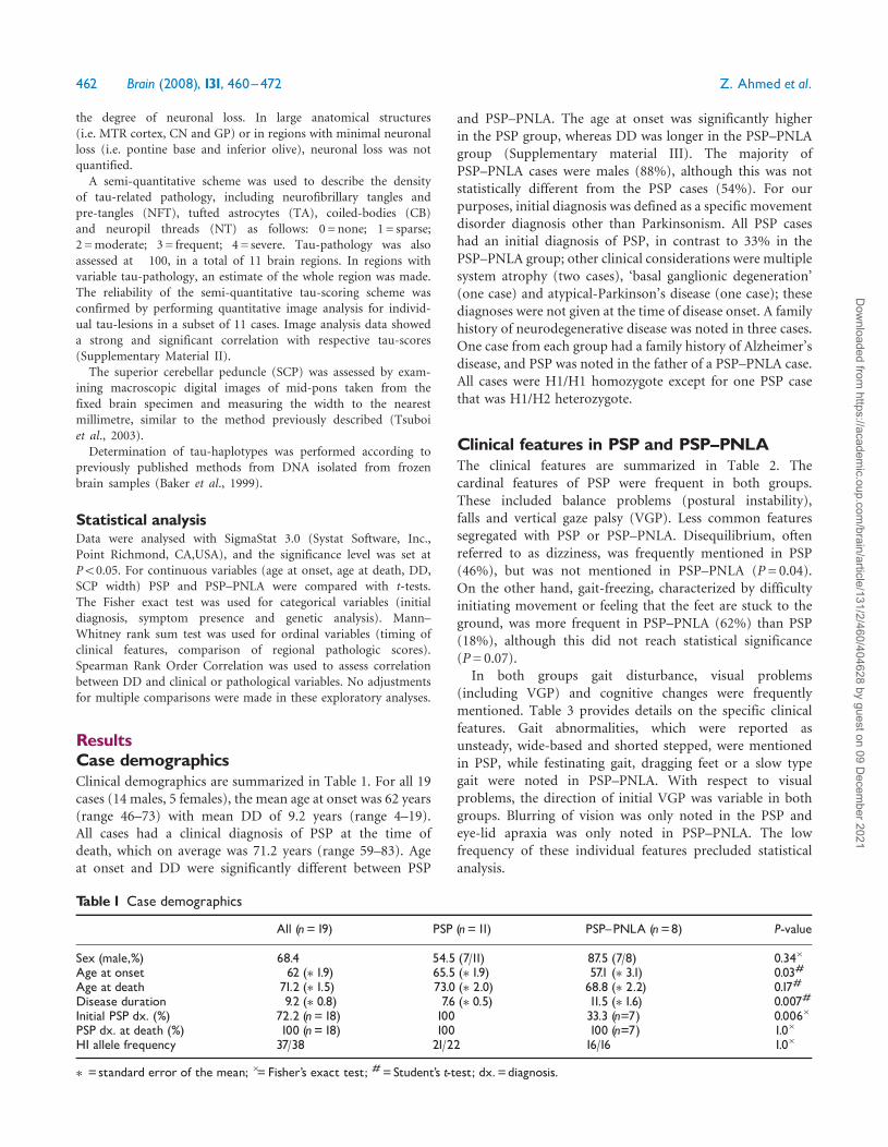
ResultsCase demographicsClinical demographics are summarized in Table 1. For all 19cases (14 males, 5 females), the mean age at onset was 62 years(range 46–73) with mean DD of 9.2 years (range 4–19).All cases had a clinical diagnosis of PSP at the time ofdeath, which on average was 71.2 years (range 59–83). Ageat onset and DD were significantly different between PSP
and PSP–PNLA. The age at onset was significantly higherin the PSP group, whereas DD was longer in the PSP–PNLAgroup (Supplementary material III). The majority ofPSP–PNLA cases were males (88%), although this was notstatistically different from the PSP cases (54%). For ourpurposes, initial diagnosis was defined as a specific movementdisorder diagnosis other than Parkinsonism. All PSP caseshad an initial diagnosis of PSP, in contrast to 33% in thePSP–PNLA group; other clinical considerations were multiplesystem atrophy (two cases), ‘basal ganglionic degeneration’(one case) and atypical-Parkinson’s disease (one case); thesediagnoses were not given at the time of disease onset. A familyhistory of neurodegenerative disease was noted in three cases.One case from each group had a family history of Alzheimer’sdisease, and PSP was noted in the father of a PSP–PNLA case.All cases were H1/H1 homozygote except for one PSP casethat was H1/H2 heterozygote.
Clinical features in PSP and PSP^PNLAThe clinical features are summarized in Table 2. Thecardinal features of PSP were frequent in both groups.These included balance problems (postural instability),falls and vertical gaze palsy (VGP). Less common featuressegregated with PSP or PSP–PNLA. Disequilibrium, oftenreferred to as dizziness, was frequently mentioned in PSP(46%), but was not mentioned in PSP–PNLA (P= 0.04).On the other hand, gait-freezing, characterized by difficultyinitiating movement or feeling that the feet are stuck to theground, was more frequent in PSP–PNLA (62%) than PSP(18%), although this did not reach statistical significance(P= 0.07).
In both groups gait disturbance, visual problems(including VGP) and cognitive changes were frequentlymentioned. Table 3 provides details on the specific clinicalfeatures. Gait abnormalities, which were reported asunsteady, wide-based and shorted stepped, were mentionedin PSP, while festinating gait, dragging feet or a slow typegait were noted in PSP–PNLA. With respect to visualproblems, the direction of initial VGP was variable in bothgroups. Blurring of vision was only noted in the PSP andeye-lid apraxia was only noted in PSP–PNLA. The lowfrequency of these individual features precluded statisticalanalysis.
Table 1 Case demographics
All (n=19) PSP (n=11) PSP^PNLA (n=8) P-value
Sex (male,%) 68.4 54.5 (7/11) 87.5 (7/8) 0.34�
Age at onset 62 (�1.9) 65.5 (�1.9) 57.1 (�3.1) 0.03#
Age at death 71.2 (�1.5) 73.0 (�2.0) 68.8 (�2.2) 0.17#
Disease duration 9.2 (�0.8) 7.6 (�0.5) 11.5 (�1.6) 0.007#
Initial PSP dx. (%) 72.2 (n=18) 100 33.3 (n=7) 0.006�
PSP dx. at death (%) 100 (n=18) 100 100 (n=7) 1.0�
H1 allele frequency 37/38 21/22 16/16 1.0�
�=standard error of the mean; �=Fisher’s exact test; #=Student’s t-test; dx.=diagnosis.
462 Brain (2008), 131, 460^472 Z. Ahmed et al.
Dow
nloaded from https://academ
ic.oup.com/brain/article/131/2/460/404628 by guest on 09 D
ecember 2021
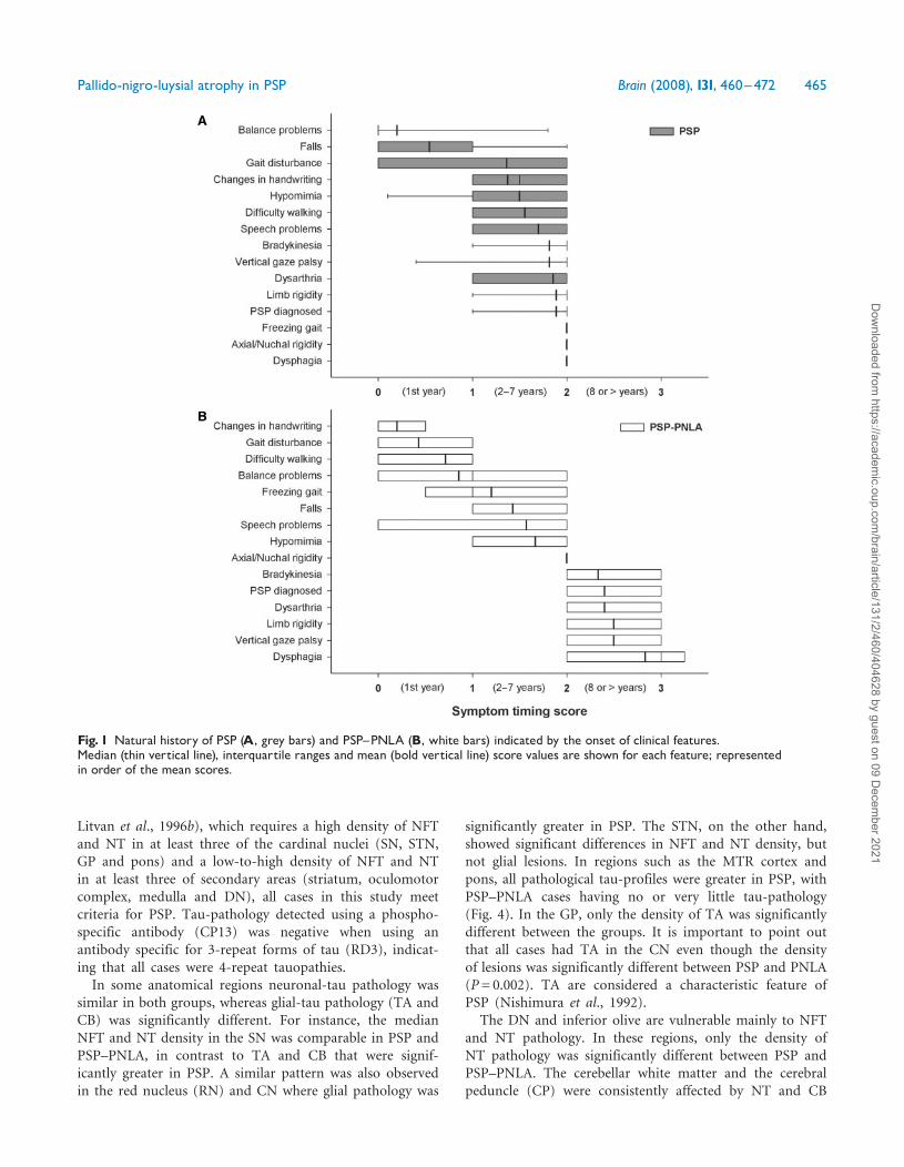
Clinical courseFor those cases in which sufficient information wasavailable, the timing of clinical features was recorded ona 4-point scale. For features present in both groups, themean and median scores for each feature are shown inTable 4. The mean score for the timing of clinical featuresis ranked (lowest to highest) for PSP and PSP–PNLA inFig. 1. In PSP, balance problems were the presentingsymptom, followed by falls within the first year and laterby progressive gait disturbance, changes in handwritingand hypomimia. Difficulty walking, speech problems andbradykinesia preceded VGP. A diagnosis of PSP was mostoften made at the time that dysarthria and rigidity werenoted. Axial or nuchal rigidity and dysphagia were, onaverage, the last clinical manifestations, as was gait-freezingin two patients. All these features were present within7 years of onset.
In PSP–PNLA, the order of clinical features differed fromPSP. Changes in handwriting preceded gait disturbance.Balance problems were followed by gait-freezing and falls.Speech problems and hypomimia presented before axialor nuchal rigidity. Bradykinesia as well as dysarthria, limbrigidity and VGP contributed to a diagnosis of PSP at thistime. Again, dysphagia was last.
In the PSP group, there was considerable variation fromcase to case as indicated by large whisker caps in Fig. 1A.In contrast, there was less variation between cases in thePSP–PNLA group as shown in Fig. 1B.
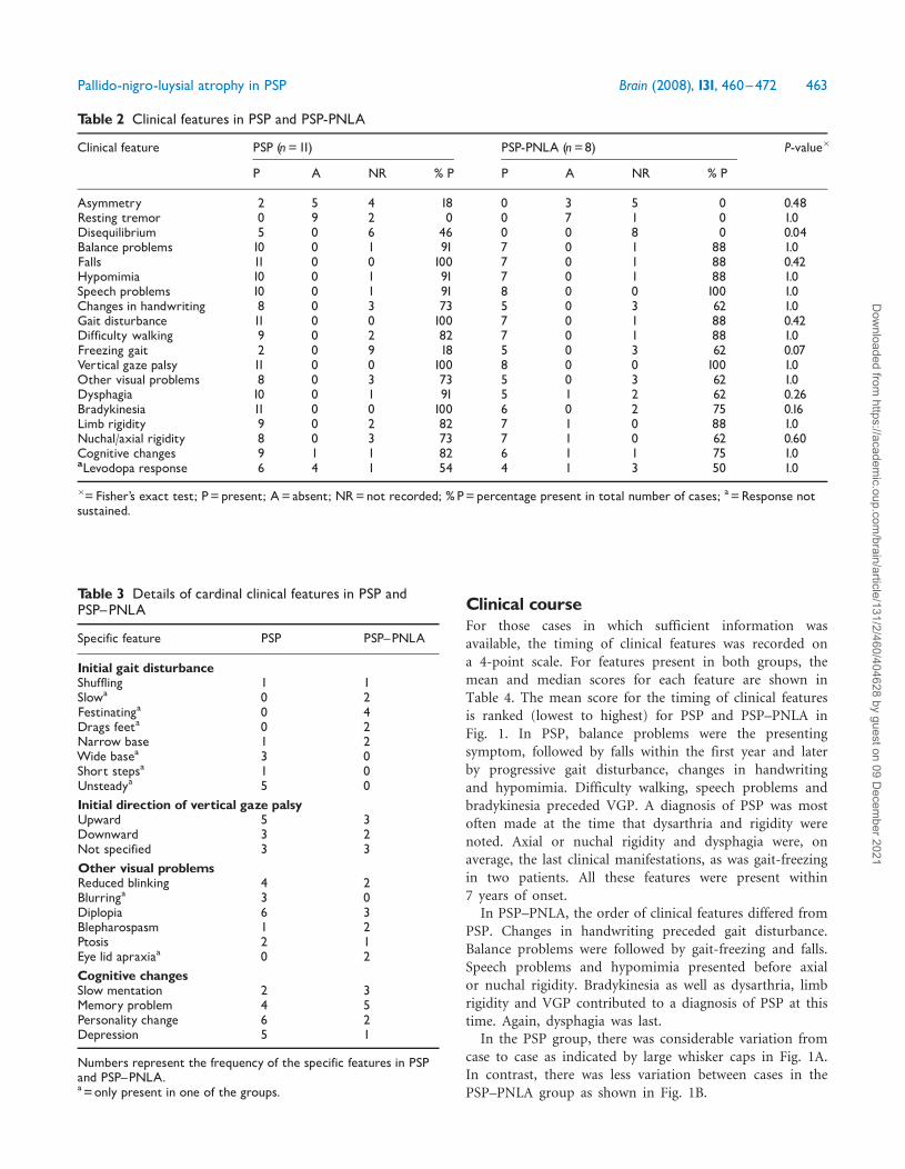
Table 2 Clinical features in PSP and PSP-PNLA
Clinical feature PSP (n=11) PSP-PNLA (n=8) P-value�
P A NR % P P A NR % P
Asymmetry 2 5 4 18 0 3 5 0 0.48Resting tremor 0 9 2 0 0 7 1 0 1.0Disequilibrium 5 0 6 46 0 0 8 0 0.04Balance problems 10 0 1 91 7 0 1 88 1.0Falls 11 0 0 100 7 0 1 88 0.42Hypomimia 10 0 1 91 7 0 1 88 1.0Speech problems 10 0 1 91 8 0 0 100 1.0Changes in handwriting 8 0 3 73 5 0 3 62 1.0Gait disturbance 11 0 0 100 7 0 1 88 0.42Difficulty walking 9 0 2 82 7 0 1 88 1.0Freezing gait 2 0 9 18 5 0 3 62 0.07Vertical gaze palsy 11 0 0 100 8 0 0 100 1.0Other visual problems 8 0 3 73 5 0 3 62 1.0Dysphagia 10 0 1 91 5 1 2 62 0.26Bradykinesia 11 0 0 100 6 0 2 75 0.16Limb rigidity 9 0 2 82 7 1 0 88 1.0Nuchal/axial rigidity 8 0 3 73 7 1 0 62 0.60Cognitive changes 9 1 1 82 6 1 1 75 1.0aLevodopa response 6 4 1 54 4 1 3 50 1.0
�=Fisher’s exact test; P=present; A=absent; NR=not recorded; %P=percentage present in total number of cases; a=Response notsustained.
Table 3 Details of cardinal clinical features in PSP andPSP^PNLA
Specific feature PSP PSP^PNLA
Initial gait disturbanceShuffling 1 1Slowa 0 2Festinatinga 0 4Drags feeta 0 2Narrow base 1 2Wide basea 3 0Short stepsa 1 0Unsteadya 5 0Initial direction of vertical gaze palsyUpward 5 3Downward 3 2Not specified 3 3Other visual problemsReduced blinking 4 2Blurringa 3 0Diplopia 6 3Blepharospasm 1 2Ptosis 2 1Eye lid apraxiaa 0 2Cognitive changesSlow mentation 2 3Memory problem 4 5Personality change 6 2Depression 5 1
Numbers represent the frequency of the specific features in PSPand PSP^PNLA.a=only present in one of the groups.
Pallido-nigro-luysial atrophy in PSP Brain (2008), 131, 460^472 463
Dow
nloaded from https://academ
ic.oup.com/brain/article/131/2/460/404628 by guest on 09 D
ecember 2021
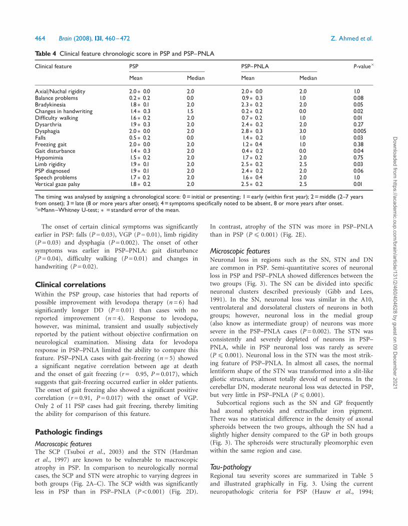
The onset of certain clinical symptoms was significantlyearlier in PSP: falls (P= 0.03), VGP (P= 0.01), limb rigidity(P= 0.03) and dysphagia (P= 0.002). The onset of othersymptoms was earlier in PSP–PNLA: gait disturbance(P= 0.04), difficulty walking (P= 0.01) and changes inhandwriting (P= 0.02).
Clinical correlationsWithin the PSP group, case histories that had reports ofpossible improvement with levodopa therapy (n= 6) hadsignificantly longer DD (P= 0.01) than cases with noreported improvement (n= 4). Response to levodopa,however, was minimal, transient and usually subjectivelyreported by the patient without objective confirmation onneurological examination. Missing data for levodoparesponse in PSP–PNLA limited the ability to compare thisfeature. PSP–PNLA cases with gait-freezing (n= 5) showeda significant negative correlation between age at deathand the onset of gait freezing (r=�0.95, P= 0.017), whichsuggests that gait-freezing occurred earlier in older patients.The onset of gait freezing also showed a significant positivecorrelation (r= 0.91, P= 0.017) with the onset of VGP.Only 2 of 11 PSP cases had gait freezing, thereby limitingthe ability for comparison of this feature.
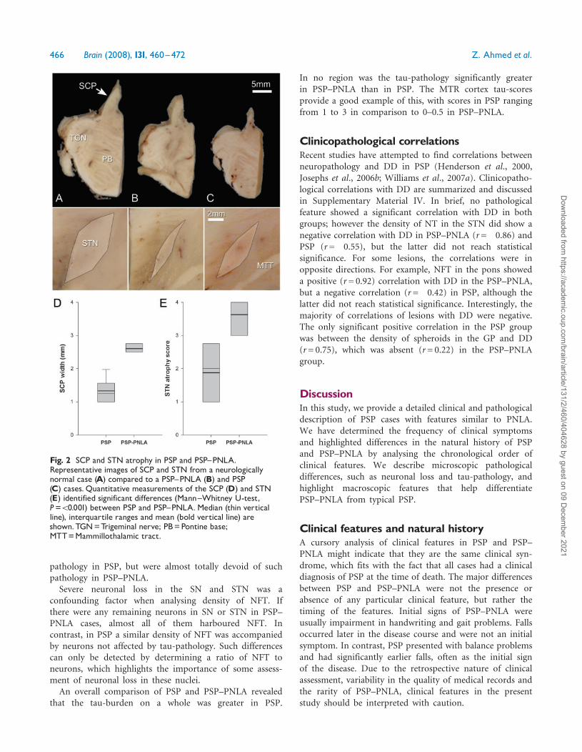
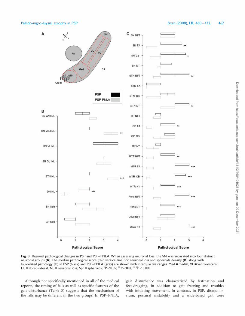
Pathologic findingsMacroscopic featuresThe SCP (Tsuboi et al., 2003) and the STN (Hardmanet al., 1997) are known to be vulnerable to macroscopicatrophy in PSP. In comparison to neurologically normalcases, the SCP and STN were atrophic to varying degrees inboth groups (Fig. 2A–C). The SCP width was significantlyless in PSP than in PSP–PNLA (P50.001) (Fig. 2D).
In contrast, atrophy of the STN was more in PSP–PNLAthan in PSP (P4 0.001) (Fig. 2E).
Microscopic featuresNeuronal loss in regions such as the SN, STN and DNare common in PSP. Semi-quantitative scores of neuronalloss in PSP and PSP–PNLA showed differences between thetwo groups (Fig. 3). The SN can be divided into specificneuronal clusters described previously (Gibb and Lees,1991). In the SN, neuronal loss was similar in the A10,ventrolateral and dorsolateral clusters of neurons in bothgroups; however, neuronal loss in the medial group(also know as intermediate group) of neurons was moresevere in the PSP–PNLA cases (P= 0.002). The STN wasconsistently and severely depleted of neurons in PSP–PNLA, while in PSP neuronal loss was rarely as severe(P4 0.001). Neuronal loss in the STN was the most strik-ing feature of PSP–PNLA. In almost all cases, the normallentiform shape of the STN was transformed into a slit-likegliotic structure, almost totally devoid of neurons. In thecerebellar DN, moderate neuronal loss was detected in PSP,but very little in PSP–PNLA (P4 0.001).
Subcortical regions such as the SN and GP frequentlyhad axonal spheroids and extracellular iron pigment.There was no statistical difference in the density of axonalspheroids between the two groups, although the SN had aslightly higher density compared to the GP in both groups(Fig. 3). The spheroids were structurally pleomorphic evenwithin the same region and case.
Tau-pathologyRegional tau severity scores are summarized in Table 5and illustrated graphically in Fig. 3. Using the currentneuropathologic criteria for PSP (Hauw et al., 1994;
Table 4 Clinical feature chronologic score in PSP and PSP^PNLA
Clinical feature PSP PSP^PNLA P-value�
Mean Median Mean Median
Axial/Nuchal rigidity 2.0� 0.0 2.0 2.0� 0.0 2.0 1.0Balance problems 0.2� 0.2 0.0 0.9� 0.3 1.0 0.08Bradykinesia 1.8� 0.1 2.0 2.3� 0.2 2.0 0.05Changes in handwriting 1.4� 0.3 1.5 0.2� 0.2 0.0 0.02Difficulty walking 1.6� 0.2 2.0 0.7�0.2 1.0 0.01Dysarthria 1.9� 0.3 2.0 2.4� 0.2 2.0 0.27Dysphagia 2.0� 0.0 2.0 2.8� 0.3 3.0 0.005Falls 0.5� 0.2 0.0 1.4� 0.2 1.0 0.03Freezing gait 2.0� 0.0 2.0 1.2� 0.4 1.0 0.38Gait disturbance 1.4� 0.3 2.0 0.4� 0.2 0.0 0.04Hypomimia 1.5� 0.2 2.0 1.7�0.2 2.0 0.75Limb rigidity 1.9� 0.1 2.0 2.5� 0.2 2.5 0.03PSP diagnosed 1.9� 0.1 2.0 2.4� 0.2 2.0 0.06Speech problems 1.7�0.2 2.0 1.6� 0.4 2.0 1.0Vertical gaze palsy 1.8� 0.2 2.0 2.5� 0.2 2.5 0.01
The timing was analysed by assigning a chronological score: 0= initial or presenting; 1=early (within first year); 2=middle (2^7 yearsfrom onset); 3= late (8 or more years after onset); 4= symptoms specifically noted to be absent, 8 or more years after onset.�=Mann^Whitney U-test; �=standard error of the mean.
464 Brain (2008), 131, 460^472 Z. Ahmed et al.
Dow
nloaded from https://academ
ic.oup.com/brain/article/131/2/460/404628 by guest on 09 D
ecember 2021
Litvan et al., 1996b), which requires a high density of NFTand NT in at least three of the cardinal nuclei (SN, STN,GP and pons) and a low-to-high density of NFT and NTin at least three of secondary areas (striatum, oculomotorcomplex, medulla and DN), all cases in this study meetcriteria for PSP. Tau-pathology detected using a phospho-specific antibody (CP13) was negative when using anantibody specific for 3-repeat forms of tau (RD3), indicat-ing that all cases were 4-repeat tauopathies.In some anatomical regions neuronal-tau pathology was
similar in both groups, whereas glial-tau pathology (TA andCB) was significantly different. For instance, the medianNFT and NT density in the SN was comparable in PSP andPSP–PNLA, in contrast to TA and CB that were signif-icantly greater in PSP. A similar pattern was also observedin the red nucleus (RN) and CN where glial pathology was
significantly greater in PSP. The STN, on the other hand,showed significant differences in NFT and NT density, butnot glial lesions. In regions such as the MTR cortex andpons, all pathological tau-profiles were greater in PSP, withPSP–PNLA cases having no or very little tau-pathology(Fig. 4). In the GP, only the density of TA was significantlydifferent between the groups. It is important to point outthat all cases had TA in the CN even though the densityof lesions was significantly different between PSP and PNLA(P= 0.002). TA are considered a characteristic feature ofPSP (Nishimura et al., 1992).
The DN and inferior olive are vulnerable mainly to NFTand NT pathology. In these regions, only the density ofNT pathology was significantly different between PSP andPSP–PNLA. The cerebellar white matter and the cerebralpeduncle (CP) were consistently affected by NT and CB
Fig. 1 Natural history of PSP (A, grey bars) and PSP^PNLA (B, white bars) indicated by the onset of clinical features.Median (thin vertical line), interquartile ranges and mean (bold vertical line) score values are shown for each feature; representedin order of the mean scores.
Pallido-nigro-luysial atrophy in PSP Brain (2008), 131, 460^472 465
Dow
nloaded from https://academ
ic.oup.com/brain/article/131/2/460/404628 by guest on 09 D
ecember 2021
pathology in PSP, but were almost totally devoid of suchpathology in PSP–PNLA.Severe neuronal loss in the SN and STN was a
confounding factor when analysing density of NFT. Ifthere were any remaining neurons in SN or STN in PSP–PNLA cases, almost all of them harboured NFT. Incontrast, in PSP a similar density of NFT was accompaniedby neurons not affected by tau-pathology. Such differencescan only be detected by determining a ratio of NFT toneurons, which highlights the importance of some assess-ment of neuronal loss in these nuclei.An overall comparison of PSP and PSP–PNLA revealed
that the tau-burden on a whole was greater in PSP.
In no region was the tau-pathology significantly greaterin PSP–PNLA than in PSP. The MTR cortex tau-scoresprovide a good example of this, with scores in PSP rangingfrom 1 to 3 in comparison to 0–0.5 in PSP–PNLA.
Clinicopathological correlationsRecent studies have attempted to find correlations betweenneuropathology and DD in PSP (Henderson et al., 2000,Josephs et al., 2006b; Williams et al., 2007a). Clinicopatho-logical correlations with DD are summarized and discussedin Supplementary Material IV. In brief, no pathologicalfeature showed a significant correlation with DD in bothgroups; however the density of NT in the STN did show anegative correlation with DD in PSP–PNLA (r=�0.86) andPSP (r=�0.55), but the latter did not reach statisticalsignificance. For some lesions, the correlations were inopposite directions. For example, NFT in the pons showeda positive (r= 0.92) correlation with DD in the PSP–PNLA,but a negative correlation (r=�0.42) in PSP, although thelatter did not reach statistical significance. Interestingly, themajority of correlations of lesions with DD were negative.The only significant positive correlation in the PSP groupwas between the density of spheroids in the GP and DD(r= 0.75), which was absent (r= 0.22) in the PSP–PNLAgroup.
DiscussionIn this study, we provide a detailed clinical and pathologicaldescription of PSP cases with features similar to PNLA.We have determined the frequency of clinical symptomsand highlighted differences in the natural history of PSPand PSP–PNLA by analysing the chronological order ofclinical features. We describe microscopic pathologicaldifferences, such as neuronal loss and tau-pathology, andhighlight macroscopic features that help differentiatePSP–PNLA from typical PSP.
Clinical features and natural historyA cursory analysis of clinical features in PSP and PSP–PNLA might indicate that they are the same clinical syn-drome, which fits with the fact that all cases had a clinicaldiagnosis of PSP at the time of death. The major differencesbetween PSP and PSP–PNLA were not the presence orabsence of any particular clinical feature, but rather thetiming of the features. Initial signs of PSP–PNLA wereusually impairment in handwriting and gait problems. Fallsoccurred later in the disease course and were not an initialsymptom. In contrast, PSP presented with balance problemsand had significantly earlier falls, often as the initial signof the disease. Due to the retrospective nature of clinicalassessment, variability in the quality of medical records andthe rarity of PSP–PNLA, clinical features in the presentstudy should be interpreted with caution.
Fig. 2 SCP and STN atrophy in PSP and PSP^PNLA.Representative images of SCP and STN from a neurologicallynormal case (A) compared to a PSP^PNLA (B) and PSP(C) cases.Quantitative measurements of the SCP (D) and STN(E) identified significant differences (Mann^Whitney U-test,P=50.001) between PSP and PSP^PNLA. Median (thin verticalline), interquartile ranges and mean (bold vertical line) areshown.TGN=Trigeminal nerve; PB=Pontine base;MTT=Mammillothalamic tract.
466 Brain (2008), 131, 460^472 Z. Ahmed et al.
Dow
nloaded from https://academ
ic.oup.com/brain/article/131/2/460/404628 by guest on 09 D
ecember 2021
Although not specifically mentioned in all of the medicalreports, the timing of falls as well as specific features of thegait disturbance (Table 3) suggests that the mechanism ofthe falls may be different in the two groups. In PSP–PNLA,
gait disturbance was characterized by festination andfeet-dragging, in addition to gait freezing and troubleswith initiating movement. In contrast, in PSP, disequilib-rium, postural instability and a wide-based gait were
Fig. 3 Regional pathological changes in PSP and PSP^PNLA.When assessing neuronal loss, the SN was separated into four distinctneuronal groups (A). The median pathological score (thin vertical line) for neuronal loss and spheroids density (B) along withtau-related pathology (C) in PSP (black) and PSP^PNLA (grey) are shown with interquartile ranges. Med=medial; VL=ventro-lateral;DL=dorso-lateral; NL=neuronal loss; Sph= spheroids; �P50.05; ��P50.01; ���P50.001.
Pallido-nigro-luysial atrophy in PSP Brain (2008), 131, 460^472 467
Dow
nloaded from https://academ
ic.oup.com/brain/article/131/2/460/404628 by guest on 09 D
ecember 2021

more common. Early falls are important in the clinicalidentification and differential diagnosis of PSP (Litvanet al., 1996a). The fact that falls were significantly later inPSP–PNLA may explain why the initial diagnosis wassomething other than PSP for many cases. In addition tofalls, VGP is probably the second most important clinicalfeature for a clinical diagnosis of PSP (Litvan et al., 1996a).In PSP–PNLA, VGP was significantly later than in PSP.Dysphagia, which was often a terminal complication in
both groups, was significantly later in PSP–PNLA. Giventhat dysphagia is an important factor leading to aspirationpneumonia, which is the most common cause of death inPSP (Litvan et al., 1996c), the later onset of swallowingdifficulties may be a factor in longevity of PSP–PNLAcompared to PSP. It is worth noting that for two of thethree PSP–PNLA cases with the shortest disease durationsthere were other serious medical problems, such as conges-tive heart failure and myocardial infarction, that could haveshortened their survival.Considering differences in disease duration alone, one
would expect age at death to also be different; however,
the age at onset was substantially younger in the PSP–PNLA cases, on average by almost 10 years, explainingwhy there was no difference in the age at death. In otherneurodegenerative disorders, earlier onset of disease issometimes attributed to genetic risk factors or familialforms of disease. In the present series, only one of thePSP–PNLA had a family history of neurologic disease (PSPin the father). Familial history of neurologic disease hasbeen reported in other cases of PNLA in the literature(Gray et al., 1981, 1985; Kawai et al., 1993). In addition,a missense mutation in the tau-gene has been identifiedin a Japanese patient with familial pallido-nigro-luysialdegeneration (Yasuda et al., 1999), but this patient hadgreater tau-pathology than PSP–PNLA cases in the presentstudy. Tau-haplotype analysis was performed in bothgroups and was consistent with frequencies previouslyreported in PSP (Baker et al., 1999).
Gait freezing was a frequent feature in PSP–PNLA, butwas also present in two cases of PSP. Although based upona small sample size (n= 5), cases of PSP–PNLA with earlygait freezing also developed VGP earlier, indicating that the
Table 5 Median and mean tau scores for different anatomical regions
Anatomical region PSP PSP^PNLA P-value�
Lesion Median 0.25 0.75 Mean Median 0.25 0.75 Mean
Substantia nigra NFT 2.0 1.0 2.0 1.6 2.0 1.0 2.0 1.6 1.0TA 1.0 1.0 2.5 1.5 0.0 0.0 0.8 0.3 0.001CB 2.0 1.0 2.8 2.0 1.0 0.3 1.0 0.8 0.01NT 1.0 1.0 1.8 1.3 1.0 0.8 1.0 1.0 0.16
Red nucleus NFT 1.0 1.0 1.8 1.3 1.0 1.0 1.0 1.0 0.33TA 1.0 1.0 2.0 1.6 0.0 0.0 0.5 0.4 0.01CB 2.0 1.3 2.0 2.0 1.0 0.0 1.0 0.8 0.02NT 2.0 1.0 2.0 1.6 1.0 1.0 1.0 0.9 0.02
Subthalamic nucleus NFT 2.0 1.0 3.0 1.9 0.5 0.5 1.0 0.7 0.004TA 0.0 0.0 1.0 0.4 0.0 0.0 0.0 0.0 0.18CB 1.5 1.0 2.0 1.7 1.0 0.0 1.0 0.8 0.07NT 2.0 2.0 3.0 2.3 1.0 1.0 2.0 1.3 0.007
Globus Pallidus NFT 1.0 0.6 1.0 0.9 1.0 0.5 1.0 0.9 0.84TA 1.0 1.0 2.0 1.2 0.0 0.0 0.4 0.1 0.006CB 2.0 1.3 2.0 1.8 1.0 0.5 2.0 1.3 0.20NT 1.0 0.6 1.0 0.8 0.5 0.0 0.9 0.4 0.06
Caudate NFT 1.0 1.0 1.0 1.1 1.0 1.0 1.0 1.0 0.46TA 3.0 2.0 3.8 2.8 1.3 1.0 2.0 1.4 0.002
Motor cortex NFT 1.0 1.0 2.0 1.5 0.5 0.0 0.5 0.4 0.001TA 3.0 2.0 3.0 2.5 0.0 0.0 0.5 0.2 0.00CB 2.0 2.0 2.8 2.1 0.0 0.0 0.3 0.2 0.00NT 2.0 1.0 2.0 1.8 0.5 0.5 0.5 0.5 0.00
Dentate nucleus NFT 2.0 2.0 3.0 2.2 1.5 1.0 2.0 1.5 0.05NT 1.0 1.0 2.0 1.5 1.0 0.8 1.0 0.9 0.01
Pontine base NFT 3.0 2.0 3.0 2.7 0.8 0.5 1.0 0.7 0.00NT 2.0 1.0 2.0 1.5 0.0 0.0 0.0 0.1 0.00
Medullar olive NFT 2.0 1.0 2.0 1.7 1.5 0.8 2.0 1.5 0.54NT 3.0 3.0 3.0 2.8 1.0 0.5 1.0 0.9 0.00
Cerebellar white matter CB 2.0 1.0 2.8 1.8 0.0 0.0 0.3 0.1 0.00Cerebral peduncle CB 2.0 1.3 3.0 2.1 0.0 0.0 0.0 0.1 0.00
�= Mann^Whitney U-test; NFT=neurofibrillary tangles; TA=Tufted astrocytes; CB=coiled bodies; NT=threads. Regions underlinedare not in Fig. 3.
468 Brain (2008), 131, 460^472 Z. Ahmed et al.
Dow
nloaded from https://academ
ic.oup.com/brain/article/131/2/460/404628 by guest on 09 D
ecember 2021
neurodegenerative process leading to these symptoms wascoincident. The onset of gait-freezing was also earlier inPSP–PNLA individuals who lived the longest.Based on clinical research criteria for PSP (Litvan et al.,
1996b), the clinical diagnosis of PSP cases would be‘clinically probable PSP’—onset at age 40 or more, posturalinstability (balance problems) and falls within the first year,and VGP. The clinical progression and demographics ofPSP cases were also similar to those reported in clinicalseries of PSP (De Bruin and Lees, 1994; Collins et al., 1995;Litvan et al., 1996c). The demographics and clinical prog-ression in PSP–PNLA cases, in contrast, departed from thatreported in PSP. Most would not meet strict clinicalresearch criteria for PSP. This is in part due to delayedonset of postural instability, falls and VGP. Interestingly,onset of falls after the first year has previously beenreported as a prognostic indicator of good survival inPSP (Litvan et al., 1996c), which was true for PSP–PNLA.PSP–PNLA cases eventually developed VGP as well as otherclinical features of PSP consistent with the final clinicaldiagnosis of PSP in all cases.
Clinical features of PSP^PNLAcompared to PAThe clinical features of our PSP–PNLA cases share somesimilarities to the syndrome of ‘pure-akinesia’ (PA), whichwas first described by Imai and co-workers (Imai andNarabayashi, 1974; Imai et al., 1987, 1993; Imai, 1996).Riley and coworkers described clinical characteristics of fivepatients with PA who developed PSP-like symptoms laterin life (Riley et al., 1994). Micrographia, gait and speechproblems were some of the initial features. All patientseventually developed speech problems, gait-freezing orfestination and postural instability that resulted in falls.The case with the longest disease duration (14 years) devel-oped bradykinesia, nuchal rigidity and VGP (Riley et al.,1994). Imai also reported that many PA cases had VGPlater in the disease course (Imai et al., 1987, 1993). In moststudies of PA, the term akinesia has been applied to notonly gait problems (i.e. freezing or difficulty with initia-tion), but also to akinesia of speech (stuttering or stammer-ing speech) and akinesia of writing (with micrographia)(Hoshino et al., 1999). Interestingly, changes in handwritingwere the earliest clinical feature in several PSP–PNLA cases.
No PSP or PSP–PNLA case had resting tremor or morethan a minimal response to levodopa therapy. Even so,many of the patients in both groups remained on levodopathroughout the disease without suffering drug-induceddyskinesia. One should note that some cases referred toas PA have been reported to respond well to levodopatherapy, and one such case had postmortem evidenceof Lewy body disease (Quinn et al., 1989). A number ofstudies also describe a clinical syndrome similar to PA, withgait-freezing and falls occurring without other Parkinsonianfeatures. These cases have been termed primary progressivefreezing gait (PPFG) (Achiron et al., 1993) or gait ignitionfailure (GIF) (Atchison et al., 1993). Some cases of PPFGdevelop clinical features suggestive of PSP (e.g. VGP, post-ural instability and dysphagia) later in the disease course,as well as pathologic changes similar to PSP at autopsy(Compta et al., 2007). In a study of nine cases, initiallydiagnosed as PPFG (Factor et al., 2006), four evolved into aclinical syndrome resembling PSP or CBD. Autopsy studiesof two of the nine cases revealed PNLA in one and diffuseLewy body disease in another (Factor et al., 2006). As inany diagnosis that is based upon a clinical syndrome, thepathology reflects distribution rather than underlyingmolecular pathology. For example, the pathologic substrateof the corticobasal syndrome includes several differentpathologic processes, in addition to corticobasal degenera-tion (Boeve et al., 1999). Thus, the neuropathology of PAand related clinical syndromes is likely to be heterogeneous.
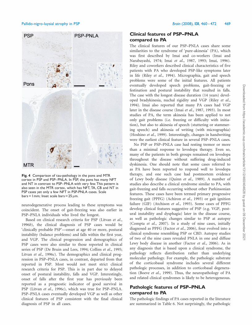
Pathologic features of PSP^PNLAcompared to PAThe pathologic findings of PA cases reported in the literatureare summarized in Table 6. Not surprisingly, the pathologic
Fig. 4 Comparison of tau-pathology in the pons and MTRcortex in PSP and PSP^PNLA. In PSP, the pons has many NFTand NT in contrast to PSP^PNLAwith very few.This pattern isalso seen in the MTR cortex, which has NFT, TA, CB and NT inPSP cases yet only a few NFT in PSP-PNLA cases. Scalebars=1mm; Inset scale bars=25mm.
Pallido-nigro-luysial atrophy in PSP Brain (2008), 131, 460^472 469
Dow
nloaded from https://academ
ic.oup.com/brain/article/131/2/460/404628 by guest on 09 D
ecember 2021
diagnoses that are most often given for PA are PSP(Takahashi et al., 1987; Matsuo et al., 1991; Mizusawa et al.,1993; Yoshikawa et al., 1997), PNLA (Takahashi et al., 1977;Inagaki et al., 1989; Konishi et al., 2005) and in onecase combined PSP and PNLA (Yamamoto et al., 1991).Macroscopic atrophy accompanied by severe neuronal loss inthe GP, SN and STN with relative sparing of the cerebellar DNand lower brainstem structures are consistent features in thesereports. In addition, NFT pathology is described in manyof these reports, mainly in regions (SN, STN, GP and DN)that are also vulnerable to PSP pathology. In comparison tosilver stains, immunohistochemistry for tau is more specificand sensitive for neuronal (including pre-tangles) and glialtau-pathology. This may explain the discrepancy in frequencyof glial pathology in reports of PA. The most recent casereport by Konishi and co-workers reported glial tau-pathology with tau-immunohistochemistry (Konishi et al.,2005). The presence of glial tau-pathology in PA supports arelationship to PSP. A major histopathological hallmark ofPSP, tufted astrocytes (Nishimura et al., 1992), was detectedin all cases of PSP–PNLA. Biochemical data also supportsa diagnosis of PSP in PNLA. Mori and coworkers reportedresults from immunoblot analysis of phosphorylated tau-protein in a patient with PNLA (Mori et al., 2001). Theyidentified major tau-bands at 64 and 68 kDa, which arebiochemical characteristics of tau in PSP.The preservation of hindbrain structures, such as the
pontine nuclei and the cerebellar DN has been noted inreports of PA (Table 6). Our study also demonstrated lesspathology in forebrain regions, such as the MTR cortex;in PSP–PNLA compared to PSP. In no region was the
tau-pathology in PSP–PNLA greater than PSP. Except forthe cerebellar DN, neuronal loss was greater in all regionsin PSP–PNLA compared to PSP and the SN and STNshowed statistical significance. Collectively this data indi-cates that the differential distribution and severity ofpathology in PSP and PSP–PNLA cannot be explainedby obvious clinical variables and implies other environ-mental or genetic variables as driving factors in the twophenotypes.
Relationship of PSP^PNLA to PAGFA detailed clinical study using pathologically confirmedPSP by Williams and co-workers suggested that PSP can beseparated into two major clinical types, termed Richardson’ssyndrome (RS) and PSP-Parkinsonism (PSP-P) (Williamset al., 2005). In the present study, PSP cases fit the syndromeof RS, with falls, balance problems or VGP within thefirst year. The same investigators reported the pathologic(Williams et al., 2007a) and clinical features (Williams et al.,2007b) of a group of patients with a clinical syndromecharacterized by PA and gait-freezing (PAGF). Comparisonof the clinical features of PAGF to the clinical features foundretrospectively in PSP-PNLA reveals many similarities(Supplementary Material V). Unfortunately, pathologicaldescriptions of PAGF had insufficient detail to know if theyhad severe pallido-nigro-luysial degeneration and axonaldystrophy (Williams et al., 2007a, b). On the other hand,a regional tau severity score was used to characterize PAGF.This score was a summary measure of individual scoresassigned for each of the lesion types (NFT, TA, CB and NT)
Table 6 Summary of pathological case reports of PA and PNLA in literature
Study Takahashi1977
Takahashi1977
Homma1987
Yamamoto1991
Matsuo1991
Matsuo1991
Mizusawa1993
Mizusawa1993
Mizusawa1993
Mizusawa1993
Yoshikawa1997
Konishi2005
Sex M F F M M M F M M M F MOnset 61 64 54 57 72 62 55 62 65 70 58 60Disease duration 5 7 11 ^ 2 45 21 47 6 2 8 10Path diagnosis PNLA PNLA PSP PSP/PNLA PSP PSP PSP PSP PSP PSP PSP PNLAGross atrophy STN,GP STN,GP STN,GP SN, STN ^ ^ ^ ^ ^ ^ Normal SN, STN,GPSCP ^ ^ ^ Normal ^ ^ ^ ^ ^ ^ ^ ^SNNL +++ +++ +++ +++ +++ +++ Y Y Y Y +++ +++STNNL +++ +++ Y +++ ++ ++ Y Y Y Y +++ +++GP NL +++ +++ Y +++ ++ ++ Y Y Y Y +++ +++DNNL N N Y ++ + + N Y Y N ^ NSNNFT ^ + Y ^ +++ +++ +++ +++ +++ ++ +++ +STNNFT ^ ^ Y ^ + + +++ +++ +++ ++ +++ +GP NFT ^ ^ Y ^ + +++ +++ +++ +++ ++ +++ +DNNFT ^ ^ Y + ^ + + ^ ^ + ^ +Pons NFT ^ ^ ^ ^ + ^ + + + ^ + +Olive NFT ^ ^ ^ ^ ^ ^ + + + ^ + +MTR tau ^ ^ ^ ^ ^ ^ ^ ^ ^ ^ ^ ^Glial tau ^ ^ ^ ^ ^ ^ ^ ^ ^ ^ ^ CbTau stain N N N N Y Y Y Y Y Y N YSilver stain ^ ^ Y Y Y Y Y Y Y Y Y Y
M=male; F= female; PNLA=pallido-nigro-luysial atrophy; PSP=progressive supranuclear palsy; Y=present; N=not present;+=Mild; ++=Moderate; +++=severe; ^=not mentioned.
470 Brain (2008), 131, 460^472 Z. Ahmed et al.
Dow
nloaded from https://academ
ic.oup.com/brain/article/131/2/460/404628 by guest on 09 D
ecember 2021
in a given region. The summary score eliminates the ability todetect differences related to a particular type of lesion, whichwe found helpful in differentiating PSP from PSP–PNLA,such as TA in the motor cortex and caudate (Table 5).While the median regional tau severity scores tended to begreater in RS than PAGF, statistical analyses showed relativelyfew differences (Williams et al., 2007a). In particular, nodifferences were noted in tau severity score in the SN, STNor GP. Although there may be less severe tau-pathologyoverall in PAGF than RS, the results of the present studysuggest that one should be cautious in concluding thatpathological severity is less, since neuronal loss in the SN andSTN might well have been as severe or worse than in RS,had this analysis been performed as in the present study ofPSP–PNLA.
Differentiation of PSP and PSP^PNLAwith imagingIn light of the possible differences in prognosis with respectto longevity, it would be useful to identify PSP–PNLA earlyin the disease course. Results of macroscopic findings inSTN and SCP suggest that this might be possible withmodern imaging techniques. We noted more severe atrophyof the STN in PSP–PNLA compared to PSP. Detailedimaging of the STN has significantly progressed in responseto advances in deep brain stimulation therapy for PDpatients (Elolf et al., 2007). We also found that the width ofthe SCP was significantly smaller in PSP than PSP–PNLA,which has also been noted in PA (Yamamoto et al., 1991).Several studies have highlighted the use of MRI techniquesin assessing SCP atrophy in PSP to increase diagnosticaccuracy (Paviour et al., 2005, 2006). Differentiation ofPSP from PSP–PNLA would theoretically be possible in apatient with atypical Parkinsonism with small STN andrelatively normal SCP.
SummaryPSP–PNLA was characterized pathologically by severedegeneration and axonal dystrophy in a pallido-nigro-luysial distribution. The present study indicates that PSP–PNLA is a clinical and pathological variant of PSP.PSP–PNLA has the clinical and pathological hallmarks oftypical PSP, yet the timing of clinical features, the age atonset, duration of disease, the density of pathology andthe distribution of pathology make it distinct. The charac-teristic pattern of atrophy suggests that ante-mortemimaging might be able to differentiate PSP–PNLA fromtypical PSP.
Supplementary materialSupplementary material is available at Brain online.
AcknowledgementsZ.A. is a Doctoral Candidate at the MRC Centre forNeurodegeneration Research, King’s College London,Institute of Psychiatry, Department of Neuroscience,De Crespigny Park, London, SE5 8AF, UK. The authorsacknowledge the valuable statistical support of Michael G.Heckman and Samuel Younkin; the assistance of StaceyMelquist and Mike L. Hutton in genetic analyses and thehistological support of Monica Casey-Castanedes, VirginiaPhillips and Linda Rousseau. Hiroshige Fujishiro helpedwith interpretation of original Japanese articles. The authorswould like to thank the Society for Progressive Supra-nuclear Palsy and the generous brain donations from PSPpatients and their families, without which these studieswould be impossible. The article is funded by NationalInstitute of Health (P50-NS40256, P01-AG17216 toD.W.D.), Society for Progressive Supranuclear Palsy(CurePSP, Inc.) and Mayo Foundation.
ReferencesAchiron A, Ziv I, Goren M, Goldberg H, Zoldan Y, Sroka H, et al. Primary
progressive freezing gait. Mov Disord 1993; 8: 293–7.
Atchison PR, Thompson PD, Frackowiak RS, Marsden CD. The syndrome
of gait ignition failure: a report of six cases. Mov Disord 1993; 8:
285–92.
Baker M, Litvan I, Houlden H, Adamson J, Dickson D, Perez-Tur J, et al.
Association of an extended haplotype in the tau gene with progressive
supranuclear palsy. Hum Mol Genet 1999; 8: 711–5.
Boeve BF, Maraganore DM, Parisi JE, Ahlskog JE, Graff-Radford N,
Caselli RJ, et al. Pathologic heterogeneity in clinically diagnosed
corticobasal degeneration. Neurology 1999; 53: 795–800.
Collins SJ, Ahlskog JE, Parisi JE, Maraganore DM. Progressive supra-
nuclear palsy: neuropathologically based diagnostic clinical criteria.
J Neurol Neurosurg Psychiatry 1995; 58: 167–73.
Compta Y, Valldeoriola F, Tolosa E, Rey MJ, Marti MJ, Valls-Sole J. Long
lasting pure freezing of gait preceding progressive supranuclear palsy: a
clinicopathological study. Mov Disord 2007; 22: 1954–8.
Contamin F, Escourolle R, Nick J, Mignot B. Atrophy of the globus
pallidus, substancia nigra, and nucleus subthalamicus. Akinetic syn-
drome with palilalia, oppositional rigidity and catatonia. Rev Neurol
1971; 124: 107–20.
De Bruin VM, Lees AJ. Subcortical neurofibrillary degeneration presenting
as Steele-Richardson-Olszewski and other related syndromes: a review of
90 pathologically verified cases. Mov Disord 1994; 9: 381–9.
Elolf E, Bockermann V, Gringel T, Knauth M, Dechent P, Helms G.
Improved visibility of the subthalamic nucleus on high-resolution
stereotactic MR imaging by added susceptibility (T2�) contrast using
multiple gradient echoes. Am J Neuroradiol 2007; 28: 1093–4.
Factor SA, Higgins DS, Qian J. Primary progressive freezing gait: a
syndrome with many causes. Neurology 2006; 66: 411–4.
Gibb WR, Lees AJ. Anatomy, pigmentation, ventral and dorsal subpopula-
tions of the substantia nigra, and differential cell death in Parkinson’s
disease. J Neurol Neurosurg Psychiatry 1991; 54: 388–96.
Gray F, De Baecque C, Serdaru M, Escourolle R. Pallido-luyso-nigral
atrophy and amyotrophic lateral sclerosis. Acta Neuropathol Suppl 1981;
7: 348–51.
Gray F, Eizenbaum JF, Gherardi R, Degos JD, Poirier J. Luyso-pallido-
nigral atrophy and amyotrophic lateral sclerosis. Acta Neuropathol 1985;
66: 78–82.
Hardman CD, Halliday GM, McRitchie DA, Morris JG. The subthalamic
nucleus in Parkinson’s disease and progressive supranuclear palsy.
J Neuropathol Exp Neurol 1997; 56: 132–42.
Pallido-nigro-luysial atrophy in PSP Brain (2008), 131, 460^472 471
Dow
nloaded from https://academ
ic.oup.com/brain/article/131/2/460/404628 by guest on 09 D
ecember 2021
Hauw JJ, Daniel SE, Dickson D, Horoupian DS, Jellinger K, Lantos PL,
et al. Preliminary NINDS neuropathologic criteria for Steele-
Richardson-Olszewski syndrome (progressive supranuclear palsy).
Neurology 1994; 44: 2015–9.
Henderson JM, Carpenter K, Cartwright H, Halliday GM. Loss of thalamic
intralaminar nuclei in progressive supranuclear palsy and Parkinson’s
disease: clinical and therapeutic implications. Brain 2000; 123: 1410–21.
Hoshino M, Kita Y, Mitani K, Bando M, Yamanouchi H. [A case report of
pure micrographia progressing over 5 years—an early sign of ‘‘pure
akinesia’’?] Rinsho Shinkeigaku 1999; 39: 615–8.
Imai H. Clinicophysiological features of akinesia. Eur Neurol 1996; 36
(Suppl 1): 9–12.
Imai H, Nakamura T, Kondo T, Narabayashi H. Dopa-unresponsive pure
akinesia or freezing. A condition within a wide spectrum of PSP?
Adv Neurol 1993; 60: 622–5.
Imai H, Narabayashi H. Akinesia - concerning 2 cases of pure akinesia.
Adv Neurol Sci 1974; 18: 787–94.
Imai H, Narabayashi H, Sakata E. ‘‘Pure akinesia’’ and the later added
supranuclear ophthalmoplegia. Adv Neurol 1987; 45: 207–12.
Inagaki T, Hashizume Y, Mitake S, Nokura K, Niimi T, Yamamoto T,
et al. [An autopsy case of pallido-nigro-luysian atrophy associated with
OPLL]. Nippon Ronen Igakkai Zasshi 1989; 26: 361–6.
Josephs KA, Boeve BF, Duffy JR, Smith GE, Knopman DS, Parisi JE, et al.
Atypical progressive supranuclear palsy underlying progressive apraxia
of speech and nonfluent aphasia. Neurocase 2005; 11: 283–96.
Josephs KA, Ishizawa T, Tsuboi Y, Cookson N, Dickson DW. A clinico-
pathological study of vascular progressive supranuclear palsy: a multi-
infarct disorder presenting as progressive supranuclear palsy. Arch
Neurol 2002; 59: 1597–601.
Josephs KA, Katsuse O, Beccano-Kelly DA, Lin WL, Uitti RJ, Fujino Y,
et al. Atypical progressive supranuclear palsy with corticospinal tract
degeneration. J Neuropathol Exp Neurol 2006a; 65: 396–405.
Josephs KA, Mandrekar JN, Dickson DW. The relationship between
histopathological features of progressive supranuclear palsy and disease
duration. Parkinsonism Relat Disord 2006b; 12: 109–12.
Kawai J, Sasahara M, Hazama F, Kuno S, Komure O, Nomura S, et al.
Pallidonigroluysian degeneration with iron deposition: a study of three
autopsy cases. Acta Neuropathol 1993; 86: 609–16.
Konishi Y, Shirabe T, Katayama S, Funakawa I, Terao A. Autopsy case of
pure akinesia showing pallidonigro-luysian atrophy. Neuropathology
2005; 25: 220–7.
Litvan I, Agid Y, Calne D, Campbell G, Dubois B, Duvoisin RC, et al.
Clinical research criteria for the diagnosis of progressive supranuclear
palsy (Steele-Richardson-Olszewski syndrome): report of the NINDS-
SPSP International Workshop. Neurology 1996a; 47: 1–9.
Litvan I, Hauw JJ, Bartko JJ, Lantos PL, Daniel SE, Horoupian DS, et al.
Validity and reliability of the preliminary NINDS neuropathologic
criteria for progressive supranuclear palsy and related disorders.
J Neuropathol Exp Neurol 1996b; 55: 97–105.
Litvan I, Mangone CA, McKee A, Verny M, Parsa A, Jellinger K, et al.
Natural history of progressive supranuclear palsy (Steele-Richardson-
Olszewski syndrome) and clinical predictors of survival: a clinicopatho-
logical study. J Neurol Neurosurg Psychiatry 1996c; 60: 615–20.
Matsuo H, Takashima H, Kishikawa M, Kinoshita I, Mori M, Tsujihata M,
et al. Pure akinesia: an atypical manifestation of progressive supra-
nuclear palsy. J Neurol Neurosurg Psychiatry 1991; 54: 397–400.
Mizusawa H, Mochizuki A, Ohkoshi N, Yoshizawa K, Kanazawa I, Imai H.
Progressive supranuclear palsy presenting with pure akinesia. Adv
Neurol 1993; 60: 618–21.
Mori H, Motoi Y, Kobayashi T, Hasegawa M, Yamamura A, Iwatsubo T,
et al. Tau accumulation in a patient with pallidonigroluysian atrophy.
Neurosci Lett 2001; 309: 89–92.
Nishimura M, Namba Y, Ikeda K, Oda M. Glial fibrillary tangles with
straight tubules in the brains of patients with progressive supranuclear
palsy. Neurosci Lett 1992; 143: 35–8.
Paviour DC, Price SL, Jahanshahi M, Lees AJ, Fox NC. Regional brain
volumes distinguish PSP, MSA-P, and PD: MRI-based clinico-
radiological correlations. Mov Disord 2006; 21: 989–96.
Paviour DC, Price SL, Stevens JM, Lees AJ, Fox NC. Quantitative MRI
measurement of superior cerebellar peduncle in progressive supranuclear
palsy. Neurology 2005; 64: 675–9.
Quinn NP, Luthert P, Honavar M, Marsden CD. Pure akinesia due to
Lewy body Parkinson’s disease: a case with pathology. Mov Disord 1989;
4: 85–9.
Riley DE, Fogt N, Leigh RJ. The syndrome of ‘pure akinesia’ and its
relationship to progressive supranuclear palsy. Neurology 1994; 44:
1025–9.
Steele JC, Richardson JC, Olszewski J. Progressive supranuclear palsy.
A heterogeneous degeneration involving the brain stem, basal ganglia
and cerebellum with vertical gaze and pseudobulbar palsy, nuchal
dystonia and dementia. Arch Neurol 1964; 10: 333–59.
Takahashi K, Nakashima R, Takao T, Nakamura H. Pallido-nigro-luysial
atrophy associated with degeneration of the centrum medianum. A
clinicopathologic and electron microscopic study. Acta Neuropathol
1977; 37: 81–5.
Takahashi H, Takeda S, Ikuta F, Homma Y. Progressive supranuclear palsy
with limbic system involvement: report of a case with ultrastructural
investigation of neurofibrillary tangles in various locations. Clin
Neuropathol 1987; 6: 271–6.
Tsuboi Y, Slowinski J, Josephs KA, Honer WG, Wszolek ZK, Dickson DW.
Atrophy of superior cerebellar peduncle in progressive supranuclear
palsy. Neurology 2003; 60: 1766–9.
Uchikado H, DelleDonne A, Ahmed Z, Dickson DW. Lewy bodies in
progressive supranuclear palsy represent an independent disease process.
J Neuropathol Exp Neurol 2006; 65: 387–95.
Williams DR, de Silva R, Paviour DC, Pittman A, Watt HC, Kilford L,
et al. Characteristics of two distinct clinical phenotypes in pathologically
proven progressive supranuclear palsy: Richardson’s syndrome and PSP-
Parkinsonism. Brain 2005; 128: 1247–58.
Williams DR, Holton JL, Strand C, Pittman A, de Silva R, Lees AJ, et al.
Pathological tau burden and distribution distinguishes progressive
supranuclear palsy-Parkinsonism from Richardson’s syndrome. Brain
2007a; 130: 1566–76.
Williams DR, Holton JL, Strand K, Revesz T, Lees AJ. Pure akinesia with
gait freezing: a third clinical phenotype of progressive supranuclear
palsy. Mov Disord 2007b; (doi: 10.1002/mds.21698) [Epub ahead of
print].
Yamamoto T, Kawamura J, Hashimoto S, Nakamura M, Iwamoto H,
Kobashi Y, et al. Pallido-nigro-luysian atrophy, progressive supranuclear
palsy and adult onset Hallervorden-Spatz disease: a case of akinesia as a
predominant feature of Parkinsonism. J Neurol Sci 1991; 101: 98–106.
Yasuda M, Kawamata T, Komure O, Kuno S, D’Souza I, Poorkaj P, et al.
A mutation in the microtubule-associated protein tau in pallido-nigro-
luysian degeneration. Neurology 1999; 53: 864–8.
Yoshikawa H, Oda Y, Sakajiri K, Takamori M, Nakanishi I, Makifuchi T,
et al. Pure akinesia manifested neuroleptic malignant syndrome: a
clinical variant of progressive supranuclear palsy. Acta Neuropathol
1997; 93: 306–9.
472 Brain (2008), 131, 460^472 Z. Ahmed et al.
Dow
nloaded from https://academ
ic.oup.com/brain/article/131/2/460/404628 by guest on 09 D
ecember 2021