Embed Size (px)
Citation preview

CompendiumClinical
Clinique
2016 - Volume 1
Restauration d’une centrale maxillaire après reconstruction osseuse - Résultats à 5 ans Maxillary central incisor restoration after bone reconstruction - 5 year ResultsRestauration des zones molaires mandibulaire et maxillaire - Résultats à 4 ans Implant supported restoration of the molar regions of the upper and lower jaws - 4 year results Agénésies multiples maxillaire et mandibulaire - Résultats à 2 ans Multiple maxillary and mandibular agenesis - 2 year resultsReprise d’un traitement implantaire en secteur esthétique Management of an implant failure in the aesthetic area
Restauration d’édentement plural - Résultats à 16 ans Multiple prosthetic restorations on implants - 16 year resultsRestauration unitaire en secteur esthétique - Résultats à 5 ans Single unit restauration in aesthetic zone - 5 year resultsRestauration complète maxillaire et zones molaires mandibulaires Full-arch upper jaw and posterior mandibular restorationsRemplacement d’une incisive centrale - Résultats à 3 ansReplacement of a central incisor - 3 year resultsProthèse complète supra-implantaire amovo-inamovible sur chapes électrogalvaniques Full-arch implant supported prosthesis on electro-galvanic copingsRemplacement d’une incisive latérale - Résultats à 8 ans Restoration of a lateral incisor - 8 year results Restauration de deux incisives centrales - Résultats à 3 ans Restoration of two central incisors - 3 year resultsRestauration complète maxillaire avec mise en charge immédiate - Résultats à 7 ans. Complete upper jaw restauration with immediate loading - 7 year resultsRestauration d’une incisive latérale - Résultats à 4 mois Lateral incisor restoration - 4 month results
Partenaire de vos chirurgies

CompendiumClinical
Clinique
2016 - Volume 1
Cas Cliniques
Restauration d’une centrale maxillaire après reconstruction osseuse - Résultats à 5 ans Maxillary central incisor restoration after bone reconstruction - 5 year ResultsDr. Jean-Pierre Axiotis P.4
Restauration des zones molaires mandibulaire et maxillaire - Résultats à 4 ans Implant supported restoration of the molar regions of the upper and lower jaws - 4 year results Dr. Pierre Bouju P.6
Agénésies multiples maxillaire et mandibulaire - Résultats à 2 ans Multiple maxillary and mandibular agenesis - 2 year resultsDr. Philippe Brunaud P.8
Reprise d’un traitement implantaire en secteur esthétique Management of an implant failure in the aesthetic areaDr. Romain Chaleil P.10
Restauration d’édentement plural - Résultats à 16 ans Multiple prosthetic restorations on implants - 16 year resultsDr. Gérard Duminil P.12
Restauration unitaire en secteur esthétique - Résultats à 5 ans Single unit restauration in aesthetic zone - 5 year resultsDr. Enguerran Lyautey P.14
Restauration complète maxillaire et zones molaires mandibulaires Full-arch upper jaw and posterior mandibular restorationsDr. David Mailhes P.16
Remplacement d’une incisive centrale - Résultats à 3 ansReplacement of a central incisor - 3 year resultsDr. Sébastien Monchanin P.18
Prothèse complète supra-implantaire amovo-inamovible sur chapes électrogalvaniques Full-arch implant supported prosthesis on electro-galvanic copingsDr. Emmanuel Pontonnier P.20
Remplacement d’une incisive latérale - Résultats à 8 ans Restoration of a lateral incisor - 8 year results Dr. Jean-Baptiste Rebouillat P.22
Restauration de deux incisives centrales - Résultats à 3 ans Restoration of two central incisors - 3 year resultsDr. Eric Schneck P.24
Restauration complète maxillaire avec mise en charge immédiate - Résultats à 7 ans. Complete upper jaw restauration with immediate loading - 7 year resultsDr. Alain Simonpieri P.26
Restauration d’une incisive latérale - Résultats à 4 mois Lateral incisor restoration - 4 month results Dr. Konstantinos D. Valavanis P.28
A lire P.30
Posters P.36

4 5
Restauration d’une centrale maxillaire après reconstruction osseuse - Résultats à 5 ans
Maxillary central incisor restoration after bone reconstruction5 year Results
Cas d’une patiente de 43 ans ayant perdu son incisive centrale (11) suite à une chute. Nous constatons la perte totale de la table osseuse vestibulaire mais n’avons pas de perte osseuse en hauteur. Nous proposons un plan de traitement avec une greffe épithélio-conjonctive libre, suivie à 3 mois, par une greffe osseuse autologue à partir
d’un prélèvement retro-molaire. A 6 mois, nous posons un implant twinkon® Ø 4 mm x lg. 10 mm en un temps chirurgical. A 4 mois, le faux-moignon est posé et la prothèse d’usage est mise en place.A 5 ans, on observe une parfaite stabilité tissulaire ainsi qu’une corticalisation de l’os périphérique sur l’image radio.
A 43 year-old patient consulted after she had lost her central incisor (11) by accident. During the diagnosis, we observe that the vestibular bone table has resorbed but the height is preserved. We propose a treatment plan including a subepithelium-connective tissue graft followed, 3 months after, by an autologous bone grafting. The bone bloc is harvested from the retromolar region.
6 months later, a Ø 4 mm x L. 10 mm twinkon® implant is placed in one surgical time. After a healing period of 4 months, the fi nal prosthesis is fabricated.At 5 year follow-up, we observe a perfect stability of the soft tissues as well as the formation of a cervical cortical seems to show on X-rays.
Dr. Jean-Pierre Axiotis (42)Situation initiale
Cicatrisation à 3 mois
Greffe selon la technique de Khoury
Réouverture
Radio post-op
Faux-moignon en place
Cas à 4 ans. Noter la stabilité tissulaire
Présence de 15, 11, 23, 25
Absence de table vestibulaire
Membrane PRF
Pose d’un implant twinkon®
Cicatrisation à 10 jours
Cas terminé
Greffe épithélio-conjonctive
Prélèvement retro-mandibulaire
Cicatrisation à 6 mois
Chirurgie en 1 temps
Cicatrisation à 3 mois
Résultats à 2 ans
Situation au 27/09/2016 (5 ans)
6 7
Restauration des zones molaires mandibulaire et maxillaire Résultats à 4 ans
Implant supported restoration of the molar regions of the upper and lower jaws4 year results
Une femme de 59 ans, cadre administratif d’entreprise, consulte car elle présente un édentement des secteurs prémolaires maxillaires et molaires mandibulaires. La patiente est porteuse d’un diabète non insulino dépendant. Elle ne supporte pas l’hypothèse de prothèses amovibles et souhaite une réhabilitation par prothèse fi xe. Après analyse, nous lui proposons une restauration implanto-portée.Après extractions nécessaires en secteurs 2 et 3, comblées en peropératoire avec de l’os BIOBank en poudre, un bilan est réalisé avec guide radiologique (et chirurgical). L’étude montre une atrophie modérée des
régions sous-sinusiennes et des branches horizontales. Après réfl exion, la patiente fait le choix d’une allogreffe apposée en mars 2011 par le Dr. V. Duguet. Septembre 2011, des implants courts In-Kone® sont placés en une séance et laissés en nourrice pendant un délai d’ostéointégration de 3 mois. Les vis de cicatrisation sont posées dans un deuxième temps puis, en mars 2012, les prothèses d’usage unitaires sont réalisées. La réhabilitation est complètement terminée en avril 2012.En aout 2016, 4 ans et 4 mois après, la patiente est aujourd’hui retraitée (65 ans) et pleinement satisfaite par les travaux réalisés.
A female patient, 59 year old, executive in a private company, consults because she is edentulous on the maxillary premolar sectors and mandible molar sectors. The patient suffers a non-insulino dependent diabet. Furthermore, she cannot accept the idea of a removable overdenture and would like a fi xed restoration alternative. After diagnosis, we propose her an implant supported prosthetic solution. Necessary extractions are performed in sectors 2 and 3. The sockets are fi lled in the same session with BIOBank cancellous bone powder. A diagnosis, driven with radiologic and surgical guides, shows a
moderate atrophy of the sub-sinus region as well as the horizontal ramus. The patient accepts to receive an allogeneic bone graft which is performed in March 2011 by Dr. V. Duguet. On September 2011, short In-Kone® implants are placed in one session and buried during a 3 month osseointegration period. The healing screws are placed on a second surgical time and, on March 2012, the fi nal single units prosthesis are fabricated. The restoration is fully completed on April 2012.On August 2016, 4 years and 4 months later, the patient who is 65 year old is now retired and fully satisfi ed with the rehabilitation.
Dr. Pierre Bouju (44)
30/05/2012 Pose de couronnes défi nitives et contrôle à 1 mois
14/09/2009 Bilan 26/03/2010 Post-extraction
Site pré-implantaire
16/09/2011 Pose des implants 20/12/2011 mise en place de vis de cicatrisation
19/04/2012 Réalisation prothétique
Prothésistes : Vincent ROCARD - Philippe TRENIT, NantesChirurgie osseuse : Pr. Vincent DUGUET, Clinique Jules VERNE, Nantes
Assistante dentaire : Mme Annick LE MOALIGOU

8 9
Agénésies multiples maxillaire et mandibulaire - Résultats à 2 ans
Multiple maxillary and mandibular agenesis - 2 year results
La patiente, 16 ans, présente des agénésies multiples, notamment au maxillaire, sans signe de dysplasie ectodermique. Elle a déjà été opérée pour une avancée du maxillaire corrigeant la classe III squelettique. Le recul orthodontique de 23 a échoué. Nous récupérons la patiente à ce stade du traitement.Nous décidons d’attendre la fi n de la croissance pour faire réaliser des greffes pariétales postérieures d’épaississement préalablement à la pose de 6 implants EVL®
maxillaires et deux implants mandibulaires.
A guérison de ceux-ci, les trois dents antérieures seront extraites, deux autres implants mis ainsi que le bridge provisoire complet sur 6 implants postérieurs.La prothèse d’usage céramo-céramique est réalisée transvissée sur les dents antérieures et scellée sur les postérieures.
A 16-year old female patient with multiple agenesis ectodermal dysplasias, particularly at the maxillary comes to our offi ce. She has already been surgically treated to correct a class III malocclusion. The orthodontic translation of 23 has failed. Our diagnosis starts from this situation. We decide to wait until the patient has grown up to undertake widening parietal grafts on the posterior area before placing 6 EVL® implants in the maxillary and 2 in the mandible.
After healing, the three anterior teeth are removed, and 2 other implants are placed. A complete provisional bridge is fabricated and loaded onto the 6 posterior implants.The fi nal prosthesis is fabricated, screw retained on the anterior teeth and cemented on the posterior ones.
Dr. Philippe Brunaud (69)Radio initiale. Ostéotomie d’avancée maxillaire
Greffes bilatérales pariétales
Comblement des gaps et couverture par membrane
Radio avec provisoires en place
2 onlays sur 16 et 26 sur les prémolaires existantes
Sourire initial
Présence de papilles
Présence de 15, 11, 23, 25
Pose de 6 implants EVL® postérieurs dans un premier temps
Provisoires sur coiffes Tempo
Coiffes céramo-céramiques scellées postérieures
2 céramiques transvissées en bas et dents antérieures reconstituées par composite
Sourire fi nal
Concavités postérieures
Extraction des dents antérieures et pose de 2 implants EVL®
Guérison bridge provisoire en place
Coiffes vissées sur piliers multi antérieures
Bridge en occlusion
Résultats à 2 ans
Radiographie à 2 ans avec une stabilité des niveaux osseux autour des implants EVL

10 11
Reprise d’un traitement implantaire en secteur esthétique
Management of an implant failure in the aesthetic area
Une patiente agée de 48 ans, vient consulter car l’émergence de ses deux implants en position de 21 et 22 est incompatible avec la réalisation de couronne supra-implantaire. D’autre part, la patiente a un bridge 11-12-13 et une couronne sur la 23. Pour une meilleure gestion esthétique, la solution thérapeutique retenue a été la pose de 2 nouveaux implants en situation de 12 et 22 avec un bridge remplaçant les 4 incisives.Tout d’abord, nous avons déposé les 2 implants et réalisé un bridge provisoire associé à une greffe conjonctive pour remodeler le contour gingival.
Ensuite, après 8 semaines de cicatrisation, nous avons posé 2 implants In-Kone® en situation de 12 et 22, et effectué une régénération osseuse guidée (os de forage, bio-oss, membrane Kreos).Puis, après 4 mois d’ostéointégration nous avons extrait la 11 et amélioré le profi l gingival par une nouvelle greffe conjonctive et un deuxième bridge provisoire.Enfi n, 8 semaines plus tard, nous avons pu poser les prothèses d’usage : bridge de 4 élements scellés sur deux faux-moignons transvissés sur implants et couronnes céramiques en 13 et 23.
A 48 year old patient consults for the emergence of two implants in positions 21 and 22 are not compatible with the fabrication of implant supported crowns. Furthermore, the patient has a bridge on 11-12-13 and a crown on 23. In order to obtain a better aesthetic result, we decide to place two new implants in positions 12 and 22 together with a bridge to restore the 4 incisors.First of all, we remove the inappropriate implants and realize a provisional bridge together with a connective tissue graft in order to shape the soft tissues contours.
Then, 8 weeks after healing, we place two In-Kone® implants in 12 and 22 and proceed to a guided bone regeneration (drill bone, bio-oss, Kreos membrane). After 4 months of osseointegration, we remove the 11 and improve the gingival profi le thanks to a new connective tissue graft together with a second provisional bridge. Finally, 8 weeks later, we fabricate the fi nal prosthesis: a 4-element bridge cemented on abutments and two ceramic crowns on 13 and 23.
Dr. Romain Chaleil (38)
Cas initial
Radio situation initiale Dépose des implants 21 et 22
Pose des implants 12 et 22 associée à une régénération osseuse guidée
Coupe coronale de l’implant 12 Panoramique de contrôle
2ème bridge provisoire à 8 semaines après le deuxième remodelage gingival
Prothèse d’usage
Premier bridge provisoire à 4 semaines
Bridge 11-12-13 et ccm 23 déposé

12 13
Restauration d’édentement plural Résultats à 16 ans
Multiple prosthetic restorations on implants16 year results
Ce patient présentait l’agénésie de 10 dents défi nitives 15, 14, 13, 23, 24, 25, 34, 35, 44 et 45. Un traitement orthodontique a maintenu les espaces jusqu’à la majorité du patient. Les dents de lait sont extraites et deux petites prothèses adjointes à base métal sont mises en place à titre temporaire.Le patient supporte diffi cilement ces prothèses. En 2000, il décide de réaliser un traitement implantaire. Un sinus lift doit préalablement être réalisé dans le secteur 20. Une première intervention réalisée sous
neuroleptique comporte la mise en place de 7 implants EVL® et le sinus lift. Les implants sont placés avec une chirurgie peu invasive.Le secteur 20 a été implanté 6 mois plus tard. Les mises en charges avec prothèses d’usage furent réalisées six mois après la pose des implants. Des cales occlusales en composite ont été réalisées sur les dents 36 et 46 pour améliorer le calage.16 ans après les cas est stable, pas de lyses osseuses, les prothèses sont toujours fonctionnelles.
This patient suffered agenesis of the teeth: 15, 14, 13, 23, 24, 25, 34, 35, 44 and 45. An orthodontic treatment kept the inter-dental spaces stable until the patient is 18. The milkteeth are removed and two small metal based overdentures are placed temporarily. The patient hardly stands these prostheses. In 2000, he decides to undertake an implant treatment. A sinus lift is needed in the area 20. A fi rst surgery is performed under neuroleptic to place 7 EVL® implants as well
as the sinus lift. The implants are inserted with a minimally invasive protocol. The area 20 will receive implants 6 months later. The loading with the fi nal prosthesis is achieved 6 months after implants placement. Composite occlusal wedges are fabricated on 36 and 46 in order to improve the bite.16 years later the case is stable, without bone absorption. The prosthesis is still functional.
Dr. Gérard Duminil (06)Panoramique de la situation initiale
Absence de calage occlusal côté gauche
Deux implants EVL® N sont posés dans le secteur 3
Secteur 3
Secteur 3 - 10 jours post-op
Absence de calage occlusal côté droit
Situation muqueuse le jour de la chirurgie
Secteur 4
Secteur 4 - 10 jours post-op
Situation clinique 16 ans après. Les composites placés sur 12 et 22 pour améliorer l’esthétique sont toujours en place.
12 et 22 riziforme. Recouvrement correct antérieur
Incision circulaire avec un biopsy punch sur le site de 34
Secteur 1
Secteur 1 - 10 jours post-op
Panoramique de contrôle 16 ans post-op

14 15
Restauration unitaire en secteur esthétique - Résultats à 5 ans
Single Unit restauration in aesthetic zone 5 year results
Une patiente, 53 ans, vient consulter pour une fi stule en regard de la 12. Après examen radiologique, nous décidons d’extraire la dent et de réaliser une greffe épithélio-conjonctive afi n de favoriser la cicatrisation de l’alvéole et d’augmenter le volume de muqueuse.Après un délai de cicatrisation de 3 mois, une greffe osseuse autogène est réalisée à partir d’un prélèvement retro-molaire selon la technique de greffe biologique du Pr. Khoury.
Pose d’un implant In-Kone® à 4 mois. La bonne stabilité primaire de l’implant nous permet une mise en esthétique immédiate. La prothèse d’usage est réalisée après un délai d’ostéointégration de 3 mois.L’examen radiologique à 5 ans nous permet de visualiser le remodelage osseux autour du col de l’implant et la stabilité des tissus péri-implantaires
A 53 year old patient consulted because of a fi stula on 12. After X-ray analysis, we decided to remove the tooth and to perform a subepithelial connective tissue graft in order to enhance the healing of the extracted site and to increase the width of the mucosa. After a healing period of 3 months, an autogenous graft was performed. The bone was harvested from the retro molar region in accordance with Pr. Khoury’s biologic grafting technique. An In-Kone® impalnt
has been placed 4 months after the bone grafting. The good primary stability allows us to immediately load the implant with a provisional restoration. The fi nal restoration is fabricated after a 3 month osseointegration period. The X-ray control after 5 years shows the bone remodelling around the neck of the implant and the stability of the peri-implant tissues.
Dr. Enguerran Lyautey (73)Fistule en regard de la 12
Couronne provisoire à partir de la dent extraite
Prélèvement osseux Préparation de la lamelle osseuse
Lamelles osseuses de 1 mm d’épaisseur
Cicatrisation à 3 mois
Mise en esthétique immédiate
Radio le jour de la pose
Radio initiale
Cone Beam à 3 mois.et planifi cation
Cone Beam de contrôle et validation du projet
Remodelage tissulaire à 3 mois
Radio à 5 ans.
Alvéole post-extractionelle
Lambeau de pleine épaisseur
Greffe osseuse biologique
Retrait des vis de greffon à 4 mois
Transfert personnalisé
Greffe épthélio-conjonctive
Ostéotomie rétro-molaire
Mise en place de l’implant In-Kone
Couronne céramo-céramique transvissée
Noter le remodelage osseux sur le col de l’implant

16 17
Restauration complète maxillaire et zones molaires mandibulaires
Full-arch upper jaw and posterior mandibular restorations
Patient de 61 ans, consulte en raison de la mobilité d’un bridge maxillaire de grande étendue et souhaite une réhabilitation complète afi n de supprimer une prothèse amovible mandibulaire et de retrouver une esthétique satisfaisante. L’étude clinique fait apparaitre des lésions parodontales terminales du bridge maxillaire et des molaires mandibulaires. Les différentes prothèses réalisées ont conduit à une inversion complète des courbes occlusales et un aspect très inesthétique du sourire.Après avulsions nécessaires, le projet prothétique est essayé à l’aide de prothèses amovibles. Malgré la correction des courbes et l’intégration esthétique, les bords libres des incisives maxillaires restent imperceptibles
sous la lèvre supérieure au repos. Il est décidé de rallonger encore ces dents. Un nouvel essayage permet de valider le projet prothétique.La prothèse d’usage réalisée est une barre usinée vissé sur 6 implants In-Kone® au maxillaire et 2 bridges céramiques vissés à la mandibule postérieure. Le choix des implants se porte sur des implants courts In-Kone® 6 mm et ultra-court twinkon® 4 mm en raison de la faible hauteur osseuse disponible au-dessus du nerf mandibulaire. L’émergence mentonnière du nerf mandibulaire gauche ne permet pas de placer un implant en 34, cette contrainte est gérée par un intermédiaire de bridge en extension.
A male 61 years old consults us because his upper bridge is instable. In the meantime, he would like a complete rehabilitation in order to replace his removable overdenture at the lower jaw as well as to recover an acceptable aesthetic aspect. The diagnosis shows a terminal periodontal disease at the terminal stage on the upper jaw as well as on the molars of the lower jaw. The different prosthesis previously fabricated lead to the inversion of the occlusion curves together with an unsightly smile.After the teeth extraction, the prosthetic project is validated thanks to a removable prosthesis. In spite of the curves correction together with the aesthetic improvement, the edges of the maxillary incisors are still
invisible under the upper lip at rest. We thus decide to lengthen further the teeth. A new trial is carried out to validate the defi nitive project. The fi nal prosthesis is fabricated on a CADCAM bar which is screw retained on 6 In-Kone® implants at the upper jaw. Two screw retained ceramic bridges are fi xed on the posterior sectors of the lower jaw. Both In-Kone® 6 mm short implants and twinkon® 4 mm ultra-short implants are placed in the latter case because of the reduced bone height available. The emergence of the dental nerve on the left prevents us from placing an implant in 34. Therefore a bridge is proposed as an alternative.
Dr. David Mailhes (31)
Parallélisme entre la ligne bi-pupillaire et le plan occlusal
Essayage du projet prothétique
Inversion de la courbe de Spee
Bords libres non perceptibles sous la lèvre au repos
Ancienne prothèse
Nécessité d’augmenter la longueur du bloc antérieur
Ligne de sourire initiale
Radio initiale : Piliers du bridge parodontolysés
L’utilisation d’une fausse gencive sera indispensable en raison du rattrapage des courbes.
Avant
Prothèses fi nales implanto-portées : barre usinée vissée maxillaire et prothèses céramiques vissées mandibulaires
Intégration esthétique de la prothèse d’usage.
Après
Nouvel essayage : les bords libres sont maintenant perceptibles
Intégration esthétique du sourire
Avant Après
18 19
Remplacement d’une incisive centrale - Résultats à 3 ans
Replacement of a central incisor3 year results
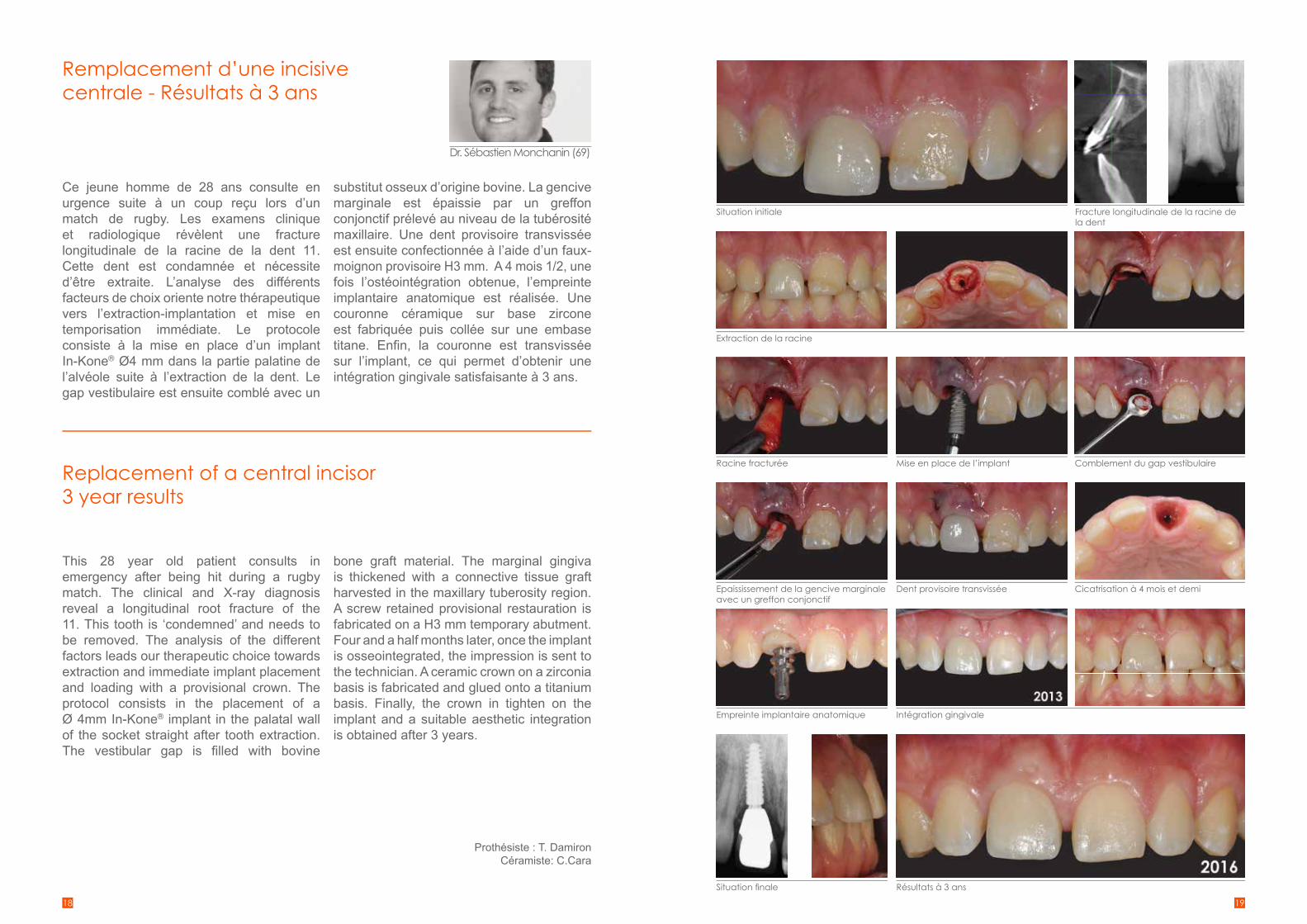
Ce jeune homme de 28 ans consulte en urgence suite à un coup reçu lors d’un match de rugby. Les examens clinique et radiologique révèlent une fracture longitudinale de la racine de la dent 11. Cette dent est condamnée et nécessite d’être extraite. L’analyse des différents facteurs de choix oriente notre thérapeutique vers l’extraction-implantation et mise en temporisation immédiate. Le protocole consiste à la mise en place d’un implant In-Kone® Ø4 mm dans la partie palatine de l’alvéole suite à l’extraction de la dent. Le gap vestibulaire est ensuite comblé avec un
substitut osseux d’origine bovine. La gencive marginale est épaissie par un greffon conjonctif prélevé au niveau de la tubérosité maxillaire. Une dent provisoire transvissée est ensuite confectionnée à l’aide d’un faux-moignon provisoire H3 mm. A 4 mois 1/2, une fois l’ostéointégration obtenue, l’empreinte implantaire anatomique est réalisée. Une couronne céramique sur base zircone est fabriquée puis collée sur une embase titane. Enfi n, la couronne est transvissée sur l’implant, ce qui permet d’obtenir une intégration gingivale satisfaisante à 3 ans.
This 28 year old patient consults in emergency after being hit during a rugby match. The clinical and X-ray diagnosis reveal a longitudinal root fracture of the 11. This tooth is ‘condemned’ and needs to be removed. The analysis of the different factors leads our therapeutic choice towards extraction and immediate implant placement and loading with a provisional crown. The protocol consists in the placement of a Ø 4mm In-Kone® implant in the palatal wall of the socket straight after tooth extraction. The vestibular gap is fi lled with bovine
bone graft material. The marginal gingiva is thickened with a connective tissue graft harvested in the maxillary tuberosity region. A screw retained provisional restauration is fabricated on a H3 mm temporary abutment. Four and a half months later, once the implant is osseointegrated, the impression is sent to the technician. A ceramic crown on a zirconia basis is fabricated and glued onto a titanium basis. Finally, the crown in tighten on the implant and a suitable aesthetic integration is obtained after 3 years.
Dr. Sébastien Monchanin (69)
Situation initiale
Extraction de la racine
Racine fracturée
Epaississement de la gencive marginale avec un greffon conjonctif
Empreinte implantaire anatomique
Situation fi nale Résultats à 3 ans
Mise en place de l’implant Comblement du gap vestibulaire
Dent provisoire transvissée Cicatrisation à 4 mois et demi
Intégration gingivale
Fracture longitudinale de la racine de la dent
Prothésiste : T. DamironCéramiste: C.Cara

20 21
Prothèse complète supra-implantaire amovo-inamovible sur chapes électrogalvaniques
Full-arch implant supported prosthesis on electro-galvanic copings
Patiente présentant un édentement complet maxillaire et partiel mandibulaire souhaitant une réhabilitation fi xe mandibulaire supra-implantaire.Après analyse des éléments cliniques et radiologiques nous proposons le plan de traitement suivant:• l’extraction des dents résiduelles mandibulaires et la pose de six implants In-Kone® par
la technique d’extraction implantation immédiate.• La pose d’une prothèse fi xe provisoire immédiate de type Brånemark.• Après trois mois de cicatrisation, pose d’une prothèse Preteau zircone amovo-inamovible
sur chapes électro-galvaniques et réalisation d’un nouvel appareil amovible maxillaire afi n de rétablir les rapports occlusaux et l’esthétique.
A female patient with a full edentulous maxillary and partial edentulous mandible is asking for a fi xed rehabilitation on implants. After clinical and X-ray diagnosis we propose the following treatment plan: • Extraction of the residual mandibular teeth and immediate implantation with 6 In-Kone®
implants.• Immediate placement of a provisional prosthesis Brånemark type.• After a healing period of 3 months, fabrication of a removable Preteau Zirconia prosthesis
stabilized by electro-galvanic copings.Then, fabrication of a new removable overdenture on the maxillary in order to restore the occlusion as well as the aesthetic aspect.
Prothésiste : Fabio Levratto
Dr. Emmanuel Pontonnier (69)Cas clinique initial
Situation initiale
Faux-moignons parallélisés à 0°
Preteau
Situation fi nale
Vue avant
Situation initiale
Extraction, pose de 6 implants In-Kone® UNIVERSAL et toronto bridge résine de temporisation
Chapes électrogalvaniques
Preteau zircone stratifi é amovo-inamovible
Cas terminé
Vue après

22 23
Remplacement d’une incisive latérale Résultats à 8 ans
Restoration of a central incisor8 year results
Une patiente, 22 ans, consulte en août 2007 avec une agénésie de la 22 compensée par un pont collé fortement dégradé. Elle présente un bon état de santé général et souhaite réhabiliter cet édentement par un implant. L’examen clinique montre un espace prothétique mésio-distal suffi sant mais des axes coronaires convergents, diagnostic confi rmé par radiographie rétro-alvéolaire. La palpation vestibulaire montre un volume osseux suffi sant pour la mise en place d’un implant. Nous adressons alors la patiente à un confrère orthodontiste pour ouvrir légèrement l’espace et redresser les dents adjacentes.La patiente consulte de nouveau en juin 2008. Elle porte une gouttière de contention. Nous réalisons un wax-up d’étude à partir duquel nous confectionnons un guide chirurgical. Un implant EVL®S 3,3/14 mm est posé en novembre 2008 en un temps chirurgical. La technique du
rouleau conjonctif a été utilisée dans le même temps opératoire pour épaissir les tissus mous vestibulaires et modifi er le biotype gingival. Une vis de fermeture plate (0,4 mm) est mise en place sur l’implant.Un contrôle d’ostéo-intégration est réalisé à 8 semaines et la vis de cicatrisation est remplacée par une vis de 3mm. Les premières empreintes sont réalisées en mars 2009 pour la confection d’une couronne transitoire réalisée à l’aide d’un pilier FM Tempo. Cette provisoire sera remaniée sur quatre séances afi n d’amener les tissus mous à maturation. Après une nouvelle prise d’empreinte de l’implant et des tissus gingivaux modelés par la couronne provisoire, une prothèse céramo-métallique transvissée est posée en octobre 2009. Des contrôles cliniques et radiographiques sont réalisés tous les ans pour le suivi habituel.
A 22 year old female patient consults in August 2007. She suffers from agenesis on 22 which is corrected by an unsightly cemented bridge. The patient is in good health and would like to restore the missing tooth with an implant. The clinical diagnosis reveals a suffi cient mesio-distal space but convergent crowns. This diagnosis is confi rmed by X-rays. The vestibular palpation indicates that the bone volume is suitable with the placement of an implant. We address our patient to an orthodontist in order to slightly open the available space and straighten the adjacent teeth.The patient who is using aligners comes back in June 2008. A diagnosis wax up is prepared and a surgical guide is fabricated. An EVL®S 3.3/14
mm is placed in November 2008 in one surgical time. The roll fl ap technique is performed in order to augment the vestibular soft tissues and to modify the biotype. A fl at cover screw (0.4 mm) is placed to close the implant. 8 weeks post-op, a X-ray is performed and the healing screw is switched with a higher one (3 mm). The fi rst impression takings are made in March 2009 in order to fabricate a temporary crown on a FM Tempo abutment. This temporary crown will be modifi ed on 4 different steps in order to guide the soft tissues to the desired architecture. After a new impression taking of the implant and the remodeled soft tissues, a trans-screwed ceramo-metal crown is placed on October 2009. Clinical and X-ray controls are made every year.
Dr. Jean-Baptiste Rebouillat (89)
Situation initialeaoût 2007
Evaluation clinique et radiographique de l’espace disponible et du volume osseux et gingival vestibulaire
Radiographie post-opératoire
Epaississement des tissus mous par technique du rouleau et gestion post-opératoire avec port de la gouttière
Situation lors de la dépose des sutures à 2 semaines
Pose de la vis de cicatrisation hauteur 3mm Radiographie et vue clinique de la prothèse transitoiremars 2009
Vue clinique et radiographie de la couronne cérame-métallique transvissée - oct. 2009
Vue clinique et radiographie de la couronne cérame-métallique transvissée - sept.-2016
Gouttière de contention avec gestion esthétique temporaire
Wax-up d’étude pré-chirurgicale Guide chirurgical issu du wax-up
Indicateur de parallélisme après passage du foret pilote.
Adéquation entre guide chirurgical et indicateur de parallélisme.
Vue occlusale de l’implant EVL-S® 3,3/14 en place - nov. 2008

24 25
Restauration de deux incisives centrales - Résultats à 3 ans
Restoration of two central incisors3 year results
Une patiente, 39 ans, vient consulter car elle n’aime pas son sourire. Lors du diagnostic, nous lui proposons l’extraction de ses deux incisives centrales déjà retraitées à de multiples reprises et de les remplacer par deux implants In-Kone®, avec mise en esthétique immédiate. Les couronnes provisoires initiales sont remplacées à 15 jours post-op par de nouvelles dents provisoires afi n d’obtenir un ajustement précis de collets par guidage tissulaire. A trois mois, la prothèse d’usage est réalisée. Mais rapidement la patiente n’est pas satisfaite par la teinte fi nale lorsque son sourire est exposé au soleil.
Après contrôle sous lumière polarisée, nous constatons effectivement le «défaut» de teinte signalée par la patiente. Nous proposons alors la réalisation de nouvelles couronnes. Sur la radio à trois ans on note la stabilité ainsi que la santé des tissus péri-implantaires. On regrettera cependant une légère cicatrice non résorbée sur la papille centrale, probablement dûe au point de suture réalisé le jour de la pose. A trois ans la patiente est, quant à, pleinement satisfaite du Résultats.
A 39 year old female patient consulted because she is not satisfi ed with her smile. After the diagnosis, we suggest to remove the already treated central incisors and to immediately place two In-Kone® implants loaded with provisional crowns. The latters are replaced by second provisional restorations two weeks post-op in order to guide the maturation of the peri-implant soft tissues to the desired architecture. 3 months later, the fi nal restoration is delivered.
But, rapidely the patient is not satisfi ed with the color of the crowns when the sun refl ects on them. After we control under polarized light, we can clearly identify the color distortion. Accordingly, we fabricate new crowns.X-rays at three years show stabilized peri-implant tissues. We regret though a small scar which remains and which is probably due to the initial suturing. Today, the patient is fully satisfi ed.
Dr. Eric Schneck (69)
Sourire initial Radio de départ
Premières prothèses d’usage Contrôle sous lumière polarisée des premières prothèses d’usage
Deuxièmes prothèses d’usage Contrôle sous lumière polarisée et validation des deu-xièmes prothèses d’usage
Extraction
Mise en esthétique immédiate
Contrôle des axes implantaires Faux-moignons en place
Nouvelles provisoires à 15 jours post-op
Contrôle radio
Contrôle radio à 3 ans

26 27
Restauration complète maxillaire avec mise en charge immédiateRésultats à 7 ans.
Complete upper jaw restauration with immediate loading7 year results
Une patiente âgée de 79 ans vient consulter car son bridge est devenu mobile. Nous lui proposons un plan de traitement avec une technique d’extraction implantation et réalisation d’une prothèse en mise en charge immédiate sur 8 implants In-Kone® dans la même séance. Notre technique opératoire est basée sur le respect d’un protocole codifi é particulièrement strict qui exige entre autre
un positionnement symétrique des implants dans de l’os natif. Dans notre technique, nous favorisons également la préservation de la muqueuse naturelle sur le plan esthétique notamment par l’apport de greffe et de PRF.A 7 ans, nous pouvons remarquer la pérennité de cette technique tant sur le plan esthétique que celui de la santé des tissus environnants.
A 79 year old female patient consulted because of her bridge mobility. After diagnosis, we propose to our patient a treatment plan including extraction and immediate placement and loading of 8 In-Kone® implants within the same session.Our operating procedure is based on a very precise surgical protocol built on a symmetric positioning of the implants in native bone.
From an aesthetic point of view, our protocol also supports the preservation of the patient mucosa together with the use of grafting combined with PRF. Long term repeatable results can be obtained under the condition that the protocol is strictly respected.At 7-year post surgery, note the quality of the results from the aesthetic point of view as well as the health of the peri-implant tissues.
Dr. Alain Simonpieri (13)Ligne du sourire
Extraction implantation immédiate. Noter la symétrie et le parallélisme des implants
Prothèse défi nitive vue cervicale et occlusale. Noter la situation des implants localisés dans le couloir prothétique
Muqueuse à 6 mois Muqueuse à 7 ans
Reconstruction céramique 6 mois post-op (2009)
Contrôle radiographique à 7 ans. Noter la corticalisation de l’os au niveau cervical
Sourire de la patiente
Contrôle à 7 ans (2016)
Situation initiale
Cicatrisation des tissus mous à 6 mois
Radiographie
Contrôle de la prothèse provisoire à 6 mois

28 29
Restauration d’une incisive latérale Résultats à 4 mois
Lateral incisor restoration4 month result
Une femme de 45 ans nous est adressée pour la pose d’un implant suite à une fracture de l’incisive latérale 22. La racine est extraite de façon atraumatique et un Implant 3.0 longueur 13 mm est alors placé dans l’alvéole dans une position tridimensionnelle adéquate. Un faux-moignon avec un profi l d’émergence Ø 3,4 mm est immédiatement connecté. Puis les sutures sont réalisées suivant le concept de « Marginal Migration ». Conformément à ce concept la limite marginale de la première couronne provisoire est placée de façon supra-gingivale afi n de gagner en volume de
tissus mous dans cette zone critique, comme en témoignent les résultats à 2 semaines post-opératoires. 3 mois après la chirurgie, une deuxième couronne provisoire est fabriquée de façon à guider la maturation de la muqueuse péri-implantaire conformément à l’architecture souhaitée. A 4 semaines, la deuxième restauration est retirée et une prise d’empreinte réalisée en vue de la fabrication de la prothèse d’usage. 2 semaines après, la prothèse d’usage est scellée en bouche. Vue du Résultats à 4 mois post-opératoire.
A 45 year old female patient is referred with a fractured hopeless lateral incisor #22 for implant therapy. The root is atraumatically extracted and a narrow 3.0 Implant x 13mm implant is then placed in the extraction alveolar in the proper 3D position. A tulip shape transgingival component with a 3,4 mm Ø prosthetic platform is connected to the implant and the grafting and suturing protocol from the Marginal Migration Concept follows. Following the same concept, the marginal portion of the fi rst provisional
restoration will be supragingival in order to gain soft tissue in this critical area as we witness already 2 weeks post-op. 3 months post-surgery a second provisional restoration is fabricated in order to guide the maturation of the peri-implant soft tissue to the desired architecture. After 4 weeks second provisional restoration will be removed and an impression will be made in order to deliver the fi nal restoration to the patient. The fi nal restoration 2 weeks post cementation and 4 months post-surgery.
Dr. Konstantinos D. Valavanis(GR)
Situation initiale
Deuxième couronne provisoire
Extraction implantation avec un implant étroit Ø 3 mm
Résultats avec prothèse d’usage à 4 mois
Mise en place de la première prothèse provisoire selon le concept de « Marginal Migration » et Résultats à 2 semaines

30 31
Early Periimplant Tissue Healing on 1-Piece Implants With a ConcaveTransmucosal Design: A Histomorphometric Study in Dogs
Rehabilitation of the Atrophic Posterior Mandible with Short (4mm) Implants: A case report
Caroline Bolle, DDS, Marie-Paule Gustin, DS, PhD, Didier Fau, DVM, PhD, Patrick Exbrayat, DDS, Georges Boivin, PhD, and Brigitte Grosgogeat, DDS, PhD
Introduction: The purpose of our study was to investigate the early healing phase of marginal bone and soft tissues around unloaded 1-piece implants with a concave transmucosal design, in a dog model.
Methods: Twenty-four 1-piece implants with a concave transmucosal neck were inserted 1 mm subcrestally in the mandibular ridge of 8 beagle dogs. Four animals were sacrifi ced after 3 and 12 weeks of healing. Histomorphometric analysis was performed to measure the height of the periimplant tissues.
Results and Discussion: The overall height of the periimplant mucosa was, respectively, 2.67 and 2.52 mm, after 3 and 12 weeks. In the connective tissue, a soft tissue Oring seal was observed in the healing area provided by the transmucosal concavity, after 12 weeks. The location of the fi rst bone-to-implant contact facing the implant shoulder was 0.00 and +0.18 mm, respectively, after 3 and 12 weeks of healing. Some bone apposition occurred on the implant shoulder during the healing.
Conclusion: Within the limits of the present study, a concave transmucosal design in 1-piece implants was associated with a short vertical value of biological width and promoted a mechanical interlocking of the implant body at the connective tissue and marginal bone levels.
Roberto Pistilli, MD, Carlo Barausse, DDS, Luigi Checchi, MD, DDS, Pietro Felice, MD, DDS, PhD
This case report describes a successful implant-prosthetic rehabilitation of an atrophic posterior mandible with 4-mm-long implants. The patient refused to undergo any reconstructive surgery, and because the available bone up to the inferior alveolar nerve was only 5 mm or less, the patient received four implant of 4-mm length. Four months after implant placement, a provisional prosthesis was put in place; after another 4 months, this was then in turn replaced with a defi nitive prosthesis. The use of such short implants allows a fi xed prosthetic solution without the need for vertically augmenting the mandibular bone. This procedure considerably reduces intra- and postoperative patient discomfort compared with reconstructive surgery for the placement of longer implants. The follow-up time was 1 year after implant loading.
Caroline Bolle: Student PhD, Laboratoire des Multimatériaux et Interfaces UMR CNRS 5615, Université Lyon 1, Université de Lyon, Villeurbanne, France; Assistant Professor, Université de Rennes 1, Faculté d’Odontologie, Université Européenne de Bretagne, Rennes, France; Hospital Practitioner, CHU de Rennes, Pôle d’Odontologie et de Chirurgie Buccale, Rennes, France. Mari-Paule Gustin: Associate Professor, Department of Public Health, Institute of Pharmacy (ISPB), EA4173, Université Lyon 1, Université de Lyon, Lyon, France; Hospital Practitioner, Service de Biostatistiques, Hospices Civils de Lyon, Lyon, France. Didier Fau: Professor, Interactions Cellule Environment UPSP ICE 2011-03- 101, Vet Agro Sup, Université de Lyon, Marcy l’Etoile, France. Patrick Exbrayat: Associate Professor, UFR Odontologie, Université Lyon 1, Lyon, France; Hospital Practitioner, Service de Consultations et de Traitements Dentaires, Hospices Civils de Lyon, Lyon, France. George Boivin: Research Director, INSERM, UMR 1033, Université de Lyon, Lyon, France. Brigitte Grosgogeat: Professor, Laboratoire des Multimatériaux et Interfaces UMR CNRS 5615, UFR Odontologie, Université Lyon 1, Université de Lyon, Lyon, France; Hospital Practitioner, Service de Consultations et de Traitements Dentaires, Hospices Civils de Lyon, Lyon, France.
Roberto Pistilli: Resident, Unit of Oral and Maxillofacial Surgery, A.C.O. San Filippo Neri, Rome, Italy.Carlo Barausse: Resident, Dental School, Unit of Periodontology ans Implantology, University of Bologna, Bologna, Italy.Luigi Checchi: Full Professor, Dental school, unit of Periodontology and Implantology, University of Bologna, Bologna, Italy.Pietro Felice: Assistant Professor, Dental School, Unit of Periodontology and Implantology, Univesity of Bologna, Bologna, Italy
ISSN 1056-6163/15/02405-001Implant Dentistry, Volume 24 _ Number 5
Int. J Periodontics Restorative Dent 2014; 34:713-718.doi: 10.11607/prd.1733
A lire A lire

32 33
A lire A lire
Soft Tissue and Marginal Bone Adaptation on Platform-Switched Implants with a Morse Cone Connection: A Histomorphometric Study in Dogs.
Posterior jaws rehabilitated with partial prostheses supported by 4.0 x 4.0 mm or by longer implants: One-year post-loading results from a multicenter Randomised controlled trial.
Caroline Bolle, DDS, Marie-Paule Gustin, DS, PhD, Didier Fau, DVM, PhD, Patrick Exbrayat, DDS, Georges Boivin, PhD, and Brigitte Grosgogeat, DDS, PhD
The purpose of this study was to investigate peri-implant tissue adaptation on platform-switched implants with Morse cone-type connection, after 3 and 12 weeks of healing in dogs. Ten weeks after mandibular premolar extraction, eight beagles dogs received three implants each. At each biopsy interval, four animals were sacrifi ced and biopsies were processed for histologic analysis. The height of the peri-implant mucosa was 2.32 mm abnd 2.88 mm, respectively, whereas the bone level in relation to the implant platform was -0.39 mm and -0.67 mm, respectively, after 3 and 12 weeks of healing. Within the limits of the present study, platform-switched implants exhibited reduced values of biologic width and marginal bone loss when compared with previous data..
Pietro Felice, MD, DDS, PhD, Luigi Checchi, MD, DDS, Carlo Barausse, DDS, Roberto Pistilli, MD, Gilberto Sammartino, MD, DDS, Irene Masi, DDS, Daniela Rita Ippolito, DDS Purpose: To evaluate wether 4.0 x 4.0 mm dental implants could be an alternative to implants at least 8.5 mm long, which were placed in posterior jaw, in the presence of adequate bone volumes.Materials and methods: One hundred and fi fty patients with posterior (premolar and molar areas) jaws having at least 12.5 mm bone height above the mandibular canal or 11.5 mm below the maxillary sinus, were randomized according to a parallel group design, in order to receive one to three 4.0 mm-long implants or one to three implants which were at least 8.5 mm-long, at three centers. All implants had a diameter of 4.0 mm. Implants were loaded after 4 months with defi nitive screw-retained prostheses. Patient were followed up to 1-year post-loading and outcome measures were prosthesis and implant failures, any complications and peri-implant marginal bone level changes.Results: Seventy-fi ve patients were randomly allocated to each group. One patient dropped out after the 4-months post-loading evaluation from the long implant group. Up to 1-year post-loading, 3 patients lost one 4.0 mm-long implant each in comparison to 2 patients who lost one long implant each (difference in proportion = 0.013; 95% CI: -0.058 to 0.087; P=0.506). All failures occurred before loading, the failed implant were replaced and the delivery of two prostheses in each group was delayed for several months (difference in proportion = 0.0004; 95% CI: -0.068 to 0.069, P=0.685). Three short implants patients experienced three complications versus 2 long Implant patients (difference in proportion = 0.013; 95% CI: -0.058 to 0.0.87; P= 0.506). Three were no statistically signifi cant differences in prostheses failures, implant failures and complication. Patients with short implants lost on average 0.53 mm of peri-implant bone and patient with longer implants lost 0.57 mm. Three were no statistically signifi cant differences in bone level changes up to 1-year between short and long implants (mean difference = 0.038 mm; 95% CI: -0.068 to 0.138; P=0.198).Conclusions: One year after loading 4.0 mm-long implants achieved similar results as 8.5 mm-long implants in posterior jaws, however 5-to 10-year post-loading data are necessary before reliable recommandations can can be made.
Caroline Bolle: Student PhD, Laboratoire des Multimatériaux et Interfaces UMR CNRS 5615, Université Lyon 1, Université de Lyon, Villeurbanne, France; Assistant Professor, Université de Rennes 1, Faculté d’Odontologie, Université Européenne de Bretagne, Rennes, France; Hospital Practitioner, CHU de Rennes, Pôle d’Odontologie et de Chirurgie Buccale, Rennes, France. Mari-Paule Gustin: Associate Professor, Department of Public Health, Institute of Pharmacy (ISPB), EA4173, Université Lyon 1, Université de Lyon, Lyon, France; Hospital Practitioner, Service de Biostatistiques, Hospices Civils de Lyon, Lyon, France. Didier Fau: Professor, Interactions Cellule Environment UPSP ICE 2011-03- 101, Vet Agro Sup, Université de Lyon, Marcy l’Etoile, France. Patrick Exbrayat: Associate Professor, UFR Odontologie, Université Lyon 1, Lyon, France; Hospital Practitioner, Service de Consultations et de Traitements Dentaires, Hospices Civils de Lyon, Lyon, France. George Boivin: Research Director, INSERM, UMR 1033, Université de Lyon, Lyon, France. Brigitte Grosgogeat: Professor, Laboratoire des Multimatériaux et Interfaces UMR CNRS 5615, UFR Odontologie, Université Lyon 1, Université de Lyon, Lyon, France; Hospital Practitioner, Service de Consultations et de Traitements Dentaires, Hospices Civils de Lyon, Lyon, France.
Int J Periodontics Restorative Dent 2016;36:221-228. doi: 10.11607/prd.2254
Pietro Felice: Assistant Professor, Dental School, Unit of Periodontology and Implantology, Univesity of Bologna, Bologna, Italy Luigi Checchi: Full Professor, Dental school, unit of Periodontology and Implantology, University of Bologna, Bologna, Italy. Carlo Barausse: Resident, Dental School, Unit of Periodontology ans Implantology, University of Bologna, Bologna, Italy.Roberto Pistilli : Resident, Unit of Oral and Maxillofacial Surgery, A.C.O. San Filippo Neri, Rome, Italy.Gilberto Sammartino: Professor, Clinic of Oral surgery, University of Naples Frederico II, Naples, ItalyIrene Masi: Resident, Department of Biomedical and Neuromotor Sciences, Unit or Periodontology and Implantology, University of Bologna, Bologna, Italy.Daniela Rita Ippolito: Postgraduate Student, Department or Orthodontics, School of Dentistry, University of Brescia, Brescia, Italy.
Eur J Oral Implantol 2016; 9(1): 35-45

34 35
A lire A lire
Success rate of the EVL Evolution implants (SERF): A fi ve-year longitudinal multicenter study.
Use of 3D cartilage scaffolds for the stabilization of implants and bone regeneration with fi t-lock technique.
Gérard Duminil, DDS, PhD; Michèle Muller-Bolla, DDS, PhD; Jean-Pierre Brun, DDS; Philippe Leclercq, DDS; Jean-Pierre Bernard, MD, PhD; David M. Dohan Ehrenfest, DDS, PhD
The purpose of this study was to evaluate the success rate of the SERF EVL evolution implants through a 5-year longitudinal multicentric study. Patients from 3 clinicians working in 3 different private practices (Grenoble, Nice, and Paris) and familiar with this implant system were included in this study; 413 patients and 1198 implants were followed over 5 years. The implant sites and implant types were recorded at the time of placement. The patients were followed yearly and controlled at the end of the study. The criterion for treatment evaluation or success was a qualitative variable related to 4 possible treatment outcomes: success, failure, ailing, and lost (dropout patients). Different variables (sex, bone quantity and quality at the implant site, location) were submitted to the chi-square test. A survival curve was established over 5 years according to the Kaplan Meyer method. The clinical follow-up was 3.1 6 1.2 years (ie, 1 to 6 years). At the end of this follow-up period, 1163 implants were classifi ed as successful, 19 as failures, 12 as ailing, and 4 as lost (dropout). This implant system thus presented an overall success rate of 97.08%, over 5 years, independent of implant location, and for patient indications commonly encountered in private practice.
Giovanni Falisi1, Massimo Galli1, Pedro Vittorini-Velasquez2, Juan C. Gallegos-Rivera3, Roberto Minasi1, Alberto De Biase1, Carlo Di Paolo1
The surgical procedures for implant applications on the lateral upper areas depend on sinus pneumatization and availability of the residual bone. In these cases, autologous bone grafting remains the gold standard. Nevertheless, because of the morbidity associated to the donor site and the post-surgical complications, several alternative bone substitutes have been introduced, which, however, imply additional costs and show limited osteoinductive properties. Such limitations can be compensated with new regeneration strategies for biological and mechanical tissue restoration, a subject which has been addressed by tissue engineering in recent years. The authors present a new therapeutic option for implant application in the upper maxilla with bone availability less than 4 mm by using 3D scaffolds obtained from antigen-free porcine cartilage in the fi t-lock technique. A longitudinal study on 18 consecutive cases was performed, with a 95.2% success rate one year after the implant. The advantages of this new technique are: 1)Functional and anatomical recovery of the maxillary antrum, 2) Immediate application of the implants; 3) Reduction of surgical times; 4) Absence of patient morbidity; 5)Local anesthesia; 6) Use of implants with a diameter > 4 mm.
Gerard Duminil: DDS, PhD, is in private practice in Nice, France.Michèle Muller-Bolla: DDS, PhD, is a professor, Department of Public Health, Faculty of Odontology at Nice University, Nice, France.Jean-Pierre Brun: DDS, is in private practice in Grenoble, France.Philippe Leclercq: DDS, is in private practice in Paris, France. Also chairman of the SIOPA (Société d’Implantologie Orale et de Prothèse Appliquée).Jean-Pierre Bernard: MD, PhD, is a professor at the Department of Stomatology and Oral Surgery, School of Dental Medicine, University of Geneva, Geneva, Switzerland.David M. Dohan Ehrenfest: DDS, PhD, is an assistant professor at Paris Descartes University, Paris.
Giovanni Falisi, Massimo Galli, Roberto Minasi, Alberto De Biase, Carlo Di Paolo: Department of Oral and Maxillo Facial Sciences, Sapienza University of Rome.Pedro Vittorini-Velasquez: Maxillo-facial Clinic Cochabamba, Bolivia.Juan C. Gallegos-Rivera: Department of Dentistry, Municipal Japanese University Hospital, Santa Cruz de la Sierra, Bolivia.
Journal of Oral Implantology, Vol. XXXIV/No. Five/2008
Vol. 26 Nº 3 / 2013 / 167-172 ISSN 0326-4815 Acta Odontol. Latinoam. 2013

36 37
CONTEXTE MATERIELetMETHODES
RESULTATS
Phasesexpérimentales
• Implanta1onrandomiséede3implantsA
et3implantsBparchien,après10semainesde
cicatrisaFon
• Prélèvementsà3et12semaines
• Extrac1onde6prémolairesmandibulaires
• SuivicliniqueSurvieimplantaire
Indicesdeplaqueetd’inflammaFongingivale(Löe
etSilness)
• RadiographieEvoluFonduniveauosseuxpéri‐implantaire
• Histologie:osnondéminéralisé
Analysedel’organisaFondelamuqueuse
Ostéo‐intégraFon(%BIC)
• MicrotomographienumériséeSkyscan1174
Architecturedel’ospéri‐implantaire
Précision26μm
Méthodesd’analyse
Suivicliniqueet
radiographique
• 100%survieimplantaire
• Indicesdeplaqueet
d’inflammaFon(LöeetSilness)
maintenusentre0et1
• A:Implantàconnec1que
cônemorseexterneetcol
transgingivalcintré
(Twinkon®Tekka)
• B:Implantàconnec1queinterne
cônemorse(In‐Kone®Tekka)
•B:
cônemorse
Implants
Histologie Microtomographie
numérisée
Deuxgroupesde
4chiensBeagle
• Similaritéosseuseavecl’homme
• GoldstandardenhistologiedesFssuspéri‐
implantaires
BIBLIOGRAPHIE
AutorisaFon
duComité
d’Ethiquede
l’Ecole
NaFonale
Vétérinairede
Lyon
Analysedes
prélèvements
encours
Becker J, Ferrari D, Mihatovic I, Sahm N, Schaer A,
Schwarz F. Stability of crestal bone level at platform
switched non-submerged titanium implants. a
histomorphometrical study in dogs. J Clin Periodontol
2009; 36: 532–539.
Berglundh T, Abrahamsson I, Welander M, Lang NP,
Lindhe J. Morphogenesis of the peri-implant mucosa :
an experimental study in dogs. Clin Oral Impl. Res. 18,
2007; 1-8
Hermann JS, Buser D, Schenk RK, Schoolfield JD,
Cochran DL. Biologic width around one- and two-piece
titanium implants. Clin Oral Implants Research 2001;
12: 559-571
Pearce Al, Richards RG, Milz S, Schneider E, Pearce
RG. Animal models for implant biomaterial research in
bone : a review. Eur Cell Mater 2007 Mar 2; 13: 1-10
Thomsen JS, Laib A, Koller B, Prohaskas S, Mosekilde
LI, Gowin W. Stereological measures of trabecular
bone structure: comparison of 3D micro computed
tomography with 2D histological sections in human
proximal tibial bone biopsies. Journal of Microscopy, vol
218, pt 2 May 2005, 171-179.
• Coupesaxiales
etlongitudinales
(ImplantA)
• Miseenplaced’unprotocole
d’hygièneorale
• Ostéo‐intégraFon
à12semaines
x5Goldnermodifié
(ImplantA)
• OrganisaFondesfibresde
collagèneà12semaines
x20Goldnermodifié,lumière
polarisée(ImplantA)
Etablissementde
l’espace
biologique
Agressions
àlajonc1on
implant‐pilier
Remodelage1ssulaire
péri‐implantaire
Développementdedesigns
implantairesinnovantsafindelimiter
lesremodelages1ssulaires
péri‐implantaires
CongrèsdelaSociétéFrançaisedeParondontologieetd’ImplantologieOrale,Strasbourg3‐5juin2010
OBJECTIF
L’objetdecetravailestdeprésenterleprotocole
d’uneétudeétudiantl’influencededeuxdesigns
implantairesinnovantssurl’établissementde
l’espacebiologique,lescaractéris1quesdela
muqueuseimplantaire,etlemain1enduniveau
osseuxpéri‐implantaire
• Radiographieà3
semaines(ImplantB)
LAZARRAetcoll.2006
Ewa Chomik DMD, PhD, Grzegorz Wasiluk DMD
INTRODUCTION
PURPOSE
CLINICAL SITUATION 1
CONCLUSIONS
CLINICAL SITUATION
Three concepts of single-unit restorations design to avoid biological complications
The risk of biological complications has been rising with the number of implants placed every year. The restorative connection to the implant can be accomplished by either screwing or cementing the restoration on standardised or customised, individually shaped CAD/CAM abutments.
Cement-retained prostheses are commonly used to restore single-unit restorations; however, there are reasonable disadvantages associated with a residual excess of cement after placing the crowns, which can lead to biological complications. Custom-made, screw-retained restorations enable
precise modelling of gingival shape in accordance with the emergence profile, which provides optimal support for soft tissues and excludes the issue of undetected cement remnants.
The aim of the study was to present three methods to restore a single implant unit that is activated by undetected residual cement rests while avoiding biological complications. Advantages of the presented concepts are discussed, based on the current literature and the utilization of the
authors’ own studies.
Decision making in restoring a single unit remains a challenge. Removal of all cement excess after crown cementation seems to be impossible, with the margin below the gingiva level, even using customized, anatomic abutments.
A few different approaches to manufacturing final restorations were introduced. The presented solutions combine advantages of screw-retained and cement-retained restorations. All of the reconstructions were manufactured according to the design of the emergence profile and
biocompatibility, which can lead to predictable and safe conditions in the implant pocket over time.
Restorative option No.3 is a full cerconia crown based on a standard
abutment milled with the screw opening. The innovation of this crown
compared to the standard procedure way of screw retained one is the
custom-prepared subgingival part, which is marked on the gingiva level. The subgingival part was designed with respect to the emergence profile
and made of unpolished cerconia due to perfect biocompatibility.
The concept No.1 is based on the individually designed and milled titanium abutment
cemented extra-orally together with CAD/CAM full cerconia crown milled with the screw
opening. The shoulder is on the level of the gingiva up to 1mm
subgingivally for better esthetic outcome. The restoration is screw-
reteined.
Concept No.2 is a custom-produced abutment based on a standard titanium base cemented with a
cerconia custom-milled part supporting the soft tissue and
creating its margin 1 mm over the gingiva level. A full ceramic crown
can be cemented on this type of custom abutment intra-orally for cement retained or cemented extra-
orally with the srew opening for screw-retained restoration.
EAO Paris - sept 2016

38 39
The Twinkon® concept
Objectives
Materials and methods
Results
Conclusion
This study evaluates the long-term impact of the design of the Twinkon® implant on the stability of peri-implant tissues.
• A study conducted on 90 Twinkon® implants in 34 patients
• 5 years after their loading
Evaluation criteria : - rate of peri-implant diseases : mucositis and
peri-implantitis - implant success according to Misch criteria - radiological assessment of bone loss around
these implants
• 98,9 % survival rate • 40,7 % mucositis • 3,5 % peri-implantitis • average bone loss near to zero
(0,00 mm ± 0,72)
• bone stability or increased bone level despite some adverse clinical situations
Radiological evaluation of marginal bone loss
D0 D0 + 5 years
• prosthetic crimping by its external morse taper (1)
• tissular crimping by its concave neck : the Tissue Creeping Profile (2)
1
2
D0 D0 + 5 years
Immediate implant placement Thin biotype
BibliographyThe design of the Twinkon® implant must influence local tissue behaviour. In certain situations, where local bone or mucous membrane-conditions didn’t enable ideal t h re e - d i m e n s i o n a l i m p l a n t positioning, we demonstrated peri-implant tissue-stability.
AHN SY, HAN CH, HEO SJ, KIM TI, SEOL YJ, LEE YM, KU Y, LEE HJ, CHUNG CP, HAN SB, RHYU IC. Soft tissue responses to differential shapes of the implant abutment. J Korean Acad Periodontol 2006;36(1):167-177.
BOLLE C, EXBRAYAT P, FAU D, GRISTCH K, GROSGOGEAT B. Protocole d’étude de l’interface implants-tissus péri-implantaires chez l’animal. Congrès de la société française de Parodontologie et d’Implantologie Orale. Strasbourg 3-5 Juin 2010.
DOUILLARD Y, BOREL JF, DUCHATELARD P. Twinkon : une révo lu t ion conceptue l le . Imp lan t Ch i ru rg ie P ro thèse . 2009;15:215-223.
Topic: Implant therapy outcomes, peri-implant biology aspects
Add Poster
Number
Can implant design influence local tissue behaviour ?
Thibaut MONEDIERE, Philippe DUCHATELARD, Jean-Luc VEYRUNE, Jean-François BOREL
PBI537
Contact info :
to access the posters online, please download the application
1st prize communications EAO 2016
EAO Paris - sept 2016EAO Paris - sept 2016

Domaines d’activitésImplantologie Chirurgie pré-implantaireChirurgie orthognathiqueChirurgie reconstructrice Chirurgie traumatologique de la faceChirurgie carcinologiqueChirurgie oraleCranio-chirurgieOrthodontieFormation
ZI de Sacuny118 avenue Marcel Mérieux69530 BrignaisFrance
tél. +33 (0)4 78 56 97 00fax +33 (0)4 78 56 01 63
www.globald.comUne société du groupe MENIX
Partenaire de vos chirurgies
00D
C-C
OM
P-F
R-E
d1-1
6